Determination of class II peptide-MHC repertoires and recognition via
large yeast-displayed libraries
by
Charles Garrett Rappazzo
B.S. Bioengineering Cornell University, 2014 M.Eng. Biomedical EngineeringCornell University, 2015
SUBMITTED TO THE DEPARTMENT OF BIOLOGICAL ENGINEERING IN PARTIAL FULFILLMENT OF THE REQUIREMENTS FOR THE DEGREE OF
DOCTOR OF PHILOSOPHY IN BIOLOGICAL ENGINEERING AT THE
MASSACHUSETTS INSTITUTE OF TECHNOLOGY JUNE 2020
© 2020 Massachusetts Institute of Technology. All rights reserved.
Signature of Author: ____________________________________________________________ Department of Biological Engineering
May 15, 2020 Certified by: ____________________________________________________________ Michael E. Birnbaum, PhD Assistant Professor of Biological Engineering Thesis supervisor Accepted by: ____________________________________________________________ Paul Blainey, PhD Associate Professor of Biological Engineering Co-chair of Graduate Program, Department of Biological Engineering
2
THESIS COMMITTEE
Michael E. Birnbaum, PhD: Assistant Professor of Biological Engineering (Thesis supervisor) K. Dane Wittrup, PhD: Carbon P. Dubbs Professor of Chemical Engineering and Biological Engineering (Thesis committee chair)
Tyler Jacks, PhD: David H. Koch Professor of Biology
Stefani Spranger, PhD: Howard S. and Linda B. Stern Career Development Assistant Professor of Biology
3
Determination of class II peptide-MHC repertoires and recognition via
large yeast-displayed libraries
by
Charles Garrett Rappazzo
Submitted to the Department of Biological Engineering on May 15, 2020 in Partial Fulfillment of the Requirements for the Degree of Doctor of Philosophy in
Biological Engineering ABSTRACT
T cells occupy essential roles throughout the immune system to prevent and limit disease. As such, breakdowns in their function and recognition underlie poor clinical outcomes across diverse maladies including pathogen infection, cancer, autoimmunity, allergies, and transplant rejection. Yet, when properly directed, T cells drive potent protective and therapeutic responses in prophylactic vaccinations and novel immunotherapies. Therefore, understanding and harnessing T cell function and recognition is of great importance to improving patient care and addressing currently unmet clinical needs.
The function and recognition of T cells are driven through their T cell receptors (TCRs), which bind with great specificity to peptide-MHCs (pMHCs), Major Histocompatibility Complex proteins displaying tissue- and disease-specific peptide antigens derived from their host cell or its surroundings. However, to specifically and comprehensively present and surveil antigens across highly divergent maladies, extreme diversity is required of both the population-level TCR and pMHC repertoires. However, this same diversity which drives T cell function also confounds generalized understanding of these repertoires, as well as their recognition. Therefore, there has been considerable recent interest in the development and application of tools to comprehensively define, predict, and screen these repertoires and their recognition at high throughput.
In this thesis, I both utilize and build upon these tools to define TCR and pMHC repertoires and explore their recognition, particularly with yeast-displayed pMHC libraries for CD4+ T cell recognition of class II pMHCs, and especially in the context of cancer. Using these technologies, I empirically define pMHC repertoires, explore the antigenic basis of TCR repertoire convergence in a preclinical tumor model, and explore the antigen reactivity of human T cells with clinical relevance. While these results provide detailed insights into the specific TCRs and pMHCs studied, they also provide guidance for future avenues in the exploration of TCR and pMHC repertoires and their recognition.
Thesis Supervisor: Michael E. Birnbaum, PhD Title: Assistant Professor of Biological Engineering
4
TABLE OF CONTENTS
Abstract...3
Acknowledgements...6
Chapter 1. Introduction 1.1 What are T cells?...8
1.2 The role and recognition of T cells...9
1.3 Drivers of TCR and pMHC repertoires...10
1.4 Determining TCR and pMHC repertoires and recognition...12
1.5 Thesis overview and motivation...14
References...14
Chapter 2. Empirical determination of class II MHC peptide repertoires for improved antigen prediction Abstract...21 2.1 Introduction...22 2.2 Results...23 2.3 Discussion...43 2.4 Methods...48 2.5 Acknowledgments...48 References...49
Chapter 3. Decoding the origin and antigen discovery of a conserved regulatory T cell response to highly immunogenic murine lung adenocarcinomas Abstract...55
3.1 Introduction...56
5
3.3 Discussion...65
3.4 Methods...66
3.5 Acknowledgments...69
References...70
Chapter 4. Design and application of yeast-displayed peptide-MHC libraries for cognate antigen discovery Abstract...73 4.1 Introduction...74 4.2 Results...75 4.3 Discussion...84 4.3 Methods...85 4.5 Acknowledgments...88 References...88
Chapter 5. Perspectives and future directions 5.1 Summary...91
5.2 Future directions...91
5.3 Closing thoughts...95
6
ACKNOWLEDGEMENTS
Writing a thesis acknowledgement section is a rare chance to editorialize and reflect on the state of science, as well as your own position within the scientific community at the outset of your career. So, here we go.
This thesis was written in its entirety from self-isolation amidst the global pandemic of SARS-Cov-2, the causative agent of Covid-19. At the time of writing, March 19, 2020, this novel coronavirus has infected 230,000 people world-wide, caused 9,300 deaths, and halted life-as-usual throughout most of the world. At this time, the only certainties are that this pandemic will get worse, and eventually, it will get better. However, in all likelihood, this pandemic will not fully resolve without the intervention of scientists.
This intervention may be in the form of an antiviral medication, a therapeutic antibody, a prophylactic vaccine, or some yet-to-be-discovered approach. Yet each of these interventions highlights the critical importance of biological research, both in the clinic and in the laboratory, in confronting the world’s greatest maladies. In light of this, I am proud to have contributed – in very small part – to the ever-growing fields of immunology and biological engineering during my years at MIT. But more importantly, I am proud to join a community of scientists who continuously strive to improve our understanding of the world, and to find new ways to treat and prevent diseases. Long after Covid-19 is a distant memory and public interest in immunology research wanes, I am confident that this community will continue to endeavor to improve patient care with the same vigor.
My own journey into science has been a somewhat winding path. When I graduated high school, I never would have envisioned that I would be writing a doctoral thesis 10 years later. However, I have been fortunate to have had the inspiration of many talented teachers, the encouragement of steadfast mentors, the loving support of my friends, family, and wife, as well as a whole lot of dumb luck along the way. The remainder of this section is dedicated to the people who made this all possible.
From my time at Cornell University, two professors, Larry Bonassar and Dave Putnam, deserve special acknowledgment. The fall of my junior year, I took a course taught by Larry and his organized, clear, and well-motivated teaching gave me an increased appreciation of biomedical engineering. Two years later, I TA’ed this same course and during this time I came to see him as a mentor. I am forever grateful for his insights into biological engineering, science, and academia, and for his encouragement, advice, and support in my applications to graduate school.
However, my applications to graduate school would have been wholly unsuccessful without my first research advisor, Dave Putnam. I met Dave when he presented on reprogramming the immune system using a bacterially derived vaccine during a senior-year rotating lecturer course. At the time of this presentation, I knew next to nothing about immunology and was still planning on applying to medical schools, not graduate programs. I’m not sure if it was the subject matter or Dave’s uniquely energetic presentation, but I was hooked. Even though I was already a senior, Dave agreed to meet with me and let me join his group to work on a universal Influenza A virus vaccine, spurring my interest in immunology and starting research career. I went on to work in the
7
group for a year and a half, staying for an additional year during a Master of Engineering degree, and have so many fond memories of this time. I am thankful to Dave for his continued mentorship, enthusiastic encouragement, his overly generous recommendation letters. I am also so thankful to the many members of the Putnam lab who taught me how to be a researcher and who inspired me to pursue a career in science, despite repeatedly advising me otherwise. In particular, I am thankful to my two mentors in the lab, Cassie and Hannah, and all the other members of team OMV, past and present.
I have also been very fortunate to have very supportive mentors and lab mates at MIT, and I could not have asked for a better thesis advisor than Michael Birnbaum. I first heard of Michael as the new faculty member just starting a lab. Although his research was well over my head at the time, he was smart, approachable, and down-to-earth, making it an easy decision to join his lab (a generous description for the time considering its limited size) that fall. The ensuing four and a half years have had their ups and downs and seen the Birnbaum lab grow from 4 members to 13 (not including the army of undergrads), but through it all, Michael has been a constant source of mentorship, encouragement, and much needed levity. Sorry for all the guff (kinda). To the members of the Birnbaum lab, past and present, thank you for many fruitful conversations and lab meetings, and for pushing me to be a better researcher. I look forward to what you will all accomplish. To my undergraduate student Ben, thank you for your trust and enthusiasm – it was a pleasure to see you grow in your knowledge and abilities. A special thank you to Christine and Anna, who keep the lab running, and to Brooke, who collaborated on nearly all my projects; none of this would have gotten done without you three.
To my committee members, Dane, Tyler, and Stefani, thank you for your consistent support, mentorship, and insights. I am very thankful to have had you as committee members and to have had the chance to collaborate with your lab members on my research throughout the years. To my friends at MIT and beyond, thank you for so many fond memories throughout these years. Once this pandemic ends, I can’t wait to have you all over for more dinners, drinks, and Lillydog time. To my family, I have no way to fully express how grateful I am for all the ways you have loved and supported me not only during my time at MIT, but every day before and hopefully every day after. A special thank you to my Mom and Dad, who have sacrificed so much to give me a good home, a good education, and a happy and healthy life - all that I accomplish is because of you. To my dog Lilly, you can’t read, but you’re a very good girl. Lastly, to my wife, Rory - thank you for being my constant companion, cheerleader, sounding board, copy editor, comfort, and joy. I can’t wait to start the next chapter with you.
8
CHAPTER I. Introduction
1.1 What are T cells?
The principal function of the immune system is to prevent and limit disease throughout the body. This function is achieved through the collective action of trillions of individual immune cells interacting with our organs, our environment, commensal microbes, and each other. When they sense danger, such as pathogens, cancers, or injury, these cells are activated to mobilize massive and coordinated protective responses. Yet these cells also have crucial roles in limiting inflammation, promoting wound healing, and maintaining homeostasis. Therefore, the immune system must be able to differentiate self from foreign, healthy from diseased, and commensal from pathogenic 1. As such, breakdowns in the function or recognition of the immune system underlie diverse ailments from pathogen infection and cancer 1, 2 to autoimmune diseases and allergies 3, 4, as well as chronic and degenerative diseases 5, 6.
The immune system is broadly partitioned by its function and recognition into the innate and adaptive immune systems; the innate immune system generates rapid responses against pathogens, toxins, and cell damage through evolutionarily conserved motifs and receptor, whereas the adaptive immune system generates slower but more specific and specialized responses through highly variable immune receptors 1. In addition, the adaptive immune system uniquely possesses memory, allowing it to generate memory cell populations that can produce larger, more rapid, more specialized, and even more specific responses upon re-exposure to a given pathogen or disease 1, 7. As such, the innate immune system serves as a first line of defense, containing potential dangers and recruiting other immune cells, while the adaptive immune system performs higher-level tasks of distinguishing between potential threats and tailoring its response accordingly. The adaptive immune system is primarily comprised of two cell types: B and T lymphocytes 1, 7. While both cell types display memory and specificity in their activation and functions, these functions are highly distinct, and are driven by their divergent recognition. In particular, B cell receptors (BCRs) are comprised of two identical recognition domains that bind directly to a diverse array of extracellular proteins. In contrast T cell receptors (TCRs) encode a single recognition site that binds to specialized cell-surface immune proteins called Major Histocompatibility Complexes (MHCs). Furthermore, upon activation, B cells can edit their receptors for higher affinity to their target through somatic hypermutation, and can secrete their receptors as antibodies, which can be oligomerized for additional binding valency 7. These antibodies can then directly bind to target far from their parent B cell to enact aggregation and direct neutralization, as well as facilitate phagocytosis and cytotoxicity. In contrast, T cells cannot edit or secrete their TCR, and their activation, localization, and function are all driven through their TCR 8. Yet T cells are uniquely capable of recognizing and responding to threats hidden within the cell, and have roles in coordinating and modulating the immune response 7.
Although T cell populations can be even further partitioned based upon their function and recognition into αβ and γδ subsets 9, as well as innate-like natural killer T (NKT) cells 10, we will focus on their most prevalent subset, αβ T cells, which recognize MHCs displaying short, linear peptides derived from within their host cell or its environment, known as peptide-MHCs (pMHCs).
9 1.2 The role and recognition of T cells
Owing to their diverse composition and recognition, T cells can fulfill many functions within the immune response across highly divergent diseases. These functions can range from directing and modulating innate and B cell responses, to enacting direct cell killing, and even suppressing other T cell responses 11. Yet these diverse functions can be largely partitioned among T cells based on their expression of two cell-surface co-receptors into CD4+ ‘helper’ and CD8+ ‘killer’ T cells, each with their own recognition 1,7.
As their name suggests, CD8+ ‘killer’ T cells – also known as cytotoxic T lymphocytes (CTLs) – primarily function to directly kill infected or diseased cells. CD8+ T cells recognize class I peptide-MHC (ppeptide-MHC) proteins, which are ubiquitously expressed by nucleated cells in the bodyand principally display peptides derived from within their parent cell 1, 12. When activated through their TCR, these cells are empowered to directly kill target cells expressing their cognate pMHC. This recognition pathway allows CD8+ T cells to monitor for intracellular infection, impaired function, and even dysregulated signaling pathways, and are therefore crucial for pathogen and tumor surveillance and control 13.
In contrast, CD4+ ‘helper’ T cells serve in broader but less direct roles, coordinating and aiding the responses of other immune cells. These cells recognize class II pMHC proteins, which are expressed by specialized antigen-presenting cells (APCs) and principally display peptides derived from their environment 1, 12. These APCs are often phagocytic members of the innate immune system – such as macrophages, dendritic cells, and monocytes – that specialize in rapidly internalizing and processing pathogens and cell debris, but also includes B cells and specialized endothelial cells 12. This antigen presentation pathway allows CD4+ T cells to respond indirectly to both extracellular and intracellular threats. Yet, in order to respond effectively to this vast array of threats, CD4+ T cells must be able to tailor their response to a threat.
As such, activated CD4+ T cells can adopt a diverse array of distinct T-helper (Th) phenotypes, each with a distinct gene expression and cytokine secretion signature, to tailor the local immune response to a broad variety of threats. A detailed description of these phenotypes – such as Th1, Th2, Th17, Tfh, and Th9 – and their functions are reviewed elsewhere 14. However, CD4+ T cells can also serve in non-inflammatory, regulatory roles. In contrast to conventional CD4+ T cell (also known as Tconv) populations, regulatory T cell (Treg) populations dampen the immune response and suppress T cell responses through a variety of indirect and direct modalities 15. While activated Tconv cells can adopt a Treg phenotype 16, most Tregs are a distinct CD4+ T cell lineage with differential recognition 17, 18.
Combined, these diverse functions allow T cells to occupy many crucial roles in the immune system simultaneously. But as such, breakdowns in T cell recognition and function can have disastrous consequences throughout the body, underlying poor clinical outcomes during viral and bacterial infections, facilitating continued outgrowth of tumors, and driving autoimmune diseases, allergies, and transplant rejection 19-23. Therefore, a detailed understanding of the diverse functions of T cells – and the recognition that drives them – is crucial to our understanding of many diseases, as well as to our ability to develop novel therapies.
10
In fact, directing, modulating, and coopting T cell function and recognition already underlies many therapies in the clinic: T cells are crucial to forming the protective memory responses required for prophylactic vaccinations 24, can be stimulated and directed by therapeutic vaccinations 25, and can be activated to fight established tumors by novel cancer immunotherapeutics 26. Furthermore, T cell function can be coopted to potent new purposes by redirecting their recognition through engineered TCRs and chimeric antigen receptors (CARs) 27, 28. But while each of these applications have already had significant impacts in the clinic, further investigation of T cell function and recognition is still needed to guide further improvements of these therapies, as well as enable future modalities to address unmet clinical needs. Yet in order to fully understand T cell function and recognition, we must first understand their common driver, the T cell receptor.
1.3 Drivers of TCR and pMHC repertoire diversity
As previously discussed, the TCR drives both the recognition and function of T cells. Accordingly, the diverse functions of T cell populations rely on diverse and distinct population-scale TCR repertoires 29. Therefore, a comprehensive understanding of T cell function and recognition requires detailed knowledge of the drivers of both the diversity and divergence observed in TCR repertoires across T cell populations, as well as how these drivers interact with diverse pMHC repertoires.
Diversity in pMHC repertoires stems from the need to comprehensively present antigenic peptides from a vast array of potential threats, both endogenous and foreign, to antigen-specific T cells, as any holes in these repertoires can blind T cells to ongoing infection or disease. To be properly sampled by both CD4+ and CD8+ T cell subsets, these threats also need to be presented across both class I and class II MHC proteins 12. In addition, these pMHC complexes must be stable and long-lived in order to be properly surveilled by potentially rare clonal T cell populations 30, which are comprised of as few as 10-100 T cells throughout the entire body 31, and therefore MHCs must be specific to their displayed peptides. Yet the number of peptides required to comprehensively represent all potential immune threats is too vast to be specifically presented by any one MHC protein.
Therefore, to facilitate broad yet specific peptide presentation, MHC proteins themselves are highly diverse 32, 33. In humans, this diversity is accomplished on an individual basis by the expression of six distinct MHC genes, three class I A, -B, and -C) and three class II (HLA-DR, -DP, and –DQ), each with their own peptide specificities 34. In addition, these MHC genes are extremely polymorphic, providing evolutionarily driven peptide-binding diversity on a population scale 32, 33. In fact, the MHC locus in humans, also known as the human leukocyte antigen (HLA) locus, is the most polymorphic region in the genome 35,with over 26,000 currently known unique HLA alleles 36. Importantly, these polymorphisms are clustered in the peptide-binding groove of MHC proteins 37, thereby imparting unique peptide-specificities. Combined, this diversity provides for the expression of up to 12 unique HLA proteins in humans (as the MHC genes are expressed co-dominantly by each chromosome) 37, and therefore up to 12 unique pMHC repertories to comprehensively present potential threats to the TCR repertoires.
To enable specific recognition of these diverse pMHC repertoires, TCR repertoires must also be highly diverse 38. This is accomplished in the TCR repertoire by VDJ recombination, a process
11
unique to B and T cells that allows receptor formation by recombination of a variable (V), diversity (D), and joining (J) region selected quasi-randomly from diverse collections, with untemplated nucleotide additions, deletions, and substitutions permitted at their junctions to provide additional diversity 1, 7. These segments are then joined to a constant (C) receptor domain, and the pairing of two independently recombined receptor chains provides additional diversity. A detailed review of this process is available elsewhere 39. This process allows each of the estimated 1012 human T cells 40 to theoretically choose between 1015-1020 unique TCR combinations 41. However, due to biases in VDJ recombination and external factors (discussed below), the true diversity of these repertoires has been recently estimated at approximately 1010 unique clones in humans 42.
As a direct result of VDJ recombination, the diversity of the TCR repertoire is greatest at the junction between the V, D, and J regions, and comprises the complementary-determining region 3 (CDR3) of each TCR chain 39. Importantly, these CDR3 regions are positioned in direct contact with the MHC-displayed peptide in TCR / pMHC complexes, and are the primary drivers of TCR binding and specificity 43, 44. In addition, these CDR3 regions contribute equally to peptide binding, and therefore the TCR alpha and beta chains are equally important to pMHC binding. The V region-encoded CDR1 and CDR2 regions of each chain contribute additional interactions with the displayed peptide – as well as the MHC peptide-binding groove – providing additional specificity to these interactions 43, 44. Therefore, the intrinsic diversity of the TCR receptor repertoire drives diverse T cell recognition, yet is essential to provide specific recognition of the even more diverse pMHC repertoire 38.
However, the TCR repertoire not only samples the pMHC repertoire, but is shaped by it. This process primarily occurs during T cell development in the thymus, but occurs to a lesser degree in the periphery following development 8. Within the thymus, T cell progenitors express both CD4 and CD8 once they successfully formed a TCR capable of signaling through VDJ recombination 1, 8. These so-called double-positive thymocytes then sample pMHC molecules displayed by specialized antigen presenting cells as well as endothelial cells that express diverse self-derived peptides 45. The strength of TCR signaling during this this developmental stage determines the T cell’s fate; excessive stimulation from strong or frequent recognition of these self-antigens drives clonal deletion to eliminate potentially autoreactive T cells, but a lack of stimulation causes the T cell to die of neglect 8, 45. Therefore, the productive TCR repertoire is derived from TCRs within this self-specificity sweet spot 46. However, the nature of this stimulation further determines the lineage of this T cell. In particular, preferential interaction with class I or class II pMHCs drives CD8+ and CD4+ differentiation, respectively, due to stabilizing co-receptor interactions, and CD4+ T cells which receive greater or more frequent TCR stimulation during this stage are preferentially driven towards Treg differentiation 1, 8.
Combined, these developmental influences underlie the ability of T cell populations to distinguish between self and foreign peptides 1, and are responsible for the distinct TCR repertoire of each T cell lineage 29. These distinct repertoires are in turn responsible for the distinct recognition of these subsets, and underlie their diverse functions throughout the immune system. However, due to their immense diversity and person-to-person variability, individual TCR and pMHC repertoires and their interactions are highly unique. Therefore, tools which help define the composition of these repertoires and their recognition are of great importance for improved understanding of T cell function – as well as our ability to coopt and redirect it – across many diseases.
12
1.4 Defining pMHC and TCR repertoires and recognition
In line with their importance to understanding and utilizing T cell function, there is great interest in developing tools to study TCR and pMHC repertoires 47, 48, as well as TCR and pMHC recognition 49. These tools can be used to define individual TCR and pMHC repertoires, screen interactions in high-throughput, and used to train computational algorithms that predict these repertoires and interactions.
As discussed, diverse TCR repertoires drive T cell function and recognition. Therefore, monitoring and defining the TCR repertoire can uncover temporal shifts or convergence in these repertoires that reveal underlying disease biology and T cell targeting. For example, temporal shifts within the TCR repertoire can reveal the temporal dynamics of viral infection and convergence in the TCR repertoire can point to shared targeting of immuno-dominant epitopes 50. These insights can then be used to guide future studies of the immune response or to design therapeutic modalities. Although early methods to define and monitor TCR repertoires through their v-region usage (via flow cytometry), and later CDR3 length (via immune spectratyping), provided partial insights into their composition, dynamics, and convergence 51, these techniques did little to define or monitor diversity in the CDR3 junctions that drive recognition. Therefore, the most powerful tool for defining these repertoires is TCR sequencing, which provides detailed coverage of entire TCR, including the CDR3. These sequencing techniques most frequently utilize reverse transcription polymerase chain reactions (RT-PCR) to amplify expressed TCR transcripts, and with the advent of next generation sequencing (NGS) can be used on a repertoire scale 47, 51. Yet while bulk TCR sequencing of T cell populations (often focused on the TCR beta chain 52) can be used to identify broad trends in repertoire dynamics and convergence, these approaches lose the linkage between TCR alpha and beta chains that is essential for defining antigen reactivity 43, 44. Therefore, paired-chain TCR sequencing techniques represent the state-of-the-art for defining TCR repertoires 47. Although these techniques were originally low throughput 53, 54, recent advances now enable thousands of T cells to be fully defined simultaneously 55-58, and may soon enable full repertoire definition in tissue- and disease-specific contexts.
In contrast, pMHC repertoires cannot be defined with sequencing, as they rely on both MHC allele and peptide diversity. However, sequencing of MHC gene usage, known as HLA typing in humans, can establish MHC allele usage in a given individual, and provide partial insights into these repertoires 59. This is because while over 19,000 unique class I and 7,000 unique class II HLA alleles have been observed in human populations 36, a small subset of these alleles are frequently observed or dominant within given ethnic groups 60. This allows researchers to partially generalize allele-specific pMHC repertoires between individuals through the expression of these over-represented alleles. However, these repertoires definitions are only partial because they are also dependent on peptide expression and antigen processing 12, 61. Therefore, pMHC repertoire definition and utilization is largely dependent on computational algorithms, called antigen prediction algorithms, that allow allele-specific prediction of individual peptide / MHC interactions and pMHC repertoires by extrapolating insights from previously curated datasets 48.
13
The allele-specific peptide datasets used to train these antigen prediction algorithms are derived from many divergent sources. However, these methods can be broadly partitioned into MHC ligand binding and ligand elution methods 62. MHC ligand binding assays often use recombinantly-expressed MHC to measure the binding of pre-selected peptides to measure quantitative metrics of binding, such as peptide-binding mode, affinity, or kinetics 48. Yet while these datasets provide detailed binding information, they are low throughput and do not incorporate antigen processing pathways. In contrast, MHC ligand elution methods utilize natively expressed and loaded pMHC molecules, and therefore capture antigen presentation biases. These methods most frequently utilize mass spectrometry (MS) to determine the sequence of the bound peptide and can be used to define pMHC repertoires in both homeostatic and disease-specific contexts 63. Furthermore, the recent development of mono-allelic engineered cell lines that express only a single MHC allele has enabled unambiguous linkage between observed peptides and their displaying MHC in MS datasets 64-66. Yet while these MS-based technologies are high throughput and can broadly define pMHC repertoires, they have notable biases 66 and provide only qualitative binding data 67. However, neither TCR or pMHC repertoire information can define or predict recognition at their interface. For this, researchers require an entirely separate set of tools. The earliest such tools use biochemical methods for T cell antigen discovery, such as the use fluorescently-labeled tetramers of recombinant pMHC proteins to identify antigen reactive T cells or mass spectrometry to identify pMHC proteins bound to a given TCR. However, these tools are low throughput and require knowledge of at least one component of the interaction 49, and therefore are incompatible with comprehensive or broad T cell antigen discovery 68. In the absence of a known antigen or eluted ligands, antigen discovery requires time- and resource-intensive ‘epitope mapping’ approaches to determine the specificity of a given TCR 69. In addition, these methods provide little-to-no information on potential antigen cross-recognition, which is essential to T cell function and comprehensive epitope coverage 38, 68. While this cross-recognition can be assessed through site-directed mutagenesis 70, or predicted in silico 71, 72 these methods are largely restricted to closely related sequences 49.
Therefore, in order to achieve both comprehensive and broad T cell antigen discovery, researchers use pMHC library technologies. Broadly, these technologies consist of a pool of unique cells expressing many copies of a single pMHC protein that are then probed with endogenously or recombinantly expressed TCR 49. To achieve single pMHC expression, the peptide and MHC are typically expressed as a single construct. The platforms used to express these pMHC library approaches can be mammalian, insect, yeast, or phage, each with unique advantages and short-comings 49. While mammalian libraries can accurately recapitulate native protein expression and antigen presentation, their potential library size is limited to 105-106 unique peptides which can limit comprehensive epitope coverage, whereas phage-displayed libraries can express up to 1012 unique variants but express very few copies of their pMHC protein 73, and can fail to express many pMHC proteins 68 While insect- and yeast-displayed pMHC libraries exist within the middle of these spectrums, achieving both improved protein expression relative to phage-displayed libraries and achieving larger library sizes than mammalian platforms, yeast-displayed libraries have larger library sizes (up to 109 unique variants 49) and have improved growth rates and modularity 74. Collectively, these advantages underlie the recent increase in use of yeast-displayed libraries for T cell antigen discovery 75-78, as well as the frequent use of yeast-display for assays and optimizations of many diverse immune proteins 74.
14 1.5 Thesis overview and motivation
In this thesis, we both utilize and build upon these tools to define TCR and pMHC repertoires and recognition, particularly with yeast-displayed pMHC libraries for CD4+ T cell recognition of class II pMHC repertoires, especially in the context of cancer.
As our lab is located within the Koch Institute for Integrative Cancer Research at MIT, understanding and treating cancer shapes our perspective of immunology, and drives many of our projects and collaborations. In particular, with the advent of potent and efficacious new cancer immunotherapies such as immune checkpoint inhibitors 26, there is great interest in the recognition of tumor-infiltrating T cells 79, which requires T cell repertoire sequencing to identify T cell clones of interest. Although early immunotherapy research focused largely on the antigen reactivity of CD8+ ‘killer’ T cells, as they directly affect tumor clearance, there is increased appreciation of the role – and therefore the recognition – of CD4+ Tconvs in the anti-tumor response 80, as well as CD4+ Tregs in suppressing tumor clearance 81. Therefore, in chapter 3 of this thesis, we use single-cell TCR sequencing to investigate the TCR repertoire of tumor-infiltrating Tregs in a preclinical model of lung adenocarcinoma and identify clones of interest. We then screen these TCRs with yeast-displayed pMHC libraries to probe for their antigen recognition.
Furthermore, the clinical successes of personalized cancer vaccines have driven great interest in improved methods for pMHC repertoire definition to facilitate improved tumor-specific antigen prediction for these vaccines 82. This is especially true for class II pMHC antigen prediction algorithms which underperform their class I counterparts 83. Therefore, in chapter 2 of this thesis, we develop a new method to define class II pMHC repertoires by modifying an existing yeast-displayed class II pMHC library platform. We then use this data to retrain existing class II antigen prediction algorithms and find that they significantly improve their performance, including in the context of candidate antigen identification for personalized cancer vaccines.
The insights we gained from TCR and pMHC repertoire definition are then applied in chapter 4 to define the native antigen reactivity of both clinically-relevant CD4+ and CD8+ T cells. Finally, through each of these projects we identified pitfalls, bottlenecks, and shortcomings in the application of our yeast-displayed pMHC libraries. Therefore, in chapter 5 of this thesis, we highlight these issues, discuss possible solutions, and discuss future applications of this powerful technology in defining TCR and pMHC repertoires and recognition.
References
1. Chaplin, D.D. Overview of the immune response. J. Allergy. Clin. Immunol. 125, S3-23 (2010). 2. Gonzalez, H., Hagerling, C., & Werb, Z. Roles of the immune system in cancer: From tumor initiation to metastatic progression. Genes Dev. 32, 1267-1284 (2018).
3. Rosenblum, M.D., Remedios, K.A., & Abbas, A.K. Mechanisms of human autoimmunity. J.
15
4. Galli, S.J., Tsai, M. & Piliponsky, A.M. The development of allergic inflammation. Nature 454, 445-54 (2008).
5. Bennett, J.M., Reeves, G., Billman, G.E, & Sturmberg, J.P. Inflammation-nature's way to efficiently respond to all types of challenges: Implications for understanding and managing "the epidemic" of chronic diseases. Front Med 5, 316 (2018).
6. Labzin, L.I., Heneka, M.T., & Latz, E. Innate immunity and neurodegeneration. Annu. Rev.
Med. 69, 437-449 (2018).
7. Bonilla, F.A. & Oettgen, H.C. Adaptive immunity. J. Allergy. Clin. Immunol. 125, S33-40 (2010).
8. Kumar, B.V., Connors, T.J., & Farber, D.L. Human T cell development, localization, and function throughout life. Immunity 48, 202-213 (2018).
9. Chien, Y., Meyer, C., & Bonneville, M. γδ T cells: First line of defense and beyond. Annu. Rev.
Immunol. 32, 121-55 (2014).
10. Wu, L. &Van Kaer, L. Natural killer T cells in health and disease. Front. Biosci. 3, 236-51 (2011).
11. Pennock, N.D., White, J.T., Cross, E.W., Cheney, E.E., Tamburini, B.A., & Kedl, R.M. T cell responses: naïve to memory and everything in between. Adv Physiol Educ. 37, 273–283 (2013). 12. Neefjes, J., Jongsma, M.L.M., Paul, P., & Bakke, O. Towards a Systems Understanding of MHC Class I and MHC Class II Antigen Presentation. Nat. Rev. Immunol. 11, 823-36 (2011). 13. Zhang, N. & Bevan, M.J. CD8+ T cells: Foot soldiers of the immune system. Immunity 35, 161–168 (2011).
14. Geginat, J. et al. Plasticity of human CD4 T cell subsets. Front Immunol 5, 630 (2014). 15. Vignali, D.A.A., Collison, L.W., & Workman, C.J. How regulatory T cells work. Nat. Rev.
Immunol. 8, 523–532 (2008).
16. Schmitt, E.G. & Williams, C.B. Generation and function of induced regulatory T cells. Front.
Immunol. 4, 152 (2013).
17. Pacholczyk, R. & Kern, J. The T-cell receptor repertoire of regulatory T cells. Immunology 125, 450–458 (2008).
18. Golding, A., Darko, S., Wylie, W.H., Douek, D.C., & Shevach, E.M. Deep sequencing of the TCR‐β repertoire of human forkhead box protein 3 (FoxP3)+ and FoxP3– T cells suggests that they are completely distinct and non‐overlapping. Clin. Exp. Immunol. 188, 12–21 (2017).
16
19. Blackwell, J. M., Jamieson, S. E., & Burgner, D. HLA and infectious diseases. Clin. Microbiol.
Rev. 22, 370-85 (2009).
20. Hadrup, S., Donia, M., Thor-Straten, P. Effector CD4 and CD8 T cells and their role in the tumor microenvironment. Cancer Microenviron. 6, 123-133 (2013).
21. Bluestone J.A., Bour-Jordan, H., Cheng, M., & Anderson, M. T cells in the control of organ-specific autoimmunity. J. Clin. Invest. 125, 2250-2260 (2015).
22. Woodfolk, J. A. T-cell responses to allergens. J. Allergy Clin Immunol. 119, 280-294 (2007). 23. Issa, F., Schiopu, A., Wood, K.J. Role of T cells in graft rejection and transplantation tolerance.
Expert Rev. Clin. Immunol. 6, 155-169 (2010).
24. Sallusto, F., Lanzavecchia, A., Araki, K., & Ahmed, R. From vaccines to memory and back.
Immunity 33, 451–463 (2010).
25. Gilbert, S.C. T-cell-inducing vaccines – what's the future. Immunology 135, 19–26 (2012). 26. Pardoll, D.M. The blockade of immune checkpoints in cancer immunotherapy. Nat. Rev.
Cancer 12, 252-64 (2012).
27. Zhao, L. & Cao, Y.J. Engineered T cell therapy for cancer in the clinic. Front. Immunol. 10, 2250 (2019).
28. Miliotou, A.N. & Papadopoulou, L.C. CAR T-cell therapy: A new era in cancer immunotherapy. Curr. Pharm. Biotechnol. 19, 5-18 (2018).
29. Izraelson, M. et al. Comparative analysis of murine T-cell receptor repertoires. Immunology 153, 133-144 (2018).
30. Baumgartner, C.K., Ferrante, A., Nagaoka, M., Gorski, J., & Malherbe, L.P. Peptide-MHC class II complex stability governs CD4 T cell clonal selection. J. Immunol. 184, 573-81 (2010). 31. Jenkins, M.K. & Moon, J.J. The role of naive T cell precursor frequency and recruitment in dictating immune response magnitude. J. Immunol. 188, 4135-40 (2012).
32. Sommer, S. The importance of immune gene variability (MHC) in evolutionary ecology and conservation. Front. Zool. 2, 16 (2005).
33. Piertney, S.B. & Oliver, M.K. The evolutionary ecology of the major histocompatibility complex. Heredity 96, 7-21 (2006).
34. Paul, S., Weiskopf, D., Angelo, M.A., Sidney, J., Peters, B., & Sette, A. HLA class I alleles are associated with peptide-binding repertoires of different size, affinity, and immunogenicity. J.
17
35. Hughes, A. & Hughes, M. Natural selection on the peptide-binding regions of major histocompatibility complex molecules. Immunogenetics 42, 233–243 (1995).
36. Robinson, J., Halliwell, J.A., Hayhurst, J.H., Flicek, P., Parham, P., & Marsh, S.G.E. The IPD and IMGT/HLA database: allele variant databases. Nucleic Acids Res. 43, D423-431 (2015). 37. Janeway C.A. et al. The major histocompatibility complex and its functions. Immunobiology:
The Immune System in Health and Disease. (Garland Science, 2001).
38. Sewell, A.K. Why must T cells be cross-reactive? Nat. Rev. Immunol. 12, 669-77 (2012). 39. Jung, D. & Alt, F.W. Unraveling V(D)J recombination; Insights into gene regulation. Cell 116, 299-311 (2004).
40. Arstila, T.P. Casrouge, A., Baron, V., Even, J., Kanellopoulos, J., & Kourilsky, P. A direct estimate of the human alphabeta T cell receptor diversity. Science 286, 958-61 (1999).
41. Laydon, D.J., Bangham, C.R.M., & Asquith, B. Estimating T-cell repertoire diversity: Limitations of Classical Estimators and a New Approach. Philos. Trans. R. Soc. Lond. B. Biol. Sci. 370, 1675 (2015).
42. Lythe, G., Callard, R.E., Hoare, R.L., & Molina-París, C. How many TCR clonotypes does a body maintain? J. Theor. Biol. 389, 214-24 (2016).
43. Zoete, V., Irving, M., Ferber, M., Cuendet, M.A. & Michielin, O. Structure-based, rational design of T cell ceceptors. Front. Immunol. 4, 268 (2013).
44. Garcia, K.C. & Adams, E.J. How the T cell receptor sees antigen--A structural view. Cell 122, 333-6 (2005).
45. Klein, L., Kyewski, B., Allen, P.M., & Hogquist, K.A. Positive and negative selection of the T cell repertoire: What thymocytes see (and don't see). Nat. Rev. Immunol. 14, 377-91 (2014). 46. Vrisekoop, N., Monteiro, J.P., Mandl, J.N., & Germain, R.N. Thymic positive selection and the mature T cell repertoire for antigen revisited. Immunity 41, 181–190 (2014).
47. Rosati, E., Dowds, C.M., Liaskou, E., Henriksen, E.K.K., Karlsen, T.H., & Franke, A. Overview of methodologies for T-cell receptor repertoire analysis. BMC Biotechnol. 17, 61 (2017). 48. Gfeller, D. & Bassani-Sternberg, M. Predicting antigen presentation-what could we learn from a million peptides? Front. Immunol. 9, 1716 (2018).
49. Gerber, H., Sibener, L.V., Lee, L.J., & Gee, M.H. Identification of antigenic targets. Trends
18
50. Ruggiero, E. et al. High-resolution analysis of the human T-cell receptor repertoire. Nat.
Commun. 6, 8081 (2015).
51. Six, A. et al. The past, present, and future of immune repertoire biology - the rise of next-generation repertoire analysis. Front Immunol 4, 413 (2013).
52. Freeman, J.D., Warren, R.L., Webb, J.R., Nelson, B.H., & Holt, R.A. Profiling the T-cell receptor beta-chain repertoire by massively parallel sequencing. Genome Res. 19, 1817–1824 (2009).
53. Han, A., Glanville, J., Hansmann, L., & Davis, M.M. Linking T-cell receptor sequence to functional phenotype at the single-cell level. Nat. Biotechnol. 32, 684-92 (2014).
54.Dash, P., Wang, G.C, & Thomas, P.G. Single-Cell Analysis of T-Cell Receptor αβ Repertoire.
Methods Mol. Biol. 1343, 181-97 (2015).
55. Zheng, G.X.Y. et al. Massively parallel digital transcriptional profiling of single cells. Nat.
Commun. 8, 14049 (2017).
56. Gierahn, T.M. et al. Seq-Well: Portable, low-Cost RNA sequencing of single cells at high throughput. Nat. Methods 14, 395-398 (2017).
57. Lee, E.S. et al. Identifying T cell receptors from high-throughput sequencing: dealing with promiscuity in TCRα and TCRβ pairing. PLoS Comput. Biol. 13, e1005313 (2017).
58. Holec, P.V., Berleant, J., Bathe, M., & Birnbaum, M.E. A Bayesian framework for high-throughput T cell receptor pairing. Bioinformatics 35, 1318-1325 (2019).
59. Hosomichi, K. Shiina, T., Tajima, A., & Inoue, I. The impact of next-generation sequencing technologies on HLA research. J. Hum. Genet. 60, 665-73 (2015).
60. Gonzalez-Galarza FF, et al. Allele frequency net 2015 update: new features for HLA epitopes, KIR and disease and HLA adverse drug reaction associations. Nucleic Acid Res. 39, D784-8 (2015).
61. Fortier, M. et al. The MHC class I peptide repertoire is molded by the transcriptome. J. Exp.
Med. 205, 595–610 (2008).
62. Vita, R. et al. The Immune Epitope Database (IEDB): 2018 update. Nucleic Acids Res. 47, D339-D343 (2019).
63. Purcell, A.W., Sri H Ramarathinam, S.H., & Ternette, N. Mass spectrometry-based identification of MHC-bound peptides for immunopeptidomics. Nat. Protoc. 14, 1687-1707 (2019).
19
64. Abelin, J. G. et al. Mass spectrometry profiling of HLA-associated peptidomes in mono-allelic cells enables more accurate epitope prediction. Immunity. 46, 315-326. (2017).
65. Sarkizova, S. et al. A large peptidome dataset improves HLA class I epitope prediction across most of the human population. Nat. Biotechnol. 38, 199–209 (2020).
66. Abelin, J. G. et al. Defining HLA-II Ligand Processing and Binding Rules with Mass Spectrometry Enhances Cancer Epitope Prediction. Immunity 51, 766-779 (2019).
67. Garde, C. et al. Improved peptide-MHC class II interaction prediction through integration of eluted ligand and peptide affinity data.Immunogenetics 71, 445-454 (2019).
68. Birnbaum, M.E., Dong, S., & Garcia, K.C. Diversity-oriented approaches for interrogating T-cell receptor repertoire, ligand Recognition, and function. Immunol. Rev. 250, 82-101 (2012). 69. Provenzano, M. et al. MHC-peptide specificity and T-cell epitope mapping: Where immunotherapy starts. Trends Mol. Med. 12, 465-72 (2006).
70. Border, E.C., Sanderson, J.P., Weissensteiner, T., Gerry, A.B., & Pumphrey, N.J. Affinity-enhanced T-cell receptors for adoptive T-cell therapy targeting MAGE-A10: Strategy for selection of an optimal candidate. Oncoimmunology 8, e1532759 (2018).
71. Dash, P. et al. Quantifiable predictive features define epitope specific T cell receptor repertoires. Nature 547, 89-93 (2017).
72. Glanville, J. et al. Identifying specificity groups in the T cell receptor repertoire. Nature 547, 94-98 (2017).
73. Pedersen, L.O. et al. Efficient assembly of recombinant major histocompatibility complex class I molecules with preformed disulfide bonds. Eur. J. Immunol. 31, 2986-96 (2001).
74. Pepper, L.R., Cho, Y.K., Boder, E.T., & Shusta, E.V. A decade of yeast surface display technology: Where are we now? Comb. Chem. High Throughput Screen. 11, 127–134 (2008). 75. Birnbaum, M.E. et al. Deconstructing the peptide-MHC specificity of T cell recognition. Cell 157, 1073-87 (2014).
76. Gee, M. H. et al. Antigen identification for orphan T cell receptors expressed on tumor-infiltrating lymphocytes. Cell 172, 549-563 (2018).
77. Sibener, L.V. et al. Isolation of a structural mechanism for uncoupling T cell receptor signaling from peptide-MHC binding. Cell 174, 672-687 (2018).
78. Saligrama, N. et al. Opposing T cell responses in experimental autoimmune encephalomyelitis.
20
79. Savage, P.A., Leventhal, D.S. & Malchow, S. Shaping the repertoire of tumor-infiltrating effector and regulatory T cells. Immunol. Rev. 259, 245-258 (2015).
80. Borst, J., Ahrends, T., Bąbała, N., Melief, C.J.M., & Kastenmüller, W. CD4 + T cell help in cancer immunology and immunotherapy. Nat. Rev. Immunol. 18, 635-647 (2018).
81. Togashi, Y., Shitara, K., & Nishikawa, H. Regulatory T cells in cancer immunosuppression — implications for anticancer therapy. Nat. Rev. Clin. Oncol. 16, 356-371 (2019).
82. Hu, Z., Ott, P.A., & Wu, C.J. Towards personalized, tumour-specific, therapeutic vaccines for cancer. Nat. Rev. Immunol.18, 168-182 (2018).
83. Jensen, K. K. et al. (2018) Improved methods for predicting peptide binding affinity to MHC class II molecules. Immunology. 154, 394-406 (2018).
21
CHAPTER 2: Empirical determination of class II MHC peptide repertoires for
improved antigen prediction.
Abstract
CD4+ ‘helper’ T cells play a central role in the immune system, coordinating the immune response in pathogen infection and cancer, but also in autoimmune diseases and allergies. Accordingly, accurate prediction of which antigens can be displayed on class II MHCs for recognition by CD4+ T cells is an important consideration for the study of immune disorders, and for the design of novel antigen-targeted vaccines and cancer immunotherapies. While many algorithms have been developed to predict peptide binding to class II MHCs, they under-perform their class I counterparts – even for well-characterized alleles – due in part to gaps and inaccuracies within their underlying training sets, and deficiencies arising from limited peptide diversity.
In this chapter, we describe a yeast-display-based platform to screen libraries of108 peptides for binding to a co-expressed class II MHC, identifying over an order of magnitude more unique binders than comparable approaches. The enriched peptide data contains strong motifs that reflect previous reports, but also highlight gaps and inaccuracies in current data collection techniques and frequently used prediction algorithms, which are validated by in vitro binding assays. We further validated that these gaps and inaccuracies are rectified when existing prediction algorithms are trained upon our yeast-display library data, providing improved prediction of peptide-binding affinity and improved antigen prediction for pathogen and tumor-associated peptides. Together, these findings demonstrate that this platform yields large, high-quality peptide-binding datasets that can be used to improve the accuracy of class II MHC prediction algorithms for improved understanding and application of CD4+ T cell recognition.
22 2.1 Introduction
T cells recognize short, linear peptides displayed by Major Histocompatibility Complexes (MHCs), or Human Leukocyte Antigens (HLAs) in humans, through their T cell receptors (TCRs). Upon recognition of a cognate peptide-MHC (pMHC) complex, the T cell is activated, initiating an immune response that can protect against infectious diseases and cancer 1, 2, but that can also potentiate autoimmunity, allergy, and transplant rejection 3-5.
As T cells occupy a central role in many diseases and underlie the successes of novel antigen-targeted vaccinations and immunotherapies 6-8, there is considerable interest in determining which peptides can be presented by MHCs for T cell surveillance. However, the highly polymorphic peptide-binding groove of MHCs and the immense diversity of potential binding peptides necessitates the use of allele-specific antigen prediction algorithms. Recent advances have described improvements of these computational algorithms 9-11, their underlying training data 12, 13, or both 15-18. But while these advances have benefited antigen prediction for both classes of MHC proteins – class I and II, which are canonically recognized by ‘killer’ CD8+ and helper CD4+ T cells, respectively – there is sustained interest in improving the performance of class II MHC prediction algorithms 19, which frequently under-perform their class I counterparts 11, 20-24.
Although this under-performance is at least partially due to a paucity of curated peptide-binding data for class II MHC alleles 25 – as under-performance is particularly pronounced for alleles with few reported binders 20, 21 – these predictions under-perform for even well-characterized alleles 20, 24. This is likely due to challenges inherent to class II MHCs, which have more degenerate peptide-binding motifs than class I MHCs 26, and have an open peptide-binding groove that requires an added algorithmic step of peptide-register determination 21, 27-29. Additionally, publically available class II MHC peptide-binding datasets contain many redundant nested peptide sets and single amino-acid variants of well-characterized peptides, limiting their effective depth and generalizability 25, 30. Therefore, we hypothesize that the under-performance of class II MHC prediction algorithms is driven primarily by deficiencies in their underlying training data that can be rectified with large and diverse high-quality peptide datasets.
In this chapter, we describe a yeast-display-based platform to screen 108 peptides for their ability to bind a co-expressed class II MHC to highlight and rectify these deficiencies. This platform generates over an order of magnitude more unique peptide data than comparable approaches for two human class II alleles, and enriches peptide motifs that mirror previous reports. However, our datasets contain additional peptide-binding information that results in consequential differences from existing prediction algorithms and other state-of-the-art data collection techniques. We demonstrate that these differences represent systemic gaps and inaccuracies in current class II MHC peptide-binding data that are rectified by training existing algorithms on yeast-display data. Finally, we show that an algorithm trained on our data improves prediction of peptide-binding affinity and improves antigen prediction for pathogen- and tumor-associated peptides. These data show the importance of large, unbiased pMHC repertoires to improve existing antigen prediction training datasets, and suggest our approach can facilitate improved understanding of CD4+ T cell recognition and improved patient benefit from antigen-targeted therapeutics.
23 2.2 Results
2.2.1 Yeast-displayed class II MHC platform identifies peptide binding
Figure 2.1. Design and validation of a yeast-display platform to identify peptide binding to a co-expressed
class II MHC. A) Structural representation (adapted from PDB 1J8H) of platform highlighting 3C protease cleavage site and Myc epitope tag within the linker connecting the peptide and MHC β1 domain. B) Schematic of validation protocol, including linker cleavage with 3C, peptide exchange at low pH in the presence of HLA-DM and high-affinity competitor peptide, and quantification of remaining bound peptide with an anti-Myc antibody. C) Time course of mean fluorescence intensity (MFI) of a fluorescently labeled anti-Myc antibody for HLA-DR401-CLIP81-101-encoding yeast without treatment (Untreated), with linker
cleavage (3C), or with linker cleavage and peptide exchange (3C + HLA-DM), as determined by flow cytometry. D) Comparison of peptide retention for HLA-DR401-CLIP81-101, -CII261-273, or -HA306-318
-encoding yeast with linker cleavage and peptide exchange, as determined by flow cytometry and normalized to MFI before treatment.
Yeast-displayed class II pMHC platforms have previously been used to identify peptides that facilitate TCR binding 31, 32. To enable this platform to determine peptide binding to the MHC, we modified the previously-described design of DR401 (HLA DRA1*01:01, HLA-DRB1*04:01) 32 to express a 3C protease site and an antibody-trackable Myc epitope tag within the flexible linker connecting the peptide to the N-terminus of the HLA β chain (Figure 2.1A). Protease treatment cleaves the linker, allowing unbound peptides to freely disassociate. Peptide exchange is then initiated at low pH in the presence of a high-affinity competitor peptide and the catalyst HLA-DM (Figure 2.1B), emulating the native endosomal environment of peptide loading
24
and exchange 33. If the original peptide is displaced by the competitor peptide, the peptide-proximal epitope tag is lost, enabling us to differentiate yeast encoding binding and non-binding peptides by flow cytometry with a fluorescently-labeled antibody directed against the peptide-proximal Myc tag.
This platform was validated through its specificity in peptide retention. Yeast expressing HLA-DR401 and the class II-associated invariable chain peptide (CLIP81-101), the peptide displaced during endogenous antigen presentation 33, exhibited significant loss of peptide-proximal epitope tag signal following linker cleavage that increased with incubation at low pH with a competitor peptide (Figure 2.1C). Consistent with its role as a peptide-exchange catalyst, the addition of DM significantly accelerated signal loss. However, yeast expressing known binders of HLA-DR401, HA306-318 34, 35 and CII261-273 35-37, exhibited retention of their peptides when treated with 3C and HLA-DM (Figure 2.1D).
2.2.2 Selection, analysis, and validation of a class II MHC peptide library
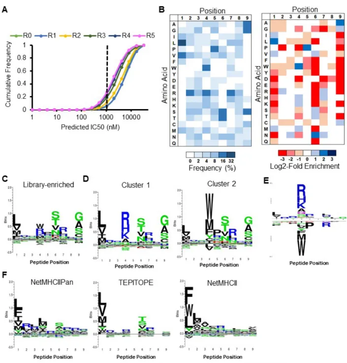
To enable large-scale identification of HLA-DR401-binding peptides, we generated a yeast library encoding 1x10⁸ random MHC-linked peptides. To simplify downstream analysis, peptides were designed as a randomized 9mer flanked by constant residues that favor binding to the MHC in a single register, as the class II MHC peptide-binding groove is open at either end and allows binding in many possible registers 22,23. The library was subjected to iterative rounds of linker cleavage, peptide exchange, and selection for epitope tag retention (Figure 2.2A), resulting in a pool of strong binders after five rounds (Figure 2.2B). Upon deep sequencing, we observed enrichment of predicted binders (Figure 2.2C). The enriched library was highly diverse, consisting of 81,422 unique peptides in the correct register, of 85,756 total peptides. To visualize positional amino acid enrichment and depletion, positional residue frequencies were benchmarked against the unselected library to generate positional log2-fold-change enrichment values. The resulting data are presented as heatmaps representing unweighted averages, as the distribution of peptide frequency in the enriched library was largely flat, with no observed correlation between individual peptide frequency and affinity (Figures 2.2D, E).
We observed the strongest enrichments at peptide positions P1, P4, P6, and P9 (Figure 2.3A), which are considered ‘anchor’ positions where the peptide backbone orients the amino acid side chain directly into pockets of the MHC surface (Figure 2.3B) 28. These enrichments largely match previous reports for HLA-DR401 37, 39-43: the deep P1 pocket favors large hydrophobic residues; the basic P4 pocket favors acidic residues; P6 favors polar residues Ser, Thr, and Asn; and the shallow P9 pocket favors Ala, Gly, and Ser. However, the observed enrichment of P9 Cys and P6 Asp do not match this consensus, and only the latter has been previously reported 43, 44. We also observed a less stringent preference for Pro and Asn at P7, which is considered to be an auxiliary anchor position 45. While the remaining positions are considered to be determinants of TCR binding 46, each displayed marked preferences, such as the uniform depletion of Trp, the enrichment of Pro and Asp at P5, the strong depletion of P2 Pro, and the previously described preference for P2 Arg 37, 40. Each described enrichment or depletion was highly statistically significant (p < 0.001).
25
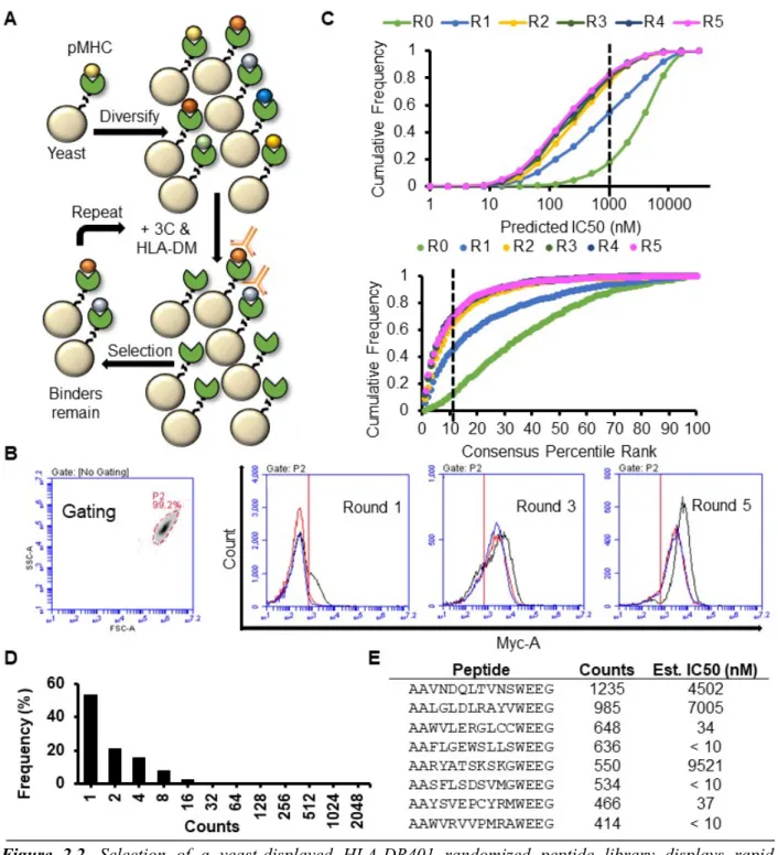
Figure 2.2. Selection of a yeast-displayed HLA-DR401 randomized peptide library displays rapid
convergence. A) Schematic of sequential rounds of library selection to eliminate non-binding peptides and enrich binders. B) Histogram of the fluorescence intensity of a labeled anti-Myc antibody for 10,000 yeast in each round of selection either before linker cleavage (Black), following cleavage (Red), or after 24h peptide exchange (Blue), with gating strategy. C) Cumulative distribution function of predicted peptide IC50
(top) and percentile rank (bottom) of 1000 peptides from each round of selection, as determined by NetMHCII and the IEDB consensus tool, respectively. Dashed lines represent previously established cut-offs for peptide binding. D) Histogram of occurrences of each unique peptide found in round 5 of selection. E) Table of the most enriched peptides found within round 5 of selection with occurrences and estimated IC50 value from two-point fluorescence polarization competition assays.
26
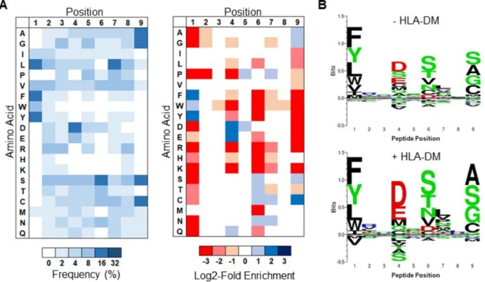
Figure 2.3. Selection of a yeast-displayed HLA-DR401 randomized peptide library reveals a strongly
enriched binding motif that differs from existing prediction algorithms. A) Unweighted heat maps of positional percent frequency and log2-fold enrichment of each amino acid in round 5 of selection (N = 81,422 unique peptides). B) Structure of HA306-318 peptide in the HLA-DR401 peptide-binding groove (PDB
1J8H), with primary peptide ‘anchor’ positions denoted in bold. (C-E) Kullback-Leibler relative entropy motifs of the core nine amino acids of HLA-DR401-binding peptides, as determined (C) empirically from our yeast-display library, (D) by clustering of binders curated on the SYFPEITHI database, or (E) by application of existing class II MHC prediction algorithms to computationally-generated peptides.
27
Notably, our overall library-enriched motif (Figure 2.3C) closely resembles that of known HLA-DR401 binders (Figure 2.2D), generated by clustering previously reported HLA-HLA-DR401-binding peptides curated on the SYFPEITHI database 30. In particular, we found stringent preferences at each anchor position that matched our own, with the exception of P9 Cys. However, these motifs were highly dissimilar to those generated by existing class II MHC prediction algorithms NetMHCII 11, NetMHCIIPan 11, TEPITOPE 48, or IEDB consensus 49 (Figure 2.2E). Comparison suggests that while these algorithms mirror the importance and nature of preferences at P1 and P6 (excepting P6 Asp), they have increased uncertainty or miscall preferences at the remaining anchor positions, P4 and P9.
In addition, we performed library selection without the addition of the endosomal class II peptide-exchange catalyst HLA-DM in order to quantify its impact on the peptide repertoire. With the exception of differences in their magnitudes, the observed enrichments and depletions were consistent with HLA-DM addition (Figure 2.4), suggesting that HLA-DM selects for the retention of high-affinity peptides uniformly across each position, but does not impart unique positional preferences, consistent with previous reports 18, 47.
Figure 2.4. HLA-DM addition increases the stringency of library selection. A) Unweighted heat maps of
the positional percent frequency and log2-fold enrichment of each amino acid in round 5 of selection without the addition of HLA-DM (N = 105,717 unique peptides). B) Kullback-Leibler relative entropy motifs of the core nine amino acids of HLA-DR401-binding peptides, determined empirically from round 5 of library selection with or without HLA-DM addition.
To quantify the consequence of these differences, we performed fluorescence polarization competition assays on selected peptides to determine their IC50 values for recombinant HLA-DR401, which correlate with affinity 50. We selected 16 peptides that were enriched by our library but deemed non-binders by both NetMHCII and the IEDB consensus tool (predicted IC50 > 1 µM,
28
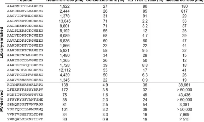
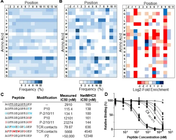
consensus rank > 10 45, 49); each contained either cysteine at P4, P7, or P9, aspartic acid at P6, or an unfavorable P1 residue (Table 2.1). Upon measurement, each of these peptides had an IC50 less than 1 µM, and 14/16 bound stronger than HA306-318 (76 nM), a known strong binder 34, 35 (Figure 2.5A). Importantly, the binding of the cysteine-containing peptides was specific, as two allele-mismatched cysteine-containing peptides did not exhibit binding (Figure 2.5B). We further identified 8 peptides from Influenza A virus [A/Victoria/3/75 (H3N2)] that both NetMHCII and IEDB consensus predicted as binders (IC50 < 200 nM, consensus rank < 5) but that did not match our enriched motif, largely due to departures at P4 and P9 (Table 2.1). Each had a measured IC50 > 2 µM, and 6/8 bound weaker than CLIP89-101. Overall, there was minimal concordance between the measured IC50 of these peptides and the predictions of NetMHCII, IEDB consensus, or TEPITOPE, and the predictions of both NetMHCII and IEDB consensus were negatively correlated with measured IC50 (Figure 2.5C).
Table 2.1. Peptides either enriched by our randomized 9mer HLA-DR401 library selections but not
predicted to bind HLA-DR401 by NetMHCII or IEDB Consensus (Library-enriched), or derived from Influenza A virus and predicted to bind HLA-DR401 but not matching our enriched motif (IAV-derived), with prediction values and IC50 measured via from fluorescence polarization competition assays.
Together, these data highlight consequential gaps and inaccuracies in current class II prediction algorithms, and suggest that our yeast-display platform enriches high-quality peptides that may rectify these deficiencies.
29
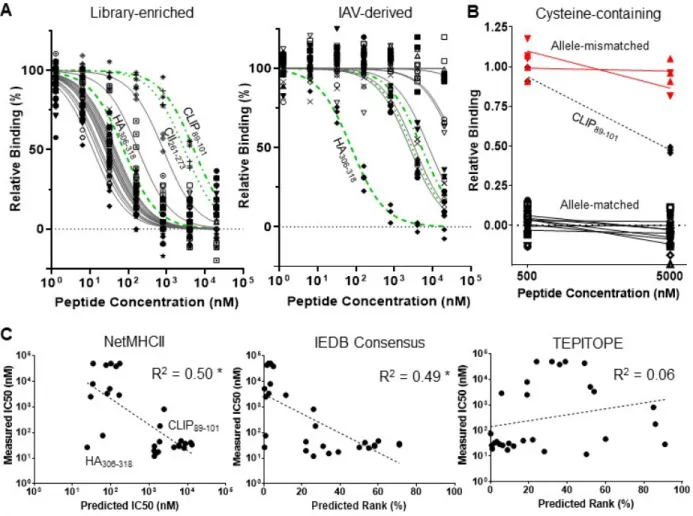
Figure 2.5. Validation of library-enriched HLA-DR401-binding motif reveals consequential gaps and
inaccuracies in class II prediction algorithms. A) Relative binding curves for HLA-DR401 in fluorescence polarization competition assays for peptides either enriched by selection of a 9mer HLA-DR401 library but not predicted to bind HLA-DR401 (Library-enriched) or derived from Influenza A virus and predicted to bind HLA-DR401 but not matching our enriched motif (IAV-derived), with selected control peptides (green). B) Relative binding of cysteine-containing peptides found in round 5 of selection of either the randomized 9mer HLA-DR401 (allele-matched) or HLA-DR402 (allele-mismatched) libraries, tested at two concentrations with HLA-DR401 in a fluorescence polarization competition assay. Curves are fit to N = 3 replicates. C) Scatterplots of algorithmic predictions versus measured IC50 with lines of best fit and their
associated coefficients of determination (R2). Asterisk denotes R2 values of negative correlations.
2.2.3 Preferences outside the peptide ‘core’ greatly affect binding
Canonically, peptide positions P1 through P9 are considered to form the ‘core’ of the interface with the class II MHC peptide-binding groove 27, 28. However, positions outside of the MHC groove, also known as peptide flanking residues (PFRs), can reportedly affect peptide binding 51-53. Most notably, modifications at position P10 can reportedly alter peptide IC50 up to two orders of magnitude 52, without altering the peptide ‘core’ or TCR interactions 46. We therefore sought to investigate peptide preferences outside the groove using a randomized peptide library.
To this end, we constructed a randomized 13mer HLA-DR401 library, which was selected analogously to the original library. While peptides from round 5 showed no initially obvious motif