HAL Id: inserm-02132560
https://www.hal.inserm.fr/inserm-02132560
Submitted on 17 May 2019
HAL is a multi-disciplinary open access
archive for the deposit and dissemination of
sci-entific research documents, whether they are
pub-lished or not. The documents may come from
teaching and research institutions in France or
abroad, or from public or private research centers.
L’archive ouverte pluridisciplinaire HAL, est
destinée au dépôt et à la diffusion de documents
scientifiques de niveau recherche, publiés ou non,
émanant des établissements d’enseignement et de
recherche français ou étrangers, des laboratoires
publics ou privés.
Direct non transcriptional role of NF-Y in DNA
replication
Paolo Benatti, Silvia Belluti, Benoit Miotto, Julia Neusiedler, Diletta Dolfini,
Marjorie Drac, Valentina Basile, Etienne Schwob, Roberto Mantovani, Julia
Blow, et al.
To cite this version:
Paolo Benatti, Silvia Belluti, Benoit Miotto, Julia Neusiedler, Diletta Dolfini, et al.. Direct non
transcriptional role of NF-Y in DNA replication. Biochimica et Biophysica Acta - Molecular Cell
Research, Elsevier, 2016, 1863 (4), pp.673-685. �10.1016/j.bbamcr.2015.12.019�. �inserm-02132560�
Direct non transcriptional role of NF-Y in DNA replication
Paolo Benatti
a,1, Silvia Belluti
a,e,1, Benoit Miotto
b,c,d, Julia Neusiedler
e, Diletta Dol
fini
f, Marjorie Drac
g,
Valentina Basile
a, Etienne Schwob
g, Roberto Mantovani
f, J. Julian Blow
e, Carol Imbriano
a,⁎
a
Dipartimento di Scienze della Vita, Università di Modena e Reggio Emilia, via Campi 213/D, 41125 Modena, Italy
b
INSERM, U1016, Institut Cochin, Paris, France
c
CNRS, UMR8104, Paris, France
d
Université Paris Descartes, Sorbonne Paris Cité, Paris, France
e
College of Life Sciences, University of Dundee, Dundee DD1 5EH, United Kingdom
fDipartimento di Bioscienze, Università degli Studi di Milano, via Celoria 26, 20133 Milano, Italy g
Institute of Molecular Genetics, CNRS UMR5535 & Université Montpellier, 1919 route de Mende, 34293 Montpellier cedex 5, France
a b s t r a c t
a r t i c l e i n f o
Article history:
Received 8 September 2015
Received in revised form 6 December 2015 Accepted 23 December 2015
Available online 28 December 2015
NF-Y is a heterotrimeric transcription factor, which plays a pioneer role in the transcriptional control of promoters containing the CCAAT-box, among which genes involved in cell cycle regulation, apoptosis and DNA damage response. The knock-down of the sequence-specific subunit NF-YA triggers defects in S-phase progres-sion, which lead to apoptotic cell death.
Here, we report that NF-Y has a critical function in DNA replication progression, independent from its transcrip-tional activity. NF-YA colocalizes with early DNA replication factories, its depletion affects the loading of replisome proteins to DNA, among which Cdc45, and delays the passage from early to middle-late S phase. Molecular combing experiments are consistent with a role for NF-Y in the control of fork progression. Finally, we unambiguously demonstrate a direct non-transcriptional role of NF-Y in the overall efficiency of DNA replica-tion, specifically in the DNA elongation process, using a Xenopus cell-free system.
Ourfindings broaden the activity of NF-Y on a DNA metabolism other than transcription, supporting the existence of specific TFs required for proper and efficient DNA replication.
© 2015 The Authors. Published by Elsevier B.V. This is an open access article under the CC BY-NC-ND license (http://creativecommons.org/licenses/by-nc-nd/4.0/). Keywords: NF-Y CCAAT-binding factor Transcription factors DNA replication Xenopus cell-free system
1. Introduction
NF-Y is a transcription factor (TF) composed of the sequence-specific subunit NF-YA and the NF-YB/NF-YC histone-fold domain—HFD—dimer
[1], which plays a pioneer role in the regulation of promoters containing the CCAAT box. NF-Y subunits are highly conserved in eukaryotes, from yeast to man, and dramatically expanded in plants. NF-Y binding affects the pattern of histone modifications, the binding of other TFs to neigh-boring sites, the recruitment of co-activators and, ultimately, the function of the RNA Pol II machinery[2–4]. It targets a multitude of genes involved in cell cycle regulation, apoptosis and DNA damage response[5–12].
NF-Y binding activity is regulated during the cell cycle due to the oscillation of NF-YA levels: while the expression of NF-YB and NF-YC subunits doesn't change during cell cycle progression, NF-YA levels,
and consequently NF-Y binding activity, are highest in S phase and lower in G2/M[9].
We, and others, recently reported that NF-YA knockdown in human and mouse cells triggers apoptosis, possibly because of DNA replication defects[13–15]. In particular, we found that NF-YA loss leads to the accumulation of BrdU negative cells with an S-phase DNA content, representing non-cycling S-phase cells that increase as apoptosis is inhibited. NF-YA inactivation delays the passage from early to late S-phase and DSBs occurred in S-phase cells. Altogether, these data raised the question whether NF-Y could have additional functions other than transcription and suggest that NF-Y could participate to S-phase progression and intra-S damage checkpoint[13].
A strong correlation between replication and transcription exists in metazoans, given the similarities between the two processes, both requiring the recruitment of multisubunit complexes to specific geno-mic sites and the association and movement of polymerases along the chromatinfiber. In addition, temporal changes in replication are coordi-nated with changes in gene transcription (for a review see Refs.[16, 17]). The interplay between transcription and replication involves both replication factors, such as ORCs, the MCM complex and Geminin
[18–22], that can be involved in transcription regulation, and,
Abbreviations: TF, transcription factor; ORC, origin recognition complex; RC, pre-replication complex; MCM, mini-chromosome maintenance; BrdU, 5-bromo-2′-deoxyuridine; PI, propidium iodide.
⁎ Corresponding author.
E-mail addresses:[email protected],[email protected](C. Imbriano).
1
These authors contributed equally to this work.
http://dx.doi.org/10.1016/j.bbamcr.2015.12.019
0167-4889/© 2015 The Authors. Published by Elsevier B.V. This is an open access article under the CC BY-NC-ND license (http://creativecommons.org/licenses/by-nc-nd/4.0/).
Contents lists available atScienceDirect
Biochimica et Biophysica Acta
j o u r n a l h o m e p a g e :w w w . e l s e v i e r . c o m / l o c a t e / b b a m c rreciprocally, sequence-specific TFs playing a role in replication origins activity, such as Myc, Spi-1/PU.1, and Hox proteins, whose depletion or overexpression affects DNA replication[23–25].
It is therefore possible that the organization of replication and transcription relies on common regulatory factors, and direct or indirect recruitment of replication proteins can be mediated by a combination of TFs. Studies of chromatin markers and DNaseI-hypersensitive regions annotated by ENCODE identified a correlation between replication timing and active chromatin modifications (H3K4me3 and H3ac)
[26–29], suggesting that an open chromatin structure may favor the binding of proteins of the pre-replication complex. In general, early-replicating origins have been found in actively transcribed genes, while origins activated in late S-phase are associated with non-transcribed and heterochromatic regions[30–32].
In this study, we investigated the role of NF-YA in DNA replication in mammalian cells and Xenopus egg extracts. We showed that NF-YA colocalizes with early-replication factories and its inactivation severely affects S phase progression: the recruitment of replisome proteins on DNA is impaired and an increase in fork asymmetry and global forks density is observed in NF-YA knocked-down cells. Through the analysis of gene expression profiles, we determined that NF-YA loss doesn't cause significant transcriptional defects in DNA replication genes cate-gories, suggesting its direct role in DNA replication.
Finally, the role of NF-Y in the advancement of replication forks has been demonstrated through a cell-free system derived from Xenopus eggs: the loss of NF-Y activity by immunodepletion or NF-Y-dominant negative expression triggers S phase-defects, associated to reduced DNA elongation and destabilization of the replisome.
2. Materials and methods 2.1. Cell lines and inactivation
Human colon carcinoma HCT116 cells were cultured in Iscove's Modified Dulbecco's Medium (IMDM) supplemented with 10% fetal calf serum (FCS). HCT116 cells were infected with shRNA lentiviral particles (MOI = 4) and harvested 48 h post-infection, as previously described[13]. Human lung adenocarcinoma epithelial A549 cells and spontaneously immortalized human keratinocyte HaCat cells were cultured in Dulbecco's Modified Medium (DMEM) supplemented with 10% FCS. The cells were infected with shRNA lentiviral particles (MOI = 6) and harvested 72 h post-infection, if not differently indicated.
Synchronization at the G1/S boundary was obtained by culturing HCT116 cells in IMDM without FCS for 24 h followed by the administra-tion of 0.5 mM of Mimosine (Sigma Aldrich) for addiadministra-tional 16 h, or by culturing A549 and HaCaT cells with 10% FCS-DMEM supplemented of 0.5 mM Mimosine for 20 h.
2.2. Cell proliferation analysis
Percentages of viable cells at the indicated time points upon NF-YA depletion versus SHC cells were determined by colorimetric cell viabil-ity assay (MTT).
2.3. Immunoblots
For Western blot analysis, NF-Y-inactivated and control cells were lysed in lysis buffer (50 mM Tris–HCl pH 8.0, 120 mM NaCl, 0.5% NP-40, 1 mM EDTA, protease and phosphatase inhibitors). Equiva-lent amount of chromatin enriched extracts, prepared as described by Mendez and Stillman [33], were resolved by SDS-PAGE, electrotransferred to PVDF membrane and immunoblotted with the fol-lowing antibodies: anti-NF-YA (sc10779, Santa Cruz), anti-NF-YB (Pab001, GeneSpin), anti-Geminin (sc13015, Santa Cruz), anti-CDC45 (sc20685, Santa Cruz), anti-ORC2 (sc28742, Santa Cruz), anti-TopBP1 (sc22858, Santa Cruz), anti-PCNA (sc56, Santa Cruz), anti-MCM7
(sc9966, Santa Cruz), anti-γH2AX (sc101696, Santa Cruz), anti-Mcm10 (A300-131A, Bethyl lab), anti-DNApolD1 (A304-005A, Bethyl lab), anti-H3 (sc8654, Santa Cruz), anti-H4 (ab10158, Abcam). 2.4. Flow cytofluorimetric cell cycle analysis
Biparametric BrdU/PI cell cycle analysis was performed 48 h post-infection as previously described[13]. DNA distribution of propidium iodide (PI)-stained cells was analyzed upon 72 h from shCTR or shNF-YA transduction by an Epics cytofluorimeter (Beckman Coulter). 2.5. Immunofluorescence
For immunofluorescence analysis, HCT116 cells were pulsed 48 h post-shRNA infection with 20μM BrdU for 20 min, treated with 2 N HCl for 30 min at room temperature, followed by the addition of 0.1 M borate buffer, pH 8.5. Cells were washed and incubated with mouse anti-BrdU antibody (Beckton Dickinson) for 1 h at 4 °C[13]. After wash-ing, cells were incubated with TRITC anti-mouse (#T5393, Sigma Aldrich) for 1 h at 4 °C. Finally, cells were stained with HOECHST (14533, Sigma Aldrich) and spotted onto microscope slides by cytospin centrifugation. Staining of BrdU/DAPI was analyzed by confocal micros-copy (Leica DM IRE2).
For NF-YA/PCNA colocalization studies, HCT116 cells were seeded on coverslips and synchronized in Early-, Middle- and Late-S phase as explained above. Cells were washed twice in PBS 1 × and soluble pro-teins were pre-extracted with Triton X-100 buffer (0.5% Triton X-100, 20 mM Hepes–KOH (pH 7.9), 50 mM NaCl, 3 mM MgCl2, 300 mM Sucrose) for 2 min. Cells were rinsed gently in PBS 1 ×,fixed in cold methanol/acetone (1:1, v/v) for 5 min, washed again and re-permeabilized with 0.05% Triton-X 100 in PBS 1 × for 10 min. After blocking with 1% BSA in PBS 1× for 15 min, samples werefirst incubated overnight at 4 °C with anti-NF-YA (sc-10779, Santa Cruz) and anti-PCNA (sc-56, Santa Cruz) antibodies (1:100 in 1% BSA in PBS1X), then for 1 h with the relative secondary antibodies, anti-rabbit FITC-conjugate (#F7512, Sigma Aldrich) and anti-mouse TRITC-conjugate (#T5393, Sigma Aldrich) diluted 1:200 in 1% (w/v) BSA in PBS 1×), and stained with DAPI. Images were obtained using a confocal microscopy (Leica DM IRE2) with a 63 × objective. The assessment of colocalization of PCNA and NF-YA was performed using NIH ImageJ Just Another Colocalization plug-in (JACoP). JACoP provides the Pearson correlation coefficient (PCC), Manders' overlap M1 and M2 coefficients and Costes' randomization p-values for a pair of images in the red and green chan-nels. Briefly, PCC measures the pattern similarities between two images, with values ranging from + 1 (complete positive correlation) to−1 (negative correlation). Mander's coefficients indicate an actual overlap of the signals and represents the true degree of colocalization: M1 is an indicator of the proportion of the red signal (PCNA) coincident with a signal in the green channel (NF-YA) and M2 is the reverse of M1. M1 and M2 have a value ranging from 0 (no colocalization) to 1 (all pixels colocalize). Costes' randomization is a statistical significance algorithm based on the PCC: random images are created by shuffling pixels in the green channel and a new PCC is calculated for each randomized image. The PCC of the original image is then compared with the PCCs of the randomized images and the significance (p-value) is calculated. The p-value (expressed as percentage) is inversely correlated to the probability that the PCC of the original image is obtain-ed by chance. For each early-S image analyzobtain-ed, the Costes' p-value from Costes' randomized analysis was 100%, indicating high probability that the colocalization observed was not due to chance.
2.6. DNA combing analysis
Cells were labeled successively with 25μM IdU (Sigma-Aldrich) for 15 min, 200μM CldU for 15 min and 200 μM thymidine for 1 h. Genomic DNA was prepared in agarose plugs (0.25 × 10⁵cells per plug) and DNA
combing on silanized coverslips was performed as described[34], ex-cept that agarose plugs were melted for 30 min at 65 °C before adding 1 U ofβ-agarase (New England Biolabs) for overnight digestion at 42 °C. IdU, CldU and single-stranded DNA were detected with mouse BrdU (347580, 1:20, Becton Dickinson) and Alexa 546 Goat anti-mouse (A11030, 1:50, Molecular Probes), rat anti-BrdU (Abc-117-7513, 1:20, Seralab) and Alexa 488 goat anti-rat IgG (A11017, 1:50, Molecular Probes), mouse anti-ssDNA (MAB3034, 1:300, Millipore) and Alexa 647 goat anti-mouse IgG2a (A21241, 1:50, Molecular Probes), respectively. IODs, fork asymmetry, fork velocity and GIFD were mea-sured as described[34]. 26 and 17 IODs, 32 and 29 fork asymmetries, 64 and 59 fork velocities, as well as 42 and 32 Forks, were measured for HCT116 shCTR and shNF-YA cells, respectively. Fork asymmetry has been calculated by the formula [(Long green track / short green track)− 1] × 100[35]. The non-parametric test of Mann–Whitney was applied to compare data sets from HCT116 shCTR and shNF-YA. 2.7. Chromatin immunoprecipitation (ChIP)
Chromatin was prepared 48 h post-shRNA infection and immuno-precipitation with anti-NF-YA, anti-NF-YB and anti-IgG (sc2027, Santa Cruz) antibodies performed as previously described[36,37]. As for ChIP shown inFig. 3, 8μg of chromatin (fragment at most 500 bp) were incubated with the relevant antibody for 8 h and an extra 2 h with Protein A-coated magnetic beads. Beads were then washed in incubation buffer, incubation buffer 500 mM salt and LiCl washing buff-er before elution in 1% SDS buffbuff-er. Revbuff-erse crosslink, protein digestion and DNA cleanup were performed under standard procedures (see Ref.[38]). PCRs were conducted in triplicate on ChIPed DNA and input DNA in a 96-well plate using SYBR green reagent on an Applied Biosystem Real-time PCR instrument. Data are presented as % Input value. ChIP experiments shown in Supplementary Fig. 3 were per-formed in triplicate with anti NF-YB antibody (Gene Spin), as previously described[13,37]. PCRs were conducted in triplicate in a 96-well plate using SYBR green reagent on a Roche Light Cycler 480 II Real-time PCR instrument.
2.8. Xenopus egg extract, replication assays and chromatin isolation Metaphase-arrested Xenopus laevis egg extract and demembranated X. laevis sperm nuclei were prepared as described in Gillespie et al.[39]. Extracts were supplemented with 250μg/ml cycloheximide, 25 mM phosphocreatine and 15μg/ml creatine phosphokinase, and incubated with 0.3 mM CaCl2for 15 min to release from metaphase arrest. For
DNA synthesis assays, sperm nuclei were incubated at 4–10 ng DNA/μl in activated extracts, supplemented with32P-dATP. When indicated,
the following recombinant proteins were added to the reaction: 100 nM p27kip1and 100μg/ml geminin, 500 nM NF-Y trimer, 500 nM NF-YA or 750 nM NF-YA dominant negative mutant (NF-YA DN)[40, 41]. DNA synthesis was assessed in extract supplemented with α-[32P]dATP by TCA precipitation, as described[39]. Immunodepletions
were obtained performing two rounds of incubation of the extract with Protein A-magnetic beads conjugated with antibodies anti-NF-YA (sc-10779, Santa Cruz), anti-NF-YB (Pab001, GeneSpin) or rabbit IgGs as a control, for 30 min at 4 °C. Chromatin isolation was performed by diluting 10μl of DNA synthesis reaction in ice-cold NIB buffer (50 mM KCl, 50 mM HEPES–KOH pH 7.6, 5 mM MgCl2, 0.5 mM spermidine,
0.15 mM spermine, 2 mM DTT + phosphatase inhibitors), under-laid with NIB +20% sucrose (w/v). After centrifugation at 2100 g for 5 min at 4 °C, the cushion was washed and chromatin was compacted by spinning at 13,000 g for 2 min. The resulting pellet was resuspended in 1× SDS sample buffer and proteins separated on 4–12% Bis–Tris gra-dient SDS–PAGE gels (Invitrogen), electrotransferred to PVDF mem-branes (GE Healthcare) and probed with antibodies against Mcm3, Mcm4, Mcm10, DNA polymeraseα, Cdc45 (produced in the lab of J.J. Blow), NF-YA (H209, sc-10779 and G2, sc-17753, Santa Cruz),γH2AX
(Upstate). Comassie staining of histone proteins was used as loading control. Elongation assay was performed as previously described[42]. 3. Results
3.1. NF-Y is associated to early DNA replication sites
To address the question whether NF-Y participates to the DNA repli-cation process, wefirst analyzed the precise cellular localization of the DNA-binding subunit NF-YA in HCT116 human cells. Proliferating cell nuclear antigen (PCNA) has been chosen to mark replication factories and follow their changes during the cell cycle. Characteristic patterns of PCNA distribution can be distinguished by confocal microscopy, as shown inFig. 1: numerous small foci scattered in the nucleus, associated with the replication of hyperacetylated transcribed chromatin, are evi-dent in early S-phase cells [43]. During mid S-phase, discrete perinuclear foci are detected, while, in late S-phase, cells show few and larger intranuclear foci[44]. A partial overlap between NF-YA and early S-replication foci was observed, while no colocalization was detected in middle and late replication domains (Fig. 1, left panel). To statistically quantify the colocalization of PCNA and NF-YA signals, we used the Pearson's correlation coefficient (PCC)[45], which shows a significant correlation in early S-cells with respect to middle and late S-cells (Fig 1, right panel). Also the Mander's coefficients, measuring what fraction of PCNAfluorescence is found in NF-YA positive area (M1) and vice versa (M2), suggested a positive relationship between NF-YA and early S-factories. These results are consistent with the idea that NF-Y chromatin binding partakes in the DNA replication process. 3.2. Depletion of NF-YA affects S-phase progression and the recruitment of replisome proteins
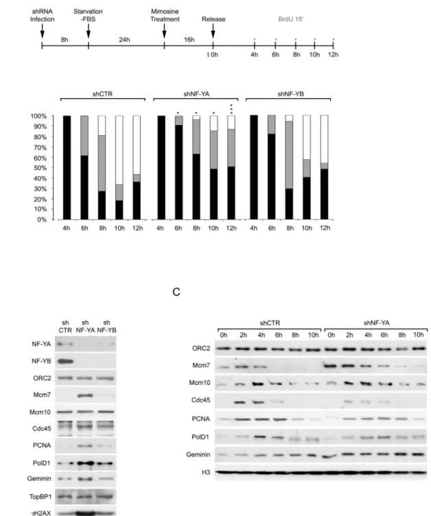
We previously showed that the depletion of NF-YA triggers an in-crease in BrdU-negative population with an S-phase DNA content, representing non cycling S cells[13]. We therefore examined the distri-bution pattern of replication foci within the nucleus, to study the effect of NF-Y depletion on the progression of the replication process. Confocal microscopy analysis of synchronized control and NF-Y-inactivated cells was performed, as illustrated inFig. 2A (upper panel). Distinct focal BrdU distributions, monitored by immunofluorescence staining, correspond to the replication of different chromatin domains during S-phase progression, as described above for PCNA. At 4 h from the re-lease, all BrdU incorporating cells showed an early S-phase pattern: the percentage of early S-phase cells was arbitrarily set at 100%, in both shCTR and shNF-Y cells (Fig. 2A, lower panels, and Supplementary Fig. 1). In control cells, early replicating population dropped to about 62% and 27% within 6 and 8 h from release, with middle S-phase cells at 38% and 54%, respectively. By 10 h,N65% of cells accumulated in late S-phase, and only 18.3% were still in early S-phase. On the contrary, NF-YA inactivation resulted in a delay in the passage from early to mid-dle S-phase: within 12 h from release, 51% of cells still showed an early S-phase pattern and only about 13% had a late S-phase foci distribution. We further investigated the effects on replication progression upon NF-YB inactivation, which does not significantly affect S-phase progression
[13]: indeed, the passage through the different S-phase stages was only marginally delayed with respect to control cells.
Western blot analysis of chromatin-enriched extracts was per-formed to investigate the effects of NF-YA or NF-YB depletion on the loading on DNA of key replication proteins, in asynchronous cells (Fig. 2B). Both NF-YA and NF-YB depletion triggered a decrease in the respective NF-Y partner, as a consequence of the failure to form the NF-Y heterotrimeric complex, which is required for the DNA binding of the transcription factor[46–48]. The chromatin binding of ORC2, one of the six subunits of the origin recognition complex, which enables the formation of the pre-replication (pre-RC) complex, was not affected by the inactivation of NF-Y subunits. A weak increase of PCNA and
DNApolD1 was detected in shNF-YB cells. Differently, the chromatin levels of the replication factors Mcm7, Cdc45, PCNA, DNApolD1 and Geminin strongly increased upon NF-YA loss (Fig. 2B). The recruitment of Mcm10 and TopBP1, which promote the loading of Cdc45 and poly-merases, didn't change either in shNF-YA or in shNF-YB cells. These data were in agreement with the existence of significant replication defects only following NF-YA loss (Fig. 2A)[13].
The chromatin levels of replication factors were then analyzed in cells synchronized at the beginning of the S phase following a time-course release from Mimosine-induced arrest (Fig. 2C). Compared to control cells, the levels of ORC2 and Mcm10 didn't change in shNF-YA cells, while Mcm7 and Geminin increased at early time points, consis-tently with previous observations. Conversely, the levels of Cdc45, PCNA and DNApolD1, which rose in asynchronous extracts, presumably as a consequence of the accumulation of cells unable to progress through and exit the S-phase, were reduced in synchronized shNF-YA with respect to shCTR cells.
A similar alteration in the loading of replication factors was observed following NF-YA inactivation in human lung adenocarcinoma epithelial A549 cells. NF-YA loss clearly impaired cell viability and increased SubG1 events and cleaved-PARP1 protein levels (Supplementary Fig. 2A–C), as previously shown in HCT116 cells[13]. A decrease in the loading of replisome proteins was even observed in asynchronous shNF-YA compared to shCTR cells (Supplementary Fig. 2D). The impairment
in chromatin recruitment of PCNA and Cdc45 was confirmed in Mimosine-released synchronous cells (Supplementary Fig. 2E). Finally, we checked whether the same defects were induced by NF-YA abrogation in a non-tumor spontaneously immortalized cell line (HaCaT). NF-YA-inactivated HaCaT cells showed increased apoptosis (Supple-mentary Fig. 3A), consistently to the expression ofγH2AX and cleaved-PARP1 (Supplementary Fig. 3C). NF-YA loss reduced BrdU-positive cell population (from 40% in shCTR to 8% in shNF-YA cells) and accumulated non-cycling S phase cells (BrdU-negative cells with an S-phase content DNA) (Supplementary Fig. 3B), as observed in HCT116 cells[13]. The levels of PCNA and Cdc45 decreased both in asynchronous and synchronous chromatin extracts (Supplementary Fig. 3D, E).
Therefore, we concluded that the loss of NF-YA affects the loading of replication proteins involved in fork progression.
3.3. Relocation of Cdc45 around origins upon NF-YA inactivation We next decided to investigate whether NF-Y could have a role in the recruitment of replication proteins at specific origins (ORIs) con-taining the NF-Y-motif. First of all, we verified by chromatin immuno-precipitation (ChIP) whether NF-Y was bound to four ORC1-associated (ORC1+) origins[28], containing CCAAT-elements, namely PCNA-P1,
PCNA-P2, cMYC and UPR/MCM4: NF-Y enrichment over negative
Fig. 1. NF-YA preferentially colocalizes with early-replication sites. Left panel: Representative confocal microscopy images of NF-YA (green) and PCNA (red) localization in early, middle and late-S HCT116 cells. The merge images show the superimposition of the green and red signals; the yellow color is indicative of colocalization of the twofluorescent probes. Right panel: The colocalization between NF-YA and PCNA, in early, middle and late-S cells, was calculated by means of PCC (Pearson Correlation Coefficient) and Mander's M1 and M2 coefficients (values shown as means ± SEM of at least 30 cells for each S-phase; statistical significance analyzed with Student's t-test, ***p b 0.0001).
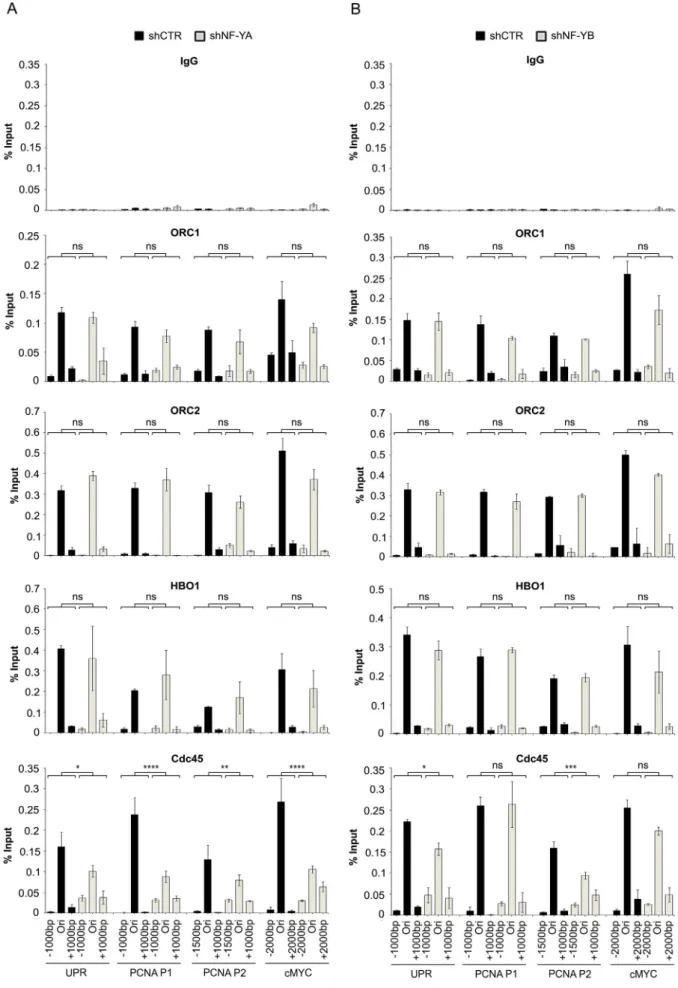
chromatin regions was observed and reduced following NF-YA inac-tivation (Supplementary Fig. 4A and B). Then, we analyzed the recruit-ment of the pre-RC proteins to ORC+/NF-Y+ regions, upon the
depletion of NF-YA (Fig. 3A) and NF-YB subunits (Fig. 3B). The binding to origins and flanking regions (−1000/+1000 bp) of ORC1 and ORC2, as well as HBO1, a HAT chromatin remodeler contributing to
pre-RC recruitment[49], was not affected by NF-Y loss. Differently, the loading of Cdc45, which converts the pre-RC into pre-IC complex and is important for fork stabilization and progression[50], was reduced at origin sites and more diffuse inflanking regions following NF-YA loss. A similar pattern was observed in shNF-YB cells, although to a lesser degree.
Fig. 2. NF-YA inactivation affects S-phase progression. (A) Upper panel: schematic representation of the experimental design for the analysis of BrdU-positive replication factories in HCT116 inactivated cells, released following Mimosine-induced G1/S arrest. Red asterisks indicate time points of BrdU pulse. Lower panel: distribution of the S phase patterns 4 h, 6 h, 8 h, 10 h and 12 h after the release from Mimosine-induced synchronization of shCTR, shNF-YA and shNF-YB cells. Black bars: Early S; gray bars: Middle S; white bars: Late S. Data are the means of three biological replicates ± SD (Suppl. Fig. 1). To compare means across shCTR and shNF-YA or shNF-YB cell distributions, Mann–Whitney test was utilized; *p b 0.05; **pb 0.01; ****p b 0.0001. (B) Expression analysis of the indicated proteins in chromatin enriched extracts of asynchronous HCT116 cells 48 h post-infection with shCTR, shNF-YA and shNF-YB. Histone H4 was used as loading control. (C) Western blot analysis of the indicated proteins in chromatin enriched extracts of shCTR and shNF-YA HCT116 cells released for the indicate time points from Mimosine-induced G1 arrest.
Fig. 3. Effects of NF-Y inactivation on chromatin binding of replication factors. qChIP analysis on chromatin from shNF-YA (A) and shNF-YB (B) HCT116 compared to shCTR cells. Antibodies used are indicated on the top of each panel and amplified regions are indicated at the bottom. The enrichment was calculated as percentage of IP recovery (% Input). p-Values were calculated for each origin as interaction between shRNA treatment and amplified region (−1000/Ori/+1000) with a two-way ANOVA; *p b 0.05; **p b 0.01; ***p b 0.001; ****p b 0.0001.
These data indicate that NF-Y binding is not necessary for recogni-tion and proper recruitment of ORC proteins, but its loss affects the load-ing of Cdc45 around origins, consistently with possible defects in the normal progression of DNA replication forks.
3.4. Characterization of DNA replication defects
To characterize the defects in DNA replication progression induced by the lack of NF-YA, we performed DNA molecular combing experi-ments in control and NF-YA-inactivated cells, and determined the den-sity and spacing of replication origins, as well as the rate and symmetry of fork progression, quantitatively and on single DNA molecules[34].
Asynchronous shCTR and shNF-YA cells were sequentially labeled by consecutive pulses of halogenated nucleotides (IdU and CIdU), DNA was purified, combed on silanized glass coverslips and nucleotides in-corporation on newly synthesized DNA was detected byfluorescence immunostaining (Fig. 4and Supplementary Fig. 5). An increase in the overall velocity of replication forks was observed in the absence of NF-YA (mean velocity 2.27 kb/min in shNF-YA versus 1.71 kb/min in shCTR cells) (Fig. 4A). We determined replication fork asymmetry, in-dicative of DNA damage-induced fork pausing, by calculating the length ratio between two divergent forks departing from the same replication origin: a significant increase in fork asymmetry was detected in shNF-YA cells, suggesting the occurrence of fork stalling (108.5% asymmetry
Fig. 4. DNA Combing analysis on shCTR and shNF-YA HCT116 cells. (A) Fork Velocity, (B) Fork Asymmetry: values were calculated as [(Long / Short tract)− 1] × 100, (C) Left panel: Global Instant Fork Density (GIFD). Right panel: Fold Increase (±SD) of the GIFD in shNF-YA versus shCTR in two biological replicates (shown inFig.4C, left panel, and Suppl. Fig. 5C), (D) Inter-Origin Distances (IODs). p-Values were calculated with two-tailed Mann–Whitney test.
in shNF-YA versus 47.89% in control cells) (Fig. 4B). When replication forks fail, previously licensed origins, that otherwise remain dormant, can be activated to allow complete genome replication (reviewed in Refs.[51,52]). To test this possibility, we analyzed the Global Instant Fork Density (GIFD), calculated as the number of forks divided by total length of DNA examined, corrected for the fraction of cells in S-phase. As shown inFig. 4C, GIFD raised from 0.82 forks/Mb in shCTR cells to 1.26 forks/Mb in shNF-YA cells: on average, an increase of 1.66-fold (± 0.17) was observed upon NF-YA depletion compared to control cells. This result can be the effect of thefiring of more origin clusters, or more origins per cluster. A reduction of IOD (intra-cluster Inter-Origin Distance) after NF-YA loss was observed (Fig. 4D), consistently with the activation of more origins per cluster, to mitigate the damaging effects of fork collapse. Nevertheless, we could not rule out that more origin clustersfired, as a consequence of defects in the replication-timing program.
All together these data corroborate that NF-YA inactivation signi fi-cantly affects DNA replication and the increase of GIFD and fork asym-metry are consistent with a role of NF-Y in forks progression.
3.5. NF-Y has a role in the DNA replication process beyond its transcriptional activity
NF-Y is a well known transcriptional regulator of cell cycle genes[13, 53,54]: the analysis of gene expression profiling of NF-Y-depleted HCT116 cells highlighted that several cell cycle genes were indeed down-regulated[6,13]. In particular, G2/M genes were found to be sig-nificantly affected following NF-YB knock down. Therefore, the defects in DNA replication following NF-Y depletion could be the consequence of transcriptional down-regulation of cell cycle genes, specifically those controlling the S phase. We analyzed the GO pathways affected by NF-YA and NF-YB inactivation with the Kobas software: specific DNA replication terms, among which DNA replication, DNA strand elon-gation, G1/S transition, replication fork, activation of the pre-replicative complex, were significantly down-regulated with p-values ≤ E-12, but only in shNF-YB cells (Fig. 5A). None of the indicated categories, except the generic DNA replication term (p-value 3.05E-02), were significantly affected by NF-YA depletion. Some of the genes belonging to DNA repli-cation pathways were validated by qRT-PCR (Fig. 5B), confirming their robust down-regulation in NF-YB-inactivated cells. Despite these tran-scriptional effects, NF-YB depletion didn't delay the G1/S transition and the progression through the S phase was only marginally affected (Fig. 2). On the other hand, NF-YA inactivation does not significantly hit the expression of genes involved in DNA replication, indicating that the defects observed in S-phase progression cannot be ascribed uniquely to its transcriptional activity.
3.6. Direct role of NF-Y in DNA replication in Xenopus cell-free system The Xenopus cell-free system is used to study the specific role of pro-teins in DNA replication, independently from interference of other DNA metabolisms, notably transcription[39]. Following fertilization, Xenopus eggs undergo 11 rounds of cell division without transcription, which takes place only after this stage[55]. Most of the proteins required for cell cycle progression are accumulated in the egg, supporting DNA rep-lication under the same cell cycle controls as occur in vivo[56]. Xenopus NF-YA and NF-YB proteins (297 aa and 207 aa, respectively) are about 90% identical to the mouse and human proteins (Supplementary Fig. 6 A)[57], and the antibodies raised against mouse/human NF-Y subunits recognize single bands corresponding to NF-YA and NF-YB proteins in Xenopus extracts (Supplementary Fig. 6B and C).
Using these antibodies, we examined whether and how NF-Y is re-cruited to chromatin during DNA synthesis in egg extract.Fig. 6A shows the timing of chromatin binding of known replication factors
[58]: Mcm3, a component of the pre-RC, is rapidly loaded onto chroma-tin to allow origin licensing, followed by the replication initiator factor
Mcm10[59], which is required for the chromatin binding of Cdc45 and DNA Polα[60]. NF-YA is recruited to chromatin at late stages of DNA replication, 30 min after the addition of sperm chromatin, building up on chromatin even when DNA replication has been completed, indi-cating that NF-Y doesn't have a role in licensing or initiation of DNA rep-lication. The association of NF-YA is prevented in egg extracts supplemented with p27Kip1, an inhibitor of the cdk2-cyclin E complex,
which allows pre-RC assembly but prevents the initiation of replication
[61], or with Geminin, which targets the pre-RC component Cdt1, thus inhibiting DNA replication[62](Fig. 6B and Supplementary Fig. 6D).
These data suggest that NF-Y binds to chromatin once replication is initiated: we therefore investigated whether NF-Y recruitment is neces-sary for efficient DNA replication, by immunodepleting NF-YA or NF-YB from Xenopus egg extracts (Supplementary Fig. 6E, F). We evaluated the kinetics of DNA replication in extracts depleted of NF-Y in comparison to control extracts depleted with non-immune antibodies (Fig. 6C), by measuring incorporation of radioactive dATP into newly synthesized DNA at different time points. The replication rate (between 120 and 180 min) of both NF-YA and NF-YB depleted extracts was reduced by about 40% compared to the control extract. However, the addition of re-combinant NF-Y to depleted extract at 120 min was not able to restore the replication efficiency (Fig. 6D), possibly because one or more repli-cation factors are co-depleted from the extract following NF-Y deple-tion. A second set of experiments was performed by adding to the egg extract a recombinant dominant negative NF-YA mutant (NF-YA-DN), which interacts with the NF-YB/YC dimer but is not able to bind to DNA[3,41]. The addition of NF-YA-DN to the extract affected DNA rep-lication, similarly to NF-Y immunodepletion, but in this case efficient replication was restored by adding back wild type (wt) recombinant NF-YA (Fig. 6E and F). The comparison of chromatin binding of replica-tion factors in control and NF-YA depleted extracts showed that the pre-RC proteins Mcm3 and Mcm4 were efficiently recruited to chromatin, while the binding of Cdc45, PCNA and DNA Polα was clearly reduced, as a consequence of NF-Y depletion. Next, we investigated whether DNA damage occurred upon the loss of NF-Y activity, as observed in mammalian cells: Western blot analysis didn't detect any increase in the expression levels ofγH2AX both in NF-Y-depleted extracts and NF-Y-DN supplemented extracts (Supplementary Fig. 6G).
Finally, to address whether NF-Y was required for fork elongation, sperm nuclei were incubated with extracts in the presence of aphidicolin, to arrest DNA replication after initiation occurred[42]. Chromatin was then isolated and used as a template in fresh extracts containing p27Kip1to prevent any further initiation event, and
elongat-ing DNA was labeled with32P ATP to monitor fork progression, either in
the presence of wt NF-YA or NF-YA-DN (Fig. 6H). The results highlight-ed that forks elongating in the presence of additional wt NF-YA progressed at the same rate as control, but in the presence of NF-YA-DN, the forks elongated less efficiently. Consistently with the observa-tions in mammalian cells, we concluded that NF-Y DNA binding is required for efficient DNA replication in Xenopus egg extract, participat-ing to the elongation process after the initiation of replication.
4. Discussion
Transcription factors (TFs) play a major role in regulating gene expression through their sequence specific binding to regulatory re-gions. In addition to transcriptional regulation, TFs influence other DNA metabolic processes, such as DNA replication. The sequence specif-ic TF NF-Y has been largely shown to regulate gene transcription in multiple organisms, from yeast to humans. Moreover, in human adeno-virus and murine gammaherpesadeno-virus, it was clearly established that the removal of CCAAT boxes affects DNA replication substantially and that NF-Y is directly implicated[63,64]. Our previous observations highlight-ed S-phase perturbation following NF-YA loss also in human cells[13], hinting at a possible function of NF-Y in DNA replication.
Here we showed that NF-YA, the CCAAT-binding subunit of the complex, is associated to early-S replication factories (Fig. 1) and its depletion delays the progression from early to middle-late S (Fig 2), suggesting that NF-Y is not involved in replication initiation, rather it may take part to the elongation step. Indeed, NF-Y is not necessary for pre-RC assembly, as ORCs are properly recruited to origins andflanking regions also when NF-Y is inactivated (Fig. 3). On the contrary, the bind-ing of Cdc45, which has a central role in the loadbind-ing of DNA Polα and interacts with the elongating DNA polymerasesδ and ε to facilitate
and increase the processive movement of the replication fork[65], is de-creased at origins and inde-creased in the surrounding regions upon NF-YA loss. These data suggest again that NF-Y depletion triggers replication forks stalling, with replication complexes not moving along the DNA and blocked as soon as replication starts, or improperly located within origins in the absence of NF-Y bookmarking.
The analysis of chromatin enriched extracts following NF-YA knock down highlights that the loading on DNA of PCNA, Cdc45 and DNA PolD1 decreases in synchronous cells, while it increases in the
Fig. 5. Transcriptional analysis of genes associated to the DNA replication process in NF-YA- and NF-YB-inactivated cells. (A) DNA replication categories and relative p-values identified with Kobas analysis among down-regulated terms in shNF-YA and shNF-YB cells. (B) Real-Time PCR validation of the indicated genes, belonging to the DNA replication categories listed above (panel A), in shCTR, shNF-YA and shNF-YB cells.
asynchronous population (Fig. 2): these results are consistent with the existence of unproductive replication forks and with the accumulation of S-phase arrested cells, which are not able to progress through and exit the S-phase.
To prove that fork progression is impaired when NF-Y activity is abrogated, we performed DNA combing analysis on single cells: surpris-ingly, the velocity of ongoing replication forks increases, but the number of origins, measured as Global Instant Fork Density, is significantly higher in shNF-YA-cells, supporting the hypothesis of increased rate level of replication fork stalling. A doubling in the percentage of fork asymmetry is also consistent with defects in fork elongation or its control. Taken together, these data suggest that NF-YA loss triggers local fork stalling and the activation of the S phase checkpoint. This presumably induces global inhibition of late-firing origins and the acti-vation of dormant origins to complete DNA replication, consistently with the observed increased GIFD. The inhibition of NF-Y activity could affect the stability of replication forks, thus leading to fork arrest but also uncontrolled progression of forks moving away at increased speed, thus resulting in faster fork velocity. Despite the local accelera-tion of fork progression and the increased number of active origins (GIFDs), this is not sufficient to compensate the overall replication length, which is significantly reduced in shNF-YA cells (Fig. 2). Changes in fork velocity of elongating forks may impact on replicativefidelity, thus triggering DNA damage and subsequent arrest of cells within S-phase, consistently with increased levels ofγH2AX and accumula-tion of non-cycling S-phase cells[13]. The role of NF-Y in the control of replication fork progression could be ascribed to the correct recruit-ment of the replication machinery, the control of the replication check-point, or the regulation of the chromatin structure. Literature data support these hypotheses: the microsequencing of NF-Y-containing fraction from CH27 cells identified a peptide that overlaps the coding sequence of RPA2 (Replication Protein A) 32 kDa, in addition to the expected peptides corresponding to NF-YA, NF-YB and NF-YC subunits
[66]. Therefore, NF-Y could interact with the p32 subunit of the heterotrimeric complex (RPA), which plays an important function in DNA replication and metabolism, such as DNA repair, recombination and DNA damage checkpoints [67]. Another possible partner interacting with NF-Y is TopBP1 (Topoisomerase IIβ Binding Protein), a protein required for initiation of DNA replication, the maintenance of replication forks in case of stalled replication, DNA repair and DNA damage signaling[68]. The existence of a complex between NF-Y and TopBP1 has been already described; in particular, TopBP1 can directly interact with the NF-YA subunit. NF-Y/TopBP1 interaction has a key role in conferring new DNA binding properties to mutant gain-of-function p53, but the complex could have additional non-transcriptional functions, such as in DNA replication, that need to be explored.
As for the last hypothesis, that is NF-Y could control DNA replication through chromatin regulation, NF-Y binding to DNA is indeed important in the establishment of some histone post-translational modifications (PTMs) associated to active promoters [69], including H3K4me3
[26–28], and might be required for replication fork progression by setting up or preserving a permissive chromatin conformation.
Since NF-Y is an important transcriptional activator of cell cycle genes, we examined the possibility that transcriptional defects triggered by the loss of NF-Y activity are the cause of DNA replication impairment. The analysis of the expression profiles from NF-Y-inactivated cells showed that NF-YB, but not NF-YA depletion, severely affects various
genes that participate to DNA replication (Fig. 5)[13]. Despite this, a se-vere effect in the progression through the S phase is observed following NF-YA, but not NF-YB inactivation. Although we cannot formally rule out that NF-YA-inactivation hits one or a handful genes required for DNA replication, these observations are not in line with a simplistic view of effects entirely secondary to the transcriptional role of NF-Y.
The experiments performed in a cell-free system derived from Xenopus eggs, in which transcription is absent[39,58], unambiguously assessed a direct role of NF-Y in DNA replication. As observed in mam-malian cells, NF-Y is necessary not for initiation, but for overall ef ficien-cy of DNA replication also in the Xenopus system (Fig. 6). The decrease of DNA synthesis by the addition of the DNA-binding NF-YA mutant indicates that DNA association is required. Moreover, the kinetics of replication observed in the DNA elongation assay with extracts added of NF-YA-DN indicates that NF-Y has a role in the elongation process, by mechanisms still unknown. It could be that NF-Y increases the efficiency of DNA replication through the interaction with other key proteins, taking into account the ineffective rescue of DNA synthesis following the administration of recombinant NF-Y in NF-Y-immunodepleted extracts.
While NF-Y depletion similarly delays DNA replication and affects the loading of Cdc45, PCNA and DNA Polymerases in human and Xenopus extracts, the activation of DNA damage is observed only in human cells. This could be the result of different degrees of replication stress induced in the two experimental systems, and therefore the acti-vation of different checkpoints. We should also consider that shNF-YA cells are analyzed 48 h post-infection, time that allows the activation of rescue measures, including a transcriptional response, not possible in the Xenopus egg system. Finally, it could be that some factors are limiting in egg extracts preparations[70], with replication stress not triggering the same biological response observed in HCT116. Thus, the discrepancy observed can be due two quantitative and qualitative differences induced by NF-Y loss in mammalian and Xenopus systems.
The data presented here put a new light on past results. NF-YA is regarded as the limiting and regulated subunit of the trimer[69]and protein levels were shown to increase at the onset of S-phase in elutri-ation experiments[9,71]: the higher levels of NF-YA in S-phase might be specifically required for optimal CCAAT-origins function, as well as the loading of the trimer on the newly synthesized DNA. In addition, while the trimer is ubiquitous in cycling cells, NF-YA, but not NF-YB, is absent in some post-mitotic cells, such as myocytes and monocytes. The down-regulation of the subunit was suggested to favor terminal differentiation by switching-off cell cycle genes[72]: a block of DNA replication might well be another key consequence of the physiological removal of NF-YA in differentiating cells.
5. Conclusions
Taken together, the data presented here suggest a new non-transcriptional mechanism through which NF-Y could regulate cell pro-liferation, corroborating the already established connection between gene transcription and DNA replication. Based on ourfindings, NF-Y may act as a factor that facilitates DNA replication participating to the elongation process, presumably through functional interactions with other cellular proteins and/or establishing or maintaining a chromatin structure that allows the progression of the replication fork. Further studies will be required to provide mechanistic insight into how NF-Y contributes to DNA replication through non-transcriptional activity.
Fig. 6. NF-YA associates to replicating chromatin and is required for proper DNA replication in Xenopus laevis. (A, B) Chromatin was isolated from a replication reaction at the indicated time-points, separated by SDS-PAGE and immunoblotted with the indicated antibodies. Histones were stained with Comassie as a loading control. When indicated, the replication reaction was supplemented with the CDK inhibitor p27kip1
or the licensing inhibitor Geminin. (C) DNA synthesis was monitored in a time-course experiment based on the incorporation ofα32P-dATP, in mock (ΔIgG) or NF-Y depleted (ΔNF-YA and ΔNF-YB) extracts. (D) Quantification of global DNA replication after addition of recombinant NF-Y protein complex toΔIgG or ΔNF-YA extracts. (E) DNA synthesis was monitored in a time-course experiment based on the incorporation of α32P-dATP in extracts with or without addition of a Dominant Negative-NF-YA mutant (NF-YA-DN). (F) Quantification of global DNA replication (±SEM) after addition of recombinant NF-YA protein to control extracts or extracts containing NF-YA-DN. (G) Time-course of replication factors association to chromatin inΔIgG and ΔNF-YA extracts. (H) Fork elongation analysis in extracts supplemented with recombinant wt NF-YA or NF-YA DN mutant, compared to control extract.
Supplementary data to this article can be found online athttp://dx. doi.org/10.1016/j.bbamcr.2015.12.019.
Transparency document
TheTransparency documentassociated with this article can be found, in online version.
Acknowledgments
We warmly thank G. S. Chadha for his technical assistance. This work was supported by the Associazione Italiana per la Ricerca sul Cancro to CI (IG No. 14210) and RM (IG No. 14130). BM is supported by Marie Curie IRG fellowship (PIRG07-GA-2010-268448). JJB acknowledges support from Cancer Research UK (grant C303/A14301).
PB is the recipient of a fellowship from the Fondazione Italiana per la Ricerca sul Cancro (FIRC).
References
[1] R. Mantovani, The molecular biology of the CCAAT-binding factor NF-Y, Gene 239 (1999) 15–27.
[2] D. Dolfini, F. Zambelli, G. Pavesi, R. Mantovani, A perspective of promoter architec-ture from the CCAAT box, Cell Cycle 8 (2009) 4127–4137.
[3] M. Nardini, N. Gnesutta, G. Donati, R. Gatta, C. Forni, A. Fossati, C. Vonrhein, D. Moras, C. Romier, M. Bolognesi, R. Mantovani, Sequence-specific transcription factor NF-Y displays histone-like DNA binding and H2B-like ubiquitination, Cell 152 (2013) 132–143.
[4] J.D. Fleming, G. Pavesi, P. Benatti, C. Imbriano, R. Mantovani, K. Struhl, NF-Y coassociates with FOS at promoters, enhancers, repetitive elements, and inactive chromatin regions, and is stereo-positioned with growth-controlling transcription factors, Genome Res. 23 (2013) 1195–1209.
[5] Q. Hu, J.F. Lu, R. Luo, S. Sen, S.N. Maity, Inhibition of CBF/NF-Y mediated transcription activation arrests cells at G2/M phase and suppresses expression of genes activated at G2/M phase of the cell cycle, Nucleic Acids Res. 34 (2006) 6272–6285.
[6] P. Benatti, V. Basile, D. Merico, L.I. Fantoni, E. Tagliafico, C. Imbriano, A balance be-tween NF-Y and p53 governs the pro- and anti-apoptotic transcriptional response, Nucleic Acids Res. 36 (2008) 1415–1428.
[7] C.Y. Kao, A. Tanimoto, N. Arima, Y. Sasaguri, R. Padmanabhan, Transactivation of the human cdc2 promoter by adenovirus E1A. E1A induces the expression and assembly of a heteromeric complex consisting of the CCAAT box binding factor, CBF/NF-Y, and a 110-kDa DNA-binding protein, J. Biol. Chem. 274 (1999) 23043–23051.
[8] I. Manni, G. Mazzaro, A. Gurtner, R. Mantovani, U. Haugwitz, K. Krause, K. Engeland, A. Sacchi, S. Soddu, G. Piaggio, NF-Y mediates the transcriptional inhibition of the cyclin B1, cyclin B2, and cdc25C promoters upon induced G2 arrest, J. Biol. Chem. 276 (2001) 5570–5576.
[9] F. Bolognese, M. Wasner, C.L. Dohna, A. Gurtner, A. Ronchi, H. Muller, I. Manni, J. Mossner, G. Piaggio, R. Mantovani, K. Engeland, The cyclin B2 promoter depends on NF-Y, a trimer whose CCAAT-binding activity is cell-cycle regulated, Oncogene 18 (1999) 1845–1853.
[10] R. Gatta, D. Dolfini, R. Mantovani, NF-Y joins E2Fs, p53 and other stress transcription factors at the apoptosis table, Cell Death Dis. 2 (2011), e162.
[11] R. Hughes, M. Kristiansen, I. Lassot, S. Desagher, R. Mantovani, J. Ham, NF-Y is essen-tial for expression of the proapoptotic bim gene in sympathetic neurons, Cell Death Differ. 18 (2010) 937–947.
[12] C. Imbriano, N. Gnesutta, R. Mantovani, The NF-Y/p53 liaison: well beyond repres-sion, Biochim. Biophys. Acta 1825 (2012) 131–139.
[13] P. Benatti, D. Dolfini, A. Vigano, M. Ravo, A. Weisz, C. Imbriano, Specific inhibition of NF-Y subunits triggers different cell proliferation defects, Nucleic Acids Res. 39 (2011) 5356–5368.
[14] A.J. Oldfield, P. Yang, A.E. Conway, S. Cinghu, J.M. Freudenberg, S. Yellaboina, R. Jothi, Histone-fold domain protein NF-Y promotes chromatin accessibility for cell type-specific master transcription factors, Mol. Cell 55 (2014) 708–722.
[15]A. Bhattacharya, J.M. Deng, Z. Zhang, R. Behringer, B. de Crombrugghe, S.N. Maity, The B subunit of the CCAAT box binding transcription factor complex (CBF/NF-Y) is essential for early mouse development and cell proliferation, Cancer Res. 63 (2003) 8167–8172.
[16] C. Maric, M.N. Prioleau, Interplay between DNA replication and gene expression: a harmonious coexistence, Curr. Opin. Cell Biol. 22 (2010) 277–283.
[17] B.D. Pope, I. Hiratani, D.M. Gilbert, Domain-wide regulation of DNA replication timing during mammalian development, Chromosom. Res. 18 (2010) 127–136.
[18] F. Del Bene, K. Tessmar-Raible, J. Wittbrodt, Direct interaction of geminin and Six3 in eye development, Nature 427 (2004) 745–749.
[19] L. Luo, X. Yang, Y. Takihara, H. Knoetgen, M. Kessel, The cell-cycle regulator geminin inhibits Hox function through direct and polycomb-mediated interactions, Nature 427 (2004) 749–753.
[20] D.T. Pak, M. Pflumm, I. Chesnokov, D.W. Huang, R. Kellum, J. Marr, P. Romanowski, M.R. Botchan, Association of the origin recognition complex with heterochromatin and HP1 in higher eukaryotes, Cell 91 (1997) 311–323.
[21] T. Triolo, R. Sternglanz, Role of interactions between the origin recognition complex and SIR1 in transcriptional silencing, Nature 381 (1996) 251–253.
[22] K. Yankulov, I. Todorov, P. Romanowski, D. Licatalosi, K. Cilli, S. McCracken, R. Laskey, D.L. Bentley, MCM proteins are associated with RNA polymerase II holoen-zyme, Mol. Cell. Biol. 19 (1999) 6154–6163.
[23] D. Dominguez-Sola, C.Y. Ying, C. Grandori, L. Ruggiero, B. Chen, M. Li, D.A. Galloway, W. Gu, J. Gautier, R. Dalla-Favera, Non-transcriptional control of DNA replication by c-Myc, Nature 448 (2007) 445–451.
[24] B. Miotto, Y. Graba, Control of DNA replication: a new facet of Hox proteins? BioEssays 32 (2010) 800–807.
[25]P. Rimmele, J. Komatsu, P. Hupe, C. Roulin, E. Barillot, M. Dutreix, E. Conseiller, A. Bensimon, F. Moreau-Gachelin, C. Guillouf, Spi-1/PU.1 oncogene accelerates DNA replication fork elongation and promotes genetic instability in the absence of DNA breakage, Cancer Res. 70 (2010) 6757–6766.
[26]C.M. Koch, R.M. Andrews, P. Flicek, S.C. Dillon, U. Karaoz, G.K. Clelland, S. Wilcox, D.M. Beare, J.C. Fowler, P. Couttet, K.D. James, G.C. Lefebvre, A.W. Bruce, O.M. Dovey, P.D. Ellis, P. Dhami, C.F. Langford, Z. Weng, E. Birney, N.P. Carter, D. Vetrie, I. Dunham, The landscape of histone modifications across 1% of the human genome infive human cell lines, Genome Res. 17 (2007) 691–707.
[27] H. Xi, H.P. Shulha, J.M. Lin, T.R. Vales, Y. Fu, D.M. Bodine, R.D. McKay, J.G. Chenoweth, P.J. Tesar, T.S. Furey, B. Ren, Z. Weng, G.E. Crawford, Identification and characteriza-tion of cell type-specific and ubiquitous chromatin regulatory structures in the human genome, PLoS Genet. 3 (2007), e136.
[28] G.I. Dellino, D. Cittaro, R. Piccioni, L. Luzi, S. Banfi, S. Segalla, M. Cesaroni, R. Mendoza-Maldonado, M. Giacca, P.G. Pelicci, Genome-wide mapping of human DNA-replication origins: levels of transcription at ORC1 sites regulate origin selec-tion and replicaselec-tion timing, Genome Res. 23 (2012) 1–11.
[29]I. Hiratani, T. Ryba, M. Itoh, T. Yokochi, M. Schwaiger, C.W. Chang, Y. Lyou, T.M. Townes, D. Schubeler, D.M. Gilbert, Global reorganization of replication domains during embryonic stem cell differentiation, PLoS Biol. 6 (2008), e245.
[30]D.M. Gilbert, Replication timing and transcriptional control: beyond cause and ef-fect, Curr. Opin. Cell Biol. 14 (2002) 377–383.
[31] I. Hiratani, S. Takebayashi, J. Lu, D.M. Gilbert, Replication timing and transcrip-tional control: beyond cause and effect–part II, Curr. Opin. Genet. Dev. 19 (2009) 142–149.
[32] M. Mechali, Eukaryotic DNA replication origins: many choices for appropriate answers, Nat. Rev. Mol. Cell Biol. 11 (2010) 728–738.
[33] J. Mendez, B. Stillman, Chromatin association of human origin recognition complex, cdc6, and minichromosome maintenance proteins during the cell cycle: assembly of prereplication complexes in late mitosis, Mol. Cell. Biol. 20 (2000) 8602–8612.
[34] E. Schwob, C. de Renty, V. Coulon, T. Gostan, C. Boyer, L. Camet-Gabut, C. Amato, Use of DNA combing for studying DNA replication in vivo in yeast and mammalian cells, Methods Mol. Biol. 521 (2009) 673–687.
[35] J. Yang, L. O'Donnell, D. Durocher, G.W. Brown, RMI1 promotes DNA replication fork progression and recovery from replication fork stress, Mol. Cell. Biol. 32 (2012) 3054–3064.
[36]V. Basile, R. Mantovani, C. Imbriano, DNA damage promotes histone deacetylase 4 nuclear localization and repression of G2/M promoters, via p53 C-terminal lysines, J. Biol. Chem. 281 (2006) 2347–2357.
[37] S. Belluti, V. Basile, P. Benatti, E. Ferrari, G. Marverti, C. Imbriano, Concurrent inhibi-tion of enzymatic activity and NF-Y-mediated transcripinhibi-tion of topoisomerase-IIalpha by bis-DemethoxyCurcumin in cancer cells, Cell Death Dis. 4 (2013), e756.
[38] B. Miotto, K. Struhl, HBO1 histone acetylase is a coactivator of the replication licens-ing factor Cdt1, Genes Dev. 22 (2008) 2633–2638.
[39] P.J. Gillespie, A. Gambus, J.J. Blow, Preparation and use of Xenopus egg extracts to study DNA replication and chromatin associated proteins, Methods 57 (2012) 203–213.
[40]C. Imbriano, A. Gurtner, F. Cocchiarella, S. Di Agostino, V. Basile, M. Gostissa, M. Dobbelstein, G. Del Sal, G. Piaggio, R. Mantovani, Direct p53 transcriptional repres-sion: in vivo analysis of CCAAT-containing G2/M promoters, Mol. Cell. Biol. 25 (2005) 3737–3751.
[41]R. Mantovani, X.Y. Li, U. Pessara, R. Hooft van Huisjduijnen, C. Benoist, D. Mathis, Dominant negative analogs of NF-YA, J. Biol. Chem. 269 (1994) 20340–20346.
[42]W.T. Poh, G.S. Chadha, P.J. Gillespie, P. Kaldis, J.J. Blow, Xenopus Cdc7 executes its essential function early in S phase and is counteracted by checkpoint-regulated pro-tein phosphatase 1, Open Biol. 4 (2014) 130138.
[43]N. Sadoni, S. Langer, C. Fauth, G. Bernardi, T. Cremer, B.M. Turner, D. Zink, Nuclear organization of mammalian genomes. Polar chromosome territories build up func-tionally distinct higher order compartments, J. Cell Biol. 146 (1999) 1211–1226.
[44] H. Nakayasu, R. Berezney, Mapping replicational sites in the eucaryotic cell nucleus, J. Cell Biol. 108 (1989) 1–11.
[45] K.W. Dunn, M.M. Kamocka, J.H. McDonald, A practical guide to evaluating colocalization in biological microscopy, Am. J. Physiol. Cell Physiol. 300 (2011) C723–C742.
[46] F. Coustry, S.N. Maity, B. de Crombrugghe, Studies on transcription activation by the multimeric CCAAT-binding factor CBF, J. Biol. Chem. 270 (1995) 468–475.
[47] F. Coustry, S.N. Maity, S. Sinha, B. de Crombrugghe, The transcriptional activity of the CCAAT-binding factor CBF is mediated by two distinct activation domains, one in the CBF-B subunit and the other in the CBF-C subunit, J. Biol. Chem. 271 (1996) 14485–14491.
[48] S. Sinha, S.N. Maity, J. Lu, B. de Crombrugghe, Recombinant rat CBF-C, the third sub-unit of CBF/NFY, allows formation of a protein-DNA complex with CBF-a and CBF-B and with yeast HAP2 and HAP3, Proc. Natl. Acad. Sci. U. S. A. 92 (1995) 1624–1628.
[49] B. Miotto, K. Struhl, HBO1 histone acetylase activity is essential for DNA replication licensing and inhibited by geminin, Mol. Cell 37 (2010) 57–66.
[50] A. Gambus, R.C. Jones, A. Sanchez-Diaz, M. Kanemaki, F. van Deursen, R.D. Edmondson, K. Labib, GINS maintains association of Cdc45 with MCM in replisome
progression complexes at eukaryotic DNA replication forks, Nat. Cell Biol. 8 (2006) 358–366.
[51] J.J. Blow, X.Q. Ge, D.A. Jackson, How dormant origins promote complete genome replication, Trends Biochem. Sci. 36 (2011) 405–414.
[52] R.C. Alver, G.S. Chadha, J.J. Blow, The contribution of dormant origins to genome stability: from cell biology to human genetics, DNA Repair (Amst) 19 (2014) 182–189.
[53] R. Elkon, C. Linhart, R. Sharan, R. Shamir, Y. Shiloh, Genome-wide in silico identifica-tion of transcripidentifica-tional regulators controlling the cell cycle in human cells, Genome Res. 13 (2003) 773–780.
[54] H. Goodarzi, O. Elemento, S. Tavazoie, Revealing global regulatory perturbations across human cancers, Mol. Cell 36 (2009) 900–911.
[55] J. Newport, M. Kirschner, A major developmental transition in early Xenopus embry-os: II. Control of the onset of transcription, Cell 30 (1982) 687–696.
[56]J.J. Blow, R.A. Laskey, Initiation of DNA replication in nuclei and purified DNA by a cell-free extract of Xenopus eggs, Cell 47 (1986) 577–587.
[57] Q. Li, M. Herrler, N. Landsberger, N. Kaludov, V.V. Ogryzko, Y. Nakatani, A.P. Wolffe, Xenopus NF-Y pre-sets chromatin to potentiate p300 and acetylation-responsive transcription from the Xenopus hsp70 promoter in vivo, EMBO J. 17 (1998) 6300–6315.
[58] J.J. Blow, Control of chromosomal DNA replication in the early Xenopus embryo, EMBO J. 20 (2001) 3293–3297.
[59] J.A. Wohlschlegel, S.K. Dhar, T.A. Prokhorova, A. Dutta, J.C. Walter, Xenopus Mcm10 binds to origins of DNA replication after Mcm2-7 and stimulates origin binding of Cdc45, Mol. Cell 9 (2002) 233–240.
[60]Y. Kubota, Y. Takase, Y. Komori, Y. Hashimoto, T. Arata, Y. Kamimura, H. Araki, H. Takisawa, A novel ring-like complex of Xenopus proteins essential for the initiation of DNA replication, Genes Dev. 17 (2003) 1141–1152.
[61] H. Toyoshima, T. Hunter, p27, a novel inhibitor of G1 cyclin-Cdk protein kinase activity, is related to p21, Cell 78 (1994) 67–74.
[62] S. Tada, A. Li, D. Maiorano, M. Mechali, J.J. Blow, Repression of origin assembly in meta-phase depends on inhibition of RLF-B/Cdt1 by geminin, Nat. Cell Biol. 3 (2001) 107–113.
[63] D. Gong, J. Qi, V. Arumugaswami, R. Sun, H. Deng, Identification and functional characterization of the left origin of lytic replication of murine gammaherpesvirus 68, Virology 387 (2009) 285–295.
[64] B. Song, C.S. Young, Functional analysis of the CAAT box in the major late promoter of the subgroup C human adenoviruses, J. Virol. 72 (1998) 3213–3220.
[65] C. Bauerschmidt, S. Pollok, E. Kremmer, H.P. Nasheuer, F. Grosse, Interactions of human Cdc45 with the Mcm2-7 complex, the GINS complex, and DNA polymerases delta and epsilon during S phase, Genes to cells: devoted to molecular and cellular mechanisms 12 (2007) 745–758.
[66]R. Hooft van Huijsduijnen, X.Y. Li, D. Black, H. Matthes, C. Benoist, D. Mathis, Co-evolution from yeast to mouse: cDNA cloning of the two NF-Y (CP-1/CBF) subunits, EMBO J. 9 (1990) 3119–3127.
[67]Y. Zou, Y. Liu, X. Wu, S.M. Shell, Functions of human replication Protein A (RPA): from DNA replication to DNA damage and stress responses, J. Cell. Physiol. 208 (2006) 267–273.
[68] V. Garcia, K. Furuya, A.M. Carr, Identification and functional analysis of TopBP1 and its homologs, DNA Repair (Amst) 4 (2005) 1227–1239.
[69] D. Dolfini, R. Gatta, R. Mantovani, NF-Y and the transcriptional activation of CCAAT promoters, Crit. Rev. Biochem. Mol. Biol. 47 (2012) 29–49.
[70] D.M. Gilbert, Replication origin plasticity, Taylor-made: inhibition vs recruitment of origins under conditions of replication stress, Chromosoma 116 (2007) 341–347.
[71] C. Borestrom, H. Zetterberg, K. Liff, L. Rymo, Functional interaction of nuclear factor y and sp1 is required for activation of the epstein-barr virus C promoter, J. Virol. 77 (2003) 821–829.
[72] A. Gurtner, I. Manni, P. Fuschi, R. Mantovani, F. Guadagni, A. Sacchi, G. Piaggio, Requirement for down-regulation of the CCAAT-binding activity of the NF-Y tran-scription factor during skeletal muscle differentiation, Mol. Biol. Cell 14 (2003) 2706–2715.
![Fig. 4. DNA Combing analysis on shCTR and shNF-YA HCT116 cells. (A) Fork Velocity, (B) Fork Asymmetry: values were calculated as [(Long / Short tract) − 1] × 100, (C) Left panel: Global Instant Fork Density (GIFD)](https://thumb-eu.123doks.com/thumbv2/123doknet/14697356.563623/8.892.141.430.284.1063/combing-analysis-velocity-asymmetry-calculated-global-instant-density.webp)