PREDICTIVE VALUE OF FRC AND RESPIRATORY
COMPLIANCE ON PULMONARY GAS EXCHANGE
INDUCED BY HIGH FREQUENCY JET VENTILATION IN
HUMANS
J. F. PITTET, D. R. MOREL, M. BACHMANN, A. FORSTER
AND P. M. SUTER
SUMMARY
To determine if functional residual capacity (FRC), compliance of the respiratory system (C), or underlying pulmonary disease are predictive for the efficacy of high frequency jet ventilation (HFJV) on pulmonary gas exchange, we investigated six adult patients within 4 h of abdominal surgery and six patients with severe adult respiratory distress syndrome. Gas exchange during intermittent positive pressure ventilation (IPPV) was compared with that during HFJV at frequencies of 100 b.p.m. (HFJV100) and 200 b.p.m. (HFJV200), resulting in a minute ventilation of about 400 ml kg'1 with both ventilatory frequencies, and in both groups of patients. Baseline FRC and C were measured during IPPV with the multiple-breath nitrogen washout method and from expiratory pressure-volume curves, respectively. Changes in the alveolar-arterial oxygen difference (?A02—Pa0J: F/Oj ratio induced by HFJV correlated negatively with C (HFJV,„,: i = -0.78, P <0.005; HFJV200'. r = -0.84, P < 0.005); that is, greater oxygenation was obtained in patients with a better compliance. Similarly, changes in arterial partial pressure of carbon dioxide (PaC0J induced by HFJV correlated negatively with C (HFJV100: x = -0.77, ?< 0.001; HFJV^: r = —0.61, P < 0.05). In contrast, there was no significant correlation between FRC measured during IPPV and changes in (PA02—?a0J:?l0l ratio or PaCOl induced by HFJV, as these changes were influenced more by the patient's pulmonary disease than by baseline FRC. These results should be interpreted in the context of different
underlying pathophysiological mechanisms re-ducing FRC in both groups of patients.
KEY WORDS
Lung: functional residual capacity, compliance, gas exchange. Ventilation: high frequency jet ventilation.
Continuous positive pressure ventilation (CPPV) or high frequency jet ventilation (HFJV) improves oxygenation in patients with acute respiratory failure and low pulmonary compliance [1]. Simi-larly, HFJV has been applied successfully in the postoperative period as an alternative to CPPV to increase functional residual capacity (FRC) and to improve pulmonary gas exchange [2, 3]. However, the relation between the effects of HFJV on gas exchange and pre-existing respiratory mechanics has not been investigated. The aim of the present study was to compare the effects of HFJV on gas exchange in two groups of patients with differing pulmonary impairment, and thus to investigate if FRC and expiratory static compliance (C) measured during conventional ventilation had any predictive value for the efficacy of HFJV.
PATIENTS AND METHODS
Patients
Two groups of patients were studied (table I). The first group comprised six selected patients
J. F. PITTET, M.D.; D . R. MOREL, M.D.; M. BACHMANN, M.D.;
A. FORSTER, M.D.; P. M. SUTER, M.D.; Division of Surgical Intensive Care, Department of Anesthesiology, University Hospital of Geneva, 1211 Geneva 4, Switzerland. Accepted for Publication: September 4, 1989.
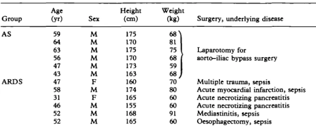
TABLE I. Patient characteristics Group AS ARDS Age (yr) 59 64 63 56 47 43 47 58 31 46 52 52 Sex M M M M M M F M F M M M Height (cm) 175 170 175 170 173 163 160 174 165 155 168 165 Weight (kg) 6 8 ^ 81 75 68 59 68 70 80 60 60 91 60
Surgery, underlying disease
Laparotomy for
aorto-Uiac bypass surgery Multiple trauma, sepsis
Acute rayocardial infarction, sepsis Acute necrotizing pancreatitis Acute necrotizing pancreatitis Mcdiastinitis, sepsis
Oesophagectomy, sepsis
(age 55 (SD 9) yr, weight 70 (9) kg) who had undergone laparotomy and aorto-iliac bypass surgery (AS). The patients needed short term postoperative ventilatory assistance and were studied within 4 h of the end of surgery. Only patients with lung function in the normal range before operation were included (vital capacity 110 (10.7)% of predicted values in the upright position, FEVt 84 (5) %). The second group of six
patients (age 47 (5.2) yr, weight 69 (12) kg) were treated in our intensive care unit for severe adult respiratory distress syndrome (ARDS) with a lung injury score of more than 2.5 as defined by Murray [4]. The study was accepted by the committee for ethics in human research of our institution. AS patients gave informed consent to the study before surgery. All patients with ARDS received sedatives and opioids and therefore informed consent was obtained from the patient's closest relative or the referring physician. Equipment
HFJV was delivered by an MK 800 respirator (Acutronic Medical Systems AG, Jona, Switzerland). Air and oxygen were supplied at a pressure of 4 bar, mixed with a blender and pulsed by an electronically-controlled solenoid valve through non-compliant tubing (120 cm long, 7 mm i.d.). This tubing was connected to a tracheal tube (Hi-Lo-Jet, NCC, Division Mallincrodt, Inc., Argyle, NY) with three sep-arate lumens: the main lumen was used for IPPV, the first auxiliary lumen (1 mm i.d.) for tracheal pressure monitoring [1], and the second auxiliary lumen (2 mm i.d.) as the "jet insufflation lumen" during HFJV. A bias flow of gas for entrainment was provided by a wide-bore tube connected to
a respirator (CV 2000 Adult Veriflo) delivering 30 litre min"1 of heated and humidified gas at the
same inspied oxygen fraction (FiOj) as for jet
ventilation. Expiration was through an open T-piece with or without a positive end-expiratory pressure (PEEP) valve. The driving pressure of the ventilator, inspiratory: expiratory time ratio, and ventilatory frequency were adjusted inde-pendently and kept constant for each study period. Intermittent positive pressure ventilation (IPPV) was administered using a volumetric respirator (CV 2000 Adult Veriflo) connected to the main lumen of the Hi-Lo-Jet tracheal tube. In ARDS patients, 10 cm H2O PEEP was applied
throughout the study using an Emerson valve and was maintained during the change of ventilator modes.
Respiratory and haemodynamic measurements Airway pressure (Paw) was measured with a pressure transducer (Hewlett-Packard (HP) PM 5E + PM 15E) and recorded on a chart recorder (HP 7754 B) via a pressure amplifier (HP 880 5D). Mean Paw was calculated electronically. Ventilatory minute volume (VE) and flow were measured using a pneumotachograph (Godart type 17212) with a Fleisch No. 2 head placed in the expiratory limb of the bias flow tubing. Total
C was measured from a multiple-step expiratory
pressure-volume curve. The curve was recorded on an X-Y recorder (HP 7041 M) following three pulmonary insufflations to obtain a peak Paw of 50-60 cm H2O. At each 100-ml decrement
vol-ume, Paw was allowed to equilibrate for 3 s. C was calculated by dividing the difference in volume corresponding to FRC and FRC+12 ml /kg body weight by the corresponding difference
in Paw on the expiratory limb of the pressure-volume loop.
FRC was measured using a computerized multiple-breath nitrogen washout method [5]. Predicted normal FRC values were taken from the tables of Bates [6] for patients in the upright position. As the patients were in the supine position, 80 % of this predicted FRC value was considered normal [6]. As the nitrogen washout method is not appropriate for measuring FRC during HFJV (the high flow delivered by the jet ventilator interferes with the precise measurement of nitrogen concentration), changes in respiratory volume relative to FRC determined with the nitrogen washout method were measured during both types of mechanical ventilation using two bellows pneumographs (Model 108, Pneumo-graph HP), detecting the change in rib cage and abdominal circumferences, respectively. One pneumograph was fixed around the rib cage at the nipple level, the other around the abdomen at the umbilical level. The bellows were connected to differential pressure transducers (HP PM 131 TC) by a semi-rigid polyethylene catheter (1.3 mm i.d.). The two signals and their sum were recorded on a chart recorder (HP 7754 B) as described previously [7,8]. Calibration was performed during IPPV by adjustment of the pressure signals to achieve a sum signal (rib cage + abdomen) corresponding to the signal of the pneumotachograph as described by Rouby and colleagues [9]. This method cannot give absolute values of FRC, but only relative FRC changes relative to apnoeic FRC [9]. The fol-lowing variables were derived from the calibrated pressure signals of the two pneumographs using an on-line data acquisition program of an Apple II microcomputer [7]: ventilatory frequency (/), effective (i.e. injected + entrained gas) tidal vol-ume ( VT = sum of the contributions of the rib cage and the abdomen), VE ( K T X / ) , rib cage contribution to tidal volume (FT,rc), relative changes in end-expiratory position of the sum of rib cage and abdomen excursions, assumed to correspond to changes in FRC.
Mean systemic arterial pressure (SAP) was measured through a radial arterial cannula con-nected to a transducer (1290 A HP) positioned at the midaxillary line. Cardiac output was measured in duplicate by thermodilution using a 7-French gauge triple lumen Swan-Ganz catheter and Edwards computer (type 9520 A).
Systemic arterial and mixed venous blood
samples were drawn for measurement of ^
P^o,> ^ c o , and pH with standard electrodes
(ABL 330, Radiometer, Copenhagen). Oxygen saturation of haemoglobin was measured with a Co-oximeter (model 182, Instrumentation Laboratories). Alveolar-arterial oxygen partial pressure difference (PAOJ —Pa^) and
intra-pulmonary venous admixture were calculated with standard formulae.
Procedure
Before the beginning of the study, the patients received morphine 0.1 mg kg"1 and pancuronium
0.1 mgkg"1 i.v. Data were collected during 30
min of HFJV at 100 and 200 b.p.m. applied randomly, separated by 30 min of IPPV. Patients were kept supine during the whole investigation. Fio was maintained constant during the study
period (0.4 for AS patients, and 0.5 for ARDS). The effective VT delivered to the lungs during HFJV was measured using bellows pneumo-graphy and set at 4 ml/kg body weight by adjusting the driving pressure at both ventilatory frequencies for each subject group. In a pre-liminary study, we found that similar ARDS patients required this effective VT in order to maintain a stable Paco < 6.7 kPa and arterial pH
> 7.3.
The following variables were recorded con-tinuously : mean and peak Paw, effective VT and
VE, VT,TC and pulmonary volume relative to FRC recorded with the bellows pneumograph system. In addition, SAP and heart rate were monitored continuously. Cardiac output was determined before and at the end of each HFJV period, when arterial and mixed venous blood samples were also collected. FRC and C were determined before and immediately after both periods of HFJV. The reliability of ventilatory volume change determinations with bellows pneumographs was confirmed in six patients (four AS, two ARDS) 60 min after the second period of HFJV, by comparing FRC measured by the nitrogen washout method alone with FRC determined by adding the changes in pulmonary volume recorded by the pneumographs to initial values of FRC (table II). Differences between methods did not exceed 80 ml.
Statistical analysis
The data (mean (SD)) of each group of patients were compared during the five periods of measurements with a one-way analysis of variance
for repeated measurements, followed by a Bonferroni test if the F ratio resulted in a P value < 0.05. For each period of measurement, both groups of patients were compared using a two-tailed unpaired Student's t test. P < 0.05 was considered significant.
RESULTS
Respiratory variables measured during IPPV differed markedly between the two groups of patients (table III, fig. 1). In patients with ARDS, FRC was significantly less than in AS patients (921 (409) ml vs 2106 (539) ml; P < 0.001). Mean and peak Paw measured during IPPV were significantly (P < 0.001) greater in ARDS than in AS patients, associated with a reduced expiratory TABLE II. Individual FRC values in six patients at the end of the study, measured with the nitrogen washout technique compared with FRC derived by adding relative changes in pulmonary volume estimated with the pneumograph system to initial measurement FRC, and difference between the two
methods
Measured Derived Difference in Group FRC (ml) FRC (ml) FRC (ml) AS AS AS AS ARDS ARDS 2320 2647 2713 1557 500 1088 2400 2647 2763 1518 560 1101 + 80 0 + 50 - 3 9 + 60 + 13
C in ARDS patients (49 (3) ml/cm H2O),
compared with AS patients (108 (25) ml/cm H2O;
P < 0.001). In order to achieve Pac0, in the same
range in both groups of patients and a clinically acceptable peak Paw < 45 cm H2O,/of IPPV was
adjusted to 8-12 b.p.m. in AS patients, and 12-22 b.p.m. in ARDS patients. Similarly, VE in ARDS patients (199 (29) ml kg"1 min"1) was almost twice
that in AS patients (115 (22) mlkg-'min"1;
P < 0.001).
Application of HFJV with the same effective VE of 400 ml kg"1 to AS and ARDS patients at both
ventilation frequencies induced consistently different responses in gas exchange in the two groups of patients.
In AS patients HFJV produced an immediate and significant two-fold increase in mean Paw (4 (1) to 9 (3) cm H2O at both frequencies;
P < 0.001) without change in peak Paw. This was associated with a 25 % increase in FRC (P < 0.001), a decrease in P a ^ , (P < 0.01) and in FT,rc (P < 0.002). These changes in respirat-ory variables were observed during both HFJV periods. PaOj, (PAOt — PaOl): -Flo, ratio, and
intra-pulmonary venous admixture were unchanged by HFJV (table III, fig. 1). HFJV100 and HFJV200
did not induce any significant differences in any measured variables other than F T , including haemodynamic data (table IV). During the second (IPPV2) and the third (IPPV3) periods of IPPV,
respiratory variables recovered to initial baseline values.
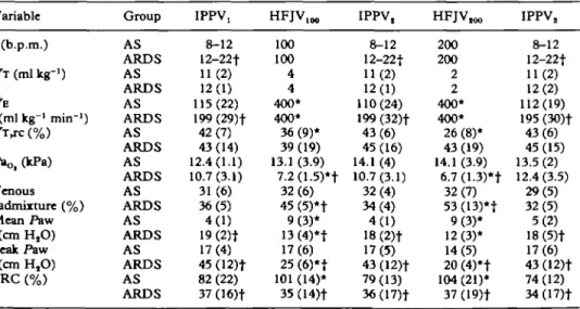
TABLE III. Respiratory variables (mean (SD) values of six patients in each group). *P < 0.05 between IPPVX and HFJV1M or IPPV, and HFJVm; TP < 0.05 between AS and ARDS groups Variable /(b.p.m.) KT (ml kg"1) VE (ml kg"1 min"1) KT,TC(%) P»o,(kPa) Venous admixture (%) Mean Paw (cm H,O) Peak Paw (cm H,O) FRC (%) Group AS ARDS AS ARDS AS ARDS AS ARDS AS ARDS AS ARDS AS ARDS AS ARDS AS ARDS IPPV! 8-12 12-22f 11(2) 12(1) 115(22) 199 (29)f 42(7) 43(14) 12.4(1.1) 10.7 (3.1) 31(6) 36(5) 4(1) 19 (2)t 17(4) 45 (12)f 82 (22) 37 (16)t HFJV1 0 0 100 100 4 4 400* 400* 36(9)* 39 (19) 13.1 (3.9) 7.2(1.5)*t 32(6) 45 (5)*t 9(3)* 13(4)*t 17(6) 25 (6)*t 101 (14)* 35 (14)t IPPV, 8-12 12-22t 11(2) 12(1) 110(24) 199 (32)f 43(6) 45 (16) 14.1 (4) 10.7(3.1) 32(4) 34(4) 4(1) 18 (2)t 17(5) 43 (12)f 79 (13) 36(17)t HFJV™ 200 200 2 2 400* 400* 26 (8)* 43(19) 14.1 (3.9) 6.7 (1.3)*t 32(7) 53(13)*t 9(3)* 12(3)* 14(5) 20(4)*f 104 (21)* 37 (19)t IPPV, 8-12 12-22t 11(2) 12(2) 112(19) 195 (30)t 43(6) 45 (15) 13.5 (2) 12.4(3.5) 29(5) 32(5) 5(2) 18 (5)f 17(6) 43 (12)t 74 (12) 34(17)t
70 ^ 60 « a.
S
*>
40 8AS patients ARDS patients
I I
I
II
3U
0-1 175 „ 1 S 0§|
1 2 5 I E 100 g - 75 O E w 50 25 X *1
• 1 •t t HFJV 1OO HFJV 200 V, HFJV 100 HFJV 200FIG. 1. Mean values (SD) of P a ^ (Pt^-PooJ-.Fi,^ FRC and static expiratory compliance in AS and ARDS patients during both types of mechanical ventilation. *Significantly different from IPPV; fsignificantly different from the same
experimental period in AS patients.
Application of HFJV in ARDS patients produced an immediate and significant decrease in both mean and peak Paw, associated with a reduced Pa^, an increase in venous admixture (P < 0.05), but no change in FRC, PaCOl, or FT,TC
(table III, fig. 1). As in AS patients, diese changes in respiratory variables were observed also throughout both HFJV periods, and respiratory variables recovered to initial baseline values during IPPV, and IPPV3.
To investigate further the different responses in gas exchange in AS and ARDS patients, in-dividual changes in the (PAOJ — Pao ): Fio ratio
and PaCOi with the application of HFJV were
plotted against baseline FRC and C values (figs 2, 3). HFJV resulted in (PAQ, - Pa^): Fio, changes
at both frequencies, depending primarily upon
HFJV 200
60 100 140 20 60 XX) 140
FRC (% predicted)
FIG. 2. Individual changes in (P/^o,-paoS):Flo, ratio and
Paco, from IPPV (open symbols) to HFJV (black symbols) at 100 b.p.m. (left panels) and 200 b.p.m. (right panels) in AS ( O — • ) and ARDS ( • — • ) patients in relation to the
per-centage of their predicted FRC values.
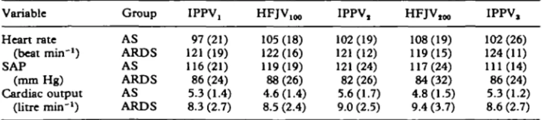
TABLE IV. Haemodynandc variables {mean (SD) values of six patients in each group); No significant
differences between IPPV and HFJV at both frequencies of ventilation
Variable Heart rate (beat min"1) SAP (mm Hg) Cardiac output (litre min"*) Group AS ARDS AS ARDS AS ARDS IPPV, 97 (21) 121 (19) 116(21) 86 (24) 5.3(1.4) 8.3 (2.7) HFJV100 105 (18) 122(16) 119(19) 88(26) 4.6(1.4) 8.5 (2.4) IPPV, 102 (19) 121(12) 121 (24) 82 (26) 5.6(1.7) 9.0 (2.5) HFJV™ 108(19) 119(15) 117(24) 84(32) 4.8(1.5) 9.4 (3.7) IPPV, 102 (26) 124(11) 111 (14) 86(24) 5.3(1.2) 8.6 (2.7)
(0 4. 1 3-n O ~ o o CO 0. 1 -1»
s
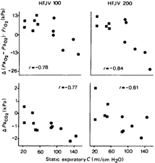
<-26-HFJV XX) • • • /•--0.78 HFJV 200 • /•--0.84 * -1 -2 /•--0.77 20 60 100 140 20 60 100 Static expiratoryC (ml/cm H2O)140
FIG. 3. Individual changes in ( P A ^ — PaoJ-.Fin ratio and PacOl induced by HFJV at 100 b.p.m. Oeft panels) and 200
b.p.m. (right panels) in AS ( # ) and ARDS ( • ) patients in relation to expiratory compliance (C).
the patient group rather than upon initial FRC values. In each ARDS patient, HFJV induced a similar increase in (PAOJ — Pa0 ): FIO J irrespective
of the baseline FRC, and without changing the latter. In contrast, HFJV increased FRC in AS patients without changing the ( P A ^ — Pa^): Fi0
ratio, even in patients with FRC values similar to those measured in patients with ARDS (fig. 2).
Similarly, pulmonary clearance of carbon di-oxide during HFJV was strongly dependent upon the patient group rather than upon initial FRC (fig. 2). Both frequencies of HFJV reduced PaCOj
and increased FRC in every AS patient. In contrast, application of HFJV in ARDS patients did not consistently modify P a ^ or FRC.
Figure 3 presents individual changes in
(PAOI — Pa^): FIO J ratio and P&co, induced by
HFJV at 100 and 200 b.p.m. in AS and ARDS patients, in relation to expiratory C. In contrast to FRC, there was no overlap of individual C values between both groups of patients. Changes in
(PAQJ — PSIQ^.FI^ ratio correlated significantly
with C(HF]V100:y = -1.26%+115.8, r = -0.78,
P< 0.005; HFJV20(): y = -1.72x + 142.5, r = -0.84, P < 0.005). Similarly, changes in P a ^
induced by HFJV also correlated significantly with C (HFJV100: y = -0.014x + 0.061,
r = -0.77, P < 0.001; HFJV,,,,,: y = -0.016x + 0.867, r = - 0 . 6 1 , P < 0.05).
In both groups of patients SAP, heart rate and cardiac output remained stable during the whole investigation (table IV).
DISCUSSION
The results of the present study suggest that FRC measured during IPPV does not predict pul-monary gas exchange during HFJV. In contrast, respiratory compliance assessed during IPPV appears to be an important variable to determine gas exchange during HFJV, as efficacy of HFJV on blood oxygenation and pulmonary elimination of carbon dioxide correlates significantly with respiratory compliance.
Accuracy of the procedure for the calibration of bellows pneumographs has been demonstrated by Rouby and colleagues [9]. In paralysed patients, rib cage and abdomen had the same contribution to F T ; therefore, their ventilatory system can be considered as an open system having a single independent variable [9]. Consequently, changes in pulmonary volumes can be derived indepen-dently from changes in both rib cage or abdominal circumference. These authors used belts con-nected to differential linear transducers with a frequency response of 60 Hz. The pneumograph system used in our study has a linear frequency response up to 10 Hz [7] that is, three times higher than the highest HFJV frequency used in this study.
We did not measure the contribution of injected, entrained and spilt volumes to FT. Using thoracic girth tranducers, Rouby and colleagues reported that, during HFJV at ventilatory frequencies of 100-600 b.p.m. gas entrainment is at least equal to the injected volume [9]. However, using a new technique which calculates tidal, entrained and spilt volumes by measuring changes in gas flow in the expiration limb of the bias flow tubing of an HFJV circuit, Young recently demonstrated that the entrained volume is only 10 % of FT at both frequencies of ventilation used in the present study [10]. Inspiratory: expiratory time ratio was set at 0.43 for both groups of patients during both HFJV sessions, as suggested as optimal by Rouby [9].
Ventilatory mechanics assessed during IPPV differed markedly in the two groups studied. In AS patients, baseline FRC was only 82 (22)% of the predicted normal value in the supine position,
most probably decreased by the effect of an-aesthesia and surgery [11]. Baseline PaOf was 12.4
(1.1) kPa with an F io of 0.4, associated with a
significant increase in (PAOI — PaOt): ^ o , r a" o and
venous admixture, whereas C and PaCOj were in
the normal range. As expected, C and FRC of ARDS patients were markedly decreased. In addition, these patients required an almost two-fold greater VE than the AS group to maintain a normal PaCOi. Arterial oxygenation of ARDS
patients was significantly less, despite a greater
Flo,-In this study we deliberately applied the same effective VE during HFJV to two groups of patients with differing ventilatory mechanics and probably differing carbon dioxide production and oxygen consumption. However, the relation be-tween the efficacy of HFJV on pulmonary clear-ance of carbon dioxide and effective VE and mean Paw markedly differed in both groups of patients. In the AS group, the increased pulmonary clearance of carbon dioxide during HFJV was related, as during IPPV, to the increment in effective VE and mean Paw. This is in accordance with results of other human studies using HFJV frequencies up to 200 b.p.m. [9, 12, 13]. This relationship suggests that a convective or axial flow transport mechanism, rather than an augmented dispersion, plays a major role in gas transport during HFJV at these frequencies of ventilation [14, 15]. However, dissociation be-tween pulmonary clearance of carbon dioxide and lung volume or mean Paw has been reported with higher frequencies [9], increased expiratory pressure resulting from short expiratory times [16, 17], or increased airway resistance [18]. This dissociation between pulmonary clearance of carbon dioxide and lung volume or mean Paw on alveolar ventilation can be explained by the expansion of the bronchial tree, resulting in an increase in anatomical deadspace [19].
HFJV produced better elimination of carbon dioxide in ARDS patients dian in AS patients. Indeed, we did not observe a significant decrease in pulmonary clearance of carbon dioxide during HFJV, despite a decreased mean Paw. These findings are in accordance with the results of Hurst, Branson and DeHaven who did not observe a relationship between improvement in clearance of carbon dioxide and increase in mean Paw during application of HFJV in ARDS patients [20]. Bulk convection alone cannot ex-plain the absence of this relationship. Other gas
transport theories such as the "Pendelluft" effect should be sought, as it may explain the more homogeneous peripheral lung gas concentrations observed during high frequency oscillation than during IPPV in patients with lung regions with significant time constant inequalities [17, 21, 22]. The relation between the efficacy of HFJV on blood oxygenation and mean Paw differed in both groups. The two groups of patients probably had differing distributions of ventilation-perfusion ( FA : Q) ratios. After operation, AS patients with a normal preoperative lung function had a decrease in PaOi associated with an increased (PAOJ — Pa^): FiOt ratio, caused possibly by lung regions with
low VA:Q ratios [23]. ARDS patients may have considerable volumes of lung with FA : Q = 0, or true shunt [24]. Arterial oxygenation in patients with ARDS depends on alveolar recruitment obtained by using a sufficiently great Paw to open alveoli and prevent them from collapsing during expiration [25]. This may explain why the HFJV-induced decrease in mean Paw in the ARDS group was associated with a worsening in Pa^ and venous admixture whereas, in AS patients, no relationship was found between mean Paw and blood oxygenation. Thus the reasons why the effect of HFJV on pulmonary gas exchange is related primarily to the patient's underlying disease and not to FRC are explained by the fact that the pathophysiological mechanisms reducing FRC are different in both groups of patients studied: in ARDS patients, the decreased FRC is caused by loss of endothelial cell integrity associated widi non-cardiogenic pulmonary oedema whereas, in patients recovering from major surgery, the reduced FRC is produced by microatelectasis and poorly ventilated lung regions.
In contrast to FRC, expiratory compliance assessed during IPPV correlated well with efficacy of HFJV on pulmonary gas exchange in the groups of patients studied. All ventilatory pressure-volume curves were linear for the seg-ment studied. In particular, no ARDS patients had a compliance curve with an inflection point, as has been described in the early stages of the disease [26]. It has been reported that HFJV has effects similar to those of conventional ventilation on lung morphology, pressure-volume curves and surface tension [27]. It must be concluded that lung stability and compliance are not affected differently during IPPV and HFJV. In the present study, application of HFJV increased alveolar
ventilation more in patients with a better C, resulting in enhanced pulmonary clearance of carbon dioxide. Both frequencies of ventilation produced a similar effect, as demonstrated by similar slopes of the relationship between changes in Pa^Oi and C. As pulmonary clearance of carbon
dioxide depends on bulk convection by direct alveolar ventilation with both ventilatory modes used in the present study, in addition to FE, clearance of carbon dioxide is mainly a function of
C [28]. As previously demonstrated [9, 29, 30],
HFJV induces a PEEP effect which depends both on ventilatory settings and on C. At fixed ventilatory settings, the importance of the HFJ V-induced PEEP effect is related to the C of the total ventilatory system [9]. In the presence of "stiff" lungs, which tend to decrease the time constant of the total system, the HFJV-induced PEEP effect is less than in patients with normal lungs [9]. Therefore, a greater change in blood oxygenation may be observed in patients with a better C, related to more effective gas distribution and gas mixing [28, 31]. This relationship was observed also in our study, as patients with the best C most decreased their (.PAOI — P a ^ : FIO J ratio during
HFJV at both ventilatory frequencies.
Application of HFJV decreased significantly the contribution of rib cage to tidal volume at both frequencies of ventilation in AS, but not in ARDS, patients. This decrease in Fr,rc was inversely related to FT values. We have not found any other study on HFJV which has examined this problem in man. Using inductance plethysmography in rabbits during ventilation by high frequency oscillations, Boynton and colleagues reported that the magnitude of ab-dominal to rib cage displacements increased with the frequency of oscillations to a maximum near 6 Hz, but thereafter diminished rapidly [32]. Moreover, abdominal relative to rib cage displace-ments decreased with increasing F T [32]. Similarly, Alleyne, Frantz and Fredberg, study-ing excised dog lungs ventilated with frequencies similar to those used in our patients, observed that distension of the lung base was favoured [14]. Decrease in Fr,rc during application of high frequency ventilation in these studies, as in our own, may be explained by the preference for the high velocity inspiratory gas stream to follow relatively straight airway paths [33]. This pref-erential axial flow to the base of the lungs was not observed in our ARDS patients. This could be related to the distribution of lung lesions in
ARDS, which are typically more severe in dependent regions of both lungs [34].
In conclusion, the comparison of patients investigated after abdominal surgery and patients with severe ARDS suggests that FRC alone measured during IPPV can not predict the changes in gas exchange produced by HFJV. In contrast, the efficacy of HFJV on pulmonary gas exchange depends upon the underlying disease. Arterial oxygenation and clearance of carbon dioxide are improved more (or impaired less) in patients with better compliance. These results have to be interpreted in the context of the different underlying pathophysiological mechanisms reducing FRC in both groups of patients studied.
REFERENCES
1. Brichant JF, Rouby JJ, Viars P. Intermittent positive pressure ventilation with either positive end-expiratory pressure or high frequency jet ventilation (HFJV), or HFJV alone in human acute respiratory failure. Anesthesia and Analgesia 1986; 65: 1135-1142.
2. Benhamou D, Bourgain JL, Rouby JJ, Viars P. High frequency jet ventilation vs continuous positive airway pressure for postoperative respiratory support. Chest 1984; 85: 733-738.
3. Shinozaki T, Deane RS, Perkins FM, Coffin LH, Ittleman FP, Mazuzan JE, Bird FM. Comparison of high frequency lung ventilation with conventional mechanical lung ven-tilation. Prospective trial in patients who have undergone cardiac operations. Journal of Thoracic and Cardiovascular Surgery 1985; 89: 269-274.
4. Murray JF, Matthay MA, Luce JM, Flick MR. An expanded definition of the adult respiratory distress syndrome. American Review of Respiratory Disease 1988; 138: 720-723.
5. Shinozaki T, Abajian JC, Burton JR, Tabakin S, Hanson JS. N, multiple breath washout, theory and clinical application of a digital nitrogen washout computer. Journal of Applied Physiology 1966; 21: 202-208. 6. Bates DV, Maklem PT, Christie RV. Respiratory Function
in Disease. An Introduction to the Integrated Study of the Lung, 2nd Edn. Philadelphia: W. B. Saunders, 1971. 7. Morel DR, Forster A, Suter PM. Noninvasive ventilatory
monitoring with bellows pneumographs in supine subjects. Journal of Applied Physiology 1983; 55: 598-605. 8. Morel DR, Forster A, Bachmann M, Suter PM. Effect of
intravenous midazolam on breathing pattern and chest wall mechanics in humans. Journal of Applied Physiology 1984; 57: 1104-1110.
9. Rouby JJ, Simonneau G, Benhamou D, Sartene R, Sardnal F, Deriaz H, Duroux P, Viars P. Factors influencing pulmonary volumes and CO, elimination during high frequency jet ventilation. Anesthesiology 1985; 63: 473-^82.
10. Young JD. Gas movement during jet ventilation. Acta Anaesthesiologica Scandinavica 1989; 33: (Suppl. 90) 72-74.
11. Froese AB, Bryan AC. Effects of anesthesia and paralysis on diaphragmatic mechanics in man. Anesthesiology 1974; 41: 242-255.
12. Maclntyre NR, Follett JV, Deitz JL, Lawlor BR. Jet ventilation at 100 breaths per minute in adult respiratory failure. American Review of Respiratory Disease 1986; 134: 897-901.
13. Waterson CK, Militzer HW, Quan SF, Calkins JM. Airway pressure as a measure of gas exchange during high frequency jet ventilation. Critical Care Medicine 1984; 12: 742-746.
14. Alleyne CM, Frantz ID, Fredberg JJ. Preferential axial flow during high frequency oscillations: effects of gas density. Journal of Applied Physiology 1989; 66: 542-547. 15. Bourgain JL, Mortimer AJ, Sykes MK. Carbon dioxide
clearance and deadspace during high frequency ven-tilation: investigation in the dog. British Journal of Anaesthesia 1986; 58: 81-91.
16. Fusciardi J, Rouby JJ, Benhamou D, Viars P. Hemodynamic consequences of increasing mean airway pressure during high frequency jet ventilation. Chest
1984; 86: 30-34.
17. Lehr J. Circulation currents during high frequency ventilation. Federation Proceedings 1980; 39: 576. 18. Saari AF, Rossing TH, Solway J, Drazen JM. Lung
inflation during high frequency jet ventilation. American Review of Respiratory Disease 1984; 129: 333-336. 19. Yamada Y, Venegas JV, Strieder DJ, Hales CA. Effects of
mean airway pressure on gas transport during high frequency ventilation in dogs. Journal of Applied Physi-ology 1986; 61: 1896-1902.
20. Hurst JM, Branson RD, DeHaven CB. The role of high frequency ventilation in post-traumatic respiratory insufficiency. Journal of Trauma 1987; 27: 236-242. 21. Allen JL, Fredberg JJ, Keefe DH, Frantz ID. Alveolar
pressure magnitude and asynchrony during high fre-quency oscillation of excised rabbit lungs. American Review of Respiratory Disease 1985; 132: 343-349. 22. Fredberg JJ, Keefe DH, Glass GM, Castile RG, Frantz
ID. Alveolar pressure nonhomogenicity during small-amplitude high frequency oscillation. Journal of Applied Physiology 1984; 57: 788-800.
23. Rehder K, Knopp TJ, Sessler AD. Ventilation-perfusion relationship in young healthy awake and anesthetized-paralyzed man. Journal of Applied Physiology 1979; 47: 745-753.
24. Dantzker DR, Brook LJ, Dehart P, Lynch JP, Weg J. Ventilation-perfusion distribution in the ARDS. American Review of Respiratory Disease 1979; 120: 1039-1052.
25. Boros SJ. Variations in inspiratory:expiratory ratio and airway pressure wave form during mechanical ventilation: the significance of mean airway pressure. Journal of Pediatrics 1979; 94: 114-117.
26. Matamis D, Lemaire F, Harf M, Brun-Buisson C, Ansquer JC, Atlan G. Total respiratory pressure-volume curves in the adult respiratory distress syndrome. Chest 1984; 86: 58-66.
27. Weinmann GG, Simon BA, Mitzner W. Lung compliance changes on high-frequency ventilation in normal dogs. Journal of Applied Physiology 1984; 56: 506-512. 28. Otis AB, McKerrow CB, Barlett RA, Mead J, Mcllroy
MB, Silverstone NJ, Radford EP. Mechanical factors in distribution of pulmonary ventilation. Journal of Applied Physiology 1956; 8: 427-443.
29. Benhamou D, Ecoffey C, Rouby JJ, Fusciardi J, Viars P. Impact of changes in operating pressure during high frequency jet ventilation. Anesthesia and Analgesia 1984; 63: 19-24.
30. Rouby JJ, Fusciardi J, Bourgain JL, Viars P. High frequency jet ventilation in postoperative respiratory failure: determinants of oxygenation. Anesthesiology 1983; 59: 281-287.
31. Knopp TJ, Kaethner T, Meyer M, Rehder K, Scheid P. Gas mixing in the airways of dog lungs during high-frequency ventilation. Journal of Applied Physiology 1983; 55: 1141-1146.
32. Boynton BR, Fredberg JJ, Buckley BG, Frantz ID. Rib cage versus abdominal displacement in rabbits during forced oscillations to 30 Hz. Journal of Applied Physiology 1987; 63: 309-314.
33. Fredberg JJ, Allen J, Tsuda A, Boynton BR, Butler J, Lehr J, Frantz ID. Mechanics of the respiratory system during high frequency ventilation. Ada Anaesthesiologica Scandinavica 1989; 33 (Suppl. 90): 39-45.
34. Gattinoni, L, Pesenti A, Bombino M, Baglioni S, Rivolta M, Rossi F, Rossi G, Fumagalli R, Marcolin R, Mascheroni D, Torresin A. Relationships between lung computed tomographic density, gas exchange, and PEEP in acute respiratory failure. Anesthesiology 1988; 69: 824-832.