Research Article
The expression levels of three raft-associated molecules in
cultivated vascular cells are dependent on culture conditions
K. Monastyrskayaa, E. B. Babiychuka, b, e, J. C. Schittnya, U. Rescherc, V. Gerkec, H.-G. Mannherzd
and A. Draegera,*
aDepartment of Cell Biology, Institute of Anatomy, University of Bern, Bühlstr. 26, 3012 Bern (Switzerland),
Fax: +41 31 631 38 07, e-mail: [email protected]
bInstitute of Physiology, University of Kiev (Ukraine)
cInstitute for Medical Biochemistry, ZMBE, University of Münster (Germany) dInstitute of Anatomy and Embryology, Ruhr University, Bochum (Germany) ePresent address: Department of Physiology, University of Liverpool (England)
Received 4 August 2003; received after revision 18 September 2003; accepted 25 September 2003
Abstract. Relaying a signal across the plasma membrane requires functional connections between the partner mol-ecules. Membrane microdomains or lipid rafts provide an environment in which such specific interactions can take place. The integrity of these sites is often taken for granted when signalling pathways are investigated in cell culture. However, it is well known that smooth muscle and endothelial cells undergo cytoskeletal rearrange-ments during monolayer culturing. Likewise affected – and with potentially important consequences for
sig-DOI 10.1007/s00018-003-3307-1 © Birkhäuser Verlag, Basel, 2003
nalling events – is the organization of the plasma mem-brane. The expression levels of three raft markers were massively upregulated, and raft-associated 5¢-nucleoti-dase activity increased in conventional monolayer cul-tures as compared with a spheroidal coculture model, shown to promote the differentiation of endothelial cells. Our data point to a shift of raft components in monolayer cultures and demonstrate potential advantages of the spheroid coculture system for investigation of raft-medi-ated signalling events in endothelial cells.
Key words. Signalling; smooth muscle; endothelial cells; 5¢-nucleotidase; annexin.
Introduction
Vasomotor activity is synergistically controlled by all vascular components and involves close cooperation of endothelial and smooth muscle cells [1]. Changes in ves-sel diameter brought about by smooth muscle cell con-traction influence the organization of endothelial plasma membranes. In turn, differences in fluid shear stress, sensed by endothelial cells, are transmitted via biochem-ical and mechanbiochem-ical pathways to the underlying muscle layer. Since endothelial and smooth muscle cells are
read-*Corresponding author.
ily perpetuated in culture, there exists a wealth of in vitro data pertaining to these signal transduction pathways. In-volving both receptors and cytoplasmic proteins, these events originate above all at the plasma membrane. Lipid rafts have been shown to mediate signalling events, and their clustering is believed to stabilize membrane-bound receptor molecules [2]. Raft-mediated signalling is known to play a role in the vasomotor response to nitric oxide (NO) [3].
Annexins are known to be ‘membrane organizers’ and be-long to a family of structurally related, anionic lipid-bind-ing proteins [4]. Annexins 2 and 6 both have an affinity for raft-associated lipids [5, 6]; annexin 2 is known to par-ticipate in the Ca2+-dependent assemblage of membrane
rafts on the inner leaflet of the plasma membrane [7]. On the outer leaflet, GPI (glycosyl-phosphatidyl-inositol)-anchored proteins have been identified as readily accessi-ble raft markers, above all in cells of the immune system [8, 9]. 5¢-Nucleotidase is one of the GPI-anchored en-zymes whose activity is believed to be regulated by the annexin 2-mediated oligomerization of lipid rafts in smooth muscle and endothelial cells [6].
Based on our observation that plasma membrane proteins undergo significant rearrangement during subculturing of cells [10], we searched for a system which combined ease of pharmacological manipulation with a more accu-rate correlation of the structural parameters we wanted to investigate than conventional monolayer cultures. Since plasma membrane rafts are known to be centres of signal transduction [2] and their reorganization is likely to affect downstream signal transduction cascades, we compared the expression levels of three known raft mark-ers in endothelial and smooth muscle cells grown under flat (i. e. conventional) and spheroidal culture conditions. Cocultured spheroids spontaneously organize into a smooth muscle core and an endothelial surface layer. This three-dimensional culturing system has been shown to promote the differentiation of endothelial cells and pre-vent their apoptosis in a smooth muscle-endothelial cell contact-dependent manner [11].
Our investigation demonstrated a massive upregulation of all raft markers investigated in flat cultures of endothelial or smooth muscle cells (single or cocultures) compared with the levels expressed in spheroids and a marked in-crease in 5¢-nucleotidase activity measured in flat as op-posed to spheroidal cultures. These findings outline a po-tentially important methodological problem in the execu-tion and interpretaexecu-tion of studies which are concerned with membrane-associated signalling in vascular cells.
Materials and methods Cell culturing
Primary cultures of human microvascular endothelial cells and human aortic smooth muscle cells were ob-tained from Promocell (Heidelberg, Germany) and culti-vated according to the distributor’s instructions. In initial experiments, primary cultures of myometrial smooth cells were prepared according to the protocol described by Ehler et al. [10]. There was no difference between vas-cular and visceral smooth muscle cells in our experi-ments. Densely confluent cultures of cells between pas-sages 2 – 6 were used throughout. For the assessment of flat cultures, smooth muscle and endothelial cells were grown either separately or together at a seeding ratio of 1:1.
Spheroid cultures (composed exclusively of endothelial or smooth muscle cells or of a mixture of the two types)
were prepared according to the protocol described by Korff et al. [11], with one modification: the spheroids were grown individually, in hanging drops, as described for the cultivation of embryoid bodies [12], these being suspended above a Petri dish filled with phosphate-buffered saline (PBS).
Smooth muscle cells, which form the core of the spheroid bodies, are unable to maintain the expression of their dis-tinct protein isoforms under anchorage-independent cul-ture conditions. Smooth muscle-associated proteins (smooth muscle-myosin heavy chain, h-caldesmon) are rapidly downregulated; thus, spheroids were used for ex-periments within 24 h of cultivation. For immunohisto-chemistry, spheroids were collected in Eppendorf tubes, pelleted in a 1:1 Tissue Tek:PBS mixture, snap frozen in liquid N2, and cryostat sections were prepared.
Immunohistochemistry
Smooth muscle biopsies were excised from the human bladder during surgical undertakings that were unrelated to muscle disease. Consent for working with this tissue was obtained from the Bernese Medical-Ethical Com-mission.
With the aid of a light microscope, they were teased into individual bundles. Ultracryomicrotomy and immunola-belling were performed as described previously [5]. For immunostaining, the cells were seeded onto glass coverslips, fixed at ambient temperature in 4 % paraformaldehyde buffered with Na+-Tyrode’s solution
(140 mM NaCl, 5 mM KCl, 1 mM MgCl2, 10 mM
glu-cose and 10 mM Hepes; pH 7.4) and then permeabilized (for 30 s at ambient temperature) within 0.5 % Triton X-100 (in Na+-Tyrode’s solution). Immunolabelling was
performed as described by Jostarndt-Fögen et al. [13]. The employed monoclonal antibodies against annexin 6 (purchased from Transduction Laboratories, Lexington, USA) and against annexin 2 [14] have been previously characterized.
Fluorescent labelling was performed using Cy3- (Jack-son, Baltimore, USA) or Alexa-conjugated (Molecular Probes, Eugene, USA) secondary antibodies. Negative controls were generated either by absorbing the antibody with purified antigen (for annexin 2 or 6) or by applying a nonbinding primary antibody.
Specimens were examined in a Zeiss Axiophot fluores-cent microscope, and images were collected using a digi-tal CCD camera (Ultrapix, Astrocam) or a Bio-Rad MicroRadiance confocal microscope.
5¢¢-Nucleotidase activity
5¢-Nucleotidase was assayed using 5¢-AMP as a sub-strate, as described by Babiychuk et al. [5]. Monolayer cultures (105cells/dish) were washed once with Na+
-Ty-rode’s solution containing 2 mM CaCl2 and then
incu-bated in 1 ml of the substrate buffer (Na+-Tyrode’s
solu-tion (pH = 8.0) containing 2 mM AMP, 2 mM MgCl2and
2 mM CaCl2) for 45 min at 37 °C. Spheroids were
col-lected in Eppendorf tubes (2250 cells/spheroid; 30 spher-oids per tube), washed and resuspended in 500 ml of sub-strate buffer, care being taken not to disrupt them during these steps, in order to monitor the activity in the intact complexes. The reaction products were quantified spec-trophotometrically (A820) as described by Parkin et al.
[15], and the activity was expressed in units of optical density (OD) per 104cells.
Isolation of RNA and synthesis of cDNA
Flat-cultured cells (endothelial cells as well as smooth muscle cells) and spheroids were washed with PBS, re-suspended in lysis buffer and lysed by centrifugation through QiaShredder columns (Qiagen).Total RNA was isolated using the RNAeasy Mini Kit (Qiagen) in accor-dance with the manufacturer’s instructions. Samples were treated on-column with RNAse-free DNAse (30 units) to avoid possible contamination with genomic DNA. Complementary DNA (cDNA) templates were prepared from 200 ng of total RNA, using the Omniscript RT kit (Qiagen), random hexamer primers (Life Technologies) being employed at a concentration of 2.5 mM in the pres-ence of an RNAseOUT RNAse inhibitor (Life Technolo-gies). Control reactions were performed without RT. The absence of DNA contamination was confirmed by poly-merase chain reaction (PCR) using the primer pairs of an-nexin 2 (see below). A product was obtained only for samples from which cDNA was synthesized following re-verse transcription.
Reverse transcription-PCR
PCR was carried out using the PE Biosystems GeneAMP 5700 Sequence Detection System in conjunction with the SYBR Green PCR kits, as recommended by the manu-facturer (Applied Biosystems). PCR pimers were de-signed [using PRIMER EXPRESS software (Applied Biosystems)] to amplify segments containing fewer than 200 bp (in order to maximize efficiency), their specificity being confirmed by measuring the size and purity of the PCR products by 4 % NuSieve agarose gel electro-phoresis.
Primer sequences were as follows: Human annexin 2 forward primer 5¢-TGGATGAGGTCACCATTGTCA-3¢ and reverse primer 5¢-GGCGAAGGCAATATCCT-GTCT-3¢, amplifying a 70-bp fragment between nt 201 and 271 of the annexin 2 cDNA; human annexin 6 for-ward primer 5¢-CTTCGGTTGGTGTTCGATGA-3¢ and reverse primer 5¢-GCTCCCCTCGGATGCTG-3¢, ampli-fying a 70-bp fragment between nt 730 and 800 of the
an-nexin 6 cDNA; human 5¢ nucleotidase forward primer 5¢-GGCTGCTGTATTGCCCTTTG-3¢ and reverse primer 5¢-CTGCAGGAACTCTCCAGTGGA-3¢, amplifying a 120-bp fragment between nt 1282 and nt 1402 of the hu-man placental 5¢ nucleotidase cDNA; 28S ribosomal RNA (rRNA) forward primer GTTGTTGCCATG-GTAATCCTGCTCAGTACG-3¢ and reverse primer 5¢-TCTGACTTAGAGGCGTTCAGTCATAATCCC-3¢, am-plifying a 132-bp fragment between nt 4535 and nt 4667 of the human 28S rRNA cDNA; CD31 endothelial cell marker protein forward primer 5¢-GACGGTGCAAA-ATGGGAAGA-3¢ and reverse primer 5¢-TGACGTGA-GAGGTGGTGCTG-3¢ amplifying a 67-bp fragment be-tween nucleotides 276 and 343 of the human endothelial cell adhesion molecule CD31 cDNA; and glyceralde-hyde-3-phosphate dehydrogenase (GAPDH) forward primer 5¢-CCCCATGTTCGTCATGGGTGT-3¢ and re-verse primer 5¢-TGGTCATGAGTCCTTCCACGATA-3¢, amplifying a 99-bp fragment between nucleotides 446 and 545 of the human GAPDH cDNA.
For 30-ml PCR, 2 ml of cDNA template was mixed with 300 nM each of forward and reverse primer and 2 X SYBR Green Master Mix (Applied Biosystems). The re-action mix was incubated at 95 °C for 10 min, followed by 40 cycles at 95 °C for 15 s and 1 min at 60 °C. The rela-tive standard curve method was used (ABI Prism 7700 SDS, User Bulletin #2) to translate the cycle number into a relative quantity. The amount of each target was divided by the amount of 28S rRNA to yield a normalized target value. Each gene-specific PCR was performed in tripli-cate, and data from three independent experiments were analyzed.
Results
Distribution of raft marker proteins
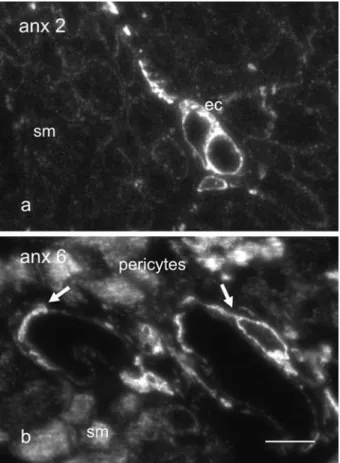
In transverse ultrathin cryosections through bundles of human urinary bladder smooth muscle, capillary en-dothelial cells are strongly stained for annexin 2, whereas the surrounding smooth muscle ones are only weakly la-belled (fig. 1 a). For annexin 6 (fig. 1 b), the labelling pat-tern is reversed: smooth muscle cells and pericytes are in-tensely stained, capillary endothelial cells but faintly so. No difference in labelling intensity was observed be-tween either raft-associated protein in flat cultures of en-dothelial and smooth muscle cells. The predominantly cytoplasmic distribution profiles for annexins 2 and 6 were comparable, occasional nuclear labelling being also encountered (fig. 2 a – d). The observed pattern of intra-cellular organization was similar to that already described for fibroblasts and epithelial cells [16 – 18]. Within smooth muscle cells, a vague but unmistakable codistrib-ution of annexins 2 and 6 with actin stress fibres was ap-parent (fig. 2 b, d).
Figure 1. Annexins 2 and 6 are differentially expressed within endothelial and smooth muscle cells in vivo. Transverse ultra-cryosection through human urinary bladder smooth muscle bundles labelled with antibodies against annexin 2 (a) and annexin 6 (b). Capillary endothelial cells (ec) stain intensely for annexin 2 (a), whereas the surrounding smooth muscle ones (sm) are but faintly immunoreactive. On the other hand, annexin 6 (b) abounds within smooth muscle cells and the pericytes (arrows) surrounding capil-laries, but it is barely visible within endothelial cells. Bar, 10 mm.
Figure 2. Localization of annexins 2 and 6 in flat cultures of endothelial and smooth muscle cells. Within monolayer cultures of endothe-lial (a, c) and smooth muscle cells (b, d), the predominantly cytoplasmic distribution patterns of annexins 2 (a, b) and 6 (c, d) are similar. Nuclear labelling is also encountered occasionally. Within smooth muscle cells (b, d), the annexins are partially codistributed with actin stress fibres. Bar (a – d), 10 mm.
We applied a coculturing system which is capable of em-ulating certain in vivo conditions [11, 19], for the investi-gation of raft-associated molecules and 5¢-nucleotidase reactivity. Endothelial and smooth muscle cells were mixed at a ratio of 1:1 and used for experiments the fol-lowing day. In spheroidal cultures, endothelial cells be-come quiescent [11]. Smooth muscle cells are unable to survive if cultivated in suspension for more than 2 days. They do not reduplicate under these conditions. Trans-verse cryostat sections of endothelial/smooth muscle mixed spheroids demonstrate the almost complete sepa-ration between the smooth muscle core and the endothe-lial surface layer, delineated with an antibody against an endothelial marker [CD31 (fig. 3 c)]. A monoclonal anti-body against annexin 2 did not distinguish between sur-face and core of the spheroid (fig. 3 a), whereas annexin 6 labelling was largely confined to the smooth muscle cells in the centre of the spheroid (fig. 3 b).
5¢¢-Nucleotidase activity
5¢-Nucleotidase is considered to be a marker for lipid mi-crodomains, and its level of activity an indicator of raft-mediated signalling events [6]. As such, it was used to gauge the influence of cell-culturing conditions on their signalling properties. In six independent measurements made using the same number of cells derived from flat cultures of either endothelial or smooth muscle cells, 5¢-nucleotidase activity was significantly higher than in a like number of corresponding cells derived from spher-oidal cultures (fig. 4). In both monolayers and spheroids, the 5¢-nucleotidase activity of endothelial cells was con-sistently lower than that in smooth muscle ones.
Gene expression
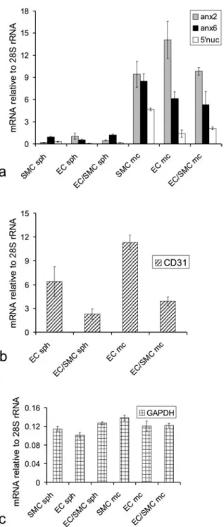
The pronounced discrepancy in 5¢-nucleotidase activity between the two culture models prompted us to investi-gate their messenger RNA (mRNA) expression levels of 5¢-nucleotidase and of annexins 2 and 6 using quantita-tive reverse transcription (RT)-PCR (fig. 5).
In accord with our biochemical data (see fig. 4), the ex-pression levels of 5¢ nucleotidase were substantially (more than 10-fold) higher in flat cultures of smooth muscle and endothelial cells, than in three-dimensional spheroids composed of the same cell types.
Likewise, in keeping with our biochemical findings, the levels of 5¢-nucleotidase mRNA were lower in endothe-lial cells than in smooth muscle ones. Mixed monolayer cultures and spheroids contained approximately the same mean amounts of mRNA from their contributing cell types (fig. 5 a).
The mixed spheroids expressed approximately the same mean amounts of all three gene products as did those con-sisting of a single cell type only (fig. 5 a). In the
cocul-Figure 3. Raft-associated proteins in spheroidal cultures. Trans-verse cryostat sections of spheroids consisting of endothelial and smooth muscle cells labelled with monoclonal antibodies against annexin 2 (a), annexin 6 (b) and CD 31 (c). Annexin 6 is largely confined to smooth muscle cells at the spheroidal centre (a), whereas annexin 2 label does not discriminate between endothelial and smooth muscle cells (b). Endothelial cell marker protein CD 31 is predominately found in the peripheral, endothelial layer (c). A small number of centrally located, labelled cells demonstrate that the separation of the different cell types is incomplete. Bar, 40 mm.
tures smooth muscle and endothelial cells were mixed at a ratio of 1:1. In order to confirm that both cell types were present, in addition to the immunofluorescence labelling (fig. 3 c), the presence and abundance of the endothelial cell-specific marker protein CD31 was examined by RT-PCR (fig. 5 b). The CD31 expression level is reduced by ~ 50 % in the mixed smooth muscle cell/endothelial cell (SMC/EC) cultures compared with the pure endothelial monolayer and spheroid cultures, indicating that the cell ratio did not change as a result of culturing conditions. The overall upregulation of the CD31 expression in the monolayer cultures might be attributed to the more active cell proliferation in the monolayers [20] vs. the cell qui-escence in spheroids [11]. On the other hand, the expres-sion levels of GAPDH remained stable irrespective of the cell culture conditions (fig. 5 c).
Discussion
Endothelial cells are strategically placed between the bloodstream and the smooth muscle wall. Serving as modulators of smooth muscle contractile responses, they play an important role in the generation and integration of signals. Indeed, they are generally the first cells to be af-fected by vascular disease. Within a spheroidal cocultur-ing system, which mimics the inside-out assembly of blood vessels, endothelial differentiation and quiescence are believed to be controlled by paracrine mechanisms emanating from the smooth muscle core [11].
Figuire 4. 5¢-Nucleotidase activity within spheroidal and
mono-layer cultures. Smooth muscle (SMC) and endothelial cells (EC) were grown in monolayers (mc) or spheroids (sph) either separately or in cocultures (EC/SMC, 1:1). Extracellular 5¢-nucleotidase
ac-tivity was measured in intact spheroids and monolayers as de-scribed in ‘Materials and methods’, using AMP as a substrate. Op-tical density [(OD) A820] units were calculated per 104cells. Data
represent the mean ± SD (bars) of six independent experiments.
Figure 5. Expression of raft-associated marker genes. Total RNA was isolated from monolayer (mc) and spheroidal (sph) cultures of endothelial (EC) and smooth muscle cells (SMC), and from their cocultures (EC/SMC). The mRNA levels for annexin 2, annexin 6, 5¢-nucleotidase (a), as well as for CD31 (b) and GAPDH (c) were
determined by quantitative RT-PCR, and the amounts expressed as relative to the 28S rRNA content of each sample. Each column rep-resents the mean ± SD (bars) of three independent experiments
per-formed in triplicate (a) or two independent experiments (b and c). The approximately equal amount of either cell type in the cocul-tures was determined by measuring the expression level of en-dothelial cell marker protein CD31 (b), which is reduced by 50 % in the mixed SMC/EC cultures compared with the individual EC monolayer and spheroid cultures. The expression levels of the raft-associated marker genes in spheroidal cultures were compared to the levels observed in monolayer cultures. The expression level of the housekeeping gene GAPDH remains largely unchanged irre-spective of culturing conditions (c).
Expression of annexins 2 and 6 in vascular cells Annexins 2 and 6 both associate with lipid mi-crodomains, the former at a higher affinity than the latter. These lateral assemblies of cholesterol and glycosphin-golipids form dynamic structures which are known as lipid rafts or detergent-insoluble glycosphingolipid com-plexes (DIGs). They are associated with characteristic sets of proteins, which are linked by GPI anchors to the extracellular face. On the inner leaflet they harbour mul-tiple acylated proteins. Membrane rafts can self-associate to form higher-order structures which are physically more stable and constitute the hubs of signalling activity [21, 22]. Recent studies have emphasized a membrane-cytoskeletal connection for annexins: annexin 6 forms a reversible link between the cytoskeleton and the sar-colemma within smooth muscle cells [5], and the F-actin binding site has been recently identified in annexin 2 [23].
Annexins 2 and 6 appear reciprocally expressed – the for-mer predominating in endothelial, and the latter in smooth muscle cells. We observed differential expression levels of mRNA for annexin 2 and annexin 6 in mono-layer and spheroidal cultures of the two cell types. These differences were reflected in the immunohistochemical reactions to monospecific antibodies of human urinary bladder smooth muscle and mimicked in the preferential outlining of the endothelial periphery by annexin 6. Oth-erwise, differences in labelling intensities between an-nexin 2 and 6 were not observed in cultured cells. It was notable, however, that the intracellular distribution of an-nexins 2 and 6 in monolayer cultures of both cell types was a predominantly cytoplasmic one, corresponding to a low intracellular [Ca2+] [24].
5¢¢-Nucleotidase activity and expression
5¢-Nucleotidase cleaves AMP into adenosine, acting as a potent vasorelaxing agent [25]. Its activity in endothelial cells is equally important, particularly within peripheral regions of the vascular tree where the marked anticoagu-lant properties of adenosine help to ensure an unre-strained blood flow [26]. As a result of arteriolar and pre-capillary dilatation, large volumes are redistributed rapidly and hemodynamic parameters are altered while blood is shunted towards another region or a different or-gan. The effects of 5¢-nucleotidase activation are equally important for smooth muscle and endothelium and di-rected at the lower end of the vasculature.
When comparing the influence of the culture conditions on enzyme activity, we demonstrated a noticeable eleva-tion of 5¢-nucleotidase activity in monolayer cultures (fig. 4). The same held true for its mRNA levels; however, the observed upregulation of gene expression was signifi-cantly higher (about 10 times in monolayer cultures, fig. 5 a). This discrepancy might be attributed to the
intracel-lular accumulation of 5¢-nucleotidase and its continuous trafficking between the surface and cytoplasmic mem-branes, which has been shown in many cell types, includ-ing endothelial cells [27 – 29]. Since we measured the en-zyme activity on the cell surface only, a significant pro-portion of the newly synthesized enzyme has probably escaped detection. It is possible that the changes in the cell membrane architecture brought about by the spheroid culture conditions have a particularly profound effect on the expression levels of the membrane-associated pro-teins. In addition to the raft associated protein genes, we have also observed upregulation of the mRNA levels of CD31 (fig. 5 b). In contrast, the expression of GAPDH was not significantly altered (fig. 5 c).
Compartmentalization of signalling
The mRNA levels for both annexins were significantly lower in spheroids than in monolayer cultures, which sug-gests that the number or size of membrane domains is de-pendent upon the culture conditions. In smooth muscle cells, the breakdown of regular, ‘contractile’ sarcolemmal architecture [30] during cultivation is well documented [10]. It is thus not surprising that membrane rearrange-ments can be observed in other cell types and that they extend beyond the microscopically discernible ‘smooth muscle macrodomains’.
It has recently emerged that signalling cascades such as those elicited by insulin [31] or those described for H-Ras [32] require well-defined microenvironments for their correct functioning. And we have shown here that alter-ations in membrane geometry can elicit significant changes in biological effects. Although the present study was not concerned with the potential influence of such disturbances relayed to downstream effectors within the cytoplasm, our data nonetheless suggest that they are likely to be considerable.
Acknowledgements. We would like to thank Dr F. Burkhard
(De-partment of Urology, Bern) for smooth muscle tissue samples, A. Hostettler and C. Allemann for excellent technical assistance, and B. Krieger for photography. This study was supported by the Swiss National Science Foundation 31-57071.99 and 31-66963.01 (to A.D.) and 7UKPJ62216 (SCOPES, to A.D. and E.B.) and the Novartis Foundation 00B30 (to A.D.).
1 Vanhoutte P. M., Rubanyi G. M., Miller V. M. and Houston D. S. (1986) Modulation of vascular smooth muscle contraction by the endothelium. Annu. Rev. Physiol 48: 307 – 320 2 Simons K. and Toomre D. (2000) Lipid rafts and signal
trans-duction. Nat. Rev. Mol. Cell Biol. 1: 31 – 39
3 Sowa G., Pypaert M. and Sessa W. C. (2001) Distinction be-tween signaling mechanisms in lipid rafts vs. caveolae. Proc. Natl. Acad. Sci. USA 98: 14072 – 14077
4 Gerke V. and Moss S. E. (2002) Annexins: from structure to function. Physiol. Rev. 82: 331 – 371
5 Babiychuk E. B., Palstra R. J., Schaller J., Kampfer U. and Draeger A. (1999) Annexin VI participates in the formation of
19 Korff T. and Augustin H. G. (1998) Integration of endothelial cells in multicellular spheroids prevents apoptosis and induces differentiation. J. Cell Biol. 143: 1341 – 1352
20 Sho E., Komatsu M., Sho M., Nanjo H., Singh T. M., Xu C. et al. (2003) High flow drives vascular endothelial cell prolifera-tion during flow-induced arterial remodeling associated with the expression of vascular endothelial growth factor. Exp. Mol. Pathol. 75: 1 – 11
21 Brown D. A. and London E. (1998) Functions of lipid rafts in biological membranes. Annu. Rev. Cell Dev. Biol. 14: 111 – 136 22 Brown D. A. and London E. (2000) Structure and function of sphingolipid- and cholesterol-rich membrane rafts. J. Biol. Chem. 275: 17221 – 17224
23 Filipenko N. R. and Waisman D. M. (2001) The C terminus of annexin II mediates binding to F-actin. J. Biol. Chem. 276: 5310 – 5315
24 Babiychuk E. B., Babiychuk V. S., Danilova V. M., Tregubov V. S., Sagach V. F. and Draeger A. (2002) Stress fibres – a Ca2+
-in-dependent store for annexins? Biochim. Biophys. Acta 1600: 154 – 161
25 Marshall J. M. (2000) Adenosine and muscle vasodilatation in acute systemic hypoxia. Acta Physiol. Scand. 168: 561 – 573 26 Kawashima Y., Nagasawa T. and Ninomiya H. (2000)
Contri-bution of ecto-5¢-nucleotidase to the inhibition of platelet
ag-gregation by human endothelial cells. Blood 96: 2157 – 2162 27 Widnell C. C., Schneider Y. J., Pierre B., Baudhuin P. and
Trouet A. (1982) Evidence for a continual exchange of 5
¢-nu-cleotidase between the cell surface and cytoplasmic mem-branes in cultured rat fibroblasts. Cell 28: 61 – 70
28 Peola S., Borrione P., Matera L., Malavasi F., Pileri A. and Mas-saia M. (1996) Selective induction of CD73 expression in hu-man lymphocytes by CD38 ligation: a novel pathway linking signal transducers with ecto-enzyme activities. J. Immunol.
157: 4354 – 4362
29 Airas L., Niemela J., Salmi M., Puurunen T., Smith D. J. and Jalkanen S. (1997) Differential regulation and function of CD73, a glycosyl-phosphatidylinositol-linked 70-kD adhesion molecule, on lymphocytes and endothelial cells. J. Cell Biol.
136: 421 – 431
30 North A. J., Galazkiewicz B., Byers T. J., Glenney J. R. Jr and Small J. V. (1993) Complementary distributions of vinculin and dystrophin define two distinct sarcolemma domains in smooth muscle. J. Cell Biol. 120: 1159 – 1167
31 Baumann C. A. and Saltiel A. R. (2001) Spatial compartmen-talization of signal transduction in insulin action. Bioessays 23: 215 – 222
32 Prior I. A., Harding A., Yan J., Sluimer J., Parton R. G. and Han-cock J. F. (2001) GTP-dependent segregation of H-ras from lipid rafts is required for biological activity. Nat. Cell Biol. 3: 368 – 375
a reversible, membrane-cytoskeleton complex in smooth mus-cle cells. J. Biol. Chem. 274: 35191 – 35195
6 Babiychuk E. B. and Draeger A. (2000) Annexins in cell mem-brane dynamics: Ca2+-regulated association of lipid
mi-crodomains. J. Cell Biol. 150: 1113 – 1123
7 Oliferenko S., Paiha K., Harder T., Gerke V., Schwarzler C., Schwarz H. et al. (1999) Analysis of CD44-containing lipid rafts: Recruitment of annexin II and stabilization by the actin cytoskeleton. J. Cell Biol. 146: 843 – 854
8 Cheng P. C., Dykstra M. L., Mitchell R. N. and Pierce S. K. (1999) A role for lipid rafts in B cell antigen receptor signaling and antigen targeting. J. Exp. Med. 190: 1549 – 1560
9 Alonso M. A. and Millan J. (2001) The role of lipid rafts in sig-nalling and membrane trafficking in T lymphocytes. J. Cell Sci.
114: 3957 – 3965
10 Ehler E., Babiychuk E. and Draeger A. (1996) Human foetal lung (IMR-90) cells: myofibroblasts with smooth muscle-like contractile properties. Cell Motil. Cytoskeleton 34: 288 – 298 11 Korff T., Kimmina S., Martiny-Baron G. and Augustin H. G.
(2001) Blood vessel maturation in a 3-dimensional spheroidal coculture model: direct contact with smooth muscle cells regu-lates endothelial cell quiescence and abrogates VEGF respon-siveness. FASEB J. 15: 447 – 457
12 Drab M., Haller H., Bychkov R., Erdmann B., Lindschau C., Haase H. et al. (1997) From totipotent embryonic stem cells to spontaneously contracting smooth muscle cells: a retinoic acid and db-cAMP in vitro differentiation model. FASEB J. 11: 905 – 915
13 Jostarndt-Fögen K., Djonov V. and Draeger A. (1998) Expres-sion of smooth muscle markers in the developing murine lung: potential contractile properties and lineal descent. Histochem. Cell Biol. 110: 273 – 284
14 Thiel C., Osborn M. and Gerke V. (1992) The tight association of the tyrosine kinase substrate annexin II with the submem-branous cytoskeleton depends on intact p11- and Ca2+-binding
sites. J. Cell Sci. 103: 733 – 742
15 Parkin E. T., Turner A. J. and Hooper N. M. (1996) Isolation and characterization of two distinct low-density, Triton- insoluble, complexes from porcine lung membranes. Biochem. J. 319: 887 – 896
16 Nigg E. A., Cooper J. A. and Hunter T. (1983) Immunofluores-cent localization of a 39,000-dalton substrate of tyrosine pro-tein kinases to the cytoplasmic surface of the plasma mem-brane. J. Cell Biol. 96: 1601 – 1609
17 Zokas L. and Glenney J. R. Jr (1987) The calpactin light chain is tightly linked to the cytoskeletal form of calpactin I: studies using monoclonal antibodies to calpactin subunits. J. Cell Biol.
105: 2111 – 2121
18 Eberhard D. A., Karns L. R., Vandenberg S. R. and Creutz C. E. (2001) Control of the nuclear-cytoplasmic partitioning of an-nexin II by a nuclear export signal and by p11 binding. J. Cell Sci. 114: 3155 – 3166