Short communication
DOTATOC: a powerful new tool
for receptor-mediated radionuclide therapy
A. Otte 1, E. J e r m a n n 1, M. Behe 1, M. Goetze 1 , H.C. B u c h e r 2, H.W. Roser 3, A. Heppeler 1, J. M u e l l e r - B r a n d 1, H.R. Maecke 1
1 Institute of Nuclear Medicine, University Hospital, School of Medicine, Basel, Switzerland 2 Department of Internal Medicine, University Hospital, School of Medicine, Basel, Switzerland 3 Department of Radiological Physics, University Hospital, School of Medicine, Basel, Switzerland Received 20 March and in revised form 4 April 1997
Abstract.
This study presents the first successful use of
a peptidic vector, DOTATOC, labelled with the [3-emit-
ting radioisotope yttrium-90, for the treatment of a pa-
tient with somatostatin receptor-positive abdominal me-
tastases of a neuroendocrine carcinoma of unknown lo-
calization. Tumour response and symptomatic relief
were achieved. In addition, the new substance DOTA-
TOC was labelled with the diagnostic chemical analogue
indium-111 and studied in three patients with histopath-
ologically verified neuroendocrine abdominal tumours
for its diagnostic sensitivity and compared with the com-
mercially available OctreoScan. In all patients the kid-
ney-to-tumour uptake ratio (in counts per pixel) was on
average 1.9-fold lower with IllIn-DOTATOC than with
OctreoScan. DOTATOC could be a potential new diag-
nostic and therapeutic agent in the management of neu-
roendocrine tumours.
Key words':
Somatostatin receptor-mediated internal ra-
diotherapy - DTPA-•-Phel-octreotide (OctreoScan) -
DOTA-D-Phel-Tyr3-octreotide (DOTATOC) - Indium-
111 - Yttrium-90
Eur J Nucl Med (1997) 24:792-795
I n t r o d u c t i o n
In recent years the use of the radiolabelled somatostatin
analogue lllin_DTPA_D_Phe l_Octreotide (OctreoScan;
DTPA: diethylene-triamine-penta-acetic acid) (Fig. 1) as
a specific radiopharmaceutical for the in vivo detection
of somatostatin receptor-positive tumours has been pro-
mulgated in clinical nuclear medicine, endocrinology
and oncology [1, 2]. Despite convincing diagnostic re-
sults, this analogue cannot be used for labelling with a
Correspondence to:
A. Otte, Institute of Nuclear Medicine, Uni-
versity Hospital, School of Medicine, Petersgraben 4, CH-4031
Basel, Switzerland
[3-emitter such as yttrium-90 for therapy. Crucial
for
such a radiotracer is the development of a peptide chel-
ator conjugate which can hold the radiometal with high
stability in vivo in order to reduce haematopoietic toxici-
ty due to bone marrow irradiation. Moreover, a hydro-
philic conjugate with predominant kidney excretion has
to be prepared. High kidney retention of the peptides and
antibody fragments - labelled with a metallic radionu-
clide - appeared to be one of the main obstacles in the
potential use of small molecules in internal radiotherapy
[3]. Therefore, we have developed a new chelator so-
matostatin analogue which can be labelled stably with
the diagnostic radionuclide 111In and its therapeutic
chemical analogue 90y. The new diagnostic radiotracer
was studied in patients for its sensitivity and compared
with the commercially available OctreoScan. In one pa-
tient, the somatostatin analogue labelled with the [3-emit-
ter 90y was tested for radionuclide therapy.
Part 1 : D i a g n o s t i c s t u d i e s
Materials, patients and methods
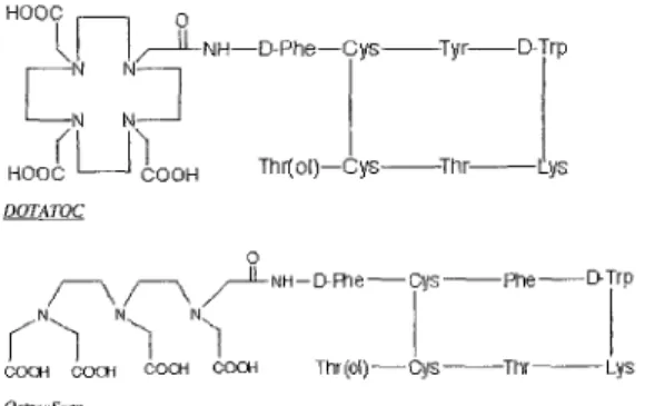
Radiotracer.
We developed a new DOTA chelated somatostatin
analogue, DOTA-D-Phel-Tyr3-Octreotide (DOTATOC; DOTA:
1,4,7,10-tetraazacyclododecane-N,N',N",N'-tetra-acetic
acid)
(Fig. 1), in a five-step synthetic procedure according to GMP
O
HO0~.
N
I/~[LNH--D-Phe--Cys
Tyr
D-Trp( ~
~]
Thr(o[)--Cys Thr
l ys
HOOC
COOH
DOTATOC~
//~IL NH-- Ev Flqe-- Ors
P h e - - D-Trp
o o[
o ~ ' ~ o o : ""to o OH N "~OOOH
Tlqr (o,)--Cys--Thr - - L~s
OctreoScan
Fig. 1. Structure formulas of DOTATOC and OctreoScan
European Journal of Nuclear Medicine
practice. IllIn-DOTATOC was prepared as follows: 8 gg of DOTATOC were dissolved in 190 gl 0.4 M sodium acetate buffer (pH 5.5) with 7 mg gentisic acid; after the addition of 6 mCi I lllnC13 (0.05 M HC1, Mallinckrodt Med., Petten, The Nether- lands), the solution was heated at 90°C for 25 min. Quality con- trol was obtained with the use of a Sep-Pak C18 cartridge and high-performance liquid chromatography (HPLC), resulting in highly pure radioligands with preserved receptor binding affinity (K D = 2.2+0.5 nM).
Patients.
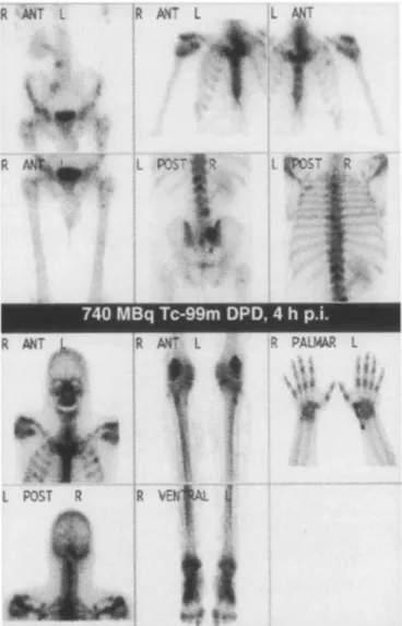
Three patients with histopathologically verified neuroen- docrine abdominal tumours were investigated after intravenous in- jection of 5 mCi 1J lin_OctreoScan and, 2 weeks later, 5 mCi 111in_ DOTATOC (specific activity: 1 Ci/gmol). Patient 1 (male, aged 46 years) had a remaining local tumour and peritoneal carcinosis af- ter hemicolectomy because of an obstructing carcinoid tumour of the terminal ileum with four of five ileocaecal nodular lymph node stations being affected with metastases. At the time of ad- mission, 5-hydroxy-3-indolacetic acid and chromogranin-B were positive. Patient 2 (male, aged 63 years) had a remaining tumour of 3x2x2 cm after subtotal splenopancreatectomy because of a malignant insulin-producing pancreatic tumour in the corpus with an initial diameter of 13 cm. Patient 3 (male, aged 43 years) had metastatic spread of a neuroendocrine tumour of unknown origin with multiple liver, abdominal and skeletal metastases (Figs. 2, 3). The patient had received two unsuccessful cycles of chemotherapy according to the PEI scheme [cisplatin (Ebewe) 20 mg/m 2 i.v. days 1-5, etoposide (Vepesid) 75 mg/m 2 i.v. days 1-5, ifosfamide (Holoxan) 1.2 g/m 2 i.v. days 1-5; PEI: platin-etoposide-ifosfa- mid@ In his past, the patient had undergone surgery with a partial resection of the left kidney due to a haemorrhagic cyst. Conven- tional computer tomography (CT) of the abdomen and thorax on the day of admission showed cystic degeneration of the left kid- ney, two large liver metastases in the segments II (4.5x4 cm) and V (9x7.5 cm) and multiple abdominal metastases. CT did not re- veal any intrapulmonary or mediastinal infiltrations. Skeletal scin- tigraphy on the day of admission detected multiple bone metastas- es in the first, second and fourth lumbar vertebrae, in the third, seventh and ninth thoracic vertebrae, in the sacrum, in the third rib on the left ventrolateral side and, additionally, in the posterior skull (Fig. 2). Neuron-specific enolase was positive (20 gg/1) on the day of admission.Methods.
Planar scintigraphic images were obtained with a large- field-of-view gamma camera (Siemens DIACAM), equipped with a medium-energy parallel-hole collimator (matrix 64x64, zoom 1). The pulse height analyser windows were centered over both ltlIn photon peaks (172 and 246 keV) with a window width of 20%. Data from both windows were then added to the acquisition frames. For the first 60 min, dynamic images were acquired from posterior views of the abdominal region with 240 frames (15 s/frame); one image consisted of four summed dynamic frames. Static images (5 min/frame) were acquired from anterior and posterior views of the abdominal region 1, 3, 4.5, 6, 24 and 48 h p.i.; in addition, static images from anterior views of the tho- racic region and skull were acquired 24 and 48 h p.i. in patient 3. One image consisted of one static frame. Between the 4- and 24-h imaging and between the 24- and 48-h imaging, all patients were treated with Prontolax (I0 mg bisacodylum). Region of interest (ROD analysis (in counts/pixel) of the background, kidneys, liver, tumour and/or metastases and spleen was obtained over the whole acquisition time. Within each patient, the ROI template was cop- ied from one time frame to the next for intra-individual standard- ization. Each ROI was normalized to the uptake (in counts/pixel)793
Fig. 2. Skeletal scintigraphy of patient 3 at the time of admission. 740 MBq 99mTc-dicarboxydiphosphonate (DPD) was adminis- tered. Note the bone metastases in the sacrum, spine, rib and skull
of the summed time frames over 5 min. All data were stored on a Siemens ICON computer system.
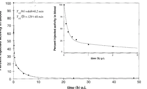
Radioactivity was measured in blood and urine over 48 h. Blood samples were obtained after 2, 5, 10, 20 and 40 min and 1, 2, 3, 6, 24 and 48 h. Urine was collected at 6-h intervals over 48 h. The chemical structure of the radioligand in blood and urine was determined by HPLC.
Results
R a d i o a c t i v i t y cleared q u i c k l y from the blood. T h e b l o o d c l e a r a n c e curve was fitted b y two e x p o n e n t i a l s (c~, ~3) o b t a i n i n g h a l f - t i m e s of ti/2(c~) = 5__+1 m i n (76% o f radio- activity) a n d
t~n(~3)
= 110_+30 m i n (24% of radioactivity) (Fig. 4). T h e r a d i o l a b e l l e d p e p t i d e s h o w e d high stability in vivo. N o b r e a k d o w n products were o b s e r v e d up to 6 h o f observation. T h e r a d i o a c t i v i t y was m a i n l y excreted via the k i d n e y a n d f o u n d intact in the u r i n e w i t h i n the first 4 h ( > 9 6 % intact, > 5 0 % of activity in the urine).794
Fig. 3. a Scintiscans of patient 3 5,5 h and 48 h after intravenous injection of 200 MBq 11 qn_DTPA_D_PheLOctreotide (OctreoScan). Pre- treatment scan:-NiSte the high kidney uptake over the entire investigated time. The left kidney is without function due to cystic degenera- tion. b Scintiscarlg:~f patient 3 1-60 rain and 1, 3, 4.5, 6, 24 and 48 h after intravenous injection of 196 MBq of the newly developed sub- stance 11 tln_DOT)X2~_Phel_Tyr3_Octreotide (DOTATOC), Pre-treatment scan 2 weeks later than the scan in a. Note the high kidney excre- tion within the first-"[2 rain in contrast to the OctreoScan in a
: _ / lOO "u 90 0 o 80 C • - 70 .-> 60 50 O • 40 L ~ C 30 20 O Q. 10 0 TI n (or) =6~0+-0.2 rain T1a (~) = 129 +-48 rain 100
8
c 75 50 25 0 t i m e (h) p.i. 10 20 30 4 0 50Fig. 4. Typical example of the blood clear- ance curve of 1111n_DOTATO C (patient l), The curve was fitted by two exponentials
(~,
13)
time (h) p.i.
Table 1. Uptake ratios 24 h after injection
Radiotracer patient k/t k/a t/a
DOTATOC 1 1.10 1.19 1.08 OctreoScan 2.00 1.28 0.64 DOTATOC 2 1.33 2.68 2.02 OctreoScan 2.54 5.80 2.28 DOTATOC 3 0.37 2.69 7.34 OctreoScan 0.70 5.11 7.30
Two metabolites were detected in the urine at later
times, the main one being [Illln](DOTA-D-Phel).
In all three patients, lllIn-DOTATOC showed the
same diagnostic precision as OctreoScan, but superior
biodistribution and faster blood and background clear-
ance (Fig. 3). The kidney-to-tumour (k/t) uptake ratio (in
counts per pixel) was on average 1.9-fold lower with
11 qn-DOTATOC than with OctreoScan (Table 1).
k, Kidney; t, tumor; b, background; 1, liver; a, injected activity (in MBq)/MBq
Part 2: Treatment
Materials and methods
Radiotracer. As a therapeutic radionuclide the pure 13-emitter 90y
was chosen. The labelling protocol was as described for 111In in Part 1 with the exception that different amounts of activity (25 mCi and 40 mCi, respectively) were used, resulting in a high- ly pure and stable radioligand with preserved receptor binding af- finity (K D = 2.6_+0.5 nM).
Patients. Due to the rapid progression of metastatic spread despite
two courses of chemotherapy and persisting pain in the lower back and abdomen in patient 3 (for a detailed description of the patient's status, see Part 1), we decided to treat this patient experi- mentally by internal radiotherapy. Before treatment, tumour do- simetry was assessed. As the limiting factor for therapy was the kidney dose, the kidney retention was estimated by the use of a kidney phantom according to the patient's anatomical setting. Hereby, it was calculated that the kidney would receive a dose of 20 Gy if approximately 80 mCi 90y-DOTATOC were applied (de- tailed data on tumour dosimetry are not presented in this short communication, but can be provided by the author). Therefore, we started fractionated treatment with two small portions each of 25 mCi 9°Y-DOTATOC and one portion of 40 mCi 90y-DOTATOC within 4 months (first treatment session: 21 October 1996; second session: 10 December 1996; third session: 3 February 1997). The treatment was approved by the Ethical Committee of the Universi- ty of Basel.
Methods. In each 9°y-DOTATOC treatment session, 1 mCi of
I llIn-DOTATOC was injected simultaneously in order to control the DOTATOC binding. Therefore, 1, 4.5, 24 and 48 h p.i. static images (5 rain/image) were acquired. In addition, 4 months after the last internal radiotherapy a follow-up I IIIn-DOTATOC scan was performed according to the protocol described in Part 1.
Results
D u r i n g the 2 m o n t h s f o l l o w i n g the last i n t e r n a l r a d i o - t h e r a p y s e s s i o n in p a t i e n t 3, r a p i d t u m o u r p r o g r e s s i o n
795 was s t o p p e d . T h i s was v e r i f i e d b y the f o l l o w - u p l t l I n - D O T A T O C scan, w h i c h e x h i b i t e d no further m e t a s t a s e s and no g r o w t h o f the k n o w n t u m o u r m a s s e s . In a d d i t i o n , the t u m o u r - t o - b a c k g r o u n d ratio o f the m e t a s t a s e s d i d not d i f f e r f r o m that b e f o r e t r e a t m e n t ( d a t a not p r e s e n t e d ) . F u r t h e r m o r e , the o n l y p o s i t i v e t u m o u r marker, n e u r o n - specific e n o l a s e , d e c r e a s e d f r o m 20 btg/1 to <10 btg/1 dur- ing this time. T h e p a t i e n t ' s c l i n i c a l status r e v e a l e d a c l e a r s u b j e c t i v e i m p r o v e m e n t after t h e r a p y ; in p a r t i c u l a r , the p a i n in his l o w e r b a c k and a b d o m e n d i s a p p e a r e d .
Conclusion
D O T A T O C , l a b e l l e d with l l l I n , is not o n l y a p o s s i b l e n e w d i a g n o s t i c a g e n t but c o u l d , g i v e n its s u p e r i o r b i o k i - netics and e s p e c i a l l y k i d n e y - t o - t u m o u r u p t a k e ratio, re- p r e s e n t a n e w t h e r a p e u t i c a l t e r n a t i v e for s o m a t o s t a t i n re- c e p t o r - p o s i t i v e t u m o u r s and m e t a s t a s e s w h e n l a b e l l e d w i t h a [3-emitter l i k e 90y. F u r t h e r studies in h u m a n s w i t h 90y are in p r o g r e s s .
Acknowledgements. The study was partially supported by the
Swiss National Science Foundation (31-42516/94) and the "Re- gionale Krebsliga'. We are further indebted to the radiotechni- cians B. Leu, L. Schwob, V. Tschanz, T. B6hler and G. Zeh for their kind help.
References
1. Krenning EP, Bakker WH, Breeman WAR et al. Localisation of endocrine-related tumours with radioiodinated analogue of somatostatin. Lancet 1989; I: 242-244.
2. Lamberts SWJ, Bakker WH, Reubi JC, Krenning ER Somato- statin-receptor imaging in the localization of endocrine tu- mours. N Engl J Med 1990; 323: 1246-1249.
3. Sivolapenko GB, Douli V, Pectasides D, et al. Breast cancer imaging with radiolabelled peptide from complementarity-de- termining region of antitumour antibody. Lancet 1995; 346:
1662-1666.