REVIEW ARTICLE
SPECT/CT in patients with painful knee
arthroplasty
—what is the evidence?
Michael T. Hirschmann&Johann Henckel&
Helmut Rasch
Received: 21 April 2013 / Revised: 13 May 2013 / Accepted: 15 May 2013 / Published online: 11 June 2013 # ISS 2013
Abstract SPECT/CT is increasingly recognized as a prom-ising imaging modality for the investigation of patients with a painful knee after knee arthroplasty. In this review article, we give an overview of the clinical value and current and future applications of SPECT/CT for patients with knee pain following joint arthroplasty. A detailed evidence-based lit-erature review is performed and presented.
Keywords Knee . SPECT/CT . SPECT . CT . Total knee arthroplasty . Replacement . Review
Introduction
Knee arthroplasty is a very successful treatment for osteo-arthritis of the knee joint, however, careful scrutiny of the orthopedic and radiological literature reveals that up to 20– 40 % of patients are not fully satisfied and symptom free following joint replacement, and complain of pain, stiffness, and loss of range of motion [1, 2]. The most common explanations for these are infection, aseptic loosening, insta-bility, malposition of the prosthetic components, arthrofibrosis,
and patellofemoral problems [2]. To date, the clinical and radiological diagnostics tools (radiographs, CT, MRI, scintig-raphy, SPECT, PET) are limited and do not clearly identify the site and cause of the patient’s symptoms [2].
Recently, the clinical value of the use hybrid imaging modalities such as PET/CT and SPECT/CT has been studied in patients with persistent symptoms after knee arthroplasty [2–13]. SPECT/CT is increasingly recognized as a clinically helpful imaging modality in patients with musculoskeletal problems [2–12]. In patients with unexplained symptoms following arthroplasty, SPECT/CT offers both radiologists and surgeons a rich source of information, combining me-chanical, structural, and metabolic data on the patient’s knee joint [2,9,14,15].
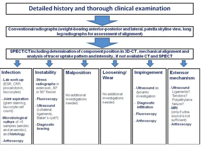
The combination of the use of three-dimensional (3D) reconstructed CT-based analysis of component position, orientation, mechanical and anatomical leg axes, as well as both the distribution and intensity of SPECT tracer uptake values has introduced a new dimension on diagnostics [2,9,
14,15]. Biomechanics and biology are brought together. In this review, we give an overview on the clinical value, and current and future applications of SPECT/CT in patients after knee arthroplasty. A detailed evidence-based literature review is performed.
Materials and methods
We performed a PubMed and MEDLINE literature search for the period 1970 to June 2012 using the terms “SPECT/CT”, “arthroplasty”, and “single-photon emission tomography” or “SPECT”. We applied no language restric-tion and also included data from both review articles and unpublished studies. In additional articles, data were re-trieved from expert consultations and personal files. These search criteria initially identified 28 published studies, one M. T. Hirschmann (*)
Department of Orthopaedic Surgery and Traumatology, Kantonsspital Baselland-Bruderholz, 4101 Bruderholz, Switzerland
e-mail: [email protected] M. T. Hirschmann
e-mail: [email protected] J. Henckel
Imperial College London, London, UK H. Rasch
Institute for Radiology and Nuclear Medicine, Kantonsspital Baselland-Bruderholz, 4101 Bruderholz, Switzerland
submitted, and one unpublished study for potential inclu-sion; all of these articles were then manually checked. All landmark studies reporting the use of SPECT or SPECT/CT in pati6ents before or after knee arthroplasty were included in our clinical review. Five articles dealing with the use of SPECT/CT and papers dealing with the application of SPECT in patients after knee arthroplasty were included.
Results
The results of the selected studies are summarized in Table1.
SPECT/CT There are currently five published studies deal-ing with the use of SPECT/CT in patients after knee arthroplasty.
Hirschmann et al. (2009) were the first to present a case series of patients with persistent pain after knee surgery [8]. They presented three such cases of pain after surgical treat-ment of knee osteoarthritis [8]. One, a non-union after high tibial osteotomy, one with tibial component loosening after medial uni-compartmental arthroplasty, and one with patellofemoral hyperpressure after total knee arthroplasty (TKA) [8]. In these patients with post-operative knee pain, SPECT/CT proved to be beneficial in establishing the diag-nosis and providing guidance for further treatment. The SPECT/CT analysis used was qualitative and descriptive [8]. The areas of increased tracer uptakes were reported and correlated with clinical symptoms [8].
In 2010, Hirschmann et al. introduced a standardized, easily applicable and reliable algorithm using SPECT/CT for patients after TKA and evaluated its inter- and intra-observer reliability [9]. Their SPECT/CT localization scheme differen-tiates the knee into nine tibial, nine femoral, and four patellar regions on standardized axial, coronal, and sagittal slices [9]. The intensity and localization of areas with increased SPECT/CT tracer uptake was semi-quantitatively assessed using a color-coded scale (0–10), in 18 consecutive patients complaining of knee pain after total knee arthroplasty [9]. The localization scheme together with measurements of TKR component position showed excellent inter- and intra-observer reliability [9]. The authors further highlighted that with the introduction of a standardized localization scheme, pathology-specific distribution patterns and intensity thresh-olds of SPECT/CT tracer uptake could be identified and detect mechanical loosening, joint instability, component malposi-tion, and patellofemoral problems [9]. The biggest limitation of this study was that only a semi-quantitative method of SPECT/CT tracer uptake analysis was used [9]. Moreover, the absolute measured intensity is only valid for each patient and not directly comparable between patients. It was only possible to compare relative values (ratios), for example the
activity in one region relative to the activity of the femoral mid-shaft of the same patient. The authors suggested using the SPECT tracer uptake value of the femoral shaft to normalize the data [9].
Graute et al. retrospectively evaluated the contribution of SPECT/CT as an adjunct to planar scintigraphy with (99m)Tc-labeled antigranulocyte antibodies for diagnosing and localizing low-grade joint infections [16]. They first evaluated the diagnostic sensitivity of scintigraphy in isola-tion and then in conjuncisola-tion with SPECT/CT in 31 patients [16]. Planar scintigraphy showed a sensitivity, specificity, and positive and negative predictive values of 0.66, 0.60, 0.4, and 0.81 [16]. SPECT/CT improved these values to 0.89, 0.73, 0.57, and 0.94 [16].
Filippi et Schillaci prospectively evaluated the usefulness of (99 m)Tc-HMPAO SPECT/CT in patients with bone and joint infections [17]. Two patient groups were investigated: group I, including patients with suspected bone infection (n=15), and group II, patients with suspected implant infec-tion (n=13). Six of the patients had their knee prosthesis greater than 12 months after primary surgery [17]. The authors found that SPECT/CT provided accurate anatomic localiza-tion for all tracer uptake-positive areas but did not help in the assessment of patients with negative scan findings [17]. They concluded that SPECT/CT with99mTc-HMPAO-labeled leu-kocytes can be a useful clinical tool to image bone and joint infections because it precisely localizes sites of leukocyte accumulation and allows the differentiation of soft-tissue from bone involvement [17].
Another landmark study piloted the validated SPECT/CT algorithm including 3D analysis of component position and tracer uptake assessment on a series of 23 knees with pain after total knee arthroplasty [2]. The authors reported that SPECT/CT significantly altered the working diagnosis and proposed treatment, and changed the previous intention to revise or treat the patients non-surgically [2]. In the patients who underwent revision surgery, the diagnosis arrived at using SPECT/CT was confirmed at surgery for all patients [2]. Patellofemoral hyperpressure, progression of patellofemoral osteoarthritis (within the first 5 years after primary TKR), tibial and or femoral loosening were reported as the largest causes of the patients problems [2]. Patients with femoral mechanical loosening demonstrated increased tracer activity around the femoral component [2]. Those with externally mal-rotated tibial components showed significantly higher tracer activity in the medial patellar facet [2]. Tibial components positioned and orientated on the tibia with posterior slopes <3° or >10° were associated with increased femoral tracer uptake [2]. Patients with patellofemoral OA as the leading cause for their knee pain had a significantly higher tracer uptake in the patella than others [2]. Patients with loosening of the femoral component showed significantly higher tracer uptake in the tibia, which extend to the prosthetic bone
interface [2]. The femoral uptake was also higher, but did not reach statistical significance [2]. The main limitation of this study was the small number of patients, and hence there was insufficient power to demonstrate a clear relationship between tracer uptake and prosthetic component position [2].
In currently unpublished data, Hirschmann et al. have extended their work in patients with painful total knee arthroplasty to a series of 100 knees. Along with the 3D
CT analysis of tibial and femoral component position (var-us-valgus, flexion-extension, internal and external rotation), the knee images were evaluated for areas of increased tracer uptake (“hotspots”). In this new study, the authors aimed to identify the typical pattern of tracer uptake distribution and intensity values in patients after total knee arthroplasty. Their findings were correlated with the shape, type, and make of the total knee arthroplasty component. They Table 1 Publications dealing with the use of SPECT/CT, SPECT, or three-phase-bone-scintigraphy in patients after knee arthroplasty
Author Year Journal Title Tracer SPECT data analysis Determination of component position SPECT/CT Hirschmann et al. 2009 Knee Surg Sports Traumatol Arthrosc
Combined single-photon emission computerized tomography and conventional computerized tomography (SPECT/CT): clinical value for the knee surgeons? 700 Mbq 99mTc-MDP Qualitative, descriptive 2D-CT measurements Hirschmann et al. 2010 Knee Surg Sports Traumatol Arthrosc
A novel standardized algorithm for evaluating patients with painful total knee arthroplasty using combined single-photon emission tomography and conventional computerized tomography
700 Mbq 99mTc-MDP
Semiquantitative, tracer uptake intensity graded 0–10, specific localization scheme 3D-CT measurements using customized software (Robin’s 3D, London, UK) Hirschmann et al. 2011 BMC Musculoskel-etal Disorders
Clinical value of SPECT/CT for evaluation of patients with painful knees after total knee arthroplasty—a new dimension of diagnostics?
700 Mbq 99mTc-HDP
Semiquantitative, tracer uptake intensity graded 0–10, specific localization scheme 3D-CT measurements using customized software (Robin’s 3D, London, UK) Hirschmann et al. 2012 BMC Medical Imaging 700 Mbq 99mTc-HDP
3D, volumetric, quantitative using customized software
(Introspect,
OrthoImagingSolutions Ltd., London, UK), specific localization scheme 3D-CT measurements using customized software (Robin’s 3D, London, UK) Hirschmann et al. – Unpublished data – 700 Mbq 99mTc-HDP (n=100)
3D, volumetric, quantitative using customized software
(Introspect,
OrthoImagingSolutions Ltd., London, UK), specific localization scheme 3D-CT measurements using customized software (Robin’s 3D, London, UK) Graute et al. 2010 Eur J Nucl Med
Mol Imaging
Detection of low-grade prosthetic joint infections using 99mTc-antigranulocyte SPECT/CT: initial clinical results
Semiquantitative, tracer uptake intensity graded 0–10, specific localization scheme
–
Filippi et al. 2006 J Nucl Med Usefulness of hybrid SPECT/CT in 99mTc-HMPAO-labeled leukocyte scintigraphy for bone and joint infections
400– 555 Mbq 99mTc- HMPAO-labeled leukocyte (n=28)
Semiquantitative, tracer uptake intensity graded 0–10, specific localization scheme
evaluated the time from primary TKR surgery, component fixation method (cemented or un-cemented arthroplasty), and included “intraoperative” findings at revision surgery (loose vs. well-fixed TKR components). The results will be published soon in a peer-reviewed journal.
In 2011, Hirschmann et al. presented a novel method of standardized volumetric 3D analysis of SPECT/CT imaging using a customized specific software solution (OrthoImagingSolutions Ltd, London, UK) [15]. To date, SPECT analysis is mainly qualitative due to variation in overall metabolic uptake among patients [15]. They illustrated the difficultly in comparing the intensity and patterns of SPECT tracer uptake between patients [15]. To address this, the authors described a fourfold approach.
Firstly, a method to quantitatively and volumetrically measure the intensity of SPECT tracer uptake in any area of the knee joint.
Secondly, a method to localize SPECT/CT data in 3D using clinically relevant anatomical landmarks and frames of reference.
Thirdly, a method to normalize orthopedic SPECT/CT data.
Fourthly, a method of thresholding SPECT/CT data, which distinguishes clinically relevant hot spots from background activity [15].
Discussion
There are a growing number of publications reporting the use of SPECT/CT in symptomatic patients prior to and following knee arthroplasty [2,3,6,8,9,12,15,16].
Taken as a whole, the current published studies and up-to-date evidence could be summarized as follows.
1. SPECT/CT is a promising imaging modality for use in patients with continued or new pain following knee arthroplasty as it offers the combined analysis of tracer uptake distribution and intensity as well as the ability to measure prosthetic component position. This ability to evaluate both the biomechanical and anatomical limb alignment of the lower limb together with the tracer uptake localization in one imaging modality makes it a feasible tool for the radiologist and surgeon in investigating unexplained pain in knee arthroplasty. SPECT/CT analysis gives rich information on the many factors known to be important for outcome after knee arthroplasty [2,18]. This combined analysis offers new dimensions of diagnostics such as investigating the influence of alignment or pros-thetic component position on tracer uptake distribution or intensity [2,14,15]. This broader knowledge about the mutual influences of these factors on SPECT tracer uptake will in future lead to new diagnostic algorithms, improving the specificity and sensitivity of diagnosing aseptic, or
septic loosening in patients after knee arthroplasty. SPECT/CT seems to be the one and only current imaging modality, which allows the assessment of the patient’s biology and mechanics [2, 14, 15]. It can become the biomechanical eye of the orthopedic surgeon.
2. In investigating patients prior to knee arthroplasty, SPECT or SPECT/CT has proven beneficial for the evaluation of osteoarthritic changes in the individual knee joint compartments. This information can assist the surgeon in his choice of the optimal type of pros-thesis. It may help him to decide wether a unicondylar, patellofemoral or total knee arthroplasty is indicated. 3. The methods of analysis of SPECT/CT data have
significantly evolved during the last 5 years [2, 14,
15]. First, there were only descriptive and qualita-tive methods to evaluate the SPECT tracer uptake data, with some authors using semi-quantitative methods to describe the intensity of tracer uptake
[2, 9]. The recent proposed 3D volumetric quantita-tive method is designed to identify pathology-specific diagnostic thresholds [15]. New customized software tools used for clinical studies could help introduce novel methods SPECT analysis into to daily practice [15]. In addition, these software tools could automatically integrate mechanical measure-ments and other determination of component posi-tion and orientation with outcomes in knee arthroplasty.
4. The natural history of the distribution and intensity of tracer uptake should be investigated for each specific type of knee prosthetic component, unicondylar, patello-femoral, or total knee arthroplasty. However, other influencing factors such as time from primary arthroplasty surgery, body mass index (BMI), the pa-tient’s individual tracer variety, and the leg’s mechanical and anatomical alignment have also to be taken into
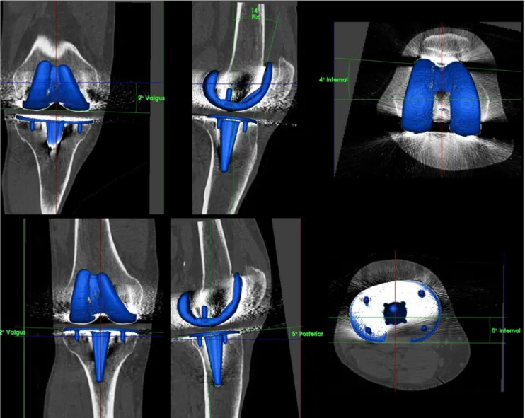
Fig. 2 Determination of tibial and femoral TKA component position (varus-valgus, flexion-extension, internal rotation–external rotation) on 3D-CT using customized software after reorientation in relation to the mechanical axis and definition of anatomical landmarks on the bone surface
consideration. A large sample size of patients will be needed to further investigate these significant questions, and should be performed in a standardized manner in a multicenter setting ideally using identical analysis tools and methods. We suggest that this research should be coordinated by an international research group focusing on SPECT/CT in orthopedics, its aim to define clear diagnostic guidelines.
In our current practice, we have recently introduced a diagnostic algorithm for unhappy patients after TKR (Fig.1). It is our opinion that there is no place for conven-tional bone scintigraphy or SPECT alone [2,9,14,19–22]. Only if there is no SPECT/CT available one should use these conventional purely nuclear medicine imaging modal-ities. Currently, we only have limited experience with arthro-SPECT/CT, but it could in future be an important adjunct in exceptional cases.
CT, either as part of SPECT/CT or alone, should be used to accurately assess femoral and tibial TKR component position (varus-valgus, flexion-extension, internal rotation– external rotation, Fig.2) [9,14]. The determination of TKR
component position should be performed on 3D-CT as this is the only accurate method [9,14].
SPECT/CT is performed using a hybrid system, which is equipped with a pair of low-energy, high-resolution colli-mators and a dual-head gamma camera and an integrated multi-slice CT scanner with a collimation of 16×0.75-mm. All patients receive a commercial 500–700 MBq, 99mTc-marked diphosphonate injection. Planar scintigraphic im-ages are taken in three phases, the perfusion phase (imme-diately after injection), the blood pool phase (from 1 to 5 min after injection) and the delayed metabolic phase (from 2 h after injection). SPECT/CT is performed with a matrix size of 128×128, an angle step of 32, and a time per frame of 25 s, 2 h after injection. For the CT, a specific protocol is used, which takes 3-mm slices of the femoral head, 0.75-mm slices of the knee joint (30 cm above and below), and 3-mm slices of the ankle joint. The whole TKR and parts of the femoral and tibial shaft, in particular important in the case of stems, have to be in the field of scanning. The protocol used was modified according to the Imperial Knee Protocol, which is a low-dose CT protocol [23]. The local-ization scheme differentiates the knee into nine tibial, nine
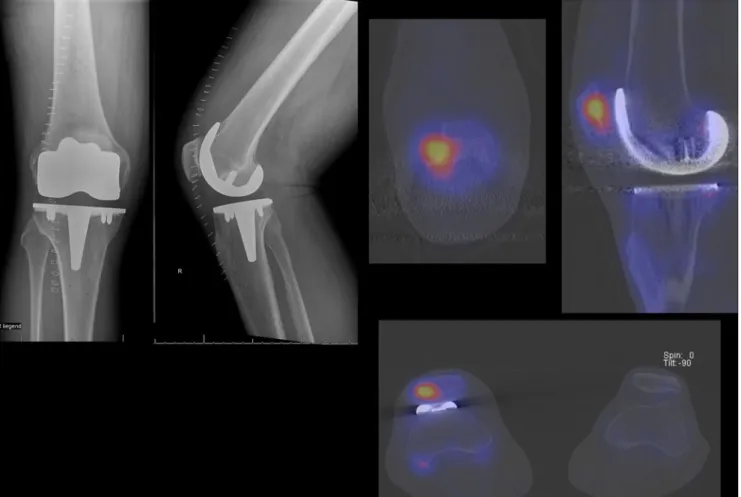
Fig. 3 Clinical example of a patient with persistent pain 1 year after total knee arthroplasty. Left—conventional postoperative anterior-pos-terior and lateral radiographs showing a total knee arthroplasty without
primary patellar resurfacing. Right—99mTc-HDP-SPECT/CT of the patient indicating a patellofemoral hyperpressure
femoral, and four patellar regions on standardized axial, coronal, and sagittal slices [9]. Along with the introduction of a standardized localization scheme, pathology-specific distribution patterns and intensity thresholds of SPECT/CT tracer uptake could be identified, which then reflect mechan-ical loosening, instability, component malposition, or patellofemoral problems (Fig.3) [9].
We acknowledge the substantial limitations of our re-view, largely reflecting the sparse body of literature in this area of research, much of which has been published by one SPECT/CT research group. A fact which is due to the limited availability of SPECT/CT systems and the low awareness in the orthopaedic fraternity.
Conflict of interest None.
References
1. Mannion AF, Kampfen S, Munzinger U, Kramers-de Quervain I. The role of patient expectations in predicting outcome after total knee arthroplasty. Arthritis Res Ther. 2009;11(5):R139.
2. Hirschmann MT, Konala P, Iranpour F, Kerner A, Rasch H, Friederich NF. Clinical value of SPECT/CT for evaluation of patients with painful knees after total knee arthroplasty—a new dimension of diagnostics? BMC Musculoskelet Disord. 2011;12:36.
3. Filippi L, Biancone L, Petruzziello C, Schillaci O. Tc-99m HMPAO-labeled leukocyte scintigraphy with hybrid SPECT/CT detects perianal fistulas in Crohn disease. Clin Nucl Med. 2006;31(9):541–2. 4. Gnanasegaran G, Barwick T, Adamson K, Mohan H, Sharp D, Fogelman I. Multislice SPECT/CT in benign and malignant bone disease: when the ordinary turns into the extraordinary. Semin Nucl Med. 2009;39(6):431–42.
5. Hirschmann MT, Adler T, Rasch H, Hugli RW, Friederich NF, Arnold MP. Painful knee joint after ACL reconstruction using biodegradable interference screws- SPECT/CT a valuable diagnos-tic tool? A case report. Sports Med Arthrosc Rehabil Ther Technol. 2010;2:24.
6. Hirschmann MT, Davda K, Iranpour F, Rasch H, Friederich NF. Combined single-photon emission computerised tomography and con-ventional computerised tomography (SPECT/CT) in patellofemoral disorders: a clinical review. Int Orthop. 2011;35(5):675–80. 7. Hirschmann MT, Davda K, Rasch H, Arnold MP, Friederich NF.
Clinical value of combined single-photon emission computerized tomography and conventional computer tomography (SPECT/CT) in sports medicine. Sports Med Arthrosc. 2011;19(2):174–81. 8. Hirschmann MT, Iranpour F, Davda K, Rasch H, Hugli R, Friederich
NF. Combined single-photon emission computerized tomography and conventional computerized tomography (SPECT/CT): clinical
value for the knee surgeons? Knee Surg Sports Traumatol Arthrosc. 2009;18(3):341–5.
9. Hirschmann MT, Iranpour F, Konala P, et al. A novel standardized algorithm for evaluating patients with painful total knee arthroplasty using combined single-photon emission tomography and conven-tional computerized tomography. Knee Surg Sports Traumatol Arthrosc. 2010;18(7):939.
10. Hirschmann MT, Schmid R, Dhawan R, et al. Combined single-photon emission computerized tomography and conventional com-puterized tomography: clinical value for the shoulder surgeons? Int J Should Surg. 2011;5(3):72–6.
11. Horger M, Eschmann SM, Pfannenberg C, et al. Added value of SPECT/CT in patients suspected of having bone infection: prelim-inary results. Arch Orthop Trauma Surg. 2007;127(3):211–21. 12. Scharf S. SPECT/CT imaging in general orthopedic practice.
Semin Nucl Med. 2009;39(5):293–307.
13. Vanquickenborne B, Maes A, Nuyts J, et al. The value of (18)FDG-PET for the detection of infected hip prosthesis. Eur J Nucl Med Mol Imaging. 2003;30(5):705–15.
14. Hirschmann MT, Konala P, Amsler F, Iranpour F, Friederich NF, Cobb JP. The position and orientation of total knee replacement components: a comparison of conventional radiographs, transverse 2D-CT slices and 3D-CT reconstruction. J Bone Joint Surg Br. 2011;93(5):629–33.
15. Hirschmann MT, Wagner CR, Rasch H, Henckel J. Standardized volumetric 3D analysis of SPECT/CT imaging in orthopaedics: overcoming the limitations of qualitative 2D analysis. BMC Med Imaging. 2012;12:5.
16. Graute V, Feist M, Lehner S, et al. Detection of low-grade prosthetic joint infections using 99mTc-antigranulocyte SPECT/CT: initial clin-ical results. Eur J Nucl Med Mol Imaging. 2010;37(9):1751–9. 17. Filippi L, Schillaci O. Usefulness of hybrid SPECT/CT in
99mTc-HMPAO-labeled leukocyte scintigraphy for bone and joint infec-tions. J Nucl Med. 2006;47(12):1908–13.
18. Hirschmann MT, Hoffmann M, Krause R, Jenabzadeh RA, Arnold MP, Friederich NF. Anterolateral approach with tibial tubercle osteotomy versus standard medial approach for primary total knee arthroplasty: does it matter? BMC Musculoskelet Disord. 2010;11:167. 19. Strobel K, Steurer-Dober I, Huellner MW, Veit-Haibach P, Allgayer B. Importance of SPECT/CT for knee and hip joint prostheses. Radiologe. 2012;52(7):629–37.
20. Hirschmann MT, Mathis D, Afifi FK, et al. Single-photon emission computerized tomography and conventional computerized tomogra-phy (SPECT/CT) for evaluation of patients after anterior cruciate ligament reconstruction: a novel standardized algorithm combining mechanical and metabolic information. Knee Surg Sports Traumatol Arthrosc. 2013;21(4):965–74.
21. Starke C, Ropke EF, Lohmann CH (2011) The third compartment in knee endoprosthetics: from denervation to replacement, which therapy is correct? Orthopade 40(10):896–898, 900–901 22. Smith SL, Wastie ML, Forster I. Radionuclide bone scintigraphy in
the detection of significant complications after total knee joint replacement. Clin Radiol. 2001;56(3):221–4.
23. Henckel J, Richards R, Lozhkin K, et al. Very low-dose computed tomography for planning and outcome measurement in knee re-placement. The imperial knee protocol. J Bone Joint Surg Br. 2006;88(11):1513–8.