Silvia R. Cottini Nicolas Lerch Marc de Perrot Miriam M. Treggiari Anastase Spiliopoulos Laurent Nicod Bara Ricou
Risk factors for reperfusion injury
after lung transplantation
Received: 6 February 2006 Accepted: 6 February 2006 Published online: 7 March 2006 © Springer-Verlag 2006
The preliminary results of the present study were presented at the 14th Annual Meeting of the European Society of Intensive Care Medicine, September 2001, Geneva, Switzerland
S. R. Cottini · N. Lerch · B. Ricou University Hospital of Geneva, Division of Surgical Intensive Care, Department of Anesthesiology, Pharmacology and Surgical Intensive Care,
Geneva, Switzerland
M. de Perrot · A. Spiliopoulos
University Hospital of Geneva, Clinic of Thoracic Surgery, Department of Surgery, Geneva, Switzerland
M. M. Treggiari
University of Washington, Department of Anesthesiology,
Seattle, WA, USA L. Nicod
University Hospital of Geneva, Division of Pulmonary Medicine, Department of Internal Medicine,
Geneva, Switzerland
S. R. Cottini (u)
Ospedale Civico di Lugano, Servizio di Anestesiologia e Cure Intensive, 6903 Lugano, Switzerland e-mail: Silvia.Cottini@eoc.ch Tel.: 0041 91 811 61 81 Fax: 0041 91811 61 82
Abstract Objective: To assess the influence of recipient’s and donor’s factors as well as surgical events on the occurrence of reperfusion injury after lung transplantation. Design and setting: Retrospective study in the surgical intensive care unit (ICU) of a university hospital. Methods: We collected data on 60 lung transplantation donor/recipient pairs from June 1993 to May 2001, and compared the demographic, peri- and postoperative variables of patients who experienced reperfusion injury (35%) and those who did not. Results: The occurrence of high systolic pulmonary pressure immediately after transplantation and/or its persistence during the first 48 h after surgery was as-sociated with reperfusion injury, independently of preoperative values. Reperfusion injury was associated
with difficult hemostasis during transplantation (p = 0.03). Patients with reperfusion injury were more likely to require the administration of catecholamine during the first 48 h after surgery (p = 0.014). The extubation was delayed (p = 0.03) and the relative odds of ICU mor-tality were significantly greater (OR 4.8, 95% CI: 1.06, 21.8) in patients with reperfusion injury. Our analysis confirmed that preexisting pulmonary hypertension increased the incidence of reperfusion injury (p< 0.01). Conclusions: Difficulties in perioperative hemostasis were associated with reperfusion injury. Occurrence of reperfusion injury was associated with postoperative systolic pulmonary hypertension, longer mechanical ventilation and higher mortality. Whether early recognition and treatment of pulmonary hyper-tension during transplantation can prevent the occurrence of reperfusion injury needs to be investigated.
Keywords Graft · Human · Ische-mia–reperfusion injury · Organ trans-plantation · Primary graft failure · Pulmonary hypertension
Introduction
Lung transplantation progressed from an early experimen-tal stage to become the mainstay of therapy for end-stage
lung disease refractory to medical treatment. Unfortu-nately, lung transplantation is limited by a significant shortage of available organs: only one among five donor organs is suited for transplantation since chest trauma,
aspiration, or neurogenic pulmonary edema cause exces-sive damage to the lung [1]. Seven to 15% of transplant candidates die each year while waiting for suitable organs in Switzerland [2]. After transplantation, 10–20% of the patients die during the first 3 months due to infection, cardiac failure and/or reperfusion injury (RI) [1, 3].
RI remains a serious complication in lung transplanta-tion and may contribute to the early mortality and mor-bidity [3, 4, 5]. Numerous terms and diagnostic criteria are used to describe ischemia–reperfusion-induced lung in-jury, and therefore the prevalence of the early graft dys-function cannot be determined precisely; indeed, a large majority of lung transplant recipients develop some degree of perihilar edema during the immediate postoperative pe-riod, and 15–35% of them will experience respiratory fail-ure and require prolonged mechanical ventilation and phar-macological support.
Prolonged ischemia [6], the type of preservation solu-tion [7, 8] and previous pulmonary hypertension [3] all seem to play an important role in the occurrence and the severity of RI; however, there are few studies investigating the risk factors for RI accounting simultaneously for sur-gical events, donor and recipient characteristics. The pur-pose of the present study was to investigate the effect of donor’s characteristics, intraoperative variables, and recip-ient’s characteristics on the development of RI.
Materials and methods
PatientsWe collected data on a retrospective cohort of 65 consec-utive lung graft recipients transplanted between June 1993 and May 2001 at the University Hospitals of Geneva. There were 17 single lung transplantations (SLT) and 48 bilateral lung transplantations (BLT). Informed consent for registry data use was obtained from the patients when placed on the waiting list. Data abstractors were blind to patient’s outcome. Age, sex, weight, height, underlying disease, arterial blood gas and 6-min walking distance were collected for all patients at the time they were put on the transplantation list. Data on cardiac catheterization (cardiac output, pulmonary arterial systolic and diastolic pressure, pulmonary capillary wedge pressure) were available for all patients with exception of eight young recipients with cystic fibrosis.
Donor selection criteria
Lung donors were identified according to standard selection criteria: < 60 years of age; PaO2/FiO2 ratio
> 300 mmHg with an inspired oxygen fraction of 1.0 and
positive end-expiratory pressure of 5 cmH2O, normal chest
X-ray; absence of abnormal findings at bronchoscopy; and
smoking history of less than 20 packs per year. Individuals who failed to meet one of these criteria were eligible for organ donation provided the remaining criteria were satis-fied. Variables collected from donor charts included: age, sex, weight, height, smoking habits, cause of death, last PaO2/FiO2 ratio, chest X-ray findings, pre-explantation
ventilatory settings (duration of ventilation, peak inspi-ratory pressure, positive end-expiinspi-ratory pressure), length of ICU stay, time from diagnosis of cerebral death to explantation (duration of brain death), and medication during the ICU stay (antibiotics, vasopressors).
Surgical technique and operative data
SLT was performed through a standard posterolateral thoracotomy approach and BLT through an anterior bilateral thoracosternotomy approach. Cardiopulmonary bypass was used for all patients with primary pulmonary hypertension and in selected cases with intraoperative hemodynamic instability or inadequate gas exchange. Ischemic time was calculated independently for each lung as the interval from the start of the pulmonary artery flush to reperfusion. Operative data included previous thoracic surgery, surgical difficulties in dissection of the native lung as reported in the surgical report, technical difficulties in vascular and/or bronchial anastomosis, difficulties of hemostasis as specifically mentioned in the report, severe bleeding, blood transfusion, lung preservation solution (modified Euro-Collins or low-potassium dextran), use of cardiopulmonary bypass, and ischemia time. Hemodynamic and ventilatory data during surgery were retrieved from anesthesia reports and included systemic and pulmonary pressures before induction of general anesthesia, during surgery, and after reperfusion of the transplanted lung. The use of vasopres-sors, inhaled nitric oxide (NO), fluid administration and transfusion, bleedings, and cardiac resuscitation during transplantation were also recorded. Vasopressors and inhaled NO were initiated in accordance with standard clinical protocols: norepinephrine was administered to maintain mean arterial pressure above 60 mmHg, and dobutamine was added if the cardiac index was below 2.5 ml/min/m2. Inhaled NO was initiated in patients with mean pulmonary artery pressure increase above 27 mmHg.
Postoperative management
All patients were intubated and ventilated for at least 12–24 h in the postoperative period. Criteria for liberation from mechanical ventilation included a reduction in ventilatory support to a pressure support < 10 cmH2O,
PEEP ≤ 5 cmH20, and FiO2< 40% with a PaO2 of at
pro-tocol was as follows: 5-day course of antilymphocyte
globulins 5–15 mg/kg (ATGAM®; Upjohn,
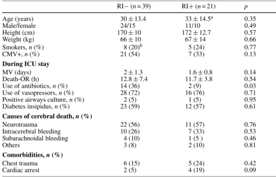
Kalama-zoo, MI) with a triple immunosuppressive therapy combining cyclosporine (CsA), azathioprine, and methyl-prednisolone. CsA was given to achieve and maintain a blood concentration of 200–250 ng/ml. Azathioprine was administered at the dose of 2 mg/kg IV and then replaced with mycophenolate mofetil (Cell cept®; Roche Pharma, Basel, Switzerland) as soon as the patient was enterally fed. Prednisone was initiated and maintained at a dose of 0.5 mg/kg during the first 3 months. After 1996, basiliximabum (Simulect®; Novartis Pharma, Basel, Switzerland) 20 mg on day 1 and day 4 replaced antilymphocyte globulins. Hemodynamic and ventilatory parameters were noted 1, 12, 24, and 48 h after admission to the intensive care unit (ICU). Chest X-rays were analyzed at 1, 12, 24, and 48 h after arrival in the ICU and graded by two critical care physicians according to the scoring system described by Anderson [9]. Dis-agreements were resolved by a joint second reading to achieve consensus. Briefly, each lung field was divided into four regions (perihilar, apical, lateral and basilar) and the degree of injury was assigned by a number between 0 and 3 (0 = normal lung and 3 = extensive alveolar infiltrate obscuring lung vessels with or without air bronchograms). The cumulative score (0–12) for each lung was the sum of the four regions. Time to extubation, length of ICU and hospital stay, and mortality in ICU, during hospital stay and after discharge were recorded. RI− (n = 39) RI+ (n = 21) p Age (years) 47± 12 41± 17 0.25 Male/female 23/16 10/11 0.40 Height (cm) 168± 9 164± 14 0.65 Weight (kg) 60± 12 59± 20 0.76 Diagnosis, n (%) Emphysema 24 (62) 3 (14) < 0.01 Pulmonary hypertension 1 (3) 8 (38) < 0.01
Idiopathic pulmonary fibrosis 3 (4) 3 (14) 0.42
Cystic fibrosis 7 (18) 4 (19) 0.92 Others 4 (10) 3 (14) 0.64 Preoperative variables, n (%) PAP systolic (mmHg) 43± 19 61± 27 < 0.01 CO (l/min) 4.9± 1.1 4.5± 1.5 0.26 PaO2/FiO2(mmHg) 270± 75 258± 58 0.58 PaCO2(mmHg) 45.7± 11.2 43.5± 14.2 0.43 6 min walk (m) 266± 124 266± 122 0.83 SaO2(%) 83± 13 86± 8 0.32 Previous surgery 5 (13) 5 (24) 0.28 Steroids> 20 mg/day 4 (10) 1 (5) 0.46 Home O2 21 (54) 10 (48) 0.64 Home NIV 1 (3) 5 (24) < 0.01
PAP pulmonary arterial pressure, CO cardiac output, NIV non invasive ventilation
Table 1 Characteristics of recipients (mean± SD, unless otherwise specified)
Bronchoscopy with bronchoalveolar lavage (BAL) was routinely performed after 48 h. Cell count and gram stain results were collected.
Definition of reperfusion injury
RI was defined as being present when the patient had a chest X-ray score ≥ 6 (diffuse alveolar opacities), a PaO2/FiO2ratio≤ 200 mmHg during the first 48 h
post-operatively, and no secondary cause of graft dysfunction was identified [3].
Statistical analysis
A complete case analysis was performed: five patients that had missing data were excluded from all analyses. De-scriptive statistics were used to examine the distribution of continuous and categorical variables among patients with and without reperfusion injury and the two groups were compared using unpaired Student t-test or Chi-square, as appropriate. Multiple logistic regression was used to es-timate the odds ratios associated with reperfusion injury and adjust for potential confounders (age, gender and body weight). For analysis of repeated observations, general-ized estimating equations (GEE) with robust estimate of the variance were used to account for dependence in the data. The level of significance was set at an alpha of 0.05 for all analyses. All data are expressed as mean± SD.
Results
Among the 60 patients analyzed, 21 (35%; 95% CI 0.23, 0.48) recipients developed RI. Demographic characteris-tics of all patients by presence or absence of RI are shown in Table 1. Patients with and without RI were not different with regard to age, gender or body mass index. The time on waiting list was not different between the two groups. Among patients with RI, eight had a history of primary or secondary pulmonary hypertension while of those without RI, only one had such history (p< 0.01). Three (11%) out of 27 patients with emphysema developed RI; the remaining 24 patients with emphysema represent the 62% of all patients without RI (p< 0.01) (Table 1). Patients with history of emphysema had a significantly
RI− (n = 39) RI+ (n = 21) p Age (years) 30± 13.4 33± 14.5a 0.35 Male/female 24/15 11/10 0.49 Height (cm) 170± 10 172± 12.7 0.57 Weight (kg) 66± 10 67± 14 0.66 Smokers, n (%) 8 (20)b 5 (24) 0.77 CMV+, n (%) 21 (54) 7 (33) 0.13
During ICU stay
MV (days) 2± 1.3 1.6± 0.8 0.14
Death-OR (h) 12.8± 7.4 11.7± 3.8 0.54
Use of antibiotics, n (%) 14 (36) 2 (9) 0.03
Use of vasopressors, n (%) 28 (72) 16 (76) 0.71
Positive airways culture, n (%) 2 (5) 1 (5) 0.95
Diabetes insipidus, n (%) 23 (59) 12 (57) 0.61
Causes of cerebral death, n (%)
Neurotrauma 22 (56) 11 (57) 0.76 Intracerebral bleeding 10 (26) 7 (33) 0.53 Subarachnoidal bleeding 4 (10) 1 (5 ) 0.46 Others 3 (8) 2 (10) 0.81 Comorbidities, n (%) Chest trauma 6 (15) 5 (24) 0.42 Cardiac arrest 2 (5) 4 (19) 0.09
aOne patient> 60 years of age
bOne patient smoking> 20 packs per year
MV Mechanical ventilation, CMV cytomegalovirus, Death-OR time between diagnosis of cerebral death
and organ explantation Table 2 Characteristics of
donors (mean± SD, unless otherwise specified
RI− (n = 39) RI+ (n = 21) p
Single lung transplantation 16 (41) 5 (24) 0.10*
Hemostasis difficulties 8 (20) 10 (48) 0.03
Mobilization difficulties 10 (26) 6 (29) 0.81
Previous thoracic surgery 5 (13) 6 (29) 0.13
Cardiopulmonary bypass 10 (26) 10 (48) 0.09
Blood transfusion> 2 BU 17 (44) 9 (43) 0.68
Ischemia time (h), 1 lung/2 lungs 3.5/4.4 3.6/4.3 0.82
Preservation solution (EC/LPD) 27/12 15/6 0.86
BU blood units, EC Euro-Collins, LPD low-potassium dextran
* single vs. double lung transplantation Table 3 Frequency of surgical
events, n (%)
lower risk of RI (odds ratio 0.10; 95% CI 0.02, 0.42). Among the preoperative variables, only pulmonary artery pressure was significantly different between patients with and without RI. None of the donor variables were associated with a significant risk of RI (Table 2). Five patients (29%) with SLT developed RI, and 16 patients (33%) with BLT developed RI (p = 0.10, Table 3). Among the surgical events that were analyzed, difficulties of hemostasis reported on the surgical record were associated with a greater risk of RI: 10 out of 21 patients with RI had hemostasis difficulties, whereas 8 out of 39 patients without RI had hemostasis difficulties (OR 3.5; 95% CI 0.95, 13.1; p = 0.03). These difficulties consisted in tendency to diffuse bleeding, without major loss of blood but requiring a meticulous surgical hemostatic measure.
Fig. 1 Time course of systolic pulmonary artery pressure (PAP), PaO2/FiO2ratio and cumulative chest X-ray scores in patients with RI (squares) and without RI (diamonds) during the first 48 h after surgery. PAP was significantly higher in patients with RI during the first 48 h after transplantation regardless of history of pulmonary hypertension. It is noteworthy that the PaO2/FiO2 ratio appears lower in the RI group already during the intraoperative period, suggesting an early onset of RI. The PaO2/FiO2ratio was omitted from the analysis as it represents one of the components of the RI definition. *p < 0.01, generalized estimating equations, with robust standard errors
Cardiopulmonary bypass, blood transfusion, type of preservation solution, and duration of ischemic time did not differ between patients who presented with RI and those who did not (Table 3). Postoperatively, the odds ratio of RI was 1.14 (95% CI 1.04, 1.26) for 1 mmHg difference in systolic pulmonary artery pressure. This remained significant regardless of preoperative pulmonary hyperten-sion, and after adjustment for catecholamine requirement (Fig. 1). It is interesting to note that one patient with preexisting pulmonary hypertension who did not experi-ence RI presented low postoperative pulmonary arterial pressures, similar to the group of patients without RI.
Systemic pressures and cardiac output did not differ between patients with and those without RI, although pa-tients with RI were more likely to require the adminis-tration of catecholamines: 10 (17%) patients with RI vs. 6 (10%) without RI required catecholamines at 12 h post-transplantation (p = 0.013), 12 (20%) patients with RI vs. 3 (5%) without RI at 24 h (p< 0.01), and 5 (8%) patients with RI vs. 1 (2%) without RI at 48 h (p = 0.014). The odds ratio for RI was 4.31 (95% CI 1.08, 17.25) for patients re-quiring catecholamines at 24 h compared with those who did not.
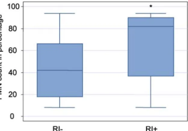
The number of neutrophils in the BAL was signifi-cantly higher in patients developing RI (Fig. 2) and was positively correlated with the severity score of the lung infiltrate on the chest X-ray (r = 0.548, p< 0.01).
The time to extubation was significantly longer for patients with RI than for patients without RI
(6.05± 5.74 days vs. 2.78 ± 3.7 days; p = 0.03); the ICU length of stay did not differ significantly between
the two groups (15± 15 days in the RI group vs.
10.8± 7.6 days in the non-RI group; p = 0.6).
ICU mortality was higher in patients with RI: 6/21 (28%) in patients with RI vs. 3/39 (8%) in patients without RI (OR of death 4.8; 95% CI 1.06, 21.8). How-ever, there was no difference in the cumulative mortality between the two groups after 6 and 12 months of
follow-Fig. 2 Box-and-whiskers plot displaying polymorphonuclear neu-trophils (PMN) in bronchoalveolar lavage fluid of patients with (RI+) and without (RI–) reperfusion injury, expressed in percentage. *p < 0.01, comparing patients with and without RI
up: 8/21 (38%) vs. 9/39 (23%) at 6 months (p = 0.2), and 8/21 (38%) vs. 10/39 (26%) at 12 months (p = 0.3).
Discussion
In this retrospective study, we investigated the effect of donor, respective recipient, and operative characteristics on the development of RI after lung transplantation. Similarly to previously published reports, we observed that the rate of RI was 35% [3, 7]. Multiple factors were found to be associated with its occurrence.
Difficulties of surgical hemostasis were associated with increased risk of RI, but not with greater requirement for blood or platelets transfusion or greater postoperative bleeding. The retrospective design of the present study does not allow exploring the nature of possible coagu-lation defects during surgery. However, this finding is consistent with recent data showing that disorders of the complex thrombin/antithrombin III are related to early graft dysfunction [10, 11].
Postoperatively, the increase and/or the persistence of preexisting high pulmonary systolic arterial pressure im-mediately and during the first 48 h after transplantation was associated with increased risk of RI, independently of previous pulmonary pressure levels. It is interesting to note that after adjustment for history of pulmonary sion, the occurrence of postoperative pulmonary hyperten-sion remained significantly associated with RI, suggesting that postoperative factors could also contribute to RI. As shown by other investigators [3, 12, 13], we found that pre-existing pulmonary arterial hypertension was associated with increased risk of RI, while recipients with pulmonary emphysema were less likely to develop RI.
There was increased short-term (ICU) mortality but not mid-term (3 months) mortality associated with RI.
The main limitation of our study is the small size of the sample; therefore, our findings may not be generaliz-able to other settings. However, all patients were treated at the same institution and received uniform treatment. In addition, despite the large number of variables collected, other unmeasured factors may influence the development of RI.
Increased level of interleukin-8 (IL-8) in the donor BAL has been described to be associated with the develop-ment of severe early graft dysfunction [14]. We could not investigate intrapulmonary factors such as IL-8 in donors’ lung because of the retrospective nature of our study. Thus, we cannot exclude the possibility that processes related to the development of the pulmonary hypertension and RI were already ongoing in the donors.
Preoperative factors. In order to determine the influ-ence of donor and recipient characteristics, some authors compared early graft function in paired recipients, each re-ceiving one lung from the same donor. Sommers et al. [12] observed that donor-related characteristics influenced only
early lung allograft function within 24 h after transplan-tation. Indeed, donor age, length of donor hospitalization, and primary pulmonary hypertension influenced the risk of primary graft failure in their study. We did not observe an association between RI and donor age or length of hos-pitalization prior to explantation. This discrepancy could possibly arise from the different definitions of graft dys-function in the two studies.
There is a controversy regarding the effect of head trauma of the donor on the development of RI. Fisher et al. [15] described an enhanced inflammatory state and graft failure when donors suffered traumatic intracranial hemorrhage. More recently, Ciccone et al. [16] observed no difference in the PaO2/FiO2ratio of recipients between
donors with traumatic and nontraumatic brain injury. Our data support the latter findings that the risk of RI does not differ between patients with and without neurotrauma (Table 2).
Perioperative factors. Snell et al. [17] observed that the duration of ischemia was a significant risk factor for graft dysfunction. In our study, because of the small size of our country and the short transport time, the ischemia time was overall less than 6 h. This lack of variation of ischemia duration could explain why we did not observe any effect of ischemic time on RI. Extracellular preserva-tion solupreserva-tions have been found to be protective against the development of RI [7, 18]. The potential beneficial role of low-potassium dextran could not be confirmed in our study because of the small numbers of graft treated with this so-lution.
Postoperative factors. In the postoperative course, pro-longed catecholamine requirement and longer duration of intubation were also found to be positively associated with the risk of RI. The presence of inflammation was indi-rectly confirmed by the higher polymorphonuclear neu-trophil count in the BAL of patients with RI than in those without RI. Furthermore, a positive correlation was ob-served between the number of neutrophils in the BAL and the extent of the pulmonary infiltrates, which supports the inflammatory nature underlying RI. As shown by de Perrot et al. [19], a possibly increased inflammatory response was observed in patients with RI with accompanying higher hemodynamic instability and prolonged requirement for pharmacological support.
The retrospective nature of our study does not permit a direct biological explanation for the present findings. However, based on prior knowledge we could speculate that RI is characterized by: (1) Early impairment in the co-agulation system, as indicated by perioperative hemostasis difficulties; (2) the activation of inflammatory cascades, as indicated by the prolonged requirement of hemodynamic support; and (3) a possible impairment in the regulation of the endogenous nitric oxide production that accompanies the endothelial dysfunction lead to a loss of NO-dependant vasodilatation [20], as indicated by the raised pulmonary pressure. Indeed, Thabut et al. [21] showed that inhaled
nitric oxide supplemented by intravenous pentoxifylline at reperfusion reduced the occurrence of RI.
Despite the several limitations of our study, the finding of a raised pulmonary hypertension early after transplanta-tion associated with RI seems to be important. Due to the observational nature of the present study, we cannot infer the cause. Whether early recognition and treatment of pul-monary hypertension during and immediately after trans-plantation can prevent the occurrence of RI needs to be investigated. In addition, our study provides data for
fur-ther research into the mechanisms that lead to RI. Com-parison across studies was particularly difficult due to the nonuniform definition of graft failure. For future studies a consensus on the definition of RI would be highly desir-able.
Acknowledgements. The authors would like to thank Paolo G. Merlani MD, Catherine Chenaud MD, Patrick Pasquina and Jean Max Granier for their assistance in data collection and during the preparation of the manuscript.
References
1. Trulock EP (1997) Lung transplan-tation. Am J Respir Crit Care Med 155:789–818
2. http://www.swisstransplant.org 3. King RC, Binns OA, Rodriguez F,
Kanithanon RC, Daniel TM, Spotnitz WD, Tribble CG, Kron IL (2000) Reperfusion injury significantly im-pacts clinical outcome after pulmonary transplantation. Ann Thorac Surg 69:1681–1685
4. Cottini S, Lerch N, de Perrot M, Spiliopoulos A, Nicod L, Ricou B (2001) Risk factors for pulmonary graft failure (PGF) after lung transplantation: retrospective analysis. Intensive Care Med 27:S93
5. Christie JD, Bavaria JE, Palevsky HI, Litzky L, Blumenthal NP, Kaiser LR, Kotloff RM (1998) Primary graft failure following lung transplantation. Chest 114:51–60
6. Snell GI, Shiraishi T, Griffiths A, Levvey B, Kotsimbos T, Esmore DS, Williams TJ (2000) Outcomes from paired single-lung transplants from the same donor. J Heart Lung Transplant 19:1056–1062
7. Thabut G, Vinatier I, Brugiere O, Leseche G, Loirat P, Bisson A, Marty J, Fournier M, Mal H (2001) Influence of preservation solution on early graft fail-ure in clinical lung transplantation. Am J Respir Crit Care Med 164:1204–1208 8. Schneuwly OD, Licker M, Pastor CM,
Schweizer A, Slosman DO, Kapanci Y, Nicod LP, Robert J, Spiliopoulos A, Morel DR (1999) Beneficial ef-fects of leukocyte-depleted blood and low-potassium dextran solutions on microvascular permeability in preserved porcine lung. Am J Respir Crit Care Med 160:689–697
9. Anderson DC, Glazer HS, Semenkovich JW, Pilgram TK, Trulock EP, Cooper JD, Patterson GA (1995) Lung trans-plant edema: chest radiography after lung transplantation – the first 10 days. Radiology 195:275–281
10. Salvatierra A, Guerrero R, Rodriguez M, Alvarez A, Soriano F, Lopez-Pedrera R, Ramirez R, Carracedo J, Lopez-Rubio F, Lopez-Pujol J, Velasco F (2001) Antithrombin III prevents early pulmonary dysfunction after lung transplantation in the dog. Circulation 104:2975–2980
11. Gu YJ, de Haan J, Brenken UP, de Boer WJ, Prop J, Van Oeveren W (1996) Clotting and fibrinolytic disturbance during lung transplantation: effect of low-dose aprotinin. Groningen Lung Transplant Group. J Thorac Cardiovasc Surg 112:599–606
12. Sommers KE, Griffith BP, Hardesty RL, Keenan RJ (1996) Early lung allograft function in twin recipients from the same donor: risk factor analysis. Ann Thorac Surg 62:784–790
13. Boujoukos AJ, Martich GD, Vega JD, Keenan RJ, Griffith BP (1997) Reperfusion injury in single-lung transplant recipients with pulmonary hypertension and emphysema. J Heart Lung Transplant 16:439–448 14. Fisher AJ, Donnelly SC, Hirani N,
Haslett C, Strieter RM, Dark JH, Corris PA (2001) Elevated levels of interleukin-8 in donor lungs is associ-ated with early graft failure after lung transplantation. Am J Respir Crit Care Med 163:259–265
15. Fisher AJ, Donnelly SC, Hirani N, Burdick MD, Strieter RM, Dark JH, Corris PA (1999) Enhanced pulmonary inflammation in organ donors following fatal non-traumatic brain injury. Lancet 353:1412–1413
16. Ciccone AM, Stewart KC, Meyers BF, Guthrie TJ, Battafarano RJ, Trulock EP, Cooper JD, Patterson GA (2002) Does donor cause of death affect the outcome of lung transplantation? J Thorac Car-diovasc Surg 123:429–434; discussion 434–426
17. Snell GI, Rabinov M, Griffiths A, Williams T, Ugoni A, Salamonsson R, Esmore D (1996) Pulmonary allograft ischemic time: an important predictor of survival after lung transplantation. J Heart Lung Transplant 15:160–168 18. Struber M, Wilhelmi M, Harringer W,
Niedermeyer J, Anssar M, Kunsebeck A, Schmitto JD, Haverich A (2001) Flush perfusion with low potassium dextran solution improves early graft function in clinical lung transplantation. Eur J Cardiothorac Surg 19:190–194 19. De Perrot M, Sekine Y, Fischer S,
Waddell TK, McRae K, Liu M, Wigle DA, Keshavjee S (2002) Interleukin-8 release during early reperfusion pre-dicts graft function in human lung transplantation. Am J Respir Crit Care Med 165:211–215
20. Cooke JP, Tsao PS (1993) Cytoprotec-tive effects of nitric oxide. Circulation 88:2451–2454
21. Thabut G, Brugiere O, Leseche G, Stern JB, Fradj K, Herve P, Jebrak G, Marty J, Fournier M, Mal H (2001) Preventive effect of inhaled nitric oxide and pentoxifylline on ischemia/reperfusion injury after lung transplantation. Transplantation 71:1295–1300