Publisher’s version / Version de l'éditeur:
Cellular Microbiology, 6, June 8, pp. 695-705, 2004
READ THESE TERMS AND CONDITIONS CAREFULLY BEFORE USING THIS WEBSITE. https://nrc-publications.canada.ca/eng/copyright
Vous avez des questions? Nous pouvons vous aider. Pour communiquer directement avec un auteur, consultez la première page de la revue dans laquelle son article a été publié afin de trouver ses coordonnées. Si vous n’arrivez pas à les repérer, communiquez avec nous à [email protected].
Questions? Contact the NRC Publications Archive team at
[email protected]. If you wish to email the authors directly, please see the first page of the publication for their contact information.
NRC Publications Archive
Archives des publications du CNRC
This publication could be one of several versions: author’s original, accepted manuscript or the publisher’s version. / La version de cette publication peut être l’une des suivantes : la version prépublication de l’auteur, la version acceptée du manuscrit ou la version de l’éditeur.
For the publisher’s version, please access the DOI link below./ Pour consulter la version de l’éditeur, utilisez le lien DOI ci-dessous.
https://doi.org/10.1111/j.1462-5822.2004.00423.x
Access and use of this website and the material on it are subject to the Terms and Conditions set forth at
Technical knockout: understanding poxvirus pathogenesis by
selectively deleting viral immunomodulatory genes
Johnston, J. B.; McFadden, Grant
https://publications-cnrc.canada.ca/fra/droits
L’accès à ce site Web et l’utilisation de son contenu sont assujettis aux conditions présentées dans le site LISEZ CES CONDITIONS ATTENTIVEMENT AVANT D’UTILISER CE SITE WEB.
NRC Publications Record / Notice d'Archives des publications de CNRC:
https://nrc-publications.canada.ca/eng/view/object/?id=a646f73c-5476-4961-9fd4-9ebff270b279
https://publications-cnrc.canada.ca/fra/voir/objet/?id=a646f73c-5476-4961-9fd4-9ebff270b279
Cellular Microbiology (2004) 6(8), 695–705 doi:10.1111/j.1462-5822.2004.00423.x
Received 25 February, 2004; revised 12 April, 2004; accepted 15 April, 2004. *For correspondence. E-mail [email protected]; Tel. (+1) 519 663 3184; Fax: (+1) 519 663 3847.
Microreview
Technical knockout: understanding poxvirus
pathogenesis by selectively deleting viral
immunomodulatory genes
J. B. Johnston and Grant McFadden*
Biotherapeutics Research Group, Robarts Research Institute and Department of Microbiology and Immunology, University of Western Ontario, London, Ontario, Canada.
Summary
The study of viral pathogens with genomes as large and complex as poxviruses represents a constant experimental challenge. Advances in recombinant DNA technologies have provided sophisticated meth-ods to produce mutants defective in one or more viral genes, termed knockout (KO) viruses, thereby facili-tating research into the impact of specific gene prod-ucts on viral pathogenesis. Such strategies have rapidly advanced the systematic mining of many pox-virus genomes and enabled researchers to identify and characterize poxvirus genes whose functions represent the culmination of host and pathogen coevolution. Of particular interest are the multiple classes of virus-encoded immunomodulatory pro-teins that have evolved specifically to allow poxvi-ruses to evade, obstruct or subvert critical elements within the host innate and acquired immune responses. Functional characterization of these viral genes by generating KO viruses and investigating the phenotypic changes that result is an important tool for understanding the molecular mechanisms under-lying poxvirus replication and pathogenesis. More-over, the insights gained have led to new developments in basic and clinical virology, provided a basis for the design of new vaccines and antivirals, and increased the potential application of poxviruses as investigative tools and sources of biotherapeutics for the treatment of human diseases.
Introduction
The poxviruses are a large family of double-stranded DNA viruses that includes members that infect vertebrates (Chordopoxvirinae) as well as insects (Entomopoxvirinae) (Moss, 2001). Poxviruses are notable among DNA viruses for their large virion size and the ability to replicate within the cytoplasm of infected cells autonomous of the host nuclear machinery. Poxviruses also possess one of the largest viral genomes, ranging in size from 135 kb to 290 kb and encoding as many as 260 open reading frames (ORFs), with termini that form covalently closed hairpin loops. In general, genes that are centrally located in the genome are relatively conserved among poxviruses and have common essential molecular functions, such as replication and virion assembly (Moss, 2001). However, increasing interest has been engendered by the products of the more variable, terminally located genes that have been shown to encode a diverse array of proteins that function in host-range restriction and modulation or inhi-bition of the host responses to infection.
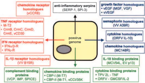
The sequences of over two dozen poxvirus genomes have been determined and multiple immunomodulatory proteins have been identified and broadly divided by func-tion into a three strategic classes: virostealth proteins, virotransducers and viromimetics (virokines and virocep-tors) (Nash et al., 1999). Virostealth encompasses a gen-eral strategy in which the visible signals of infection are masked in order to reduce the ability of cell-mediated immune responses to recognize and eliminate infected cells. Virotransducers are viral proteins that act intracellu-larly to inhibit innate antiviral pathways and the signal transduction cascades that mediate host range. Virokines and viroceptors represent virus-encoded proteins that mimic host cytokines or their receptors, respectively, thereby blocking extracellular communication signals and promoting a protected microenvironment for the virus within immuno-exposed tissues (Fig. 1). Generally, viro-ceptors, which can be either secreted or localized to the surface of infected cells, are related to cellular receptors and act by competing for ligands that promote antiviral
immune or inflammatory processes. In contrast, virokines are generally secreted viral proteins that mimic host mol-ecules, such as cytokines, complement regulators or their inhibitors.
KO poxvirus genes – strategies, considerations and effective analyses
Although the obvious sequence similarity between some poxvirus genes and the cDNA versions of related cellular counterparts provides insight into their function, the evo-lutionary origins of other ORFs are more obscure (Moss, 2001). Thus, genetic analyses employing recombinant DNA technologies are essential tools for studying the con-tribution of specific viral genes and proteins to virus–host interactions and viral pathogenesis. Much of what is known about viral pathogenesis can be traced back to discoveries made with knockout (KO) viruses, mutant viruses in which the targeted disruption of a specific viral gene produces phenotypic changes reflective of the nor-mal biological function of its protein product (Coen and Ramig, 1996). The typical method for generating KO mutants of poxviruses (Fig. 2) employs a step-wise approach that harks back to early marker-transfer mutagenesis strategies and closely resembles the ances-tral retrotranscription/recombination events by which many immunomodulatory proteins were likely acquired by poxviruses from their vertebrate hosts. The information that can be obtained from KO analyses is not sufficient to definitively assign a particular function to a viral gene product. Rather, the biological activity of purified proteins must be confirmed in relevant in vitro assays to provide further insight into how poxviral immunomodulatory pro-teins interact with their targets. Several recent reviews have provided in depth accounts of the advances made using these techniques for the study of poxviruses and the reader is referred to select examples for more
infor-mation (Moss and Shisler, 2001; Turner and Moyer, 2002; Seet et al., 2003).
The requirement for an animal model for in-depth anal-yses renders KO strategies ineffective for the study of poxviruses like molluscum contagiousum virus (MCV), an obligate human pathogen for which neither animal models nor tissue culture systems are available (Smith and Skel-ton, 2002). Consequently, investigation of the properties of poxviral immunomodulatory proteins using KO viruses has largely employed murine models in the study of ortho-poxviruses, such as vaccinia virus (VV), and rabbit models in the study of leporipoxviruses, such as myxoma virus (MV). VV exhibits a broad host range that includes several mammalian species, but the natural reservoir for the virus is unknown (Moss, 2001). The route of inoculation also significantly impacts upon disease course and VV infec-tion is achieved under experimental condiinfec-tions using intra-nasal, intracerebral, intraperitoneal or intradermal routes. In contrast, MV is an obligate rabbit pathogen that estab-lishes only a localized infection in its natural host species, the S. American Sylvilagus rabbit, but a lethal dissemi-nated disease (myxomatosis) in European Oryctolagus rabbits (Nash et al., 1999). Moreover, MV is transmitted under natural conditions by arthropod vectors and intrad-ermal injection is the most common mechanism for intro-ducing virus. Despite these differences, both VV and MV cause generalized disseminated infections characterized by the formation of a primary lesion at the initial site of inoculation and a viremia that spreads the infection through the host lymphoreticular system to establish inter-nal and exterinter-nal lesions in secondary organs and tissues. As a consequence of their immunosuppressive capacities, infection with VV and MV supports the development of supervening bacterial infections that ultimately lead to the death of host. Thus, targeted disruption of poxviral immu-nomodulatory genes can impact greatly on disease pro-gression in these models.
Fig. 1. Virokines and viroceptors produced by
poxvirus-infected cells. Indicated are select poxvirus viromimetics representing cytokine receptor homologues (red), cytokine and com-plement binding proteins (green), viral homo-logues of immune molecules (blue) and the anti-inflammatory poxviral serpins (black). IFN, interferon; IL, interleukin; TNF, tumour necrosis factor; VCP, vaccinia complement protein; IMP, inflammation modulatory protein; SPICE, smallpox inhibitor of complement enzymes; CBP, chemokine binding protein; vEGF, viral epithelial growth factor; vVEGF, viral vascular endothelial growth factor.
Viromimicry – seeing a familiar face
The MV and VV immunomodulatory genes that have been investigated using KO viruses are summarized in Table 1 and addressed in greater detail below with relevant exam-ples from other poxviral species. In a recent minireview,
we focused on the role of virostealth and virotransduction in poxvirus immune evasion (Johnston and McFadden, 2003). Thus, we emphasize here the large body of litera-ture devoted to gene KO analysis of poxviral virokines and viroceptors.
TNF viroceptors
Tumour necrosis factor (TNF) is a potent proinflammatory and proapoptotic cytokine secreted by macrophages and activated T-cells. To inhibit the activities of this cytokine, many poxviruses encode soluble proteins that resemble secreted versions of the extracellular domains of cellular TNFR, termed vTNFRs (Cunnion, 1999). vTNFRs func-tion primarily as molecular scavengers that bind to and sequester TNF, thereby blocking the interaction between the ligand and its native receptor. The T2-like vTNFRs, found in the leporipoxviruses MV and Shope fibroma viruse (SFV), are secreted glycoproteins that bind TNF with high affinity (Sedger and McFadden, 1996). SFV T2 has been reported to bind both TNF-a and TNF-b from several species (Smith et al., 1991), but MV T2 exhibits specificity for rabbit TNF-a (Schreiber et al., 1996). The orthopoxvirus cytokine response modifier (Crm)-like vTN-FRs, of which four major classes (CrmB, C, D and E) have been identified, also vary widely in distribution and biolog-ical activity (Saraiva and Alcami, 2001; Cunnion, 1999). CrmD is found primarily in poxviruses that lack CrmB and CrmC (Alcami et al., 1999), while functional CrmE homo-logues has been identified only in cowpoxvirus (CPV) and select VV strains (Reading et al., 2002; Saraiva and Alcami, 2001). Recently, members of a fifth Crm-like vTNFR family closely resembling CD30 have also been identified in CPV and ectromelia virus (EV) (Panus et al., 2002; Saraiva and Alcami, 2001).
KO studies. Despite the importance of TNF in host anti-viral responses, naturally arising MV strains deficient in M-T2 have been reported (Saint et al., 2001) and M-T2 KO viruses retain the capacity to infect susceptible hosts. However, rabbit lymphocytes infected in vitro with MV disrupted in both copies of the M-T2 gene undergo apo-ptosis and abortive infection (Macen et al., 1996). Consis-tent with this finding, the M-T2 KO virus is markedly attenuated in vivo, exhibiting decreased lethality and a pathology in which opportunistic bacterial infections are less frequent, primary lesions are smaller and less pro-nounced and secondary lesions are largely absent (Upton et al., 1991). Both the in vivo and in vitro observations suggest that deletion of M-T2 impairs virus replication and spread in an immunocompetent host, emphasizing the importance of inhibiting TNF to poxviral pathogenesis. KO analysis of Crm-like vTNFRs is hindered by virus strain-dependent variability in their expression; thus, the roles these proteins play in pathogenesis are more commonly
Fig. 2. General strategy for constructing knock-out poxviruses. A
fragment of the poxvirus genome containing the gene to be disrupted and flanking sequences of varying lengths is cloned and used as a template for subsequent manipulations. Two subfragments are ampli-fied by PCR using primers designed to selectively delete a portion of the targeted ORF while introducing restriction sites (RE) for subclon-ing the fragments. Each fragment is cloned in series into a transfer vector containing a selection marker under a poxvirus promoter (green fluorescence protein shown), such that the marker sequences further disrupt the target ORF. The vector encoding the poxvirus sequences is transfected into permissive cells that are infected at low multiplicity with wild-type virus. Within infected-transfected cells, homologous recombination occurs between the transfected sequences and wild-type virus genomes. KO viruses are isolated from wild-type virus and serially purified according to the selection criteria conferred by the selection marker.
studied by expressing their ORFs in a background that normally lacks them. For example, recombinant VV expressing CPV CrmB, CrmC or CrmE is more virulent in mice than wild-type virus, causing rapid weight loss and mortality (Reading et al., 2002). The contribution of the two vTNFRs encoded by VV (strain USSR), CrmE and A53R, however, has been assessed in both intradermal and intranasal mouse models using KO viruses. Deletion of A53R did not impact on virulence following infection by either route, but loss of CrmE resulted in marked attenu-ation of the virus when delivered by the intranasal route (Reading et al., 2002). Thus Crm-like vTNFRs likely con-tribute to pathogenesis, but in a manner that reflects the complex regulation of TNF in the host.
IFN viroceptors
The integral role played by interferons (IFNs) in host anti-viral responses is underscored by the fact that all poxvi-ruses employ at least one mechanism to disrupt its activity (Sen, 2001). As with TNF, a common strategy is
to encode soluble viral mimics of both Type I (a/b) and Type II (g) IFN receptors (IFN-R) that bind to and seques-ter these cytokines. For example, the B8R genes of both VV and EV encode proteins that bind IFN-g from several species (Alcami and Smith, 1995), although only the B8R homologue of EV inhibits murine IFN-g despite the ability of both viruses to infect mice (Smith and Alcami, 2002). Both viruses also encodes a protein (B18R) that closely resembles the IL-1 receptor but actually strongly interacts with Type I IFNs from several species (Smith and Alcami, 2002; Symons et al., 1995). Of note, the VV B18R prod-uct has been detected as both a secreted protein and localized to the surface of infected and uninfected cells (Alcami et al., 2000), suggesting that it protects infected cells from the direct action of IFN-a/b and uninfected cells from IFN-induced resistant to infection. MV encodes an IFN-g receptor homologue, M-T7, whose anti-IFN properties is rabbit-specific (Mossman et al., 1996), but purified M-T7 protein also exhibits the surprising ability to bind to diverse families of human chemokines (Lalani et al., 1997).
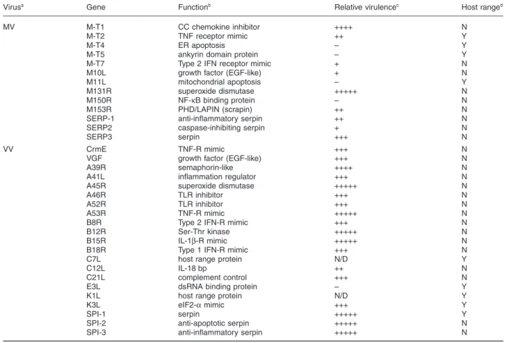
Table 1. Effects of disrupting select immunomodulatory genes on MV and VV pathogenesis.
Virusa Gene Functionb Relative virulencec Host ranged
MV M-T1 CC chemokine inhibitor ++++ N
M-T2 TNF receptor mimic ++ Y
M-T4 ER apoptosis – Y
M-T5 ankyrin domain protein – Y
M-T7 Type 2 IFN receptor mimic + N
M10L growth factor (EGF-like) + N
M11L mitochondrial apoptosis – Y
M131R superoxide dismutase +++++ N
M150R NF-kB binding protein – N
M153R PHD/LAPIN (scrapin) ++ N
SERP-1 anti-inflammatory serpin ++ N
SERP2 caspase-inhibiting serpin + N
SERP3 serpin +++ N
VV CrmE TNF-R mimic +++ N
VGF growth factor (EGF-like) +++ N
A39R semaphorin-like ++++ N
A41L inflammation regulator +++ N
A45R superoxide dismutase +++++ N
A46R TLR inhibitor +++ N
A52R TLR inhibitor +++ N
A53R TNF-R mimic +++++ N
B8R Type 2 IFN-R mimic +++ N
B12R Ser-Thr kinase +++++ N
B15R IL-1b-R mimic +++++ N
B18R Type 1 IFN-R mimic +++ N
C7L host range protein N/D Y
C12L IL-18 bp ++ N
C21L complement control +++ N
E3L dsRNA binding protein – Y
K1L host range protein N/D Y
K3L eIF2-a mimic +++ Y
SPI-1 serpin +++++ Y
SPI-2 anti-apoptotic serpin +++++ N
SPI-3 anti-inflammatory serpin +++++ N
a. Select immunomodulatory genes from myxoma (MV) and vaccinia (VV) virus for which KO analyses have been performed are shown. b. TNF, tumour necrosis factor; ER, endoplasmic reticulum; IFN, interferon; TLR, toll-like receptor; IL, interleukin.
c. Virulence exhibited by KO viruses is compared to wild-type virus on a descending scale from fully virulent (+++++) to avirulent (–). N/D, not done. d. Deleting a gene restricts (Y) or does not affect (N) replication in a permissive host or cell line.
KO studies. Deletion of both copies of M-T7 from MV attenuates the virus and leads to elevated inflammatory responses in primary lesions (Mossman et al., 1996). Given the contribution of chemokines to inflammation, however, this finding must be viewed in the context of the reported capacity for M-T7 to bind both chemokines and IFN. Deletion of the VV B8R gene has been reported to either enhance or not affect virulence in mice (Symons et al., 2002a; Verardi et al., 2001), although B8R-deletants are attenuated in other rodent species (Verardi et al., 2001). The increased virulence reported in some mice infected with B8R KO virus is particularly surprising because the protein does not bind mouse IFN, possibly indicating that like M-T7 this protein has multiple activities (Alcami and Smith, 1995). The VV B18R gene product also exhibits low affinity for murine IFN-a/b in vitro, but deletion of this gene from VV significantly attenuates vir-ulence in mice resulting in minimal disease symptoms, limited mortality and loss of neuroinvasiveness (Symons et al., 1995).
IL-1b viroceptors
The VV B15R ORF encodes a secreted interleukin (IL)-1b receptor that was shown to bind to both murine (Spriggs et al., 1992) and human (Alcami and Smith, 1992) IL-1b and to block the activity of the former in a functional bioassay (Spriggs et al., 1992). Although deletion of B15R influences VV pathogenesis in mice, the extent of the effect has been shown to vary according to the route of inocu-lation. Intracranial injection of VV deleted for B15R results in significant attenuation compared to wild-type virus (Spriggs et al., 1992), but B15R KO virus delivered intra-nasally exhibits lethality comparable to wild-type VV despite the earlier emergence of clinical symptoms (Alcami and Smith, 1992). In fact, B15R-deleted virus delivered intranasally induced greater fevers and was mar-ginally more pathogenic. Thus, the effects of deleting a specific virus gene can vary according to non-genotypic factors such as the inoculation route, likely reflecting regional differences in host immune responses. The patho-genic contribution of the IL-1b receptors encoded by other orthopoxviruses such as CPV and EV has yet to be deter-mined in the context of an in vivo infection.
Viral IL-18 binding proteins
Poxviruses also target the IFN pathway indirectly through proteins that scavenge IL-18, a pleiotrophic pro-inflammatory cytokine that induces IFNg production. Mammalian IL-18 binding protein (IL-18 BP) is a natural antagonist of IL-18 and many poxviruses have been shown or are predicted to encode soluble homologues of this protein (Smith et al., 2000; Xiang and Moss, 1999).
The IL-18BPs encoded by orthopoxviruses that are able to infect rodents, such as VV, EV and CPV, exhibit much greater affinity for murine IL-18 than human (Calderara et al., 2001). Of the three putative IL-18BPs encoded by the human poxvirus MCV (MC51L, 53 L and 54 L), only MC54L appears to bind to IL-18 (Xiang and Moss, 1999). This interaction exhibits high affinity for human and murine IL-18, but its role in pathogenesis can not be assessed in the context of MCV infection for the reasons detailed above.
KO studies. Recombinant murine IL-18 introduced sys-temically by intravenous injection has been shown to decrease the development of lesions and increased the activity of cytolytic immune cells following VV infection of mice (Tanaka-Kataoka et al., 1999). Deletion of the C12L gene encoding the VV IL-18 BP produces similar attenu-ation in mice inoculated intranasally (Symons et al., 2002b), further supporting the importance of IL-18 in con-trolling VV infections. In contrast, disruption of the EV IL-18 BP gene (p13) by insertional mutagenesis has little impact on pathogenic outcome beyond an observed increase in NK cell activity that correlates with moderate increases in the clearance of infected cells (Born et al., 2000). It should be noted that the presence of other intact immunomodulatory factors, such as the EV IFN-gR described above, may have compensated for the loss of IL-18 BP activity.
Viral IL-10
IL-10 is a multifunctional cytokine with both immunostim-ulatory and immunosuppressive effects. Homologues of IL-10 have been identified in the genomes of orf virus (ORFV), Yaba-like disease virus (YLDV) and lumpy skin disease virus (LSDV), but only the ORFV IL-10 has been characterized to date and shown to have biological activity similar to that of ovine IL-10 (Fleming et al., 1997). In vitro, ORFV IL-10 promotes thymocyte proliferation, costimu-lates mast cell growth and suppresses macrophages acti-vation (Imlach et al., 2002), suggesting a role in immune evasion that involves mimicking the suppressive effects of host IL-10 on Th1-mediated responses. More recently, ORFV IL-10 has also been implicated in the impairment of acquired immunity by inhibiting the maturation of and antigen presentation by dendritic cells (Lateef et al., 2003), possibly explaining why ORFV can repeatedly infect the same host. In a sheep model, deletion of ORFV IL-10 was shown to produce elevated levels of IFN-g in infected tissue compared to wild-type virus (Haig et al., 2002).
Viral growth factors
Many poxviruses encode proteins that resemble mamma-lian growth factors, most notably the homologues of
epi-dermal growth factor (EGF) detected in members of virtually all poxvirus genera and vascular endothelial growth factor (VEGF) found only in parapoxviruses (Seet et al., 2003). The EGF homologues encoded by VV and MV, termed VGF and MGF, respectively, are secreted pro-teins produced early in infection that compete with cellular EGF for receptors (EGFR) expressed on epithelial cells overlying sites of infection (Opgenorth et al., 1992; Stroobant et al., 1985). The functional consequences of this interaction include promoting EGFR autophosphory-lation and the generation of mitotic responses, as well as reducing EGFR downregulation and degradation to pro-long the duration of proliferative signals. The parapoxvi-ruses, ORFV and pseudocowpoxvirus (PCPV), encode biologically active homologues of mammalian VEGF-A that have been shown to stimulate proliferation of vascular endothelial cells and promote vascular permeability (Wise et al., 1999). Moreover, they exhibit a receptor binding profile that is unique among VEGF family members in its apparent specificity for VEGFR-2 and neuropilin-1 (Meyer et al., 1999). Thus, their function in pathogenesis is likely to contribute to the proliferative and highly vascularized nature of parapoxvirus lesions.
KO studies. Deletion of VGF affects VV pathogenesis in both mice and rabbits. For example, KO virus delivered by an intracranial route exhibits decreased neurovirulence in mice, whereas virus introduced intradermally produces less localized cellular proliferation in lesions (Buller et al., 1988). Similarly, infection of rabbits with MV KO virus lacking MGF was found to result in less mortality and milder disease symptoms, including less hyperplastic lesions and fewer secondary bacterial infections (Opgenorth et al., 1992). Disruption of the VEGF-like gene in ORFV results in the loss of the three VEGF activities associated with the parent virus: mitogenesis of vascular endothelial cells, induction of vascular perme-ability and activation of VEGF receptor 2 (Savory et al., 2000). In vivo, loss of viral VEGF does not impact greatly on viral replication, but it does result in lesions character-ized by decreased genesis of blood vessels at sites of infection, reduced inflammatory cell influx and abrogation of epidermal cell proliferation (Savory et al., 2000). Viral semaphorins
Semaphorins represent a family of cellular regulatory pro-teins implicated in both neuronal development and activa-tion of B and T lymphocytes. Included in this family are several poxviral proteins, most notably the products of the VV and EV A39R genes, which have been shown to possess the defining 500-amino acid ‘sema’ domain (Comeau et al., 1998). Moreover, these proteins interact with a novel virus-encoded semaphorin protein receptor, termed VESPR, a plexin family cell surface receptor for
which the natural ligand is unknown. Surprisingly, studies into the function of the EV A39R protein have suggested pro-inflammatory properties that manifest as increased recruitment of immune cells to sites of infection because of IL-6 and -8 upregulation (Comeau et al., 1998). This strategy likely favours virus dissemination within the host by attracting immune cells that can be subsequently infected.
KO studies. The VV (strain Western Reserve) A39R gene product is naturally truncated and insertion of full-length A39R from VV (strain Copenhagen) was shown to have only minimal effect on pathogenesis in mice (Gardner et al., 2001). Consistent with its pro-inflammatory poten-tial, moderately increased inflammation leading to larger lesions that were slower to resolve was observed following infection with virus containing A39R (Gardner et al., 2001). However, disease symptoms and viral titres remained unaffected. Similarly, deletion of the intact gene from VV (strain Copenhagen) did not influence pathogen-esis, suggesting that poxviral semaphorins may promote, but are not essential to, infection.
Viral anti-inflammatory serpins: SERP-1 and SPI-3 Serpins are a family of serine proteases inhibitors that regulate complex proteinases-dependent pathways involved in such processes as inflammation, apoptosis and tissue remodelling. The MV SERP-1 is the first pox-viral serpin shown to be secreted and therefore it is tech-nically a virokine (Upton et al., 1990). The orthopoxvirus SPI-3 is not secreted from RPV-infected cells and shares limited sequence homology with SERP-1, but the pres-ence of a common P1 Arg residue in the active site of the protein confers a similar inhibitory profile in vitro (Turner et al., 2000). Both proteins inhibit a range of trypsin-like serine proteinases in vitro, including tissue plasminogen activator, urokinase, plasmin, thrombin and factor Xa (Turner et al., 2000; Nash et al., 1998; Lomas et al., 1993), suggesting that the primary function of these serpins is to modulate host inflammatory responses to infection. Despite these similarities, however, SERP-1 and SPI-3 are not functionally interchangeable (Wang et al., 2000). The precise targets and receptors through which SERP-1 acts in vivo are not yet defined, although purified SERP-1 has recently been shown to interact with native vascular urokinase-type plasminogen activator receptors to inhibit inflammatory cell responses in a mouse model (Dai et al., 2003).
KO studies. Deleting the SPI-3 gene from the genome of orthopoxviruses such as CPV and VV has limited effect on pathogenesis in vivo and KO viruses exhibit only mod-erate reductions in virulence murine models (Thompson et al., 1993). In contrast, SERP-1 is an important
viru-lence factor for MV in its rabbit host. Deletion of SERP-1 markedly reduces the lethality of MV compared to wild-type virus and produces a pathology characterized by rapid induction of host inflammatory responses, reduced leukocyte infiltration and the development of fewer sec-ondary lesions (Upton et al., 1990). These observations suggest that SERP-1 is important to the dissemination of MV in vivo as well as the control of host inflammatory responses to infection.
Manipulation of chemokine function by poxviruses Chemokines (chemoattractant cytokines) are small, secreted cytokines that contribute to the host efforts to limit virus infections by coordinating the activation and mobilization of leukocytes that mediate inflammatory responses in areas of infection. Consequently, all poxvi-ruses attempt to modulate chemokine activity by encoding chemokine receptor homologues and secreted ligand mimics and chemokine binding proteins (CBPs) (Mahal-ingam and Karupiah, 2000). Bioinformatic analyses have identified putative G-protein-coupled chemokine receptor (GPCR) homologues and chemokine ligand mimics in the genomes of several poxviruses (Alcami, 2003). With the exception of the product of the MCV MC148R gene, which has been shown to function as a selective antagonist of human CCR8 (Ishikawa-Mochizuki et al., 1999), func-tional studies on these proteins are limited and their role in pathogenesis in vivo remains speculative. Greater insight has been gained into the role of poxvirus CBPs, however. Classified as either Type I (low affinity) or Type II (high affinity), their roles in poxvirus virulence have been examined extensively using KO viruses.
Type I poxviral CBPs
The dual-function IFNgR homologue of MV, M-T7, is the sole Type I poxvirus CBP identified to date. The capacity for M-T7 to inhibit the activity of a broad spectrum of chemokines, in addition to IFN-g, likely arises from its ability to interact with the heparin binding domains com-mon to many chemokines (Lalani et al., 1997). In doing so, M-T7 has the potential to interfere with generalized chemokine binding to glycosaminoglycans and disrupt the localization of a large number of C, CC and CXC chemok-ines in tissues. Deletion of the M-T7 gene produces marked attenuation that prevents dissemination of the virus to distal sites of infection (Mossman et al., 1996). Locally, loss of MT-7 function is associated with leukocyte infiltration into the primary dermal sites of viral replication and activation of leukocytes in secondary immune tissues, such as the lymph nodes and spleen (Mossman et al., 1996). Naturally, the question remains as to whether this phenotype reflects loss of binding to chemokines, IFN-g or both classes of molecules.
Type II poxviral CBPs
Type II CPB, also termed CPB-IIs or vCCIs, have been identified in several poxvirus species and are exemplified by the 35 kDa vCCI encoded by many orthopoxviruses (Smith et al., 1997) and the product of the M-T1 gene of MV (Graham et al., 1997). Despite lacking sequence sim-ilarity with known mammalian proteins, type II CBPs target the GPCR binding conserved among many CC chemok-ines to competitively inhibit their ability to interact with diverse cellular receptors (Seet and McFadden, 2002). Like the high binding affinities exhibited by these proteins, this property likely reflects the unique structure of type II CPBs that distantly resembled the collagen-binding domain of the Staphylococcus aureus adhesin molecule (Carfi et al., 1999). The proposed function of poxviral CBPs is based on in vitro studies that support the ability of these proteins to bind chemokines and impede leuko-cyte migration. However, loss of type II CBP activity appears to have limited effect on the virulence in vivo. For example, infection of rabbits with MV lacking M-T1 differs from wild-type infections only in the development of height-ened localized cellular inflammation in primary lesions, together with a moderate increase in infiltrating monocytes and macrophages early in infection (Lalani et al., 1999). Similarly, the virulence of a rabbitpox (RPV) KO virus lacking the 35 kDa CBP-II gene was shown to differ little from wild-type virus in mice (Martinezpomares et al., 1995). These results are perhaps unsurprising when the considerable redundancy in chemokine function and the combined effects of other poxviral immunomodulators that indirectly impact on chemokine function are considered.
Control of the complement system by poxviruses The complement system is an integrated network of cell-associated effector proteins and secreted regulatory pro-teins that participate in the identification and destruction of invading pathogens, as well as the initiation and ampli-fication of inflammatory responses. The VV complement control protein (VCP) typifies poxviral strategies to modu-late this system, with genes encoding similar products identified several other orthopoxviruses (Kotwal, 2000). VCP targets both the classical and alternative complement activation pathways by directly and indirectly promoting the decay of the C3 convertase (Sahu et al., 1998; Kotwal et al., 1990). A similar mechanism of action has been demonstrated for the smallpox inhibitor of complement enzymes (SPICE), the VCP homologue of the variola virus (VaV) (Rosengard et al., 2002). Given the virulence of VaV in humans compared to VV, the finding that SPICE inhibits human C3 activity nearly 100-fold more than VCP is of particular interest (Rosengard et al., 2002).
assess the function of the CPV VCP homologue, the inflammation modulatory protein (IMP). Although results varied extensively with host strain and route of inoculation, these studies suggest that modulation of host comple-ment contributes little to poxvirus virulence. For example, the IMP KO virus was not attenuated in BALB/c mice inoculated using either footpad injections (Miller et al., 1997) or a connective tissue air pouch model (Kotwal et al., 1998), exhibiting lethality comparable to the wild-type. However, inflammation and mononuclear cellular infiltration at sites of infection were greater in animals infected with the IMP KO virus compared to wild-type CPV. Because BALB/c mice express only low constitutive levels of C3, mice that were either fully deficient in C3 or expressed high levels of C3 were also studied (Kotwal et al., 1998). Infection with either wild-type or KO virus produced similar disease pathologies in C3-deficient mice, but the differences in inflammatory responses elic-ited by the viruses was comparable to that observed in BALB/c mice despite greater levels of host C3 expression.
Overview
The examples provided above illustrate the important con-tribution of immunomodulatory genes in the progression and resolution of poxvirus infections and their ability to impact on host range, virulence and pathogenicity. How-ever, it is difficult to predict the effect of deleting a specific gene because of the redundancy inherent to many poxvirus immune evasion strategies and the capacity for individual immunomodulatory proteins to have multiple functions. Consequently, the phenotype of a KO virus may not nec-essarily reflect the full extent to which a gene product contributes to pathogenesis. Despite these limitations, cer-tain trends do manifest when KO viruses are compared on the basis of the function of the gene disrupted. As shown in Table 1, deletion of genes whose products reg-ulate host antiviral responses that influence survival at the level of the infected cell, such as apoptosis, more pro-foundly affect virulence than genes encoding proteins that modulate more global host antiviral responses, such as chemokine and complement networks. Although manipu-lation of the latter strategies are important to the efficient spread of the virus once an infection has been established, failure to block innate defence mechanisms evolved to remove infected cells prevents the infection from being established at all. Thus, it is not surprising that poxviral proteins that modulate these critical host responses are determinants of host range as well as virulence.
Applications of the information gained from KO viruses
The immunomodulatory strategies of poxviruses are so
effective that new avenues of research, collectively known as virotherapeutics, have emerged in the attempt to exploit viral immunomodulatory proteins for the treatment of human diseases (Smith and Kotwal, 2001). For example, several of the poxviral proteins described in this review have been used in animal models to prevent allograft and xenograft transplant rejection, and to inhibit adverse immune responses in models of arterial injury following balloon angioplasty (Dabbagh et al., 2000; DeBruyne et al., 2000; Liu et al., 2000; Lucas et al., 1996; Maksy-mowych et al., 1996). In many of these applications, viral proteins are used as purified biotherapeutics outside the context of the intact virus and pathogenesis is not a con-sideration. However, other virotherapies based on live pox-viruses that have been modified to be less virulent or to exhibit a specific phenotype are becoming increasingly prevalent. These include the use poxviruses in vaccines (Mayr, 2003) and as therapeutic vectors and oncolytic agents (Kwak et al., 2003; Vanderplasschen and Pastoret, 2003). The rational design of such therapies requires detailed information about how modification of the viral genome impacts on the biology of poxviruses and empha-sizes the importance of KO virus analyses. In addition to the therapeutic benefit afforded by the characterization of poxvirus immunomodulators, the study of immune modu-lation by viruses contributes greatly to our understanding of how the immune system responds to infection and the selective pressures that drive the coevolution of virus and host. Greater understanding of the role individual genes play in poxvirus pathogenesis also has the potential to provide insights into novel targets on which to base anti-viral strategies, information of particular relevance given the potential use of smallpox as a bioterrorism agent.
Acknowledgements
We thank J. Barrett and S. Nazarian for critically reviewing the manuscript. We regret that space constraints prevent the citation of more literature pertinent to this subject and extend our apologies to those whose contributions we were unable to recognize.
References
Alcami, A. (2003) Viral mimicry of cytokines, chemokines and their receptors. Nat Rev Immunol 3: 36–50.
Alcami, A., Khanna, A., Paul, N.L., and Smith, G.L. (1999) Vaccinia virus strains Lister, USSR and Evans express soluble and cell-surface tumour necrosis factor receptors. J Gen Virol 80: 949–959.
Alcami, A., and Smith, G.L. (1992) A soluble receptor for interleukin-1 beta encoded by vaccinia virus: a novel mech-anism of virus modulation of the host response to infection. Cell 71: 153–167.
Alcami, A., and Smith, G.L. (1995) Vaccinia, cowpox, and camelpox viruses encode soluble gamma interferon
recep-tors with novel broad species specificity. J Virol 69: 4633– 4639.
Alcami, A., Symons, J.A., and Smith, G.L. (2000) The vac-cinia virus soluble alpha/beta interferon (IFN) receptor binds to the cell surface and protects cells from the antiviral effects of IFN. J Virol 74: 11230–11239.
Born, T.L., Morrison, L.A., Esteban, D.J., VandenBos, T., Thebeau, L.G., Chen, N.H., et al. (2000) A poxvirus protein that binds to and inactivates IL-18, and inhibits NK cell response. J Immunol 164: 3246–3254.
Buller, R.M., Chakrabarti, S., Cooper, J.A., Twardzik, D.R., and Moss, B. (1988) Deletion of the vaccinia virus growth factor gene reduces virus virulence. J Virol 62: 866–874. Calderara, S., Xiang, Y., and Moss, B. (2001) Orthopoxvirus
IL-18 binding proteins: Affinities and antagonist activities. Virology 279: 22–26.
Carfi, A., Smith, C.A., Smolak, P.J., McGrew, J., and Wiley, D.C. (1999) Structure of a soluble secreted chemokine inhibitor vCCl (p35) from cowpox virus. Proc Natl Acad Sci USA 96: 12379–12383.
Coen, D.M., and Ramig, R.F. (1996) Viral genetics. In Fields Virology. Fields, B.N., Knipe, D.M., Howley, P.M., Cha-nock, R.M., Melnick, J.L., Monath, T.P., et al. (eds). New York: New York Lippincott-Raven, pp. 113–152.
Comeau, M.R., Johnson, R., DuBose, R.F., Petersen, M., Gearing, P., VandenBos, T., et al. (1998) A poxvirus-encoded semaphorin induces cytokine production from monocytes and binds to a novel cellular semaphorin recep-tor, VESPR. Immunity 8: 473–482.
Cunnion, K.M. (1999) Tumor necrosis factor receptors encoded by poxviruses. Mol Genet Metab 67: 278–282. Dabbagh, K., Xiao, Y., Smith, C., Stepick-Biek, P., Kim, S.G.,
Lamm, W.J., et al. (2000) Local blockade of allergic airway hyperreactivity and inflammation by the poxvirus-derived pan-CC-chemokine inhibitor vCCI. J Immunol 165: 3418– 3422.
Dai, E., Guan, H., Liu, L., Little, S., McFadden, G., Vaziri, S., et al. (2003) Serp-1, a viral anti-inflammatory serpin, regu-lates cellular serine proteinase and serpin responses to vascular injury. J Biol Chem 278: 18563–18572.
DeBruyne, L.A., Li, K., Bishop, D.K., and Bromberg, J.S. (2000) Gene transfer of virally encoded chemokine antag-onists vMIP-II and MC148 prolongs cardiac allograft sur-vival and inhibits donor-specific immunity. Gene Ther 7: 575–582.
Fleming, S.B., McCaughan, C.A., Andrews, A.E., Nash, A.D., and Mercer, A.A. (1997) A homolog of interleukin-10 is encoded by the poxvirus orf virus. J Virol 71: 4857–4861. Gardner, J.D., Tscharke, D.C., Reading, P.C., and Smith,
G.L. (2001) Vaccinia virus semaphorin A39R is a 50–55 kDa secreted glycoprotein that affects the outcome of infec-tion in a murine intradermal model. J Gen Virol 82: 2083– 2093.
Graham, K.A., Lalani, A.S., Macen, J.L., Ness, T.L., Barry, M., Liu, L.Y., et al. (1997) The T1/35 kDa family of poxvirus-secreted proteins bind chemokines and modulate leuko-cyte influx into virus-infected tissues. Virology 229: 12–24. Haig, D.M., Thomson, J., McInnes, C., McCaughan, C., Imlach, W., Mercer, A., and Fleming, S. (2002) Orf virus immuno-modulation and the host immune response. Vet Immunol Immunopathol 87: 395–399.
Imlach, W., McCaughan, C.A., Mercer, A.A., Haig, D., and Fleming, S.B. (2002) Orf virus-encoded interleukin-10 stim-ulates the proliferation of murine mast cells and inhibits
cytokine synthesis in murine peritoneal macrophages. J Gen Virol 83: 1049–1058.
Ishikawa-Mochizuki, I., Kitaura, M., Baba, M., Nakayama, T., Izawa, D., Imai, T., et al. (1999) Molecular cloning of a novel CC chemokine, interleukin-11 receptor alpha-locus chemokine (ILC), which is located on chromosome 9p13 and a potential homologue of a CC chemokine encoded by molluscum contagiosum virus. FEBS Lett 460: 544–548. Johnston, J.B., and McFadden, G. (2003) Poxvirus
immuno-modulatory strategies: current perspectives. J Virol 77: 6093–6100.
Kotwal, G.J. (2000) Poxviral mimicry of complement and chemokine system components: what’s the end game? Immunol Today 21: 242–248.
Kotwal, G.J., Isaacs, S.N., McKenzie, R., Frank, M.M., and Moss, B. (1990) Inhibition of the complement cascade by the major secretory protein of vaccinia virus. Science 250: 827–830.
Kotwal, G.J., Miller, C.G., and Justus, D.E. (1998) The inflam-mation modulatory protein (IMP) of cowpox virus drastically diminishes tissue damage by down-regulating cellular infil-tration resulting from complement activation. Mol Cell Bio-chem 185: 39–46.
Kwak, H., Horig, H., and Kaufman, H.L. (2003) Poxviruses as vectors for cancer immunotherapy. Curr Opin Drug Dis-cov Devel 6: 161–168.
Lalani, A.S., Graham, K., Mossman, K., Rajarathnam, K., Clarklewis, I., Kelvin, D., and McFadden, G. (1997) The purified myxoma virus gamma interferon receptor homolog M-T7 interacts with the heparin-binding domains of chemokines. J Virol 71: 4356–4363.
Lalani, A.S., Masters, J., Graham, K., Liu, L.Y., Lucas, A., and McFadden, G. (1999) Role of the myxoma virus solu-ble CC-chemokine inhibitor glycoprotein, M-T1, during myxoma virus pathogenesis. Virology 256: 233–245. Lateef, Z., Fleming, S., Halliday, G., Faulkner, L., Mercer, A.,
and Baird, M. (2003) Orf virus-encoded interleukin-10 inhibits maturation, antigen presentation and migration of murine dendritic cells. J Gen Virol 84: 1101–1109. Liu, L.Y., Lalani, A., Dai, E.B., Seet, B., Macauley, C., Singh,
R., et al. (2000) The viral anti-inflammatory chemokine-binding protein M-T7 reduces intimal hyperplasia after vas-cular injury. J Clin Invest 105: 1613–1621.
Lomas, D.A., Evans, D.L., Upton, C., McFadden, G., and Carrell, R.W. (1993) Inhibition of plasmin, urokinase, tissue plasminogen activator, and C1S by a myxoma virus serine proteinase inhibitor. J Biol Chem 268: 516–521.
Lucas, A., Liu, L., Macen, J., Nash, P., Dai, E., Stewart, M., et al. (1996) Virus-encoded serine proteinase inhibitor SERP-1 inhibits atherosclerotic plaque development after balloon angioplasty. Circulation 94: 2890–2900.
Macen, J.L., Graham, K.A., Lee, S.F., Schreiber, M., Bosh-kov, L.K., and Mcfadden, G. (1996) Expression of the myx-oma virus tumor necrosis factor receptor homologue and M11L genes is required to prevent virus-induced apoptosis in infected rabbit T lymphocytes. Virology 218: 232–237. Mahalingam, S., and Karupiah, G. (2000) Modulation of
chemokines by poxvirus infections. Curr Opin Immunol 12: 409–412.
Maksymowych, W.P., Nation, N., Nash, P., Macen, J., Lucas, A., McFadden, G., and Russell, A.S. (1996) Amelioration of antigen induced arthritis in rabbits treated with a secreted viral serine proteinase inhibitor. J Rheumatol 23: 878–882.
Martinezpomares, L., Thompson, J.P., and Moyer, R.W. (1995) Mapping and investigation of the role in pathogen-esis of the major unique secreted 35-kDa protein of rabbit-pox virus. Virology 206: 591–600.
Mayr, A. (2003) Development of a non-immunising, paraspe-cific vaccine from attenuated pox viruses: a new type of vaccine. New Microbiol 26: 7–12.
Meyer, M., Clauss, M., Lepple-Wienhues, A., Waltenberger, J., Augustin, H.G., Ziche, M., et al. (1999) A novel vascular endothelial growth factor encoded by Orf virus, VEGF-E, mediates angiogenesis via signalling through VEGFR-2 (KDR) but not VEGFR-1 (Flt-1) receptor tyrosine kinases. EMBO J 18: 363–374.
Miller, C.G., Shchelkunov, S.N., and Kotwal, G.J. (1997) The cowpox virus-encoded homolog of the vaccinia virus com-plement control protein is an inflammation modulatory pro-tein. Virology 229: 126–133.
Moss, B. (2001) Poxviridae: the virus and their replication. In Fields Virology. Knipe, D.M., and Howley, P.M, (eds). Phil-adelphia: Lippincott Williams and Wilkins, pp. 2849–2883. Moss, B., and Shisler, J.L. (2001) Immunology 101 at
poxvi-rus U: immune evasion genes. Semin Immunol 13: 59–66. Mossman, K., Nation, P., Macen, J., Garbutt, M., Lucas, A., and Mcfadden, G. (1996) Myxoma virus M-T7, a secreted homolog of the interferon-gamma receptor, is a critical vir-ulence factor for the development of myxomatosis in Euro-pean rabbits. Virology 215: 17–30.
Nash, P., Barrett, J., Cao, J.X., Hota-Mitchell, S., Lalani, A.S., Everett, H., et al. (1999) Immunomodulation by viruses: the myxoma virus story. Immunol Rev 168: 103–120. Nash, P., Whitty, A., Handwerker, J., Macen, J., and
McFad-den, G. (1998) Inhibitory specificity of the anti-inflammatory myxoma virus serpin, Serp-1. J Biol Chem 273: 20982– 20991.
Opgenorth, A., Strayer, D., Upton, C., and McFadden, G. (1992) Deletion of the growth factor gene related to EGF and TGF alpha reduces virulence of malignant rabbit fibroma virus. Virology 186: 175–191.
Panus, J.F., Smith, C.A., Ray, C.A., Smith, T.D., Patel, D.D., and Pickup, D.J. (2002) Cowpox virus encodes a fifth mem-ber of the tumor necrosis factor receptor family: a soluble, secreted CD30 homologue. Proc Natl Acad Sci USA 99: 8348–8353.
Reading, P.C., Khanna, A., and Smith, G.L. (2002) Vaccinia virus CrmE encodes a soluble and cell surface tumor necrosis factor receptor that contributes to virus virulence. Virology 292: 285–298.
Rosengard, A.M., Liu, Y., Nie, Z., and Jimenez, R. (2002) Variola virus immune evasion design: expression of a highly efficient inhibitor of human complement. Proc Natl Acad Sci USA 99: 8808–8813.
Sahu, A., Isaacs, S.N., Soulika, A.M., and Lambris, J.D. (1998) Interaction of vaccinia virus complement control protein with human complement proteins – Factor I-medi-ated degradation of C3b to Ic3b(1) inactivates the alterna-tive complement pathway. J Immunol 160: 5596–5604. Saint, K.M., French, N., and Kerr, P. (2001) Genetic variation
in Australian isolates of myxoma virus: an evolutionary and epidemiological study. Arch Virol 146: 1105–1123. Saraiva, M., and Alcami, A. (2001) CrmE, a novel soluble
tumor necrosis factor receptor encoded by poxviruses. J Virol 75: 226–233.
Savory, L.J., Stacker, S.A., Fleming, S.B., Niven, B.E., and Mercer, A.A. (2000) Viral vascular endothelial growth factor
plays a critical role in orf virus infection. J Virol 74: 10699– 10706.
Schreiber, M., Rajarathnam, K., and Mcfadden, G. (1996) Myxoma virus T2 protein, a tumor necrosis factor (TNF) receptor homolog, is secreted as a monomer and dimer that each bind rabbit TNF alpha, but the dimer is a more potent TNF inhibitor. J Biol Chem 271: 13333–13341. Sedger, L., and McFadden, G. (1996) M-T2: a poxvirus TNF
receptor homologue with dual activities. Immunol Cell Biol
74: 538–545.
Seet, B.T., Johnston, J.B., Brunetti, C.R., Barrett, J.W., Ever-ett, H., Cameron, C., et al. (2003) Poxviruses and immune evasion. Annu Rev Immunol 21: 377–423.
Seet, B.T., and McFadden, G. (2002) Viral chemokine-binding proteins. J Leukoc Biol 72: 24–34.
Sen, G.C. (2001) Viruses and interferons. Annu Rev Micro-biol 55: 255–281.
Smith, V.P., and Alcami, A. (2002) Inhibition of interferons by ectromelia virus. J Virol 76: 1124–1134.
Smith, V.P., Bryant, N.A., and Alcami, A. (2000) Ectromelia, vaccinia and cowpox viruses encode secreted interleukin-18-binding proteins. J Gen Virol 81: 1223–1230.
Smith, C.A., Davis, T., Wignall, J.M., Din, W.S., Farrah, T., Upton, C., et al. (1991) T2 open reading frame from the Shope fibroma virus encodes a soluble form of the TNF receptor. Biochem Biophys Res Commun 176: 335–342. Smith, S.A., and Kotwal, G.J. (2001) Virokines: novel
immu-nomodulatory agents. Expert Opin Biol Ther 1: 343–357. Smith, K.J., and Skelton, H. (2002) Molluscum contagiosum:
recent advances in pathogenic mechanisms, and new ther-apies. Am J Clin Dermatol 3: 535–545.
Smith, C.A., Smith, T.D., Smolak, P.J., Friend, D., Hagen, H., Gerhart, M., et al. (1997) Poxvirus genomes encode a secreted, soluble protein that preferentially inhibits beta chemokine activity yet lacks sequence homology to known chemokine receptors. Virology 236: 316–327.
Spriggs, M.K., Hruby, D.E., Maliszewski, C.R., Pickup, D.J., Sims, J.E., Buller, R.M., and VanSlyke, J. (1992) Vaccinia and cowpox viruses encode a novel secreted interleukin-1-binding protein. Cell 71: 145–152.
Stroobant, P., Rice, A.P., Gullick, W.J., Cheng, D.J., Kerr, I.M., and Waterfield, M.D. (1985) Purification and charac-terization of vaccinia virus growth factor. Cell 42: 383–393. Symons, J.A., Adams, E., Tscharke, D.C., Reading, P.C., Waldmann, H., and Smith, G.L. (2002b) The vaccinia virus C12L protein inhibits mouse IL-18 and promotes virus vir-ulence in the murine intranasal model. J Gen Virol 83: 2833–2844.
Symons, J.A., Alcami, A., and Smith, G.L. (1995) Vaccinia virus encodes a soluble type I interferon receptor of novel structure and broad species specificity. Cell 81: 551–560. Symons, J.A., Tscharke, D.C., Price, N., and Smith, G.L. (2002a) A study of the vaccinia virus interferon-gamma receptor and its contribution to virus virulence. J Gen Virol
83: 1953–1964.
Tanaka-Kataoka, M., Kunikata, T., Takayama, S., Iwaki, K., Ohashi, K., Ikeda, M., and Kurimoto, M. (1999) In vivo antiviral effect of interleukin 18 in a mouse model of vac-cinia virus infection. Cytokine 11: 593–599.
Thompson, J.P., Turner, P.C., Ali, A.N., Crenshaw, B.C., and Moyer, R.W. (1993) The effects of serpin gene mutations on the distinctive pathobiology of cowpox and rabbitpox virus following intranasal inoculation of Balb/c mice. Virol-ogy 197: 328–338.
Turner, P.C., Baquero, M.T., Yuan, S., Thoennes, S.R., and Moyer, R.W. (2000) The cowpox virus serpin SPI-3 com-plexes with and inhibits urokinase-type and tissue-type plasminogen activators and plasmin. Virology 272: 267– 280.
Turner, P., and Moyer, R. (2002) Poxvirus immune modula-tors: functional insights from animal models. Virus Res 88: 35.
Upton, C., Macen, J.L., Schreiber, M., and McFadden, G. (1991) Myxoma virus expresses a secreted protein with homology to the tumor necrosis factor receptor gene family that contributes to viral virulence. Virology 184: 370–382. Upton, C., Macen, J.L., Wishart, D.S., and McFadden, G.
(1990) Myxoma virus and malignant rabbit fibroma virus encode a serpin-like protein important for virus virulence. Virology 179: 618–631.
Vanderplasschen, A., and Pastoret, P.P. (2003) The uses of poxviruses as vectors. Curr Gene Ther 3: 583–595.
Verardi, P.H., Jones, L.A., Aziz, F.H., Ahmad, S., and Yilma, T.D. (2001) Vaccinia virus vectors with an inactivated gamma interferon receptor homolog gene (B8R) are atten-uated in vivo without a concomitant reduction in immuno-genicity. J Virol 75: 11–18.
Wang, Y.X., Turner, P.C., Ness, T.L., Moon, K.B., Schoeb, T.R., and Moyer, R.W. (2000) The cowpox virus SPI-3 and myxoma virus SERP1 serpins are not functionally inter-changeable despite their similar proteinase inhibition pro-files in vitro. Virology 272: 281–292.
Wise, L.M., Veikkola, T., Mercer, A.A., Savory, L.J., Fleming, S.B., Caesar, C., et al. (1999) Vascular endothelial growth factor (VEGF)-like protein from orf virus NZ2 binds to VEGFR2 and neuropilin-1. Proc Natl Acad Sci USA 96: 3071–3076.
Xiang, Y., and Moss, B. (1999) IL-18 binding and inhibition of interferon gamma induction by human poxvirus-encoded proteins. Proc Natl Acad Sci USA 96: 11537–11542.