CD44/CD24 Expression in recurrent
gastric cancer: a retrospective analysis
The MIT Faculty has made this article openly available. Please share
how this access benefits you. Your story matters.
Citation
Yong, Ching-Shya et al. “CD44/CD24 Expression in Recurrent Gastric
Cancer: a Retrospective Analysis.” BMC Gastroenterology 12.1
(2012): 95.
As Published
http://dx.doi.org/10.1186/1471-230X-12-95
Publisher
BioMed Central Ltd
Version
Final published version
Citable link
http://hdl.handle.net/1721.1/73914
Terms of Use
Creative Commons Attribution
R E S E A R C H A R T I C L E
Open Access
CD44/CD24 Expression in recurrent gastric cancer:
a retrospective analysis
Ching-Shya Yong
1, Chih-Ming Ou Yang
1*, Yenn-Hwei Chou
1, Chao-Sheng Liao
1, Chung-Wei Lee
2and Chin-Cheng Lee
3Abstract
Background: To correlate CD44/CD24 expression with gastric cancer recurrence and prognosis. Gastric cancer is the second leading cause of cancer mortality due to the high recurrence rate, of which the molecular signature has not yet been identified.
Methods: We retrospectively reviewed the hospital records of patients with gastric cancer. Among 500 patients receiving curative resection, 95 patients had recurrence. Twenty patients from the recurrence group (95 patients) and 20 patients from the non-recurrence group (405 patients) were randomly selected and identified as“study” and “control” groups, respectively. We reviewed patients’ histological study of CD44/CD24 expression by performing immunohistochemistry and recurrence rate.
Results: Study group had higher TNM stage (III-IV) than control group (80% vs. 25%, P = 0.001). Proportion of lymph node metastasis was significantly higher in study group than that in control group (90% vs. 45%, P = 0.002), and proportion of patients with 5 or more metastatic lymph nodes was also significantly higher in study group than in control group (45% vs. 15%, P = 0.007). Univariate analysis revealed no difference in risk of gastric cancer recurrence between CD44+ and CD44- patients (OR = 1.00, 95% CI: 0.29-3.45, P =1.000). CD24+ patients showed no greater significance of gastric cancer recurrence than CD24- patients (OR = 1.86, 95% CI: 0.52-6.61, P = 0.339). After adjusting for other risk factors, the association of CD44 expression (aOR = 0.66, 95% CI: 0.10-4.26, P = 0.658), CD24 expression (aOR = 0.09, 95% CI: 0.01-1.35, P = 0.081) or combined (CD44/CD24) with gastric cancer recurrence were not significant.
Conclusion: Neither individual expression of CD24 or CD44, nor combined expression of CD44/CD24 was associated with recurrence of gastric carcinoma.
Keywords: CD44, CD24, Prognosis, Recurrent gastric cancer
Background
Gastric cancer (GC) is the fifth leading cause of cancer death in Taiwan, although its incidence and mortality rate have been declining in the past five decades. Glo-bally, GC is fourth most common among all types of cancer diagnoses, and is the second leading cause of cancer mortality [1], despite improvements in surgical techniques and development of new chemotherapeutic regimens. Annual deaths have reached 700,000 world-wide and 42% are reported in China alone [1]. Even after
curative resection, 40% of patients with advanced gastric cancer die of recurrence [2]. The prognosis for patients after curative surgery remains poor due to the high re-currence rate. The overall 5-year survival rate for patients who undergo curative surgical resection for gas-tric carcinoma ranges from 47% to 60.4%, and the recur-rence rate ranges from 15.4% to 37% [3].
Genetic susceptibility variants and molecular altera-tions related to environmental and lifestyle factors are known to contribute to development of GC, but even though many studies have investigated molecular mar-kers for the disease, the true mechanisms of GC carcino-genesis remain obscure [3]. Recurrence mechanisms also lack definitive explanation [4]. Even though we know * Correspondence:[email protected]
1
Department of Surgery, Shin Kong Wu Ho-Su Memorial Hospital, No.95, Wen Chang Road, Shih Lin District, Taipei City 11120, Taiwan
Full list of author information is available at the end of the article
© 2012 Yong et al.; licensee BioMed Central Ltd. This is an Open Access article distributed under the terms of the Creative Commons Attribution License (http://creativecommons.org/licenses/by/2.0), which permits unrestricted use, distribution, and reproduction in any medium, provided the original work is properly cited.
gastric carcinoma is prone to recur despite curative re-section, no molecular biomarker is currently available to predict gastric carcinoma recurrence after resection. While clinical predictive factors, such as tumor staging, can predict recurrence of advanced gastric cancer and are well recognized as essential predictors of prognosis, is there any molecular-based biomarker that can serve as a useful predictor for recurrence of advanced gastric cancer after curative resection (R0 resection)?
Both CD44 and CD24 are known to contribute to cel-lular signaling and cell adhesion, and their role in can-cer recurrence has been investigated. In a review of existing literature on the role of CD44/CD24 in recur-rent human cancer, investigators showed positive asso-ciations between CD44+/CD24- and prognosis, especially in breast cancer; and the CD44+/CD24-phenotype of breast cancer cells was also associated with invasive properties. [5] CD44 and CD24 have been shown to regulate invasion and metastasis of breast cancer cells either positively or negatively. Tumorigenic breast cancer cells that express high levels of CD44 and low or undetectable levels of CD24 (CD44+/CD24-/low) may be resistant to chemotherapy and therefore respon-sible for cancer relapse [6]. CD44 was also highly expressed in gastric adenocarcinoma and its expression correlated with poor prognosis in patients with the in-testinal type of gastric adenocarcinoma [7]. Based on the implications of these previous studies, we hypothe-sized that CD44+/CD24- expression might be corre-lated with gastric cancer recurrence.
To our knowledge, current studies report no valid adhesion molecule to predict disease recurrence after patients undergo curative resection for gastric carcin-oma. Because the significance of CD44+/CD24- is probably not unique to breast cancer, and CD44 has been highly expressed in gastric cancer, we might speculate that similar or other molecules may also regulate the process of recurrence for gastric cancer. Therefore, we decided to investigate the expression of adhesion molecule CD44/CD24 in recurrent gastric cancer and its possible predictive relevance in future clinical practice. The purpose of this study was to evaluate the correlation of CD44/CD24 expression with recurrent gastric cancer and to determine its prognos-tic significance.
Methods Patient selection
The protocol for this study was reviewed and approved by the internal review board of Shin Kong Memorial Hospital. Data were obtained from a retrospectively maintained database consisting of patients evaluated for gastric cancer from 1993 to 2007. Tumors were staged according to the criteria of the American Joint
Commission for Cancer (AJCC 6th edition) for gastric cancer and were classified histologically according to the WHO criteria; Lauren’s classification was also applied (intestinal type GC corresponds to well- or moderately differentiated tumors; diffuse type corresponds with poorly differentiated tumors). A retrospective review of patients’ medical records was also completed and data were collected for age, sex, and final pathologic diagnosis.
During the period from January 1993 to December 2007, 500 patients received curative resection for gas-tric cancer at the Department of Surgery of Shin-Kong Wu Ho-Su Memorial Hospital. Among these, 95 patients (19%) developed a recurrence during long-term follow-up.
Forty patients with gastric cancer were randomly selected as subjects in our study. We retrospectively ana-lyzed CD44/CD24 expression in patients’ post-operative pathologic specimens. The patients were divided into two groups; 20 patients with recurrent gastric cancer were defined as the study group and the other 20 patients without recurrent gastric cancer were categor-ized as the control group. Among patients in the non-recurrence control group, the minimum disease-free sur-vival was 4 years (2002–2006) and the maximum disease-free survival was 18 years (1994–2012). A total of 14 patients from the 20 non-recurrence group are still living today. All included patients provided signed informed consent to participate in the study.
Tissue preparation
Immunohistochemistry stains for CD44 and CD24 were performed for all specimens. The cancer tissues were fixed with formalin and embedded in paraffin. After paraffin removal and rehydration, antigen retrieval was performed by placing sections into a beaker containing adequate amounts of citrate buffer (pH 6.0), then heat-ing in pressure cooker for 10 min and coolheat-ing to room temperature. After 10 min, 3% H2O2 was added. Pri-mary antibody and secondary antibody (Envision Detec-tion Kit, Wonderful Life Science Co. Ltd, Taiwan) were added after 60 minues and 90 minutes, respectively, coupled with PBS washing in between the addition of antibodies. DAB substrate solution was subsequently added and washed with running water before counter-staining with hematoxylin. Finally, tissues were washed with distilled water, dehydrated and mounted for micro-scopic evaluation. The intensity (0, 1+, 2+, 3+) of tumor cell staining was independently evaluated by two pathologists, and discrepant results were resolved by reviewing the cases together and agreeing on the scores. A complete negative staining was scored as negative (0). A weak staining (1+) was defined as minimal, but unequivocal staining in less than 10% of tumor cell.
Yong et al. BMC Gastroenterology 2012, 12:95 Page 2 of 7 http://www.biomedcentral.com/1471-230X/12/95
Stronger or more extensive staining was scored as mod-erately/strongly positive (2+/3+) (Figure 1). Tumors with weak and moderate/strong staining were defined as having positive expression, while those with negative staining only were defined as negative expression.
Statistical analysis
Data were presented as mean ± SD for continuous vari-ables, and frequencies with percentages for categorical variables. Differences between the study group (recur-rence) and the control group (non-recur(recur-rence) were ana-lyzed using independent t-test test for continuous variables, and Chi-square test or Fisher’s exact test for categorical variables as appropriate. To investigate the association of CD44 and CD24 expression as well as other risk factors with gastric cancer recurrence, the point estimates and 95% confidence intervals (CIs) of odds ratios (ORs) were calculated by univariate and multivariate logistic regression models. Multivariate lo-gistic regression with backward selection was applied, wherein variables that did not improve the model fit at P<0.05 were discarded; however, CD44 and CD24 ex-pression as well as age were always forced into the model. Two multivariate models were applied to evaluate the association of CD44 and CD24 expression with gas-tric cancer recurrence: Model 1 considered CD44 and CD 24 as two variables, while Model 2 considered the combined CD44/CD24 expression as one variable. All statistical analyses were performed with SAS software version 9.2 (SAS Institute Inc., Cary, NC, USA). A two-tailed P<0.05 indicated statistical significance.
Results
A total of 40 patients were enrolled in this study, includ-ing 19 males and 21 females with mean age of 68.4 ± 11.4 years old (ranging from 44 to 88 years old). Comparison of demographic and clinical characteristics between the study group and the control group are shown in Table 1. Study group subjects were more likely to be classified as higher TNM stage (III-IV) than con-trol group subjects (80% vs. 25%,P = 0.001). In addition, the proportion of lymph node metastasis was signifi-cantly higher in the study group than that in the control group (90% vs. 45%,P = 0.002). Moreover, the proportion of patients with five or more metastatic lymph nodes was also significantly higher in the study group than in the control group (45% vs. 15%, P = 0.007). No signifi-cant differences were found in other characteristics be-tween the study group and the control group. (Table 1).
Univariate and multivariate regression analysis
The univariate and multivariate associations of CD44 and CD24 expression, as well as other risk factors with gastric cancer recurrence, are shown in Table 2. In uni-variate analysis, patients with CD44+ showed no differ-ence in risk of gastric cancer recurrdiffer-ence compared to those who were CD44- (OR = 1.00, 95% CI: 0.29-3.45, P = 1.000). Compared to patients who were CD24-, those with CD24+ had higher likelihood to have gastric cancer recurrence but without significance (OR = 1.86, 95% CI: 0.52-6.61, P = 0.339). Compared to patients with com-bined expression of CD44-/CD24-, the unadjusted ORs for CD44-/CD24+, CD44+/CD24-, and CD44+/CD24+ were 4.00 (95% CI: 0.55-29.10), 1.60 (95% CI: 0.30-8.49),
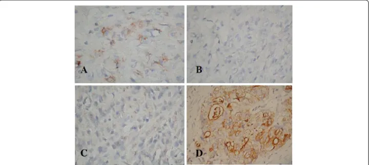
Figure 1 Immunohistochemical staining of C44/CD24 protein expression in recurrent gastric cancer tissue (400x). A: CD44 2+, B: CD24 -, C: CD44 -, D: CD24 3+.
and 1.60 (95% CI: 0.30-8.49), respectively. No signifi-cance was shown in the crude association of CD44/ CD24 expression with gastric cancer recurrence. Also,
higher TNM stage (III-IV), lymph node metastasis and higher numbers of metastasis lymph nodes, were all sig-nificantly associated with gastric cancer recurrence.
In multivariate model 1, when CD44 and CD24 ex-pression and age were forced into the model, only gen-der (male vs. female, aOR = 39.64, 95% CI: 1.85-848.44, P = 0.019), TNM stage (Stage III-IV vs. I-II, aOR = 9.63, 95% CI: 1.05-88.76,P = 0.046), and lymph node metasta-sis (Yes vs. No, aOR = 20.92, 95% CI: 1.20-365.55, P = 0.037) achieved a significance level allowing them to be retained in the multivariate logistic regression model. However, after adjusting for other risk factors, the asso-ciation of CD44 expression (aOR = 0.66, 95% CI: 0.10-4.26, P = 0.658) and CD24 expression (aOR = 0.09, 95% CI: 0.01-1.35, P = 0.081) with gastric cancer recurrence were still not significant.
In multivariate model 2, when combined CD44/CD24 expression and age were forced into the model, only gender (male vs. female, aOR = 21.59, 95% CI: 1.24-377.26, P = 0.035) and TNM stage (Stage III-IV vs. I-II, aOR = 41.01, 95% CI: 3.62-464.91, P = 0.003) achieved a significance level allowing them to be retained in the multivariate logistic regression model. Compared to patients with combined expression of CD44-/CD24-, the adjusted ORs for CD44-/CD24+, CD44+/CD24-, and CD44+/CD24+ were 0.60 (95% CI: 0.03-10.67), 1.61 (95% CI: 0.15-17.36), and 0.06 (95% CI: 0.002-2.31), respectively. After adjusting for other risk factors, the association of CD44/CD24 expression with gastric cancer recurrence was still not significant.
Discussion
Evaluation of the correlation of CD44/CD24 expression with recurrent gastric cancer revealed no differences in risk of gastric cancer recurrence between CD44+ patients and CD44- patients. Although CD24+ patients had a higher likelihood of gastric cancer recurrence than CD24- patients, significance was not demonstrated. Our study results suggest that these molecules do not appear to be clinically useful for prediction of gastric carcinoma recurrence after curative resection.
The role of CD44 and CD24 expression in gastric car-cinoma has been explored for nearly thirty years. Nu-merous studies have focused on the diagnostic and prognostic significance of CD44 expression in human tumors, especially gastric cancer. In 1982, CD44 was identified as a surface glycoprotein and a lymphocyte homing receptor found on lymphoid and epithelial cells [8]; its main function on lymphocytes is mediating inter-action with the endothelium, but its function on epithe-lial cells is not entirely understood [9]. The CD44 proteins belong to a family of type I transmembrane gly-coproteins that are encoded by a single, highly con-served gene located on the short arm of chromosome 11
Table 1 Comparison of demographic and clinical characteristics between patients with / without gastric cancer recurrence
Characteristics Study Group Control Group P-value (n = 20) (n = 20) Recurrence Non-recurrence Gender, n(%) Male 12 (60.0) 7 (35.0) 0.113† Female 8 (40.0) 13 (65.0) Age (years) mean ± SD 71.2 ± 10.6 65.7 ± 11.7 0.128{ Age group, n(%) ≤65 6 (30.0) 10 (50.0) 0.197† >65 14 (70.0) 10 (50.0) Histology, n(%) Moderately differentiated (intestinal type) 9 (45.0) 6 (30.0) 0.327† Poorly differentiated (diffuse type) 11 (55.0) 14 (70.0) Tumor location, n(%)
Upper third of stomach 5 (25.0) 10 (50.0) 0.191} Middle third of stomach 1 (5.0) 0 (0.0)
Lower third of stomach 14 (70.0) 10 (50.0) TNM Stage, n(%)
I-IIa 4 (20.0) 15 (75.0) 0.001*† III-IV 16 (80.0) 5 (25.0)
Lymph node metastasis, n(%)
No 2 (10.0) 11 (55.0) 0.002*† Yes 18 (90.0) 9 (45.0)
Number of lymph node metastasis, n(%)
0 2 (10.0) 11 (55.0) 0.007*† 1-4 s 9 (45.0) 6 (30.0) ≥5 9 (45.0) 3 (15.0) CD44 expression, n(%) CD44- 10 (50.0) 10 (50.0) 1.000† CD44+ 10 (50.0) 10 (50.0) CD24 expression, n(%) CD24- 10 (50.0) 13 (65.0) 0.337† CD24+ 10 (50.0) 7 (35.0) CD44 / CD24 expression, n(%) CD44-/CD24- 5 (25.0) 8 (40.0) 0.622} CD44-/CD24+ 5 (25.0) 2 (10.0) CD44+/CD24- 5 (25.0) 5 (25.0) CD44+/CD24+ 5 (25.0) 5 (25.0) a
Two control patients with TNM Stage 0 were grouped into Stage I-II. *P<0.05.
† Chi-square test; { independent t-test; }Fisher’s exact test.
Yong et al. BMC Gastroenterology 2012, 12:95 Page 4 of 7 http://www.biomedcentral.com/1471-230X/12/95
in humans; two molecular sizes have been identified: low MrCD44 (80–90 x 103) is expressed in lymphoid tissue
and high Mr CD44 (130–160 x 103) is expressed in tumor cells and keratinocytes [10]. Some aggressive tumors are reported to be associated with the expression of CD44. Overexpression of CD44, defined by apparently increased expression of CD44 protein, has been linked to poor prognosis with tumor progression and meta-static potential in several human malignancies, including
gastric cancer [7], colorectal cancer, breast cancer [5,6], uterine cancer, ovarian cancer, bladder cancer, lung can-cer, hematopoietic malignancies, and gliomas [11]. A retrospective study of 100 patients with gastric cancer evaluated the expression of CD44 and its prognostic im-portance, concluding that this cell adhesion molecule is highly expressed in gastric adenocarcinoma [7]. In that study, expression of CD44 correlated with a poor prog-nosis in patients with the intestinal type of gastric
Table 2 Univariate and multivariate associations of CD44/CD24 expression and other risk factors with gastric cancer recurrence
Characteristics Recurrence rate Univariate P-value Model 1† P-value Model 2{ P-value OR (95%CI) aOR (95%CI) aOR (95%CI)
CD44 expression CD44- 50.0% (10/20) 1.00 (reference) – 1.00 (reference) – CD44+ 50.0% (10/20) 1.00 (0.29-3.45) 1.000 0.66 (0.10-4.26) 0.658 CD24 expression CD24- 43.5% (10/23) 1.00 (reference) – 1.00 (reference) – CD24+ 58.8% (10/17) 1.86 (0.52-6.61) 0.339 0.09 (0.01-1.35) 0.081 CD44 / CD24 expression CD44-/CD24- 38.5% (5/13) 1.00 (reference) – 1.00 (reference) – CD44-/CD24+ 71.4% (5/7) 4.00 (0.55-29.10) 0.171 0.60 (0.03-10.67) 0.731 CD44+/CD24- 50.0% (5/10) 1.60 (0.30-8.49) 0.581 1.61 (0.15-17.36) 0.695 CD44+/CD24+ 50.0% (5/10) 1.60 (0.30-8.49) 0.581 0.06 (0.002-2.31) 0.131 Gender Male 63.2% (12/19) 2.79 (0.77-10.04) 0.117 39.64 (1.85-848.44) 0.019* 21.59 (1.24-377.26) 0.035* Female 38.1% (8/21) 1.00 (reference) – 1.00 (reference) – 1.00 (reference) – Age (years)
≤65 37.5% (6/16) 1.00 (reference) – 1.00 (reference) – 1.00 (reference) – >65 58.3% (14/24) 2.33 (0.64-8.54) 0.201 7.77 (0.89-67.99) 0.064 4.07 (0.49-34.16) 0.196 Histology
Moderately differentiated (intestinal type) 60.0% (9/15) 1.91 (0.52-7.01) 0.330 Poorly differentiated (diffuse type) 44.0% (11/25) 1.00 (reference) – Tumor location
Upper third + Middle third 37.5% (6/16) 1.00 (reference) – Lower third 58.3% (14/24) 2.33 (0.64-8.54) 0.201 TNM Stage
I-IIa 21.1% (4/19) 1.00 (reference) – 1.00 (reference) – 1.00 (reference) –
III-IV 76.2% (16/21) 12.0 (2.70-53.3) 0.001* 9.63 (1.05-88.76) 0.046* 41.01 (3.62-464.91) 0.003* Lymph node metastasis
No 15.4% (2/13) 1.00 (reference) – 1.00 (reference) – Yes 66.7% (18/27) 11.0 (2.00-60.5) 0.006* 20.92 (1.20-365.55) 0.037* Number of lymph node metastasis
0 15.4% (2/13) 1.00 (reference) – 1-4 s 60.0% (9/15) 8.25 (1.33-51.2) 0.024* ≥5 75.0% (9/12) 16.5 (2.25-121.2) 0.006* † In multivariate Model 1, CD44 and CD24 expression were considered as two variables.
{ In multivariate Model 2, CD44 and CD24 expression were combined as one variable to show different combinations. a
Two control patients with TNM Stage 0 were grouped into Stage I-II. Note: OR, odds ratio; aOR, adjusted odds ratio.
adenocarcinoma. These investigators suggested that CD44 could be utilized as a prognostic marker for this group of patients. In this study, the proportion of lymph node metastasis was significantly higher in the GC study group than that in the control group and the proportion of patients with 5 or more metastatic lymph nodes was also significantly higher in the study group than in the control group. However, while CD44 was definitely linked with GC in our study and prognostic to some de-gree, we could not verify its predictive capability in terms of GC recurrence after tumor resection.
CD24, a mucin-type GPI-linked cell surface molecule on human neutrophils and pre-B lymphocytes, plays an important role in the margination and adhesion of cells under shear force of blood flow [11]. Positive CD24 ex-pression is found to occur in a subset of GC and to correl-ate with lymphatic invasion, blood vessel invasion and poor survival. The clinicopathological significance of CD24 expression in human gastric adenocarcinoma was evaluated by Chou and colleagues, who concluded that cytoplasmic expression of CD24 was associated with inva-siveness and poorer prognosis and can serve as a novel target for prognostic prediction and adjuvant treatment of patients with diffuse-type gastric adenocarcinoma after tumor resection [12]. Further studies are needed to inves-tigate other combinations of adhesion molecules.
In the present study, CD44 and CD24 expression, in-dependently and in combination, were not associated with GC recurrence. Carcinoma of the intestinal type are more frequently CD44s and CD44v6 positive than carcinomas of the diffuse type, and the importance of subclassifying tumor types in investigations of CD44 in human cancer has been demonstrated [13]. When cyto-plasmic CD24 expression was studied in diffuse-type gastric adenocarcinoma, it was shown to be associated with invasiveness, lymph node metastasis and poorer prognosis, but not specifically associated with recurrence after tumor resection; however, no significant differences were seen in tumor stage or lymph node metastasis be-tween mixed-type GC with or without CD24 expression [12]. Although there is no ideal cross-comparison be-tween different histological grading systems, we did clas-sify GC according to the WHO histological classification and Lauren’s classification in the present study. We had no cases of lymphoepithelial-like carcinoma or mucosa-associated lymphoid tissue (MALT) lymphoma.
Another possible explanation of lack of significant associations between CD44 and CD24 and recurrence may be the particular isoform of CD44 identified in our study. Up to seven molecular forms have varying func-tional roles in vivo such as having different abilities to bind hyaluronate, while their most important common feature is their expression on tumor cells and correlation with metastases [10]. These forms can be identified
more readily by sequencing mRNAs and only a few are identified by protein analysis. The distribution of CD44 and CD24 may also differ and cellular sites in normal tissue have only been identified in animal models, and are not confirmed in normal human tissue. However, larger forms are found as minor components in normal tissue and low Mr forms are associated with lymphoid
cell types in tissue [14]. The function of CD44/CD24 is complex and changes in CD44, in particular, have been noted in carcinogenesis, including gene expression modulation, splicing of RNA and altered glycosylation [13]. Any of the above factors may account for the ap-parent lack of association with recurrence in our study. Clearly, more research is needed.
Molecular pathways involved in GC have been identi-fied over the past twenty years [15]. Current investiga-tions of molecular markers for gastric progenitor cells and gastric stem cells may hold promise for learning more about GC, its progression and propensity for re-currence, and eventually for treatment applications. This possibility is primarily because gastric tissue, as well as intestinal tissue undergoes constant epithelial cell re-placement and because stem cells and progenitor cells play important roles in the renewal of gastric glands and in epithelial repair following tissue injury [16]. Zhang et al. [17] examined CD44 and CD24 in gastric cells lines, AGS and gastric cancer tissues and identified the tumorigenic properties, self-renewal and differentiated progeny in CD44 + CD24+ and CD44-CD24- cell popu-lations. As few as 200 of the positive combination of cells injected into mice generated tumors in 50%, while many thousands more negative combined cells were needed to form a tumor in a second group of mice, sug-gesting that the subpopulation of CD44 + CD24+ gastric cell lines (AGS tumor cell lines) is GC stem cells [17]. In protein studies, CD24, which is associated with tumor metastasis, and Galectin-1 expression, which is asso-ciated with immune response and tumor progression, were studied in GC patients, giving particular attention to staining intensity and clinicopathologic variables; the researchers concluded that these proteins were inde-pendent prognostic indicators of poor survival (though not specifically associated with recurrence) and could be useful as therapeutic targets [18]. Since we are not yet studying the unique progenitor and stem cell popula-tions in human models, we should continue exploring tumor-specific protein expression that may be associated with GC and recurrent GC, seeking to find a molecular basis for this globally prevalent disease and new paths to diagnosis, prognosis and treatment.
Limitations
This study has several limitations, with the main limita-tion being its retrospective nature and limited sample
Yong et al. BMC Gastroenterology 2012, 12:95 Page 6 of 7 http://www.biomedcentral.com/1471-230X/12/95
size. In addition, the functional significance of CD44 +/CD24- cells is still unclear. Their importance in me-tastasis, and thus in disease recurrence and gastric can-cer mortality, is not yet well understood. This small retrospective series served as an initial study to investi-gate the potential association between CD44 and CD24 and gastric cancer recurrence, but a study with a larger sample is needed to confirm our preliminary results. We must also acknowledge that we were unable to confirm intratumor heterogeneity because there were only lim-ited numbers of surgically resected specimens of the re-current or metastatic tumors for repeat CD44/CD24 staining, which may be considered in future studies.
Conclusion
In conclusion, neither individual expression of CD24 and CD44 nor combined expression of CD44/CD24 was associated with recurrence of gastric carcinoma. Since adhesion molecules CD44 and CD24 do not appear to be clinically useful for prediction of gastric carcinoma recurrence after curative resection, future research is warranted to both confirm these results and to investi-gate other possible biomarkers.
Competing interests
The authors declare that they have no competing interests. Authors’ contributions
CSY, CMOY, and CWL performed majority of experiments; CMOY and YHC collected all human material; CCL reviewed pathologies; CSL analyzed data; CSY and CMOY designed the study and wrote the manuscript. All authors read and approved the final manuscript.
Acknowledgments
The authors would like to thank Dr. Cecile Logan for reviewing the immunostaining results and Ms. Chin Yu-Chun for her technical assistance. Author details
1
Department of Surgery, Shin Kong Wu Ho-Su Memorial Hospital, No.95, Wen Chang Road, Shih Lin District, Taipei City 11120, Taiwan.2Department of
Biological Engineering, Massachusetts Institute of Technology, Massachusetts, USA.3Department of Pathology, Shin Kong Wu Ho-Su Memorial Hospital,
No.95, Wen Chang Road, Shih Lin District, Taipei City 11120, Taiwan.
Received: 27 March 2012 Accepted: 17 July 2012 Published: 28 July 2012
References
1. Parkin DM, Bray F, Ferlay J, Pisani P: Global cancer statistics 2002. CA Cancer J Clin 2005, 55:74–108.
2. Moriguchi S, Maehara Y, Korenaga D, Sugimachi K, Nose Y: Risk factors which predict pattern of recurrence after curative surgery for patient with advanced gastric cancer. Surg Oncol 1992, 1:341–346. doi:10.1016/ 0960-7404(92)90034-I. [PMID: 1341269.
3. Samson PS, Escovidal LA, Yrastorza SG, Veneracion RG, Nerves MY: Re-study of gastric cancer: analysis of outcome. World J Surg 2002, 26:428–433. 4. Resende C, Thiel A, Machado JC, Ristimäki A: Gastric cancer: Basic aspects.
Helicobacter 2011, 16(Suppl 1):38–44.
5. Sheridan C, Kishimoto H, Fuchs RK, Mehrotra S, Bhat-Nakshatri P, Turner CH, Goulet R Jr, Badve S, Nakshatri H: CD44+/CD24- breast cancer cells exhibit enhanced invasive properties: an early step necessary for metastasis. Breast Cancer Res 2006, 8:R59–[PMID: 17062128. doi:10.1186/bcr165. 6. Abraham BK, Fritz P, McClellan M, Hauptvogel P, Athelogou M, Brauch H:
Prevalence of CD44+/CD24/low cells in breast cancer may not be
associated with clinical outcome but may favor distant metastasis. Clin Cancer Res 2005, 11:1154–1159.
7. Ghaffarzadehgan K, Jafarzadeh M, Raziee HR, Sima HR, Esmaili-Shandiz E, Hosseinnezhad H, Taghizadeh-Kermani A, Moaven O, Bahrani M: Expression of cell adhesion molecule CD44 in gastric adenocarcinoma and its prognostic importance. World J Gastroenterol 2008, 14:6376–6381. doi:10.3748/wjg.14.6376. PMID: 19009655.
8. Gallatin WM, Weissman IL, Butcher EC: A cell-surface molecule involved in organ-specific homing of lymphocytes. Nature 1983, 13:30–34. 9. Idzerda RL, Carter WG, Nottenburg C, Wayner EA, Gallatin WM, St John T:
Isolation and DNA sequence of a cDNA clone encoding a lymphocyte adhesion receptor for high endothelium. Proc Nat Acad Sci 1989, 86:4654– 4658.
10. Stamenkovic I, Aruffo A, Amiot M, Seed B: The hematopoietic and epithelial forms of CD44 are distinct polypeptides with different adhesion potentials for hyalouronate-bearing cells. EMBO J 1991, 10 (2):343–348.
11. Darwish NS, Kim MA, Change MS, Lee HS, Lee BL, Kim YI, Kim WH: Prognostic significance of CD24 expression in gastric carcinoma. Cancer Res Treat 2004, 36:298–302.
12. Chou YY, Jeng YM, Lee TT, Hu FC, Kao HL, Lin WC, Lai PL, Hu RH, Yuan RH: Cytoplasmic CD24 Expression Is a Novel Prognostic Factor in Diffuse-Type Gastric Adenocarcinoma. Ann Surgl Oncol 2007, 14:2748–2758. doi:10.1245/s10434-007-9501-X. PMID: 17680316.
13. Hong RL, Lee WJ, Shun CT, Chu JS, Chen YC: Expression of CD44 and its clinical implication in diffuse- and intestinal-type gastric
adenocarcinomas. Oncology 1995, 52:334–339.
14. Kennel SJ, Lankford TK, Foote LJ, Shinpock SG, Stringer C: CD44 expression on murine tissues. J Cell Sci 1993, 104:373–382.
15. Wu K, Nie Y, Guo C, Chen Y, Ding J, Fan D: Molecular basis of therapeutic approaches to gastric cancer. J Gastroenterol Hepatol 2009, 24:37–41. 16. Qiao XT, Gumucio DL: Current molecular markers for gastric progenitor
cells and gastric cancer stem cells. J Gastroenterol 2011, 46:855–865. doi:10.1007/s00535-011-0413-y. PMID: 21626457.
17. Zhang C, Li C, He F, Cai Y, Yang H: Identification of CD44 + CD24+ gastric cancer stem cells. J Cancer Res Clin Oncol 2011, 137:1679–1686. doi:10.1007/s00432-011-1038-5. [PMID: 21882047.
18. Bektas S, Bahadir B, Ucan BH, Ozdamar SO: CD24 and Galectin-1 expressions in adenocarinoma and clinicopathologic significance. Pathol Oncol Res 2010, 16:569–577. doi:10.1007/s12253-010-9248-8]. [PMID: 20177845.
doi:10.1186/1471-230X-12-95
Cite this article as: Yong et al.: CD44/CD24 Expression in recurrent gastric cancer: a retrospective analysis. BMC Gastroenterology 2012 12:95.
Submit your next manuscript to BioMed Central and take full advantage of:
• Convenient online submission • Thorough peer review
• No space constraints or color figure charges • Immediate publication on acceptance
• Inclusion in PubMed, CAS, Scopus and Google Scholar • Research which is freely available for redistribution
Submit your manuscript at www.biomedcentral.com/submit