Characterization of Lipid Membrane
Properties for Tunable Electroporation
The MIT Faculty has made this article openly available.
Please share
how this access benefits you. Your story matters.
Citation
Cho, H. Jeremy, Shalabh C. Maroo, and Evelyn N. Wang.
“Characterization of Lipid Membrane Properties for Tunable
Electroporation.” ASME 2012 Third International Conference on
Micro/Nanoscale Heat and Mass Transfer, 3-6 March 3, 2012,
Atlanta, Georgia, ASME, 2012. © 2012 by ASME
As Published
http://dx.doi.org/10.1115/MNHMT2012-75321
Publisher
ASME International
Version
Final published version
Citable link
http://hdl.handle.net/1721.1/120297
Terms of Use
Article is made available in accordance with the publisher's
policy and may be subject to US copyright law. Please refer to the
publisher's site for terms of use.
CHARACTERIZATION OF LIPID MEMBRANE PROPERTIES FOR TUNABLE ELECTROPORATION
*H. Jeremy Cho, *#Shalabh C. Maroo & *Evelyn N. Wang
*Department of Mechanical Engineering, Massachusetts Institute of Technology (MIT), Cambridge MA 02139 #presently at Department of Mechanical & Aerospace Engineering, Syracuse University, Syracuse NY 13244
ABSTRACT
Lipid bilayers form nanopores on the application of an electric field. This process of electroporation can be utilized in different applications ranging from targeted drug delivery in cells to nano-gating membrane for engineering applications. However, the ease of electroporation is dependent on the surface energy of the lipid layers and thus directly related to the packing structure of the lipid molecules. 1,2-dipalmitoyl-sn-glycero-3-phosphocholine (DPPC) lipid monolayers were deposited on a mica substrate using the Langmuir-Blodgett (LB) technique at different packing densities and analyzed using atomic force microscopy (AFM). The wetting behavior of these monolayers was investigated by contact angle measurement and molecular dynamics simulations. It was found that an equilibrium packing density of liquid-condensed (LC) phase DPPC likely exists and that water molecules can penetrate the monolayer displacing the lipid molecules. The surface tension of the monolayer in air and water was obtained along with its breakthrough force.
INTRODUCTION
Lipid bilayers are a fundamental molecular structure in biology where they constitute cell membranes. Phospholipid molecules are a type of lipid molecule with a hydrophilic head and hydrophobic tail. As shown in Figure 1a, the head is usually phosphatidylcholine and the tail is made up of two fatty acid chains, which can be saturated (no
double bonds between carbon atoms) or unsaturated (one or more double bonds between carbons). The head and tail are joined together in a glycerol backbone. When immersed in water, phospholipids orient themselves such that heads are in contact with water, while tails are attracted to each other. Thus, three possible phospholipid structures exist: liposome, micelle, and lipid bilayer as shown in Figure 1c. Cell membranes take the form of the bilayer.
(a) (b) (c) Figure 1: (a) Basic components of a phospholipid, (b) molecular structure of DPPC, and (c) different aggregates formed from phospholipids when exposed in water.
The physical and chemical characteristics of lipid bilayers are of critical importance for understanding cell membrane functions. One such complex function is the transport across lipid bilayers under electric fields. This phenomenon, called electroporation, is the permeabilization of the bilayer by the formation of nanopores. Electroporation has
Proceedings of the ASME 2012 3rd Micro/Nanoscale Heat & Mass Transfer International Conference MNHMT2012 March 3-6, 2012, Atlanta, Georgia, USA
been utilized for DNA, gene transfection, and targeted drug delivery since the early 1990s. To perform electroporation on cells, however, much of the parameters such as electric pulse voltage and frequency must be determined in an ad hoc fashion and the applicability is limited to only a few cell types. Thus, a more thorough and fundamental understanding of the electroporation phenomenon could improve the efficacy of cell based electroporation applications in medicine and molecular biology.
The electroporating lipid bilayer also has attractive capabilities that could be useful in other engineering applications. Such a membrane could be actively controlled as an “on or off” switch, and the effective pore size could be varied. As a gate, flow can be regulated and/or filtering can be activated according to desired timing. Size based exclusion of particles could also be actively achieved. Such behavior could be useful for pulsatile drug delivery and regulation of flow in microfluidic systems.
Figure 2: Formation of a pore involves the creation of a pore edge surface (2πrhγedge) and the loss of bilayer
surface (πr2γ
bilayer).
Electroporation behavior greatly depends on the surface energy of the lipid membrane [1]. According to the thermodynamic energy of a pore in a two dimensional film, the work required to form a pore is the balance between the energy required to create the pore edge and the surface energy lost due a decrease in surface area. The applied electric field increases the dipole interaction between water molecules across the bilayer. This causes water penetration through the bilayer at certain locations to form pores, which results in the creation of a water-tail interface (pore edge energy) and the loss of a water-head interface (bilayer surface energy) (Figure 2). The work to form a pore should then be strongly dependent on the tail surface energy of the hydrophobic interior [2]. Thus, the surface energy is an important parameter that determines the relative ease of electroporation.
The packing structure of lipid molecules can directly affect surface energy of the lipid layer and hence alter electroporation behavior, which is the focus of this work. Lipid monolayers, deposited on hydrophilic substrates using the Langmuir-Blodgett
technique, can be crystalline or non-crystalline, and have different packing densities, roughness, and thicknesses [3-6]. The average spacing and orientation of these molecules would likely affect the van der Waals interactions between them as well as alter the permeability of the layer to water. Thus, different lipid packing arrangement would lead to changes in wetting behavior and the underlying molecular interactions. The goal of this study is to characterize the surface energy and molecular packing of lipid monolayers through wetting experiments, AFM force spectroscopy, and molecular dynamics simulations. Understanding the physical behavior and surface energy of lipid monolayers will help infer the electroporation behavior of a lipid bilayer.
EXPERIMENTAL AND SIMULATION METHODS
Monolayers composed of
1,2-dipalmitoyl-sn-glycero-3-phosphocholine (DPPC) lipid molecules
were studied in this work via experiments and simulations. The monolayers were deposited on a mica substrate using the Langmuir-Blodgett technique at different packing densities and analyzed using atomic force microscopy (AFM). The wetting behavior of these monolayers was investigated by contact angle measurement and molecular dynamics simulations.
Langmuir-Blodgett Deposition
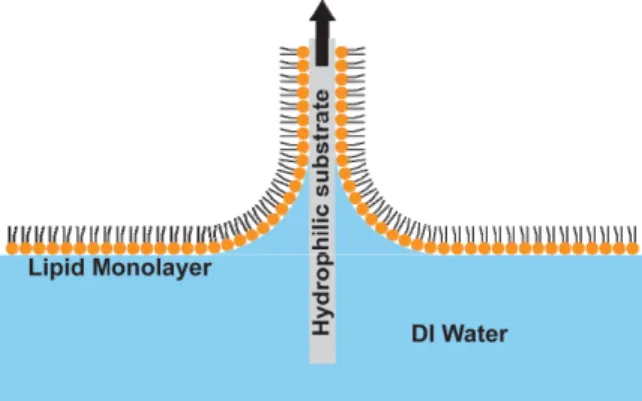
DPPC (Avanti Polar Lipids) was obtained in powder form and dissolved in heptane with drops of ethanol to a concentration of 1.91 mg/ml. Freshly cleaved grade V-1 muscovite mica (SPI Supplies) was used as a hydrophilic substrate. A Langmuir trough (601, NIMA Technologies) was used for surface pressure measurements and monolayer depositions. Prior to addition of 20 µl of DPPC to the trough, a mica substrate was lowered into the water. The mica was compressed to a target surface pressure between 5 and 45 mN/m and held for 60 minutes. The substrate was then pulled out of the water at a rate of 5 mm/min (Figure 3). All tests on samples were performed within one week of deposition, although no deterioration of sample quality was noticeable after longer periods. Surface pressure-area isotherm of DPPC as obtained in the trough is shown in Figure 4.
Contact Angle Measurement
Contact angle was measured on an automatic microscopic contact angle meter (MCA-3, Kyowa). A small printer inkjet head or 5 µm glass needle was used to deposit droplets on the order of
tens of picoliters. A video camera recorded images and were processed using FAMAS software. A temperature controlled stage was used to keep the substrate temperature at 20°C.
Figure 3: Schematic of Langmuir-Blodgett (LB) deposition of a lipid monolayer on a hydrophilic (mica) substrate by pulling.
Figure 4: Surface pressure-area isotherm of DPPC showing different phases LC (liquid condensed), LE (liquid expanded) [7], and collapse pressure.
Atomic Force Microscopy
Atomic force microscopy was performed on either a Veeco Dimension 3100 or Multimode IV . For tapping mode, tips with 42 N/m spring constant (RTESP, Bruker) at scan rates of 1 Hz and drive amplitudes of 100 mV were typically used. For force spectroscopy, Veeco MLCT tips with 0.07 N/m stiffness were used at ramp rates of 2 Hz.
Molecular Dynamics Simulation
GROMACS, a widely used simulation package for molecular dynamics simulation [8], was used in this work to perform the molecular modeling.
The force field used was a combination of lipid and GROMOS parameters. The standard parameters of the GROMOS force field were applied for all bonds, valence angles, improper dihedrals, as well as for the dihedral angles in the head-group region. The non-bonded interactions were computed as:
12 6 1 ( ) 4 4 N ij ij i j i j ij ij o ij q q U r r r r
σ
σ
ε
πε
> ⎡⎛ ⎞ ⎛ ⎞ ⎤ ⎢ ⎥ = ⎜⎜ ⎟⎟ −⎜⎜ ⎟⎟ + ⎢⎝ ⎠ ⎝ ⎠ ⎥ ⎣ ⎦∑
where the first term represents the Lennard-Jones interactions and the second term the Coulomb interactions. For Lennard-Jones interactions, the OPLS parameters were implemented. The Lennard-Jones parameters and electrostatic fractional charges for lipid, which includes the interaction parameters for the united CHn groups referenced as pentadecane,
are listed elsewhere [9]. The single point charge (SPC) model was used for water molecules. Lennard-Jones and Coulomb interactions were modeled using a shift-type function with a cutoff radius of 1.0 nm. The force interactions have been verified in a prior work by the authors where a fully hydrated DPPC bilayer was equilibrated and results compared with experiments [10]. To understand the effect of packing density on lipid-tail interaction with water in this work, a lipid monolayer was constructed on a silicon dioxide surface to achieve the desired orientation of the lipid molecules with the heads close to the surface and tails away from it. This system was relaxed for 0.5 ns. After equilibration, a water slab of height ~20 nm was added on top of the lipid monolayer to model its interaction with water molecules. Two lipid packing densities were simulated separately, 50.73 Å2/lipid and 94.56 Å2/lipid. The water slab was
pressurized using a moving platinum wall to avoid cavitation at the lipid-water interface. In all computations, the silicon dioxide molecules were stationary, while the lipid and water molecules were coupled to Berendsen thermostats to maintain their temperatures at 298 K. All boundaries in x, y and z directions were periodic.
RESULTS AND DISCUSSION
Film Quality and Domain Boundaries
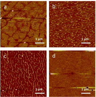
DPPC deposited onto mica at 5, 10, 15, 20, 25, 30, 35, 40, and 45 mN/m surface pressure were scanned using AFM tapping mode. All depositions above 5 mN/m correspond to the LC phase in the monolayer film. Above 30 mN/m, the topology was flat and uniform with the exception of a few adsorbed vesicles (Figure 5d). Below 30 mN/m, the morphology changed significantly. At 15, 20, and
LC
LC-LE
LE Collapse
25 mN/m, ridge-like structure was noticeable (Figure 5b and c). These ridges are approximately 1 nm and do not correspond to the height of DPPC (2.5 nm). Upon closer inspection of these structures, the ridges are shown to be compliant. The surface was probed harder by lowering the amplitude set point of the tip. As a result, the ridges become valley-like defects (Figure 6). These ridges/valleys likely correspond to low density LE phase regions whereas the area between them, which cover the bulk of the area, is the high density LC phase. Thus, low density regions serve as domain boundaries between LC phase domains which is known to pack in a hexagonal packed pattern [4], [11].
Figure 5: AFM height topography of DPPC monolayer on mica from LB deposition. At (a) 5 mN/m, two distinct phases are visible (LC and LE). At (b) 15 mN/m and (c) 25 mN/m ridge-like domain boundaries form. At (d) 35 mN/m, the lipid coverage is free of defects. Circles in the scan are due to vesicles adsorbed on the surface
In addition, it was found that monolayer films were not stable when exposed to water. Significant defects arise after exposure to water (Figure 7), which is consistent with studies of similar lipid films [12], [13]. Since the height of these defects correspond to the height of a DPPC molecule (2.5 nm), these defects are characterized by physical removal of lipids off the surface. Because of the strong polar interaction between water and the hydrophilic mica substrate, water likely penetrates through the monolayer in order to come in contact with substrate. To prove this idea, lipid monolayers were also exposed to polar and non-polar liquids,
formamide and diiodomethane, respectively. It was found that formamide introduced similar defects. However, diiodomethane did not change the surface morphology at all.
Figure 6: AFM height topography of DPPC monolayer on mica deposited at 25 mN/m at (a) high and (b) low amplitude set points. The ridge-like defects are compliant and become valley-like when imaged at a higher tip force (lower amplitude set point) suggesting such structures are actually domain boundaries (DB) between LC phases.
Figure 7: DPPC height profile after contact with water showing high defect density.
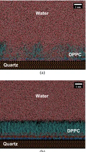
The molecular dynamics simulations also show the importance of packing density of the lipids on its interaction with water. As mentioned earlier, two separate simulations were performed with packing densities of 50.73 Å2/lipid (high packing)
and 94.56 Å2/lipid (low packing). Figure 8 shows the
interaction of water at these packing densities. For the low packing density, water molecules easily penetrate the lipid monolayer by displacing the lipid molecules and forming columns of water. However, for the high packing density, no such water penetration in the lipid monolayer was obtained. The lipid-water surface tension was evaluated from the high packing density simulation and was found to be 10.88 mN/m. The surface tension was completely dominated by the van der Waals forces compared to Coulomb forces which were over six orders of magnitude smaller. This is as expected since lipid tails are only carbon atoms which do not carry any induced charges. LC LC LC DB DB LC LC LC DB DB
a
b
d
c
1 µm 1 µm 1 µm 1 µm 200 nm 200 nm 1 µma
b
(a)
(b)
Figure 8: Lipid-water interaction at packing density of (a) 94.56 Å2/lipid (low packing), and (b) 50.73
Å2/lipid (high packing). The low packing allows
water molecules to penetrate through the monolayer, which is not the case with the well packed lipids.
Contact Angle
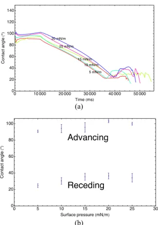
The contact angles of small droplets of DI water were measured during evaporation. Initially, the contact angle is high ~90°; however, during evaporation, pinning causes contact angle to relax during this time. As such, a contact angle relaxation profile can be attained (Figure 9). Significant relaxation of ~30-70° occurs at all surface pressures. After wetting, a droplet placed at the same spot has a lower initial contact angle. Thus, exposure to water affects the wetting behavior by removal of lipids off the substrate. In all cases, water penetrates the layer to come in contact with the mica substrate. The shape of the relaxation profile changes from concave up to concave down as surface pressure increases and as domain boundaries (DB) disappear after 30 mN/m. A concave up profile suggests a faster penetration of water through the layer occurs, which is likely due to the increase in defects at lower surface pressures. Once LE-LC transition occurs at 5 mN/m, the contact angle is lowered significantly.
Figure 9: Droplet contact angle relaxation during evaporation shows a dependence on surface pressure (means of ten runs). Below 30 mN/m, where domain boundaries exist, droplet relaxation has initial decaying behavior while above 30 mN/m, where domain boundaries (DB) do not exist, droplets relax similarly with a concave down curve.
Advancing and receding contact angle tests were also performed by gradual addition of water and evaporation. During fluid addition, the contact angle is relatively constant. After addition, evaporation causes relaxation and eventually recession (Figure 10a). The advancing and receding contact angles of have a weak correlation with surface pressure (Figure 10b). The advancing and receding contact angles of a heterogeneous surface are usually associated with the hydrophobic and hydrophilic components respectively [14] (Figure 11). Here, the surface heterogeneity arises due to defects and exposed mica (hydrophilic zones) within the lipid film (hydrophobic zone). During the advancing state, when the three phase contact line is at a point where the surface energy transitions from a low contact angle region (mica) to a high contact angle region (LC phase monolayer), pinning occurs until the high contact angle is satisfied. As the three phase contact line continues to advance over the high contact angle region, the contact angle remains at this high contact angle value. As soon as the three phase contact line reaches another low contact angle region, the local contact angle diminishes to the low contact angle associated with it. However, this causes a local curvature change, which lowers the pressure locally (according to the Young-Laplace equation) and leads to an instability thereby advancing the three phase contact line very quickly across the low contact angle region until it reaches the next high contact angle region. Because the droplet quickly moves across the low contact angle region, more time is spent along the high contact angle region and hence the advancing contact angle measured is more associated with the high contact angle material. The fact that the
Water DPPC Quartz Water DPPC Quartz
advancing contact angle is relatively invariant with surface pressure implies that the surface energy of crystalline lipid region is also constant. Thus, the packing density and orientation of crystalline LC phase DPPC even in the fractured state at low surface pressure is mostly independent of surface pressure, and an equilibrium packing density likely exists.
(a)
(b)
Figure 10: Results of advancing and receding contact angle tests. Advancing and receding angles are not strongly dependent on the surface pressure.
Using the Lifshitz-van der Waals acid-base (LWAB) approach, one can determine surface energy of the lipid monolayer tails from experimental contact angle measurements from three probe liquids (water, formamide, diiodomethane) [15], [16]. According to this theory, the total surface energy is a combination of Lifshitz-van der Waals interaction and a Lewis acid-base interaction.
= + where the acid-base interaction can be expressed in
terms of an electron donor, γ-, and an electron
acceptor, γ+, component.
= 2
Figure 11: (a) Advancing liquid-vapor interface profile across a heterogeneous surface where the local curvature near the three-phase contact line across the low contact angle zone is unstable, causing the higher contact angle to be observed macroscopically. (b) Receding liquid-vapor interface profile across a heterogeneous surface where three-phase contact line across high contact angle zone is unstable, causing the lower contact angle to be observed macroscopically.
The work of adhesion between solid and liquid can be expressed in terms of the individual LWAB components.
, = 1 + cos =
2 + 2 + 2
Between three probe liquids of known γLW,
γ+, and γ-, three different sessile drop wetting
experiments can be performed where three different contact angles can be measured. By using (1) for each case, a system of three equations can be used to solve three unknown surface energy components γLW,
γ+, and γ-. Using (2) and (3), one can determine the
total surface energy. The obtained results are listed in Table 1 and Table 2. For DPPC monolayers deposited on mica by Langmuir-Blodgett (LB), the surface energy obtained is 20.6 ± 2.8 mJ/m2, which is
similar to what is reported in literature [18]. The result is that the tails have a total surface energy that is mostly dominated by Lifshitz-van der Waals interactions rather than electrostatic interactions. Lifshitz-van der Waals interaction dominate because the fatty acid tail is highly nonpolar and should be mostly dominated by nonpolar interactions, similar to that obtained in molecular simulations.
(a)
Table 1: LWAB components of surface energies for water, formamide, and diiodomethane [17] (surface tensions in mN/m, contact angle in degrees).
Probe liquid γ γLW γ+ γ- γAB Contact Angle
Water 72.8 21.8 25.5 25.5 51.0 97 ± 3
Formamide 58.0 39.0 2.28 39.6 19.0 88 ± 2
Diiodomethane 50.8 50.8 0 0 0 68 ± 2
Table 2: LWAB surface energies for DPPC monolayer tail (surface tensions in mN/m).
γ γLW γAB
20.6 ± 2.8 24.0 ± 1.1 -3.4 ± 2.8
Force Spectroscopy
In order to determine the mechanical nature of the supported lipid monolayers, force spectroscopy was performed to obtain the breakthrough force of the films. This involves lowering the AFM tip into the sample and recording the tip deflection and the resulting force. At some point after initial contact with the surface, the tip breaks through the monolayer and a small drop-off in force is observed. Characterizing the mechanical breakthrough force should likely correlate with the packing nature of DPPC where lower density region should have a smaller breakthrough force. Thus, we seek to characterize the breakthrough force of all LC phase regions. It was hypothesized earlier that the LC phase has an equilibrium packing density that is independent of surface pressure; thus all LC phases should have the same breakthrough points regardless of the surface pressure. The LE phase, being of lower density should have a lower breakthrough point. Preliminary results on a uniform film of DPPC deposited at 25 mN/m in water have been obtained (Figure 12).
Figure 12: Force-displacement curve of AFM tip on DPPC monolayer coated mica in water. Small drop-off in tip force after contact with surface indicates tip breaking through DPPC film.
Using a thermodynamic model to explain the energy of a hole, it is possible to describe the probability of rupture as a thermally activated event [19]. The free energy of a hole of radius r in a film prior to rupture is
= + +
2
where γ is the total surface energy and F/2πR is the compressed elastic energy of the film from the AFM tip with radius of curvature R. After rupture, the energy of the hole is
= + 2 Γ
where Γ is the edge energy of the hole. Thus, the change in free energy during film rupture is
= 2 Γ +
2
The maximum change in free energy occurs at a critical radius when the parabolic function is maximized. This maximum change in energy is the activation energy, Ea, required to rupture the film.
= 2 Γ R
+ 2
Assuming that rupture is a thermally activated event requiring an energy change of ΔU, it can be shown that the probability of finding an intact (non-ruptured) film is [19]
ln =
′ ′
Figure 13: Histogram of breakthrough events in a single spot on a DPPC monolayer coated mica with probability distribution model fit. A mean breakthrough force occurs at around 1 nN.
Solving for P(F) and differentiating with respect to F gives the probability distribution function (-dP/dF) which can be fitted to the histogram as shown in Figure 13 where γ and Γ are fitting parameters. Assuming a nominal AFM tip radius of 20 nm as given by Bruker, the total surface energy obtained is 5.8 mN/m.
CONCLUSION
The surface energy, and hence the packing density, of lipid molecules is directly dependent on the electroporation of lipid membranes. In order to study the behavior of packing structure on surface energy, lipid monolayers were deposited on a mica substrate using the Langmuir-Blodgett technique at different surface pressures. The monolayers were then analyzed using AFM, and two distinct phases, ridge-like domain boundaries and defect-free lipid coverage was observed based on the deposition surface pressure. Exposure of these monolayers to water introduced significant defects as water was able to displace and penetrate through the lipid molecules, which was confirmed through molecular dynamics simulations. The surface tension of lipid tails-water interface was obtained to be 10.88 mN/m from the simulations. The Lifshitz-van der Waals acid-base (LWAB) approach was used to determine the surface tension of the lipid monolayer tails from experimental contact angle measurements from three probe liquids (water, formamide, diiodomethane) and was found to be 20.6 ± 2.8 mN/m. The mechanical strength of the supported lipid monolayers was also determined using force spectroscopy from which a surface tension of around 5.8 mN/m can be inferred. The three values of surface tension obtained from three different methods are within the same order of magnitude, which suggests that a true value surface tension should be approximately in this range as well. However, further studies with different lipids must be conducted in order to determine whether all of these methods capture the same trends with regard to changing lipid parameters such as tail length and composition.
ACKNOWLEDGEMENTS
The authors would like to thank Diane Kayitesi for assistance in contact angle experiments and depositions. Use of the Langmuir-Blodgett trough was possible thanks to Timothy Swager’s lab at MIT. In addition, the authors would like to acknowledge the Institute for Soldier Nanotechnologies at MIT for the use of an AFM. Computer workstations for MD simulations were provided for by the Intel Higher Education Grant.
Finally, this work was supported in part by the MRSEC Program of the National Science Foundation under award number DMR - 0819762.
REFERENCES
[1] M. Pavlin, T. Kotnik, D. Miklavcic, P. Kramar, and A. Macek Lebar, “Chapter Seven Electroporation of Planar Lipid Bilayers and Membranes,” Advances in
Planar Lipid Bilayers and Liposomes, vol.
6, pp. 165–226, 2008.
[2] D. Tieleman, “The molecular basis of electroporation,” BMC biochemistry, vol. 5, no. 1, p. 10, 2004.
[3] Z. V. Leonenko, E. Finot, H. Ma, T. E. S. Dahms, and D. T. Cramb, “Investigation of Temperature-Induced Phase Transitions in DOPC and DPPC Phospholipid Bilayers Using Temperature-Controlled Scanning Force Microscopy,” Biophysical Journal, vol. 86, no. 6, pp. 3783–3793, Jun. 2004. [4] X. Zhai and J. Kleijn, “Molecular structure
of dipalmitoylphosphatidylcholine Langmuir-Blodgett monolayers studied by
atomic force microscopy,” Thin Solid
Films, vol. 304, pp. 327–332, 1997.
[5] J. Korlach, P. Schwille, W. Webb, and G. Feigenson, “Characterization of lipid bilayer phases by confocal microscopy and fluorescence correlation spectroscopy,”
Proceedings of the National Academy of Sciences of the United States of America,
vol. 96, no. 15, pp. 8461–8466, 1999. [6] C.H. Huang, P.Y. Hsiao, F.G. Tseng, S.K.
Fan, C.C. Fu, and R.L. Pan, “Pore-Spanning Lipid Membrane under Indentation by a Probe Tip: A Molecular Dynamics Simulation Study,” Langmuir, vol. 27, no. 19, pp. 11930–11942, 2011. [7] C. McConlogue and T. Vanderlick, “A
close look at domain formation in DPPC monolayers,” Langmuir, vol. 13, no. 26, pp. 7158–7164, 1997.
[8] D. Van der Spoel, E. Lindahl, B. Hess, G. Groenhof, A. Mark, and H. Berendsen, “GROMACS: Fast, flexible, and free,”
Journal of Computational Chemistry, vol.
26, no. 16, pp. 1701–1718, 2005.
[9] O. Berger, O. Edholm, and F. Jähnig, “Molecular dynamics simulations of a fluid bilayer of dipalmitoylphosphatidylcholine at full hydration, constant pressure, and constant temperature,” Biophysical
Journal, vol. 72, no. 5, pp. 2002–2013,
[10] S. C. Maroo, H. J. Cho, and E. N. Wang, “Wetting characteristics of a phospholipid membrane using molecular dynamics simulation,” in Proceedings of the ASME
International Mechanical Engineering Congress & Exposition, Vancouver,
Canada, 2010.
[11] K. Voitchovsky, J. J. Kuna, S. A. Contera, E. Tosatti, and F. Stellacci, “Direct mapping of the solid-liquid adhesion energy with subnanometre resolution,”
Nature Nanotechnology, vol. 5, no. 6, pp.
401–405, 2010.
[12] M. Benz, T. Gutsmann, N. Chen, R. Tadmor, and J. Israelachvili, “Correlation of AFM and SFA measurements concerning the stability of supported lipid bilayers,” Biophysical Journal, vol. 86, no. 2, pp. 870–879, 2004.
[13] É. Kiss, J. Szalma, Z. Keresztes, E. Kálmán, M. Mohai, and I. Bertóti, “Adhesional stability of cadmium arachidate Langmuir–Blodgett layers,” in
From Colloids to Nanotechnology, vol.
125, M. Zrínyi and Z. D. Hórvölgyi, Eds. Springer Berlin / Heidelberg, 2004, pp. 127–133.
[14] Van P Carey, Liquid Vapor Phase Change
Phenomena: An Introduction to the Thermophysics of Vaporization and
Condensation Processes in Heat Transfer Equipment, Second Edition, 2nd ed. Taylor
& Francis, 2007, p. 600.
[15] C. Van Oss, M. Chaudhury, and R. Good, “Interfacial Lifshitz-van der Waals and polar interactions in macroscopic systems,”
Chemical Reviews, vol. 88, no. 6, pp. 927–
941, 1988.
[16] C. J. van Oss, “Acid--base interfacial interactions in aqueous media,” Colloids
and Surfaces A: Physicochemical and Engineering Aspects, vol. 78, pp. 1–49,
Oct. 1993.
[17] Z. Li, R. F. Giese, W. Wu, M. F. Sheridan, and C. J. van Oss, “The Surface Thermodynamic Properties of Some Volcanic Ash Colloids,” Journal of
Dispersion Science and Technology, vol.
18, no. 3, pp. 223–241, 1997.
[18] E. Chibowski and M. Jurak, “Interaction energy of model lipid membranes with water and diiodomethane,” Colloids and
Surfaces A: Physicochemical and Engineering Aspects, vol. 383, no. 1, pp.
56–60, Jun. 2011.
[19] H.-J. Butt and V. Franz, “Rupture of molecular thin films observed in atomic force microscopy. I. Theory,” Physical
Review E, vol. 66, no. 3, p. 031601, Sep.
![Table 1: LWAB components of surface energies for water, formamide, and diiodomethane [17] (surface tensions in mN/m, contact angle in degrees)](https://thumb-eu.123doks.com/thumbv2/123doknet/14242107.486945/8.918.485.811.713.987/components-surface-energies-formamide-diiodomethane-surface-tensions-contact.webp)
