HAL Id: tel-02165949
https://tel.archives-ouvertes.fr/tel-02165949
Submitted on 26 Jun 2019HAL is a multi-disciplinary open access archive for the deposit and dissemination of sci-entific research documents, whether they are pub-lished or not. The documents may come from teaching and research institutions in France or abroad, or from public or private research centers.
L’archive ouverte pluridisciplinaire HAL, est destinée au dépôt et à la diffusion de documents scientifiques de niveau recherche, publiés ou non, émanant des établissements d’enseignement et de recherche français ou étrangers, des laboratoires publics ou privés.
Time-resolved Multiplexed Förster Resonance Energy
Transfer for Nucleic Acid Biosensing
Jiajia Guo
To cite this version:
Jiajia Guo. Time-resolved Multiplexed Förster Resonance Energy Transfer for Nucleic Acid Biosens-ing. Optics / Photonic. Université Paris Saclay (COmUE), 2019. English. �NNT : 2019SACLS162�. �tel-02165949�
Time-resolved Multiplexed Förster
Resonance Energy Transfer for
Nucleic Acid Biosensing
Thèse de doctorat de l'Université Paris-Saclay préparée à l'Université Paris-Sud
École doctorale n°575:
Electrical, Optical, Bio: PHYSICS_AND_ENGINEERING
(EOBE)
Spécialité de doctorat: Physique (Electronique et Optoélectronique, Nano et Microtechnologies)
Thèse présentée et soutenue à Orsay, le 18 Juin 2019
Mme Jiajia GUO
Composition du Jury : Mme Rachel Méallet-Renault
Professeure, Université Paris-Sud Présidente Mme Julia Pérez-Prieto
Professeure, University of Valencia Rapporteur M. Marcus Wilhelmsson
Professeur, Chalmers University Gothenburgh Rapporteur M. Andreas Reisch
Maître de conférences, Université de Strasbourg Examinateur M. Niko Hildebrandt
Professeur, Université Paris-Sud Directeur de thèse
NNT : 201 9S A C LS 16 2
Université Paris-Saclay
Espace Technologique / Immeuble Discovery
Route de l’Orme aux Merisiers RD 128 / 91190 Saint-Aubin, France
Titre: Transfert d'énergie par résonance de type Förster résolu en temps pour la bio-détection multiplexée des acides nucléiques
Mots clés: FRET, complexe de lanthanide, multiplexage, résolue dans le temps, acides nucléiques Résumé: Les biomarqueurs à base d’acides
nucléiques, qui sont impliqués dans le contrôle de l'expression génétique, sont spécifiques de nombreux types de cancers. Les applications basées sur le transfert d'énergie par résonance de Förster (FRET) sont parmi les plus prometteuses pour la biodétection d’acides nucléiques. Comme la détection simultanée de plusieurs acides nucléiques est très demandée et que le multiplexage spectral est limité par des interférences (optical crosstalk), le multiplexage temporel est utilisé ici pour ajouter de nouvelles possibilités de multiples détections simultanées. La thèse porte sur le développement de systèmes comprenant différentes distances entre molécules donneuses et acceptrices de FRET (Terbium vers fluorophores) pour créer des
signaux d'intensité spécifiques correspondant à différentes séquences d'acides nucléiques. Les distances Tb-to-dye peuvent être arrangées en positionnant spécifiquement le donneur Tb sur des molécules d’ADN de différentes longueurs. Les technologies d'amplification d’acides nucléiques, telles que la réaction d'hybridation en chaîne (HCR) et l’amplification circulaire de l’ADN (RCA), ont été utilisées pour obtenir simplicité, rapidité, sélectivité et sensibilité dans la détection d’acides nucléiques. Le multiplexage temporel du signal de FRET a également été combiné avec le multiplexage spectral (couleur) pour le démultiplier. De plus, la possibilité d'un multiplexage temporel à base de nanoparticules a été démontrée.
Title: Time-resolved Multiplexed Förster Resonance Energy Transfer for Nucleic Acid Biosensing Keywords: FRET, lanthanide complex, multiplexing, time-resolved, nucleic acid
Abstract: Nucleic acid biomarkers, which involve in gene expression control, are found specific for many kinds of cancers. Förster Resonance Energy Transfer (FRET) based applications are one of the most promising for nucleic acid biosensing. As parallel detection of multiple nucleic acids is highly demanded and spectral multiplexing is limited by optical crosstalk, temporal multiplexing is used for opening another dimension of the multiplexing. The thesis focuses on developing different Tb-to-dye FRET distances to create specific intensity signals corresponding to different nucleic acid sequences. The Tb-dye distances
can be tuned by specific location of the Tb donor using different lengths of DNA. Amplification technologies, such as hybridization chain reaction (HCR) and rolling circle amplification (RCA), are used to achieve simplicity, rapidity, selectivity, and sensitivity of nucleic acid detection. Temporal multiplexing FRET was also combined with spectral (color) multiplexing for higher order multiplexed detection. Moreover, a single Tb-QD FRET modeling demonstrated the possibility of nanoparticle-based temporal multiplexing.
Acknowledgment
I would like to convey my heartfelt gratitude and sincere appreciation to all people who have helped and inspired me during my doctoral study. This thesis would not have been possible without those supports from many people.
First, I would like to express my special appreciation and thanks to my supervisor Professor Dr. Niko Hildebrandt, for his trust in my abilities, the liberties in the laboratory, opportunities to attend to different conferences and his endless support. His perpetual energy and enthusiasm in research extremely motivated me in my studies. I appreciate all his invaluable contributions of time and advice on both research and life in the last four years. I could not have imagined having a better supervisor than him.
My sincere thanks also go to Prof. Dr. Julia Pérez-Prieto and Prof. Dr. Marcus Wilhelmsson for being the reviewers of my Ph.D. thesis.
I also want to thank our collaborator Dr. Igor Medintz for his kindness and scientific support, and all the co-authors of my publications.
Many thanks to all former and current members of the NanoBioPhotonics group, special thanks to Dr. Xue Qiu, Dr. Shashi Bhuckory and Dr. Yu-tang Wu for their continuous guidance, patience and valuable time devoted in sharing their technical and scientific knowledge with me. I also want to thank Ms. Jingyue Xu for spending a lot of happy dinner and weekend’s time with me together, Dr. Thomas Plénat for improving my drive skills.
My gratefully acknowledge to Dr. Monique Chan-Hout for teaching me French. I learned a lot from her, not only French, but also her positive and optimistic attitude towards life.
I thanks China Scholarship Council for supporting the funding of my Ph.D. study.
Last but not least, my deepest gratitude goes to my parents, my boyfriend Bingrun Liu and my best friend Haiwa Zhang for their unflagging love and support throughout my life.
List of publications
Original publications
1. Jiajia Guo, Carlos Mingoes, Xue Qiu, and Niko Hildebrandt. "Simple, Amplified, and Multiplexed Detection of MicroRNAs Using Time-Gated FRET and Hybridization Chain Reaction." Analytical Chemistry 91, no. 4 (2019): 3101−3109.
2. Jiajia Guo, Xue Qiu (co-first author), Carlos Mingoes, Jeffrey R. Deschamps, Kimihiro Susumu, Igor L. Medintz, and Niko Hildebrandt. "Conformational Details of Quantum Dot-DNA Resolved by Förster Resonance Energy Transfer Lifetime Nanoruler." ACS Nano 13, no.1 (2018): 505-514.
3. Xue Qiu, Jiajia Guo (co-first author), Jingyue Xu, and Niko Hildebrandt. "Three-Dimensional FRET Multiplexing for DNA Quantification with Attomolar Detection Limits." The journal of physical chemistry letters 9, no. 15 (2018): 4379-4384.
4. Xue Qiu, Jingyue Xu, Jiajia Guo, Akram Yahia Ammar, Nikiforos-Ioannis Kapetanakis, Isabelle Duroux-Richard, Julia Unterluggauer et al. "Advanced microRNA-based cancer diagnostics using amplified time-gated FRET." Chemical Science 9, no. 42 (2018): 8046-8055. 5. Xue Qiu, Jiajia Guo, Zongwen Jin, Alexandra Petreto, Igor L. Medintz, and Niko Hildebrandt. "Multiplexed Nucleic Acid Hybridization Assays Using Single‐FRET‐Pair Distance‐Tuning." Small 13, no. 25 (2017): 1700332.
Oral presentations
1. Jiajia Guo, Xue Qiu, Carlos Mingoes, Jeffrey Deschamps, Igor Medintz, Niko Hildebrandt. Temporal multiplexing FRET: using single Tb-to-quantum dot pair distance-tuning. SPIE Photonics West, San Francisco, USA, 2019 (Invited talk)
2.Jiajia Guo, Xue Qiu, Carlos Mingoes, Jingyue Xu, Niko Hildebrandt.Temporal multiplexing of microRNA detection using Tb-dye FRET and amplifications. 10th International Conference on f-Elements (ICFE-10), Lausanne, Switzerland, 2018 (Invited talk)
3. Jiajia Guo, Qiu, Xue, Zongwen Jin, Igor L. Medintz, and Niko Hildebrandt. Lifetime-multiplexed microRNA detection based on distance-dependent FRET from Terbium-Quantum Dot. Fluorescent Biomolecules and Their Building Blocks. Tianjin, China, 2016 (Short talk)
Poster presentations
1. Jiajia Guo, Qiu, Xue, Zongwen Jin, Igor L. Medintz, and Niko Hildebrandt. One Quantum Dot Based Time-Gated Förster Resonance Energy Transfer for MicroRNA Duplexing. Förster resonance energy transfer in life sciences. Göttingen, Germany, 2016
2. Jiajia Guo, Qiu, Xue, Zongwen Jin, Igor L. Medintz, and Niko Hildebrandt. Multiplexed Nucleic Acid Hybridization Assays Using Single Tb-to-Quantum Dot FRET Pair Distance-Tuning. 15th Conference on Methods and Applications in Fluorescence (MAF 15). Bruges, Belgium, 2017
CONTENTS
1. Introduction ... 1
2. Background ... 6
2.1 Nucleic acid biomarkers ... 6
2.1.1 MicroRNA biomarker ... 7
2.1.2 Cancer-associated miRNA ... 8
2.1.3 MicroRNA detection methods ... 9
2.2 Förster resonance energy transfer ... 10
2.2.1 FRET theory ... 11
2.2.2 FRET applications ... 15
2.2.3 FRET for nucleic acid detection ... 16
2.3 Lanthanides ... 17
2.3.1. Introduction ... 17
2.3.2. Luminescence lanthanide complexes ... 18
2.3.3. Lanthanide complex applications... 22
2.4 Fluorescent dyes ... 26
2.4.1 Cyanine dyes ... 27
2.4.2 Cyanine dyes applications in FRET ... 27
2.5 Quantum dots ... 29
2.5.1 Photophysical properties ... 29
2.5.2 Bioconjugation ... 31
2.5.3 QDs applications in FRET ... 33
3. Temporal duplexing Tb-Dye HCR-FRET microRNA detection assay ... 36
3.1 Introduction ... 36
3.2 Materials and methods ... 37
3.2.1 Materials ... 37
3.2.2 HCR-FRET miRNA/ssDNA single target assays. ... 39
3.2.3 HCR-FRET miRNA/ssDNA duplexed assays. ... 40
3.2.4. Gel Electrophoresis. ... 40
3.3 Results and discussion ... 41
3.3.1 The principle of duplexed HCR-FRET ... 41
3.3.2. MicroRNA-Specific HCR-TG-FRET ... 42
3.3.3 PL decay fit results and distance calculations. ... 45
3.4. Conclusion ... 53
4. Spectral-temporal quadruplexing Tb-Dye RCA-FRET ssDNA detection assay ... 55
4.1 Introduction ... 55
4.2 Materials and methods ... 55
4.2.1 Nucleic acid probes and exogenous targets. ... 56
4.2.2 Photophysical analysis ... 56
4.2.3 RCA-FRET single target assays ... 56
4.2.4 Temporal duplexing ... 57
4.2.5 Spectral-temporal multiplexing... 57
4.3 Results and discussion ... 58
4.3.1 The principle of multiplexed RCA-FRET ... 58
4.3.2 Spectral-temporal multiplexing RCA-FRET assay development ... 60
4.4 Conclusion ... 68
5. Temporal multiplexing Tb-QD FRET for QD-DNA conformation analysis ... 70
5.1. Introduction ... 70
5.2 Materials and methods ... 72
5.2.1 Materials ... 72
5.2.2 Spectroscopic characterization ... 73
5.2.3 Tb-QD FRET assays ... 73
5.3. Results and discussion ... 74
5.3.1 Photophysical properties of Tb and QD and R0 calculation... 74
5.3.2 FRET DNA-Conjugates ... 74
5.3.3 PL decay curves and fit results of Tb and QD for Tb-QD-DNA configurations ... 75
5.3.4 Time-Resolved Tb-to-QD FRET DNA-Nanoruler and Distance Modeling. ... 89
5.3.5 Prototypical DNA Hybridization Assays ... 94
5.4 Conclusion ... 95
6. Summary and Outlook ... 96
7. Appendix ... 98
7.1 Abbreviations ... 98
7.2 LTC-ssDNA conjugation protocol ... 100
7.3 Cy 3.5-ssDNA conjugation protocol ... 101
7.4 Cy 5.5-ssDNA conjugation protocol ... 102
7.5 Instrument ... 103
8. Bibliography ... 104
1
1. Introduction
Cancer kills millions of people every year and it is one of the greatest health challenges of human beings. The 2018 Nobel Prize in Physiology or Medicine was awarded to James P. Allison and Tasuku Honjo for their discovery of cancer therapy by inhibition of negative immune regulation. If cancer can be diagnosed at an early stage, it will greatly increase the chance for successful treatment. Recognizing possible warning signs of cancer and taking prompt action leads to early diagnosis.
One of the best ways to diagnose cancer early is to use serum or tissue biomarkers. Cancer biomarkers can be DNA, RNA, proteins, metabolites, or processes such as apoptosis, angiogenesis or proliferation[1]. Such biomarkers are produced either by the tumor itself or by other tissues and can be found in a variety of fluids, tissues and cell lines, in response to the presence of cancer or other associated conditions. MicroRNAs (miRNAs) which are small (~22bp) highly conserved non-coding RNAs endogenously expressed in every cell type, have emerged as a new type of cancer-specific biomarker[2]. They function as key regulators of gene expression by silencing target transcripts through base-pair complementation. MicroRNAs can be secreted from cells and are found in a variety of body fluids including blood, saliva, and urine, where they are quantifiable and extremely stable[1]. For these reasons, miRNAs are becoming ideal candidates as non-invasive biomarkers for cancer.
Förster resonance energy transfer (FRET)-based nucleic acid detection is one of the most promising techniques that can meet almost all the demands for a fast, simple, and multiplexed diagnostic[3]. FRET is a non-radiative energy transfer from an excited donor (FRET-donor) molecule to a ground state acceptor molecule (FRET-acceptor) through dipole-dipole interactions[4]. The principal conditions for FRET are the donor and acceptor should be in close proximity to each other and have a spectral overlap between the donor emission and the acceptor absorption. Due to their strong distance-dependence, FRET biosensors are very sensitive for analyzing biological interactions and structural changes in a nanoscale range of ca.1-20 nm. For most FRET-based nucleic acid detection assays, both donor and acceptor are conjugated with DNA, the target miRNA can complement them to change the distance between donor and acceptor, thereby changing the FRET efficiency and lifetime. MicroRNAs can be qualitatively and quantitatively detected by the change of FRET efficiency and photoluminescence (PL) intensity of donor or acceptor[5]–[9].
2
As the increasing application of FRET, many kinds of fluorophores have been used as FRET donor. Compared to other traditional fluorophores, luminescent terbium complex (LTC) as FRET donor has unique advantages for multiplexing detection in combination to different FRET acceptors, such as organic dyes and quantum dots (QDs)[10], [11]. Due to the forbidden intra-configurational f-f transitions in the terbium ions, LTC provides distinguished photophysical properties, like large Stokes shift, multiple narrow emission bands and extremely long excited-state lifetimes (up to the milliseconds’ range) which allow for time-gated (TG) measurements[12]. The distinction between the FRET signal and short-lived auto-fluorescence background and high detection sensitivity for biological applications all benefit from time-gated measurements. The emission bands of Tb are well separated and therefore superior for spectral multiplexing; the long and distinguished decay time of the sensitized acceptors due to different FRET efficiencies provide a great potential for temporal multiplexing.
Fluorescent dyes, also known as reactive dyes or fluorophores, are non-protein molecules that absorb light and re-emit it at a longer wavelength. They have been used in biological studies for decades because they are easy to handle and less toxic. Fluorescent dyes offer higher photostability and brightness and do not require a maturation time. Fluorescent dyes are popular because of their high quantum yields, solubility, and ease of bioconjugation. The large variety and wide commercial availability of fluorescent dyes have made them the most frequently applied class of fluorophores for FRET[13], [14]. The combination of LTC donors with fluorescent dye acceptors has been used for multiplexing FRET biosensing[15], [16]. Although multiplexed LTC-to-dye FRET is possible and can provide high sensitivity, the poor separation of the absorption and emission spectrum affect spectral crosstalk between different dye acceptors in spectral multiplexing, and appropriate correction is demanded.
Regarding much narrower emission bands and size-tunable emission peaks, QDs are excellent FRET acceptor candidates for multiplexing. QDs have several unique intrinsic photophysical properties: relatively high quantum yields; the size- and material-tunable QDs can be used to give better spectral overlap with their FRET partner; 10 to 100 times higher extinction coefficient than fluorescent dyes which are extremely important for spectral overlap with the LTC emission[17], [18]. Förster distances R0 of more than 10 nm can be achieved by
LTC-to-QD FRET pairs which are significantly larger than the R0-values for conventional D-A
pairs[19], [20]. The combination of LTC and QDs for FRET has a very high potential for the use in biosensing analysis with the possibility of high sensitivity and large Förster radii.
3
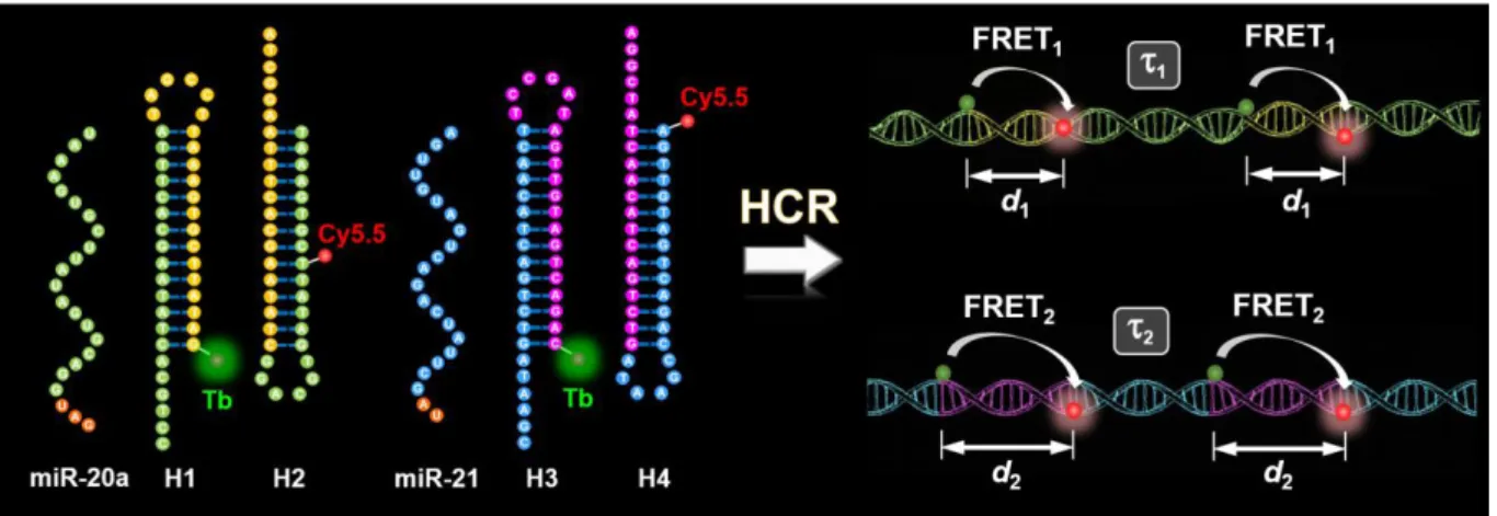
This thesis is divided into six chapters and completed by the appendix and the bibliography. Following the background of this work, three individual studies are presented. Each chapter is corroborated with introduction, materials and methods, results and discussion, and conclusion. The first study (Figure 1.1) presents a temporal duplexing assay for miRNA detection using a single Tb-dye FRET pair and hybridization chain reaction (HCR). I demonstrated the implementation of time-gated FRET between terbium donors and dye acceptors into HCR for multiplexed quantification of miRNAs (miR-20a and miR-21) and their DNA analogs. HCR-TG-FRET provided washing-free nucleic acid quantification with very low limits of detection (LOD) down to 240 attomoles (1.7 pM) of miRNA and 123 attomoles (0.88 pM) of DNA. Efficient distinction from very homologous miRNAs demonstrated high target specificity. Multiplexing with a single measurement, a single excitation wavelength, and a single FRET pair allowed for simultaneous quantification of miR-20a and miR-21 at concentrations between 30 pM and 300 pM from the same sample. HCR-TG-FRET showed similar performance for serum-free and serum-containing samples without the use of RNase inhibitors. Our results present a significant improvement in current HCR approaches regarding simplicity, sensitivity, and multiplexing. The versatile diagnostic performance of HCR-TG-FRET even in challenging biological environments presents an important advantage for advanced nucleic acid biosensing.
Figure 1.1. Schematic representation for duplexed (miR-20a and miR-21) detection of miRNAs using HCR-TG-FRET.
In the second study (Figure 1.2), I combined temporal multiplexing with spectral multiplexing, developed a quadrupling Tb-Dye FRET and rolling circle amplification (RCA) assay for single-stranded DNA (ssDNA) detection. We demonstrated time-gated FRET from a long-lifetime Tb-complex to Cy3.5 and Cy5.5 dyes for spectrotemporal multiplexing of four different DNA targets in the same sample by two time-gated windows for two different colors. We used rolling circle amplification (RCA) for high sensitivity and for placing Tb donors and dye acceptors at
4
controlled distances within the amplified DNA concatemers. We selected four different short ssDNAs (DNA-20a, DNA-20b, DNA-21, and DNA-191), which are the DNA-equivalents of the miRNAs hsa-mir-20a/20b/21/191, all of which have been found to be related to breast cancer. The principle of RCA-FRET is each DNA target is recognized by the terminal sequences of a specific padlock probe. After this hybridization step, a ligase closes the padlock nick over the target splint, which transfers the target into a primer for subsequent DNA synthesis. DNA amplification by RCA is performed at 37 °C for 2 h, and the resulting rolling circle product is labeled (specific hybridization) by Tb-donor and dye-acceptor DNA probes. The precise distance tuning led to target-specific PL decays of the FRET pairs and simple, separation-free, and higher-order multiplexed quantification of DNA. The RCA-FRET DNA assay could distinguish very homologous target sequences and provided limits of detection between 40 and 570 zeptomoles (0.3 to 4.1 fM) for four different DNA targets. We demonstrated the combination of the spectral and temporal dimensions of PL for higher-order multiplexing with the intensity dimension of PL for quantification of the four different DNAs at low femtomolar concentrations from the same sample, using a single excitation source and only two distinct fluorescence colors.
Figure 1.2. The principle of spectrotemporal quadruplexing RCA-FRET assay and PL multiplexing in three dimensions. x-axis: lifetime; y-axis: color; z-axis: intensity
In the third study (Figure 1.3), I investigated the Tb-to-QD temporal-multiplexing FRET by tuning the length of DNA. I analyzed 11 configurations of a 31 nt long DNA, which attached to QD by peptide-His6 sequence (peptide-DNA), with different Tb-conjugated complementary
5
biomolecular modeling. Our results show that the increasing Tb-to-QD distances led to increasing FRET-sensitized lifetimes of the decay curves, all 11 configurations can be distinguished without lifetime fitting of the curves. The temporally distinct PL decays of the different DNA-FRET-configurations can be used for prototypical DNA hybridization assays. Such fine-tuning of PL decay curves via displacement of the Tb donor by only changing a few nucleotides demonstrates the high distance sensitivity of FRET and the possibility to extend nanoparticle-based temporal multiplexing.
Figure 1.3. Top: Schematic representation for Tb-to-QD temporal-multiplexing FRET. Bottom: Biomolecular modeling was used to calculate Tb-to-QD distance ranges in parallel and vertical DNA-surface attachment configurations.
6
2. Background
2.1 Nucleic acid biomarkers
The term “biomarker”, short for “biological marker”, refers to an accurate and reproducible
measurable indicator of some biological state or condition. In 1998, the National Institutes of Health Biomarkers Definitions Working Group defined a biomarker as “a characteristic that is objectively measured and evaluated as an indicator of normal biological processes, pathogenic processes, or pharmacologic responses to a therapeutic intervention”[21]. WHO International Programme on Chemical Safety gave an even broader definition of a biomarker as “any substance, structure, or process that can be measured in the body or its products and influence or predict the incidence of outcome or disease”[22]. Biomarkers have a range of potential clinical utilities, including identifying the presence of disease and characterizing disease subtype (diagnosis), determining disease prognosis, aiding in the selection of appropriate therapeutic doses, predicting clinical benefit or adverse response to therapy, and monitoring treatment outcomes[23]. In recent decades, promising candidate biomarkers such as proteins, metabolites, carbohydrates, steroids, lipids, peptides, nucleic acid and cells involved in different diseases such as cancer, metabolic disorders, inflammatory disorders, and diseases of nervous system and cardiovascular system have been reported[24], [25]. The focus of this work is on cancer-related nucleic acids biomarkers. The cancer diagnosis is the starting point of patient treatment, and a fast and accurate diagnosis is highly demanded.
Nucleic acids include deoxyribonucleic acid (DNA) and ribonucleic acid (RNA), which are essential for all forms of life. DNA stores genetic information and encodes the amino acid sequences of proteins responsible for cellular function. RNA plays various important roles in the coding, decoding, regulation, and expression of genes. Nucleic acids are used as important biomarkers for biological studies and medical diagnostics because sequence-specific hybridization and amplification technologies allow highly accurate and sensitive assessment of these biomarker levels over a broad dynamic range. In addition to their biological roles, nucleic acids can be used as high-affinity recognition molecules for ions, small molecules, proteins, and even whole cells[26], [27]. Nucleic acids can also be coupled with organic or inorganic materials for assembly and construction purposes[28].
7
2.1.1 MicroRNA biomarker
MicroRNAs are one of the most important nucleic acids biomarker group. MicroRNAs are a class of ~22 nucleotide-long non-coding RNAs that play a central role in the regulation of gene expression by translational repression or degradation of messenger RNAs (mRNAs)[2]. Biogenesis of miRNA occurs through a multi-step process requiring both a nuclear and a cytoplasmic phase (Figure 2.1). Firstly, miRNA genes are transcribed into primary miRNAs (pri-miRNA) with a cap and a poly-A tail by pol II or pol III[29]. Pri-miRNA is characterized by a secondary hairpin structure. Then, pri-miRNAs are processed into short 70-110 nucleotides stem-loop structures known as precursor miRNAs (pre-miRNAs) by microprocessor complex, which consists of the RNase III enzyme Drosha, and a double stranded-RNA binding protein, Di George syndrome critical region 8 gene (DGCR8)[30]. Further processing of pre-miRNAs by the RNase III enzyme Dicer generate ~22 nucleotide miRNA duplexes (ds-miRNAs). This duplex binds to the active RNA-induced silencing complex (RISC) that performs gene silencing. Argonaute 2 (AGO2) unwinds the duplex, and removes the passenger strand retaining the mature miRNA molecule[31]. The mature miRNA functional strand is loaded into the RISC complex and AGO2. RISC-AGO2 complex guides the functional strand to target the 3’UTR of the target mRNA, causing translational inhibition or promoting their degradation.
8
Figure 2.1. Biogenesis of microRNA: miRNA is transcribed in the nucleus as a pri-miRNA and then is micro-processed by Drosha, a class 2 ribonuclease III enzyme, and transported to the cytoplasm by exportin 5 (XPO5) where the hairpin structure is removed by Dicer and a single-stranded mature miRNA is produced, which binds to RISC and induces gene silencing. (Reference [32], copyright 2018 SAGE Journals Publishers.)
2.1.2 Cancer-associated miRNA
Cancer is one of the leading causes of death worldwide. Subsequent investigations demonstrated that almost all cancers have an alternative miRNA expression profile compared to their adjunct normal tissues. Significant progress has been made on the relationship between miRNAs and cancers. For example, miR-181-5p, miR-361-5p, and miR-320b were significantly elevated in plasma exosomes of non-small cell lung cancers (NSCLC) patients[33]. The levels of miR-16-5p, miR-21-5p, and miR-199a-5p declined in plasma and tumor tissues of triple-negative breast cancer (TNBC) patients compared with both non-TNBC and healthy individuals, as well as miR-92a-3p and miR-342-3p elevated[34].
Table 2.1 summarizes the blood-based circulating miRNA expression in the major cancer types. It clearly shows that some miRNAs are overexpressed in cancers, for example, miR-21 is highly expressed in breast cancer. In contrast, some miRNAs, for example, miR-128 is downregulated in brain cancer. More importantly, some miRNAs have different expression profile pattern in different cancer types. Like miR-103 is downregulation in breast cancer, but it is upregulation in prostate cancer. This alternative and unique expression pattern allows miRNAs to become good biomarker for early detection of cancers.
9
Table 2.1. Examples of the miRNA expression in the major cancer types.
*(Reference [35], copyright 2018 Elsevier Publisher.)
2.1.3 MicroRNA detection methods
Detection of miRNA expression can help to identify miRNAs that regulate a range of vital processes and discover miRNA based biomarkers for diverse molecular diagnostic applications in cancer. Traditional strategies for detection of miRNA, including northern blotting, microarrays, and quantitative RT-PCR (qRT-PCR), have their relative strengths and weaknesses. Northern blotting is complex and requires radiolabeling, which can introduce significant contamination and has a low detection efficiency. Microarray is less expensive but does not have a lower sensitivity and dynamic range, therefore, it is often used as screening tool rather than as quantitative assay platform. qRT-PCR has the widest dynamic range and highest
Cancer type miRNAs Expression statu Expression status
7, 15b, 17-3p, 18a, 19a, 19b, 19a-3p, 20a, 21, miR-25,miR-29a, miR-92, miR-92a, miR-92a-3p, miR-96, miR-106a, miR-106b, miR-133a, miR-139-3p, miR-143, miR-145, miR-155, miR-182, miR-183, miR-196a, miR-200c, miR-206, miR-210, miR-214, miR-221, miR-223-3p, miR-320a, miR-335, miR-372, miR-378, miR-409-3p, miR-423-5p, miR-431, miR-486, miR-720, miR-1180
Upregulated
miR-7, miR-24, miR-26a-5p, miR-29b, miR-30b, miR-93, miR-103, miR-106a, miR-107, miR-221, miR-124, miR-127-5p, miR-138, miR-142-3p, miR-143, miR-145, miR-146a, miR-151-5p, miR-191, 194, 199-3p, 218, 222, 320a, 375, 382, 409-3p, 422a, miR-423-3p, miR-423-5p, miR-601, miR-652, miR-760, let-7d
Downregulated
miR-16, miR-19b, miR-21, miR-22a, miR-25, miR-29c, miR-92a, miR-96-5p, miR-127-3p, miR-133a, miR-145, miR-148a, miR-148b, miR-155, miR-199a, miR-222, miR-324-3p, miR-376a, miR-376c, miR-382, miR-409-3p, miR-424, miR-505-5p, miR-652, miR-801,
Upregulated
miR-103, miR-107, miR-195, miR-205, miR-200c, miR-451, let-7d, let-7i Downregulated 16, 21, 34b, 92a, 92b, 103, 107, 130b, 141, 145,
miR-155, miR-197, miR-200a, miR-200c, miR-210, miR-221, miR-301a, miR-326, miR-328, miR-331-3p, 375, 432, 485-3p, 486-5p, 501-3p, 551b, 562, 574-3p, miR-622, miRr-625*, miR-636, miR-640, miR-766, miR-885-5p, miR-1285, miR-2110
Upregulated
miR-30, miR-181a-2, let-7a, Let-7c, let-7e Downregulated 15b-5p, 22, 26b, 28, 34b, 125a-5p, 125b, 145, 146a,
miR-191, miR-200b, miR-200c, miR-203, miR-205, miR-340, miR-425, miR-429, miR-590-3p, miR-766, miR-944, miR-3662
Upregulated
15, 16, 16-5p, 17b-5p, 19-3p, 20, 20a-5p, 25, 28, 34b, 92-3p, 148a, 148b, 152, 193a-5p, 203, 218, 323-3p, miR-450b-5p, miR-485-3p, miR-566, miR-642, miR-661, miR-1290, let-7c
Downregulated
miR-210, miR-211 Upregulated
miR-29c, miR-324-3p, miR-509-3p, miR-509-5p, miR-4706, miR-4731 Downregulated miR-26b-5p, miR-144-5p, miR-148b-3p, miR-152, miR-205, miR-374-5p, miR-663b Upregulated miR-15b-5p, miR-27a-3p, miR-30a-5p, miR-92a, miR-100, miR-143, miR-497, miR-3187-3p Downregulated miR-15b, miR-20a, miR-21, miR-106a, miR-128, miR-181b, miR-210, miR-220, miR-221 Upregulated miR-29, miR-125b, miR-128, miR-342-3p, miR-497 Downregulated 10b, 16, 17-5p, 18a, 20a, 21, 22-3p, 24, 25, 26b,
miR-27a-3p, miR-30c, miR-99a, miR-106b, miR-134, miR-146a, miR-150, miR-155, miR-181a, miR-181b, miR-182, miR-185, miR-191, miR-192, miR-194, miR-196a, miR-196b, miR-210, miR-212, miR-221, miR-375, miR-378, miR-484, miR-486-5p, miR-628-3p, miR-636, miR-642b, miR-885-5p, miR-938, miR-1290, miR-1825, miR-6075, miR-6836-3p
Upregulated
26b-3p, 34a, 106b-3p, 122, 125a-3p, 126*, 145, 223, miR-492, miR-505, miR-663a, miR-885-5p, miR-4294, miR-4476, miR-4530, miR-6799-5p, miR-6880-5p
Downregulated Bladder Brain Pancreas Colon Breast Prostate NSCLC Melanoma
10
accuracy and is the only method that can easily provide absolute miRNA quantification, however, it suffers from throughput issues. Except for these conventional detection methods, over the past decade, various methods have been implemented for sensitive, selective and high-throughput detection of miRNAs, such as nanoparticle-derived probes, isothermal amplification, electrochemical methods, optical methods and other emerging technologies as summarized in Figure 2.2.
Figure 2.2. Detection methods of miRNA. (Reference [36], copyright 2018 Elsevier Publisher.)
2.2 Förster resonance energy transfer
Förster resonance energy transfer is a well-established photophysical phenomenon of non-radiative resonance energy transfer of excitation energy (not electron) from an excited donor fluorophore to a proximal ground-state acceptor. It was first established theoretically in 1948 and was named after the German scientist Theodor Förster. FRET is analogous to near-field communication, in that the radius of interaction is much smaller than the wavelength of light emitted. In the near-field region, the excited chromophore emits a virtual photon that is instantly absorbed by a receiving chromophore. These virtual photons are undetectable since their existence violates the conservation of energy and momentum, and hence FRET is known as a radiationless mechanism[37]. The principal conditions for FRET are: (i) the energy of the electronically excited state of donor must be higher than the possible energy level of acceptor; (ii) there should be a spectral overlap between the donor emission and the acceptor absorption; (iii) the donor and acceptor should be in close proximity to each other (ca. 1-20 nm). The
11
applications of FRET have expanded tremendously in the last 30 years, and the technique has become a staple technique in many biological and biophysical fields. FRET is a highly distance-dependent phenomenon and can be used as molecular ruler[38] to measure the distance between two positions of interest on a biological molecule by attaching appropriate donor-acceptor groups. If the molecule only involves one donor and one acceptor group, the distance between the donor and the acceptor can be measured when there is no conformational change. When the molecule has a big conformational change, FRET can also measure the dynamical activities between two sites on the molecule, such as protein interactions[39], [40]. FRET is widely applied in many other fields such as single-molecule experiments, biosensors and DNA mechanical movements[41]–[44]. This chapter provides the required theoretical background for successful use of FRET in biological applications.
2.2.1 FRET theory
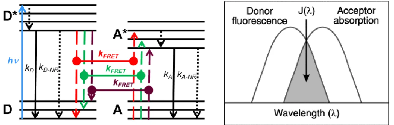
FRET is understood as a non-radiative transfer through dipole-dipole interactions that allows electronic energy to be transferred from an excited donor D to a ground-state acceptor A[45], [46]. The donor and acceptor molecule can be seen as a group of coupled electrical oscillators. Donor oscillators will oscillate and generate their own electromagnetic field. Energy transfer only occurs when the acceptor oscillators are sensitive to the donor oscillators’ electromagnetic field and “share” electronic transitions, which enable the excitation of the molecule from its ground state to the excited state. These electronic transitions are presented in a simplified Jablonski diagram in Figure 2.3 (left) showing the basic principle of FRET. The donor is brought to an excited state (D*) from an electronic ground state (D) by light excitation (h𝜈), followed by inner relaxation (dotted arrows) to an excited electronic ground state and finally to the ground state by radiative decay (kD), non-radiative decay (kD-NR) or FRET (kFRET, dashed
lines referring to possible resonant transitions). FRET (horizontal lines with dots on each end) occurs when the energy transitions of D* and A are in energetic resonance. After FRET, the acceptor is in an excited state (A*), followed by radiative decay (kA) and non-radiative decay
(kA-NR) to its ground state. In spectroscopic terms, there should be a spectral overlap between
the donor emission and the acceptor absorption spectra (Figure 2.3 right). So choosing a suitable FRET pair is the first step for successful FRET, as shown in Figure 2.4, there are many different fluorescent materials can be excellent FRET donor and acceptor. The selection of a correct FRET pair depends on the actual biological question to be investigated, the type of biological specimen, the available instrumentation and the technique applied to measure FRET. Various fluorescent proteins, organic dyes with improved photostability and excellent spectral
12
characteristics as well as quantum dots have been developed and used as FRET donor and acceptor.
Figure 2.3. (Left) Simplified Jablonski diagram representing the energy levels of the donor (D) and acceptor (A) molecules. (Right) Spectral overlap between the donor emission and the acceptor absorption.
Figure 2.4. Examples of available FRET donor and acceptor candidates. (Reference [14], copyright 2006 Wiley-VCH Publisher.)
FRET theory depends on the distance between the donor and the acceptor, the spectral overlap and their relative orientations, it can be summed up in below equations[47].
13
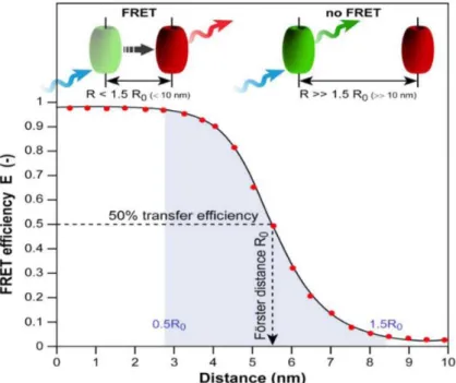
The rate of energy transfer is traditionally described as:
𝑘𝐹𝑅𝐸𝑇 = 1 𝜏𝐷( 𝑅0 𝑟) 6 (2.1)
where 𝜏𝐷 is the excited-state lifetime of the donor in absence of the acceptor; 𝑅0 is the Förster
distance representing a transfer efficiency of 50 %; r is the donor-to-acceptor distance (Figure 2.5).
Then, the FRET efficiency is
𝜂𝐹𝑅𝐸𝑇 = 𝑘𝐹𝑅𝐸𝑇
𝑘𝐹𝑅𝐸𝑇+𝜏𝐷−1 =
𝑅06
𝑅06+𝑟6 (2.2) The Förster distance R0 can be calculated by the spectral overlap integral between the emission
spectrum of the donor and the absorption spectrum of the acceptor:
𝑅0 = ( 9(ln 10)𝜅2Φ𝐷 128𝜋5𝑁 𝐴𝑛4 𝐽(𝜆)) 1 6⁄ (2.3)
where 𝑁𝐴 is Avogadro’s number (6.023 × 10²³ mol-1); n is the refractive index of the medium;
𝜅2 is the orientation factor of the coupled donor and acceptor transition dipole moments which
is usually taken as 2/3; Φ𝐷 is the quantum yield of the donor; 𝐽(𝜆) is the spectral overlap between the donor emission and acceptor absorption. A FRET pair with a large R0 value is
generally favored, because of increased likelihood of FRET occurrence. For a given FRET-pair, the larger spectral overlap results in larger Förster distance.
Figure 2.5. FRET efficiency 𝜂𝐹𝑅𝐸𝑇 variation as a function of the donor-acceptor distance (r) shows the sixth power dependency and a measurable range of the FRET efficiency between 0.5 R0 and 2.0 R0 with a steep curve around R0. (Reference [48], copyright 2012 MDPI Publisher)
14
To enhance the FRET efficiency, the donor group should have good abilities to absorb photons and emit photons. That means the donor group should have a high extinction coefficient and a high quantum yield. The overlap of the emission spectrum of the donor and absorption spectrum of the acceptor means that the energy lost from the excited donor to the ground state could excite the acceptor group. The energy matching is called the resonance phenomenon. Thus, the more overlap of spectra, the better a donor can transfer energy to the acceptor. The spectral overlap can be described as:
𝐽 = ∫ 𝐼̅ (𝜆)𝜀𝐷 𝐴(𝜆)𝜆4𝑑𝜆 (2.4)
Where 𝜀𝐴(𝜆) is the acceptor extinction coefficient, 𝐼̅ (𝜆) is the donor emission spectrum 𝐷
normalized to unity and is given by:
∫ 𝐼̅ (𝜆)𝑑𝜆=1 (2.5) 𝐷 In addition, the Förster distance strongly depends on the orientation factor 𝜅2:
𝜅2 = (cos 𝜃𝑅− 3 cos 𝜃𝐷cos 𝜃𝐴)2 (2.6)
where 𝜃R is the angle between the donor and acceptor transition dipole moments, 𝜃𝐷 and 𝜃𝐴 are
the angles between the respective dipole moments and the line connecting the donor and the acceptor.
Orientation factor 𝜅2 is in the range 0 to 4. 0 corresponds to a perpendicular orientation between the donor and acceptor transition dipoles, 1 is parallel and 4 a collinear orientation (Figure 2.6). In cases when the rotational reorientation of the chromophores is faster compared to the donor excited state lifetime, 𝜅2 averages to a value of 2/3. Since the sixth root is taken to calculate the
distance, variation of 𝜅2 does usually not result in major errors in the calculated distances except
when 𝜅2 is close to 0. In most biological FRET experiments, the orientation factor is assumed
15
Figure 2.6. The orientation of emission dipole moment of donor and absorption dipole moment of acceptor. (Reference [49], copyright 2015 MDPI Publisher)
2.2.2 FRET applications
The major constituents of all living beings are nano-sized molecular species like lipids, proteins, carbohydrates, and nucleic acids. These molecules are constantly in communication with each other through complex yet specific interactions in a crowded molecular environment, regulating the biological functions in an organism.[50], [51]. Due to the biological nanometer working distance and the high sensitivity for small distance changes, FRET has been widely applied in the study of structure elucidation, cellular processes, and diagnostics. The occurrences of the binding, diffusion, and mobility of molecular structures in situ are easily investigated under the spatial and temporal resolution of FRET[52]. Combined with microscopy and spectroscopy, FRET imaging allows scientists to study a variety of phenomena that produce changes in molecular proximity, thereby leading to many significant findings in the life sciences. FRET imaging is a powerful tool in life-science research[52]–[55]. Unlike the usual two molecule studies, FRET measurements have been made between single-molecules by labeling fluorophores to two different sites of the same molecule (i.e. single-molecule FRET; sm-FRET) to investigate the intra activities of the molecule[56]–[59]. FRET is increasingly being used in the area of “sensing/recognition” molecules as a simple, rapid, and sensitive transduction mechanism[60], [61]. Nearly every target of interest, like cancer markers, pathogen markers, microbes, cells, nucleic acids, toxins, drugs, can be sensed using FRET by exploiting the wide
16
field of recognition molecules, such as small organic molecules, polymers, carbohydrates, antibodies, proteins and peptides, nucleic acids and aptamers[60], [62]. Most sensing techniques are based on the binding or cleavage of donor and acceptor in the presence of the target, which can be measured mainly by changes of the donor/acceptor emission intensity. One of the main targets of interest which FRET has played a significant role in clinical analysis is nucleic acid testing.
2.2.3 FRET for nucleic acid detection
FRET has a significant impact on molecular diagnostic technologies for detecting specific, relatively short DNA or RNA sequences (typically identified during DNA sequencing) for infectious disease diagnosis, genotyping, and pharmacogenomics[63]–[65]. These technologies rely on the detection of target nucleic acid hybridization and/or amplification. Real-time, homogeneous, and high-throughput FRET-based technologies are desired. The hybridization reaction is one of the most important and sensitive tools in FRET-based nucleic acid assay (Figure 2.7A). It is real-time observation directly in solution, as a homogenous type without washing procedures. The high speed of hybridization in solution and the detection of only the hybridized probe signal makes it possible to perform in vivo hybridizations directly in live cells. Multiple formats of FRET hybridization probes have been developed[66]. Molecular beacon is one of the latest developments in the area of hybridizing DNA FRET (Figure 2.7B). Molecular beacons are designed as hairpin-shaped oligonucleotides. The double-stranded stem carries the donor and acceptor dyes. The loop fragment can hybridize to the target nucleic acid sequence. When the probe is not hybridized to the target, the donor and acceptor are brought in close proximity by the stem formation, resulting in the FRET-based quenching of the donor. However, in the presence of the target sequence, the FRET transfer is disrupted by the probe, which forms longer and stronger hybrids with the target. Additionally, FRET-based nucleic acid amplification technology has emerged from the marriage between polymerase chain reaction (PCR) and real-time monitoring of fluorescent chemistry. The TaqMan method was developed to allow the real-time monitoring of the reaction. The principle (Figure 2.7C) depends on a DNA-based probe with a fluorescent reporter at one end and a quencher of fluorescence at the opposite end of the probe. The close proximity of the reporter to the quencher prevents the emission of its fluorescence; hydroxylation of the probe by the 5' to 3' exonuclease activity of the Taq polymerase releases the reporter and thus allows emission of fluorescence. Therefore, an increase in the product targeted by the reporter probe at each PCR cycle causes a proportional
17
increase of fluorescence. Fluorescence is detected and measured in a real-time PCR machine[67].
Figure 2.7. Designs of FRET probes for hybridization detection and TaqMan qPCR. (Reference [68], copyright 2015 Elsevier Publisher and Wikipedia)
2.3 Lanthanides
2.3.1. Introduction
Lanthanides are one of the cornerstones of our modern technology. They play a major role in lighting industry[69], electroluminescent materials, telecommunication[70], biological assays[71] and medical imaging purposes[72], [73]. Their growing importance in biological and medical research has been highlighted in a number of recent reviews[74]–[78]. This versatile utilization of lanthanides is due to their unique electrical, optical and magnetic properties, which will be discussed in this chapter.
According to the International Union of Pure and Applied Chemistry (IUPAC) recommendations (1968), lanthanides (Ln) include elements lanthanum (La) and 14 other elements (Ce, Pr, Nd, Pm, Sm, Eu, Gd, Tb, Dy, Ho, Er, Tm, Yb, and Lu) which are listed separately at the bottom of the periodic table. They are called lanthanides because the elements in the series are chemically similar to lanthanum. These fifteen lanthanides, along with similar chemical behavior scandium and yttrium, are often collectively known as rare-earth elements. Their similar chemical properties are due to the electronic configuration of the lanthanide atoms and their corresponding ions. The general electronic configuration of lanthanide atoms are usually denoted as [Xe]4fn5dm6s2, where [Xe] represents the electronic configuration of the
18
noble gas xenon, n represents the number of electrons from 0 to 14, and m represents the number of electrons 0 or 1. Xenon has an electronic configuration of 1s2 2s2 2p6 3s2 3p6 4s2 3d10 4p6 5s2
4d10 5p6 and is abbreviated as [Xe]. The lowest energy orbitals are filled first with electrons and
orbital energy follow the sequence:
1s˂2s˂2p˂3s˂3p˂4s˂3d˂4p˂5s˂4d˂5p˂6s˂4f˂5d˂6p˂7s˂5f˂6d˂7p˂8s
Depending on the relative energy levels, lanthanum, cerium, gadolinium, and lutetium belong to the electronic configuration type [Xe]4fn6s2, and the rest of the lanthanides to the type [Xe] 4fn5d16s2 (for terbium both configurations are energetically close to each other, so either one may be adopted). The 4f orbitals have low radial expansion, and they are shielded from the surroundings by the filled energetically lower 5s and 5p sub-shells which results in very weak perturbations of the spectroscopic and magnetic properties of Ln ions by the surrounding microenvironment. Additionally, this shielding effect causes the interaction between Ln ions (the Ln ion being the central ion) with ligands to form complexes to be electrostatic in nature rather than covalent.
2.3.2. Luminescence lanthanide complexes
Most of the trivalent lanthanide ions are luminescent. The luminescence intensity of the lanthanide ion is dependent on the quantum yield, which is related to the ratio of radiative and nonradiative relaxation processes of the excited energy levels. The quantum yield is also related to the extent of the energy gap between the lowest excited energy level of the ion and the highest sublevel of its ground state multiplet [79]. The partial energy level diagram for aqueous lanthanide ions, illustrating also the energy gaps, is presented in Figure 2.8. Judged by the energy gap criterion, Eu3+, Tb3+, and Gd3+ ions have the strongest luminescence. The most commonly used lanthanide ions in bioanalytical applications are Eu3+ and Tb3+. In addition to their intense emission in the visible wavelengths, their particularly long lifetime is a very important reason[80].
19
Figure 2.8. Partial energy diagrams for the lanthanide aquo ions. The main luminescent levels are drawn in red, while the fundamental level is indicated in blue. (Reference [79], copyright 2005 RSC Publisher.)
The most common oxidation state of lanthanide ions in aqueous solvents is Ln 3+. The trivalent lanthanide ions may be implied in three types of electronic transitions:
(1) Charge transfer transition: Both ligand-to-metal and metal-to-ligand transitions are allowed by Laporte's selection rule. Their energy is usually very large so that they appear in the UV[81]. (2) 4f-5d transitions: They correspond to the promotion of a 4f electron into the 5d sub-shell are also allowed by the parity rule, resulting in sizable oscillator strengths with absorption coefficients in the range 102-103 M-1cm-1, that is, comparable to those of the charge transfer transitions. Their energy depends largely upon the metal environment because 5d orbitals are external and they interact directly with ligand orbitals; however, these transitions are also quite energetic[82].
(3) f-f transitions: The very low level of direct excitation of the lanthanide ions as well as their extremely long excited state lifetimes come from the Laporte’s selection rule and spin-multiplicity rule. Most of the transitions of the Ln3+ ions involve redistribution of electrons within the 4f sub-shell, but f-f transitions are parity-forbidden[83]. That explains their faint intensities and very low molar extinction coefficients (˂1 M-1cm-1)[84].
To get more excited, lanthanide ions usually require indirect excitation from a sensitizing structure, which can be an organic ligand containing a light absorbing chromophore structure and a moiety that coordinates the central ion, or an inorganic crystal structure containing a sensitizer. The absorption can be promoted by operators linked to the nature of light, the odd-parity electric dipole (ED) operator, the even-odd-parity magnetic dipole (MD) and electric
20
quadrupole (EQ) operators. Laporte’s parity selection rule states that levels with the same parity cannot be connected by electric dipole transitions, therefore, f-f transitions are forbidden by the ED mechanism. However, under the influence of a ligand-field, non-centrosymmetric interactions result in the mixing of electronic states of opposite parity, which somewhat relaxes the selection rule and the transitions become partially allowed. Together with the parity-allowed magnetic dipole transitions, the lanthanide ions are greatly sensitized in a three-step mechanism. Light is first absorbed by the immediate surrounding of the lanthanide, then energy is transferred to the Ln ion and then the Ln ion emits light. The overall process is quite complex and involves several mechanisms and energy levels. When sensitized by an inorganic matrix or an organic ligand, the molar absorption coefficients can be compared with that of organic fluorophores and they can emit multiple, characteristic narrow emission bands. The specific, narrow lanthanide emission bands are a result of the sharp transitions between the well-defined J-levels (be degenerated from the electronic configuration based on Coulomb interaction and spin-orbit coupling) of lanthanide ions due to the shielding of the 4f orbitals by the filled 5p66s2 sub-shells. The J-levels can be further split into sublevels (which can be seen as the fine structure of the main emission bands of the lanthanide ion) if the lanthanide ions are in a coordinating environment[85]. Eu3+ ion is taken as an example in Figure 2.9.
Figure 2.9. The interactions lead to the different electronic energy levels for the [Xe] 4f6 5d0 configuration of Eu3+. (Reference [85], copyright 2005 Science Reviews 2000 Ltd.)
21
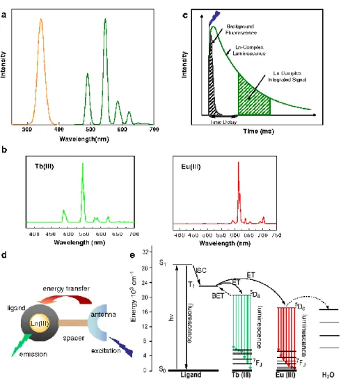
Luminescent lanthanide complexes offer several distinct advantages. First, their sensitized emission gives rise to a large Stokes shift between the absorption of the antenna and the emission of the lanthanide, thus avoiding concentration-dependent self-absorption problems (Figure 2.10a). Second, their emission spectra are characterized by very narrow bands (ca. 20 nm wide), therefore, lanthanide probes can be concurrently employed to monitor several analytes simultaneously (Figure 2.10b). Lastly, lanthanide emissions are Laporte forbidden and therefore are characterized by extremely long luminescence lifetimes, in the millisecond range for europium and terbium and the microsecond range for samarium and dysprosium[12], [79]. In comparison, the lifetimes of most organic dyes are in the nanosecond range, whereas those of biological media are typically in the sub-microsecond range. The very long luminescence lifetime is ideal for the direct, time-gated detection of analytes in a complex biological medium. The time gate allows no auto-luminescence interference before measuring the luminescence of the complex (Figure 2.10c). For a high luminescent lanthanide complex, introducing a suitable light-harvesting chromophore or ‘‘antenna” is essential. The function of this antenna is to absorb energy from UV-Vis radiation and transfer it to the Ln3+ ion, then result in the high luminescence of lanthanide complex which can reach to 104-105 M−1cm−1, being essentially determined by the molar absorption coefficients of the surrounding ligands (Figure 2.10d, e). This effective way to sensitize the Ln3+ ion is the so-called ‘‘antenna effect”. The
commonly used antenna molecules are organic ligands bearing aromatic chromophores, such as bipyridine, terpyridine, triphenylene, quinoline, or substituted phenyl and naphthyl groups.
22
Figure 2.10. Principles of lanthanide luminescence. a) Stokes shift between the absorption of an antenna (orange) and the emission of terbium (green). b) Emission of terbium and europium. c) principle of time-gated spectroscopy. d) Indirect excitation of a lanthanide via an antenna. e) Simplified Jablonski diagram showing the main energy flow paths during sensitization of lanthanide luminescence via its antenna. (Reference [86], copyright 2009 Springer Publisher.)
2.3.3. Lanthanide complex applications
Luminescent lanthanide complexes play a crucial role in biological applications, such as probes for diagnostics, and bioconjugates for medical imaging and the photodynamic treatment of cancer[87], [88]. They are widely used in Magnetic Resonance Imaging for diagnostic purposes or for monitoring the effects of therapy, usually applied for patients with cancers, cardiovascular problems, central nervous system, and other conditions[89], [90]. Moreover, lanthanide complexes are good factors for luminescence imaging because of their emission in the near-infrared region (NIR) which can be used to study the biological structures at different depths and resolutions with the accuracy of analysis[91]. Lanthanide complexes offer several advantages over conventional organic fluorophores in bioanalysis due to their unique
23
spectroscopic properties, such as large Stokes shifts, long luminescence lifetimes, and narrow emission bands. The large Stokes shifts contribute to the low background signal since there is minimal crosstalk between excitation and emission signals. The long-lived luminescence of lanthanide complex can be collected using time-resolved fluorescence measurement, while the interference of short-lived background fluorescence can be eliminated. Furthermore, narrow emission can improve the detection sensitivity of luminescent probes for analytes, hence, lanthanide-based luminescent probes have favorable signal-to-noise ratios and could serve as sensitive probes for various targets. Some of them have been successfully commercialized within detection systems for immunoassays and DNA assays based on luminescent Tb3+ or Eu3+ complexes to detect various clinical biomarkers in biomedical diagnostics and imaging[11], [92]. The conjugation of lanthanide complexes to biomolecules can be established by covalently bound functional groups on the ligand. The most popular conjugation strategies are using Ln-complexes functionalized with NHS-esters or maleimide groups, which selectively bind to amino or thiol groups, respectively.
Multiplexing
Multiplexing has stimulated the renewed interest because it can save time and reagents, increasing demand for the simultaneous measurement of multiple biological parameters (e.g., concentrations or distances) in a single sample. The most frequently used technique to detect multiple parameters from the same sample is spectral (or color) multiplexing with different fluorophores, such as organic dyes[93], [94], fluorescent proteins[95] and quantum dots[96]. Luminescent lanthanide complex-based FRET is a valuable tool for sensitive and versatile multiplexing. In preference to other lanthanide complexes, luminescent Tb complexes have been the major players in this field because of their large quantum yields, extremely long lifetime and multiple emission bands.
In recent years, different multiplexed LTC-based FRET biosensors have been developed, most of them use the principle of FRET from one LTC donor to different acceptor molecules or particles (spectral multiplexing)[8], [97]. As shown in Figure 2.11, the multiplexing capacity of LTC donor is due to the narrow and well-separated emission bands, where we can put several different acceptors in between or beyond the LTC emission bands.
24
Figure 2.11. LTC-donor-based multiplexed FRET. Top: Broad spectral overlap between LTC emission and the absorption of several different acceptors (Left: organic dyes; Right: QDs). Bottom: Separation of the single Tb emission bands allows the measurement of different acceptors in wavelength ranges with very low Tb intensity. Left: organic dyes, Oregon Green(blue), Alexa Fluor555 (green), Alexa Fluor568 (orange), Cy5 (red) and Alexa Fluor700 (brown); right: QDs, Qdot525 (blue), Qdot565 (green), Qdot605 (orange), Qdot655 (red) and QDot705 (brown), LTC-PL-spectra in black. (Reference [10], copyright 2014 Elsevier Publisher.)
The number of spectral multiplexing is still limited. Thus, the big challenge in multiplexing is to create additional distinguishable dimensions.The multiplexing based on lifetime (temporal multiplexing) is a very promising concept. LTC based FRET is well suited in temporal multiplexing. Using the extremely long PL lifetimes of LTC and different donor-acceptor distances for a single donor-acceptor pair would allow for designing different FRET-quenched and FRET-sensitized PL decay times.
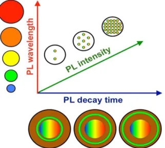
If different PL decay times can be distinguished, then temporal and spectral multiplexing could be reliably combined. When different acceptor PL intensities can also be distinguished and controlled, this parameter can be used as a third multiplexing dimension (Figure 2.12).
25
Figure 2.12. Photoluminescence multiplexing in three dimensions. The combination of PL wavelengths (e.g., from different QD sizes) with PL decay times (e.g., from different LTC-donor to QD-acceptor distances) and PL intensities (e.g., from different concentrations of the LTC-QD hybrid nanoparticles) can offer different distinguishable signals for multiplexed sensing or optical barcoding. (Reference [10], copyright 2014 Elsevier Publisher.)
Although the advantages of LTC for multiplexed FRET are numerous, and theoretical construction of a multiplexed LTC-based FRET is relatively easy, the practice is much more challenging. Pulsed excitation and time-gated or time-resolved detection must be combined with different wavelength separation technologies. Determination of conformational changes as well as distance distributions within a biological FRET system can most often only be determined by time-resolved measurements[3]. Time-resolved measurement provides more dynamic information that is available from the steady-state data. Time-dependent intensities are measured following short pulsed excitation of light. Data collection starts after the excitation pulse has decayed and fluorescent samples with long lifetimes even allow an almost background-free detection after the complete decay of detrimental effects such as sample auto-fluorescence.
Each fluorophore requires a distinct detection channel and excitation wavelength and spectral overlap, which needs to take into account the entire spectrum of each fluorophore (not only the full-width-at-half-maximum, FWHM), imposes fundamental limits to color multiplexing. Even the spectra of quantum dots (with FWHM of ~30 nm) extend over >100 nm, which limits the spectral range of 400 to 800 nm to approximately four completely distinct signals.
26
2.4 Fluorescent dyes
In the last decades, the popularity of organic dyes as fluorescent probes has steeply risen in biophysical research. Fluorescent dyes are often used as fluorescent labels attached to specific locations within the biomolecule. The message of the labeled object is qualitatively or quantitatively transferred by detecting the fluorescence intensity. The majority of common fluorophores such as fluoresceins, rhodamines, 4,4´-difluoro-4-bora-3a,4a-diaza-s-indacenes (BODIPY dyes) and cyanines have been widely used in bioimaging, biosensing, medical diagnosis, and environmental detection. Fluorescent dyes have many good chemical and physical properties, such as, easy chemical structure tuning, good biocompatibility, low-toxicity, easy metabolism in the biological system, and non-radiation which are based on their slight structure, high molar absorption coefficient, moderate-to-high fluorescence quantum yield, etc. However, their comparatively not narrow absorption and emission bands can favor crosstalk between different dye molecules and small Stokes shifts (typically less than 25 nm) can lead to serious self-quenching and fluorescence detection errors because of excitation backscattering effects. Fluorescence dye probes have been used to detect the structures of RNA and DNA, study the remedy of DNA damaged basic group, identify the status of the amino group and the active area of the protein molecule, distinguish nucleic acids with different conformation and the chemical reactive activities of related drugs.
Figure 2.13. Examples of available fluorescent dye. (Reference [14], copyright 2006 Wiley-VCH Publisher)
27
2.4.1 Cyanine dyes
Cyanine dyes are small organic molecules with two nitrogen-containing aromatic heterocyclic rings linked via a polymethine bridge, which are also named polymethine cyanine dyes. Cyanine dyes are broadly used in the life sciences and other biologically related disciplines as optical probes of membrane potential[98], organelle[99], [100], labels for neuron pathway tracing[101] and as probes for membrane structure and dynamics. Moreover, their applications as fluorescent labels for proteins, nucleic acids and other demanding fluorescence microscopy applications made cyanine dyes more popular in the last two decades because of their remarkable photostability, good fluorescence efficiency, and commercial availability as derivatives for covalent labeling of proteins and nucleic acids. The small organic dyes with spectral properties are covalently or non-covalently bound to biomolecules. The interaction of small organic dyes with proteins or nucleic acids giving a detectable signal is a very useful method for the investigation of biological processes on the molecular level.
2.4.2 Cyanine dyes applications in FRET
Cyanine dyes as energy acceptors have been widely used in a variety of FRET biophysical applications. Lee[102] reported dye cyanine 3-labeled human serum albumin can be applied to construct QDs-based FRET systems by their high affinity interaction with CdZn SeS/ZnS QDs capped with multifunctional polymer ligands containing dihydrolipoic acid (DHLA). This dye-based FRET protein complex can serve as a sensitive sensor for probing the interaction of clofazimine with proteins using fluorescence spectroscopic techniques (Figure 2.14A). Löhmannsröben[103] developed an aptasensing system based on FRET from a lanthanide terbium complex to a cyanine 5 to detect L-selectin in PBS. FRET signal was measured by time-resolved luminescence spectroscopy. Using their system, L-selectin can effectively be recognized in the concentration range of 10-500 ng/ mL that covers the completely relevant pathological range for Alzheimer's disease (Figure 2.14B ). Bald[104] investigated the FRET between the donor fluorescein and the acceptor cyanine 3 to establish a DNA origami-based dye array for light harvesting, signal amplification, and sensing. For the last 15 years, the most popular FRET probes in nucleic acid studies is cyanine dyes (Figure 2.14C).
28
Figure 2.14. Cyanine dyes-based FRET biosensor, cyanine dyes as the acceptor with different donors.
(A) QD to Cy3 FRET sensor. (Reference [102], copyright 2016 ACS Publisher.)
(B) The L-selectin detection based on FRET from a Tb-complex to a cyanine dye. (Reference [103], copyright 2015 Elsevier Publisher.)
(C) DNA origami-based fluorescein to Cy3 FRET array. (Reference [104], copyright 2018 ACS Publisher.)
29
The disadvantage of organic dyes for FRET applications is their crosstalk, which results from direct acceptor excitation due to the relatively broad absorption bands of organic dyes and needs to render tedious correction of measured signals in multiplexing detection. Lifetime multiplexing can overcome this problem. It can be performed by making use of fluorophore-specific decay behavior, measured at a single excitation and a single emission wavelength to discriminate different decays. This approach has no crosstalk from different acceptors but requires sufficiently different lifetimes. Lifetime multiplexing, as well as combining spectral multiplexing can be used to realize multiplexing detection for nucleic acids detection in this thesis.
2.5 Quantum dots
Luminescent semiconductor nanocrystals, also known as quantum dots (QDs), are generally composed of atoms from groups II-VI (ZnO, ZnS, CdS, CdSe, CdTe) and III-V (GaN, GaP, GaAs, InP, InAs) elements in the periodic table and are defined as particles because of small physical dimensions between 1 and 12 nm [18]. QDs are nanometer-sized crystals with unique photochemical and photophysical properties, which are not available from either isolated molecules or bulk solids. For example, symmetrical emission spectra, high quantum yield, broad absorption spectra, good chemical, and photo-stability and size-dependent emission wavelength tunability. QDs can be covalently linked with biorecognition molecules such as peptides, antibodies, nucleic acids, or small-molecule ligands for use as biological labels. High-quality QDs are also well suited for optical encoding and multiplexing applications due to their broad excitation profiles and narrow emission spectra. QDs have been successfully used as luminescent probes with a considerable advantage over conventional organic fluorophores in many biological fields.
2.5.1 Photophysical properties
QDs have unique photophysical properties, including tunable photoluminescent emission as a function of core size and quantum confinement effects for binary combinations of semiconductors[17], [18]; broad absorption spectra with high molar extinction coefficients (10-100 times larger than those of conventional dyes)[105]; narrow symmetric PL spectra (full width at half maximum ~25-40 nm) spanning the UV to near-IR with relatively high quantum yields, large Stokes shifts and high resistance to both photobleaching and chemical degradation [106], [107]. Compared with conventional organic fluorophores (Table 2.2), QDs present considerable advantages, especially broadband excitation and narrow bandwidth emission are


![Figure 2.7. Designs of FRET probes for hybridization detection and TaqMan qPCR. (Reference [68], copyright 2015 Elsevier Publisher and Wikipedia)](https://thumb-eu.123doks.com/thumbv2/123doknet/12888447.370490/28.892.107.787.179.488/designs-hybridization-detection-reference-copyright-elsevier-publisher-wikipedia.webp)
![Figure 2.9. The interactions lead to the different electronic energy levels for the [Xe] 4f 6 5d 0 configuration of Eu 3+](https://thumb-eu.123doks.com/thumbv2/123doknet/12888447.370490/31.892.244.654.641.997/figure-interactions-lead-different-electronic-energy-levels-configuration.webp)