HAL Id: tel-01249393
https://tel.archives-ouvertes.fr/tel-01249393
Submitted on 4 Jan 2016HAL is a multi-disciplinary open access archive for the deposit and dissemination of sci-entific research documents, whether they are pub-lished or not. The documents may come from teaching and research institutions in France or abroad, or from public or private research centers.
L’archive ouverte pluridisciplinaire HAL, est destinée au dépôt et à la diffusion de documents scientifiques de niveau recherche, publiés ou non, émanant des établissements d’enseignement et de recherche français ou étrangers, des laboratoires publics ou privés.
Structure-function studies of human ribosome complexes
Heena Khatter
To cite this version:
Heena Khatter. Structure-function studies of human ribosome complexes. Genomics [q-bio.GN]. Université de Strasbourg, 2014. English. �NNT : 2014STRAJ072�. �tel-01249393�
UNIVERSITÉ DE STRASBOURG
ÉCOLE DOCTORALE des Sciences de la Vie et de la Santé
Centre de Biologie Intégrative, IGBMC, UMR7104, Illkirch
THÈSE
présentée par :Heena KHATTER
Soutenue le : 18 Septembre 2014
pour obtenir le grade de :
!"#$%&'($')*%+,-$&.,#/'($'0#&1.2!%&3
Discipline/ Spécialité
: Biophysique et biologie structurale
(Biophysics and Structural biology)
Structure-function studies of human
ribosome complexes
Etudes structure-fonction de complexes
du ribosome humain
THÈSE dirigée par :
Dr. KLAHOLZ Bruno P. DR, CNRS, IGBMC, Illkirch. RAPPORTEURS :
Dr. JENNER Lasse B. Associate professor, Aarhus University, Denmark.
Dr. MECHULAM Yves DR, École Polytechnique, Paris.
AUTRES MEMBRES DU JURY :
Dedicated to my parents, Chander and Renu, for their unconditional love and
support.
We are kept from our goal, not by obstacles, but by a clear path to a
lesser goal.
-Quote 9.24
Bhagavad- !"#
Acknowledgements
My PhD thesis has been possible only because of constant support and participation of several people. Some of the people who helped me in accomplishing this task deserve the credit equally.
First and foremost, I would like to thank my thesis advisor Dr. Bruno Klaholz, for giving me this opportunity to work on the fascinating subject of human ribosomes. With his constant support and guidance, the past four years have been a great learning experience. Sincere thanks for being so patient with my amateur and overtly enthusiastic attitude. I am glad that you never underestimated my potential and helped me to become a better scientist.
I would take this opportunity to express my gratitude to all my Jury members, Dr. Lasse Jenner, Dr. Yves Mechulam, Dr. Eric Westhof and Dr. Albert Weixlbaumer who enthusiastically agreed to read my thesis and evaluate my work. I am grateful for your presence here.
I am thankful to the people at the cell culture service, especially Betty and Leslie, who were always cooperative for trying different HeLa culture conditions. Thanks for providing HeLa cells every time I needed them.
I am thankful to Stefano, who helped in setting-up the ribosome purification; he was indeed a great support in the first year. I am also thankful to Anne-Sophie, who worked with me for two years. It was a valuable experience for me, and it was her willingness to learn that made it such a pleasure to work together. Also, I would like to thank all the Klaholz team members, past and present, for their help and assistance during the past four years.
I am indebted to those who made working here at CEBGS and now CBI, a pure joy. Kareem, Sacha and Kundhavai for helping me learn crystallography and for all the useful advices in data processing, Judith and Marie, for being supportive all the time and for the much needed pep-talks, Alastair for all his nagging and bossing around, Pierre for his assistance at the crystallisation platform, Morgan for all the delicious cakes and help with translations, Anna, Karima, for being around in the lab and for the enthusiastic tea-time discussions. I am thankful to all of you for creating a fabulous work environment.
I am grateful to Alexey, Adam and Kundhavai for patiently correcting my thesis and bringing it to its presentable form.
! "#$%&! '%(#! %)*+! ,#! ,-'.*! /&'0! 1#2! '%%! ,-+! 12$),1$%! &)(3$(()#.(! '.&! 45-0).6! 3-',(7! 8)(! contagious enthusiasm and high spirits made research a lot more motivating.
I would take this opportunity to thank my friends who have made this journey truly enjoyable and memorable. Kamal, Alexey, Dhiraj, Iskander, Thanuja, Anna, Karima, Grigory, Firas, 9%'&)0)2:! /%'(,')2:! 2).'7!;+! 0+,! '(! (,2'.<+2(:! =$,! ),6(!=+3'$(+! #1!,-+0!,-',! %)>ing in
Strasbourg has been so much fun. Also, Priyanka and Saumya, who have been just a phone call away to share my happiness and troubles alike.
Most importantly, I would like to thank my parents, my brother and my entire family for being my support and strength all the time, without which this work would never have been accomplished. Leaving the comfort of home and homeland was not easy for me and I $.&+2(,'.&! ),! "'(.6,! +'(?! 1#2! 0?! 1'0)%?! '(! "+%%7! @).3+2+! ,-'.*(! 1#2! %+,,).<! 0+! 1#%%#"! 0?! dreams. No matter in which part of the world I would be, I know that I can always rely on you all for encouragement.
Last, but not the least I would like to thank Sasha for teaching me the nuances of cryo-EM, for sharing creative ideas and his experience in this enthralling field of translation. He has been a pillar of support and motivation all throughout. I am indebted to him for being a constructive critic, for believing in me, for never letting me yield to the circumstances and keeping my spirits high in testing times.
Summary
Protein synthesis in the cell is catalyzed by the ribosome and is regulated by protein factors that bind transiently to the ribosome during the different phases of translation-initiation, elongation, termination and recycling. My work focussed on structural and functional aspects of these huge (2-4 MDa) protein synthesising machines. When I started working on this project in October 2010, a lot of structural studies had been done on the prokaryotic ribosome, both by crystallography and by cryo electron microscopy (cryo-EM) of functional ribosome complexes with tRNA, mRNA, and protein factors (for example Chandramouli et al., 2008; Marzi et al., 2007; Spahn et al., 2004). However, human ribosomes were not studied to atomic resolution because of their particularly complex structure. Also, they are inherently difficult to prepare to high homogeneity, which is a key requisite for high-resolution structural work.
The aim of my project was two-fold:
1. Human ribosome structure determination using X-ray crystallography and cryo-EM to provide insights into the specific mechanism of protein synthesis and regulation, like with respect to antibiotic side effects.
2. Elucidate the molecular mechanism of translation termination by forming in-vitro termination complexes for cryo-EM analysis and co-crystallize the 2 eukaryotic release factors (eRF1 and eRF3).
I established the purification of homogenous 80S ribosomes from HeLa cells using sucrose density gradients and polyethylenglycol (PEG) precipitation. The ribosomes were characterized biophysically by Analytical Ultracentrifugation, Mass spectrometry, Multiangle Laser Light Scattering and cryo-EM imaging and 3D reconstruction.
Human 80S samples from different preparations were frozen in the hydrated state as thin vitreous ice films (Dubochet et al., 1988) and cryo-EM images were collected on the high-resolution in-house Tecnai Polara F-30 electron microscope. 3D density maps were calculated from the selected particles at a medium resolution using EMAN2 to address the ribosome conformation and the potential presence of endogenous factors or tRNAs.
Moreover, cryo-EM was used to screen samples with modified conditions to obtain samples with homogenous distribution on grid. With the aim of getting a high-resolution structure I worked on the crystallization of human ribosomes. I setup crystallization trials with well-characterized, homogenous 80S samples in drops as well as capillaries, using various screens like PEG/Ion, Index from Hampton Research; MPD suite, PEGs suite from Qiagen. Initial hits were obtained in capillaries with a few conditions; however, they did not diffract X-rays. These conditions were reproduced and optimized in sitting drops after a series of trials; since the principle of counter diffusion in capillaries is different from vapour diffusion used in sitting drops. Plate-like crystals were obtained which diffracted at SLS (Swiss Light Source) synchrotron up to 26 Å resolution. This part of the work is described in my article (Khatter et al., 2014) in Nucleic Acids Research, 2014, providing a very promising basis for future high-resolution work on the human ribosome.
In the recent past, there have been interesting advances in eukaryotic ribosome studies using crystallography (Ben-Shem et al., 2010, 2011; Rabl et al., 2010; Klinge et al., 2011) and cryoAEM of functional ribosome complexes with tRNA, mRNA, and protein factors (Taylor et al., 2012; Anger et al., 2013; Pallesen et al., 2013). Previously, our lab has worked on prokaryotic initiation and termination of translation. Now we wanted to focus on eukaryotic termination to reveal the interactions between the 2 eukaryotic release factors (eRF1 and eRF3). eRF1 (Class I release factor) indentifies the stop codon and binds to the translating ribosome, followed by eRF3 (Class II release factor) association and release of the nascent peptide chain.
The structures of the isolated eRF1 and eRF3, and of the eRF1/3 complex (missing the catalytic GTPase domain) has been determined already, but N terminus and G domain of human eRF3 have not been well studied. I cloned the eukaryotic release factors (eRF1 and eRF3) in suitable vectors for expression in E. coli. Both the proteins were expressed, purified to biochemical homogeneity and concentrated to 7-15 mg/ml. The interaction of the two proteins was confirmed using Microscale thermophoresis and dissociation constant (kd) was determined to be 150 nM consistent with values found in the literature. (yes, values were determined to be 200nM for full erf1/3 complex, in 2010) These factors were co-crystallized, and a few hits were obtained with protein crystals. During screening at SLS beamline (Villigen, Switzerland), it was found that out of all these crystals obtained with different
precipitants, only one condition with Lithium sulphate as precipitant gave diffraction up to 4 Å resolution. Data sets were collected for this crystal and a few others which diffracted up to 8 Å. These crystals, for the condition with Li2SO4, belong to space group P412121 with 2 molecules of eRF1 and 1 copy of eRF3 in the asymmetric unit, which could be because of eRF1 existing as a dimer in solution. Higher resolution data sets need to be obtained, but a first molecular replacement solution has been found.
The purified ribosomes from HeLa cells (Khatter et al., 2014) fulfil an essential requirement for forming functional complexes to investigate the missing links in translation like termination. The ribosomal termination complex was assembled using purified 80S, eRF1, eRF3, mRNA, tRNA (uncharged) and characterized using cryo-EM. For analysis, 80 000 particles were selected and structure refined by image processing, revealing that it contains the tRNA in the E (exit) site rather than in the P (peptidyl transferase) site. This could be due to misplacement of mRNA or complete absence of mRNA in this structure. Also, ribosomes exist in 2 major states, ratcheted and non-ratcheted, which is due to movement of the small subunit, leading to heterogeneity in the sample set. The data were analysed using the image processing softwares, EMAN2 and Relion with splitting the particle sets, to look for even a small population which might have tRNA in P site. This is being evaluated. This structure obtained will be analysed by fitting the structures determined by crystallography. It should provide accurate information on the functional specificity of eukaryotic ribosomes, with a prospect of developing specific antibiotics preferentially targeting the function of the prokaryotic ribosome.
Alongside, in collaboration with the group of Yves Mély at the faculty of pharmacy, Illkirch, we have been addressing the question whether interactions exist between the ribosome and the viral proteins GAG (viral polyprotein) and NCP7 (a constituent of GAG). HeLa cells transfected with GAG or NCP7, were lysed, spun briefly to remove cell debris and enveloped organelles (mitochondria and nuclei) and the supernatant was used for analysis. I performed 20-50% sucrose density gradients for polysome analysis and checked co-sedimentation of ribosomes with GAG. Western blot analysis revealed co-localization of GAG with polyribosome fractions. Even purified 80S ribosomes, when incubated with NCP7 peptide, co-precipitated, showing that NCP7 binds to the ribosome and could be involved in its own translational regulation.
Future Perspectives
My work on human ribosomes has established a new hybrid way of obtaining crystals for inherently challenging molecules, based on interplay between biochemistry, cryo-EM and crystallization assays. Sample visualisation for obtaining feedback on homogeneity and stability of sample is a novel aspect which is important to consider for the study of complex macromolecular assemblies in integrated structural biology. Here, it clearly highlights the advantage of teaming up two major structural techniques, which can be used in future for other biomolecules.
With the new in-house Titan Krios electron microscope installed recently better data sets can be collected like in the movie mode for achieving improved structures with less number of particles. High-resolution structure for termination complex will shed light on the exact mode of interaction of eRF1 and eRF3 with the peptidyl transferase center (PTC) and the decoding center, with the ultimate aim of providing insights into the molecular basis of stop codon recognition by the class-I release factors.
Résumé de thèse
Dans la cellule, la synthèse protéique est catalysée par le ribosome, et régulée par des facteurs protéiques qui interagissent de manière transitoire, pendant les différentes B-'(+(!&6).),)',)#.:!C%#.<',)#.:!,+20).')(#.! +,! 2+3?3%'<+7! D#.! ,2'>')%! B#2,+! ($2! %6C,$&+! &+(! aspects fonctionnels et structuraux de cette énorme (2-4 MDa) machinerie de synthèse. E#2(F$+! G6')! 3#00+.3C! H! ,2'>')%%+2! ($2! 3+! B2#G+,! +.! #3,#=2+! IJKJ:! B%$()+$2(! Ctudes structurales avaient été publiées, à la fois en cristallographie aux rayons-X et en cryo-microscopie électronique, du ribosome procaryote fonctionnel, seul ou en complexe avec des ARN de transfert (ARNt), ARN messagers (ARNm) ou facteurs protéiques (Chandramouli et al., 2008; Marzi et al., 2007; Spahn et al., 2004). Cependant, et en raison de sa structure B'2,)3$%)L2+0+.,! 3#0B%+M+:! %+! 2)=#(#0+! -$0').! .6'>'),! '%#2(! B'(! C,C! C,$&)C! H! 2C(#%$,)#.! atomique. De plus, sa purification homogène Aun prérequis maG+$2! B#$2! %6C,$&+! H! -'$,+! résolutionA est tout particulièrement difficile à accomplir.
Le but de mon projet était double :
1. Résoudre la structure du ribosome humain par cristallographie aux rayons-X et cryo-microscopie électronique, afin de poser les bases structurales de la synthèse B2#,C)F$+!+,!&+!('!2C<$%',)#.:!B'2!%6'3,)#.!&6'.,)=)#,)F$+(!B'2!+M+0B%+7
2. Elucider le mécanisme moléculaire de la terminaison, en formant in vitro des 3#0B%+M+(! &+! ,+20).')(#.! B#$2! %6C,$&+! B'2! 32?#-microscopie électronique, et en co-cristallisant les deux facteurs de terminaison eucaryotes (eRF1 et eRF3).
N6')! 0)(! '$! B#).,! %'! B$2)1)3',)#.! &+! 2)=#(#0+(! OJ@! -#0#<L.+(:! H! B'2,)2! &+! 3+%%$%+(! HeLa, en u,)%)('.,! &+(! <2'&)+.,(! &+! &+.(),C! &+! ($32#(+! ').()! F$6$.+! B2C3)B),',)#.! '$! polyéthylène glycol (PEG). Les ribosomes ont ensuite pu être caractérisé par plusieurs 0C,-#&+(! =)#B-?()F$+(:! ,+%%+(! F$+! %6$%,2'3+.,2)1$<',)#.! '.'%?,)F$+:! %'! (B+3,2#0C,2)+! &+! masse, la diffusion de lumière-laser dynamique à plusieurs angles (MALLS) et la cryo-microscopie électronique avec reconstitution tridimensionnelle. Des échantillons de 2)=#(#0+!OJ@!&+!B2CB'2',)#.(!&)11C2+.,+(!#.,!C,C!3#.<+%C(!H!%6C,',!-?&2',C!+.!1).+!3#$3-+! de glace vitrifiée (Dubochet et al., 1988) et des images par cryo-microscopie électronique ont été collectées à haute résolution, sur le microscope Tecnai Polara F-30 installé dans
notre laboratoire. Des cartes de densité tridimensionnelles, à résolution moyenne, ont pu P,2+! 3'%3$%C+(! H! B'2,)2! &+(! B'2,)3$%+(! (C%+3,)#..C+(! H! %6')&+! &$! %#<)3)+%! QD/RI:! B#$2! déterminer la conformation du ribosome et la présence potentielle de facteurs endogènes #$!&6/SR, (Figure I).
Figure I:(A) La structure en cryo-microscopie électronique du ribosome humain 80S vide et (B) avec un ARNt dans le site E ; les deux ont été purifiés à partir de cellules HeLa, à haute et faible concentration en KCl pendant le traitement à la puromycine, respectivement ; code couleur : 40S en jaune, 60S en bleu, ARNt en rouge.
De plus, la cryo-microscopie électronique a été mise à profit pour cribler des C3-'.,)%%#.(! &'.(! &)11C2+.,+(! 3#.&),)#.(:! '1).! &6#=,+.)2! $.+! &)(,2)=$,)#.! -#0#<L.+! ($2! %+(! <2)%%+(7!T'.(!%+!=$,!&6#=,+.)2!$.+!(,2$3,$2+!H!2C(#%$,)#.!',#0)F$+:!G6')!C<'%+0+.,!,2'>')%%C!H! %'! 32)(,'%%)(',)#.! &$! 2)=#(#0+! -$0').7! N6')! 0)(! '$! B#).,! &+(! +((')(! &+! 32)(,'%%)(',)#.! H! B'2,)2! &6C3-'.,)%%#.(! &+! 2)=#(#0+(! B$2(! +,! B2C'%'=%+0+.,! 3'2'3,C2)(C(! B'2! %+(! &)11C2+.,(! 0#?+.(! énoncés précédemment, en gouttes assises, suspendues, mais également en capillaires, avec différents kits commerciaux de criblage (PEG/Ion et Index de Hampton Research, MPD suite et PEGs suite de Qiagen). Des premiers cristaux ont été obtenus en capillaires dans plusieurs conditions. Cependant, ils ne diffractaient pas les rayons-X, même sur les lignes intenses des synchrotrons. Le principe de contre-diffusion en capillaire étant différent de
celui de diffusion de vapeur en gouttes assises, ces conditions ont dû être re-optimisées pour la mise en place de gouttes assises. Des cristaux en forme de plaques ont été obtenus, et leur diffraction à la source synchrotron SLS (Swiss Light Source) a atteint 26 Å de résolution, illustrant la première obtention de cristaux du ribosome humain capables de diffracter les rayons-X (Figure IIU7! V+,,+! B'2,)+! &+! 0#.! ,2'>')%! '! 1'),! %6#=G+,! &6$.! '2,)3%+! (Khatter et al., 2014) dans Nucleic Acids Research, 2014, fournissant une base très prometteuse pour les futurs travaux à haute résolution sur le ribosome humain.
Figure II: !"#$%&'(&%)#*%()+,-.#/-#%(01)1'-',(3#4567#10+&3-)#/,3)#8&)#*,9(88,(%&)#/:-3b,;-&# /&# *%()+,88(),+(13# <%=)+,8>,%9?@# A"# <%()+,-.# &3# B1%'&# /&# 98,;-&# %&9%1/-(+)# &3# C1-++&# ,))()&7# diffractant à 26 Å. La plupart sont visualisés sur leur tranche et donnent ,(3)(# 8:('9%&))(13# /&# baguettes. (C) Le spectre de diffraction montre un réseau réciproque complet avec des 9,%,'D+%&)#/&#',(88&#/:&3E(%13#,#F#G5H#I7#0#F#J4K#I#&+#*#F#LJJ#I@#M&)#*&%*8&)#/&#%N)18-+(13)#,BB(*2N)# sont à 23, 30 et 40 Å. Le cliché montre des +O*2&)#/&#/(BB%,*+(13#P-);-:Q#RH#I#/&#%N)18-+(13@
Récemment, des avancées intéressantes dans le domaine des ribosomes eucaryotes ont vu le jour, grâce à la cristallographie aux rayons-X (Ben-Shem et al., 2010, 2011; Rabl et al., 2010; Klinge et al., 2011) ou à la cryo-microscopie électronique (Taylor et al., 2012; !"#$% #&% '()*% +,-./% 0'((#1#!% #&% '()*% +,-.2)% 0'$% (#% 3'114*% !5&$#% ('65$'&57$#% 18#1&% 95!9#!&$4% 3$7!973'(#:#!&% 1;$% (87!7&7'&75!% #&% ('% &#$:7!'715!% <#% ('% &$'<;9&75!% 3$59'$=5&#)% >5;1% '?5!1% donc souhaité faire un pas en avant et étudier la terminaison de la traduction chez les #;9'$=5*% #&% !5&'::#!&% (87!&#$'9&75!% 561#$?4#% #!&$#% (#1% <#;@% A'9&#;$1% <#% &#$:7!'715!% #BC-%#&%#BC.)%#BC-%DA'9&#;$%<#%&#$:7!'715!%<#%9('11#%-2%#1&%9'3'6(#%<87<#!&7A7#$%(#1%95<5!1% sto3%#&%<#%1#%(7#$%';%$76515:#%#!%3E'1#%<84(5!"'&75!)%#BC.%DA'9&#;$%<#%&#$:7!'715!%<#%9('11#% 2) est alors recruté, et permet la libération de la chaîne peptidique néosynthétisée.
F#1%1&$;9&;$#1%<8#BC-%#&%#BC.%715(41*%'7!17%G;#%<;%95:3(#@#%#BC-H.%D1'!1%(#%<5:aine 9'&'(=&7G;#%IJ0'1#2%5!&%<4KL%4&4%3;6(74#1*%:'71%!5;1%:'!G;5!1%<87!A5$:'&75!1%95!9#$!'!&% (8#@&$4:7&4%':7!5-terminale ainsi que le domaine G de la protéine eRF3 humaine (Figure III). M8'7% <5!9% #!&$#3$71% <#% 9(5!#$% #!% ?#9&#;$1% 6'9&4$7#!1% (#1% <#;@% "N!#1% #$f1 et erf3. Après expression en bactérie E. coli*%K8'7%3;%715(#$%(#1%<#;@%3$5&47!#1%#&%56&#!7$%<#1%95!9#!&$'&75!1% <#%3$5&47!#%3;$#%'(('!&%<#%O%L%-P%:"H:F)%F87!&#$'9&75!%#!&$#%(#1%<#;@%A'9&#;$1%'%4&4%:#1;$4#% par thermophorèse à micro-échelle et la constante de dissociation (kd) a été estimée à 150 nM. Ce résultat corrobore les valeurs décrites dans la littérature. Ces facteurs ont alors été co-cristallisés, et quelques cristaux ont pu être obtenus dans différentes conditions. F8#!1#:6(#% <#1% 9$71&';@% '% '(5$1% été testé à la source synchrotron SLS avec une diffraction atteignant les 8 Å de résolution et des premiers jeux de données ont été enregistrés. Apres optimisation de tous les cristaux obtenus avec différents agents précipitants, seule une condition avec d;%1;(A'&#%<#%(7&E7;:%'%3#$:71%<856&#!7$%;!#%<7AA$'9&75!%L%Q%R%<#%$415(;&75!)% S!% K#;% <#% <5!!4#1% '% <5!9% 4&4% 95((#9&4% 35;$% 9#% 9$71&'(% G;7% '33'$&7#!&% ';% "$5;3#% <8#13'9#% P412121*% '?#9% <#;@% :5(49;(#1% <8#BC-% #&% ;!#% :5(49;(#% <8#BC.% 3'$% ;!7&4% '1=:4&$7G;#)% T#97% peut 18#@3(7G;#$%3'$%(8561#$?'&75!%A'7&#%<#%<7:N$#1%<8#BC-%#!%15(;&75!)%U#1%K#;@%<#%<5!!4#1% à plus haute résolution sont nécessaires pour obtenir une structure plus détaillée, mais un premier remplacement moléculaire a pu être effectué sur ces jeux à moyenne résolution.
Figure III: (A) Représentation schématique des protéines eRF3 de S. pombe et humaine. (B) La structure cristallographique de eRF1 montre les motifs catalytiquement actifs NIKS et GGQ, marqués en rouge dans le domaine N (vert) et M (jaune), res9&*+(E&'&3+@#<1/&#/:,**&))(13#$SA#T# 1DT9. (C) La structure cristallographique de eRF3 sans le domaine amino-terminal, montrant les /1',(3&)#U7#R#&+#V#&3#08&-7#%1)&#&+#',C&3+,7#%&)9&*+(E&'&3+@#<1/&#/:,**&))(13#$SA : 1R5B.
Les ribosomes purifiés à partir de cellules HeLa (Khatter et al., 2014) remplissent les 95!<7&75!1% #11#!&7#((#1% L% ('% A5$:'&75!% <#% 95:3(#@#1% A5!9&75!!#(1*% 'A7!% <84&;<7#$% (84&'3#% encore mal comprise de la terminaison. Le complexe de terminaison ribosomal a été assemblé in vitro, en mélangeant du ribosome humain 80S purifié, eRF1, eRF3, un ARNm et un ARNt non chargé, et ce complexe a pu être caractérisé par cryo-microscopie électronique. Pour son analyse, 80000 particules ont été sélectionnées, et la structure, 'AA7!4#%3'$%&$'7&#:#!&%<87:'"#1*%'%:5!&$4%G;#%(8 B>&%184&'7&%3517&75!né dans le site de sortie (E) plutôt que dans le site peptidyl-transférase (P) (Figure IV). Ceci peut être dû à un mauvais '!9$'"#% <#% (8 B>:*% ?57$#% 15!% '61#!9#% <;% 95:3(#@#)% U#% 3(;1*% (#1% $76515:#1% #@71&#!&%
majoritairement sous deux états, « ratcheted » et « non-ratcheted », dus à une légère rotation de la petite sous-;!7&4*% 3$5?5G;'!&% '7!17% ;!#% E4&4$5"4!47&4% <#% (849E'!&7((5!)% F#1% <5!!4#1%5!&%4&4%&$'7&4#1%L%(8'7<#%<#1%(5"797#(1%VW >+%#&%B#(75!*%#!%1;6<7?71'!&%(#1%K#;@%<#% données, afin de mettre en évidence une population, aussi minoritaire soit-elle, de $76515:#1%3$41#!&'!&%(8 B>&%<'!1%(#%17&#%0)%T#97%#1&%#!%95;$1%<#%&$'7&#:#!&%#&%('%1&$;9&;$#% obtenue le cas échéant sera analysée par intégration des structures cristallographiques disponibles. Ceci devrait fournir des informations précises quant à la spécificité fonctionnelle des ribosomes eucaryotes, avec la perspective de pouvoir développer des antibiotiques spécifiques, ciblant préférentiellement les ribosomes procaryotes.
Figure IV: Le complexe de terminaison reconstitué in vitro7#,E&*#-3&#*1-%+&#)N;-&3*&#/:!WX'@# !"# Vue de côté du complexe, montrant le facteur de terminaison dans son site de liaison, eRF1 en E(18&+7#&WYV#&3#%1-C&#&+#8:!WX+#/,3)#8&#)(+&#Z#&3#E&%+@# A"#[-&#/&#*\+N#/- complexe montrant eEF2. (C) La séparation des densités électroniques montre les deux sous unités du ribosome ainsi que les deux facteurs de terminaison. La structure cristallographique du complexe eRF1-eRF3 superposée *1/&# /:,**&))(13# $SA : 3J5Y) montre ;-&# 8&# '1+(B# ]]^# ):1%(&3+&# &+# 91(3+&# Q# 8:1991)N# /-# $_<#
(Centre peptidyl-transférase). (D) La densité correspondant à eEF2 avec la structure *%()+,881C%,92(;-&#)-9&%91)N&# *1/&#/:,**&))(13#$SA : 3DNY).
0'$'((N(#:#!&*%#!%95(('65$'&75!%'?#9%(#%"$5;3#%<8X?#1%Mély à la faculté de pharmacie <8Y((Z7$9E*%!5;1%'?5!1%?5;(;%$435!<$#%L%(8E=35&EN1#%<87!&#$'9&75!1%#!&$#%(#1%$76515:#1%#&%(#1% protéines virales GAG (polyprotéine virale) et NCP7 (un constituant de GAG). Des cellules HeLa transfectées avec GAG ou NCP7, ont été lysées, brièvement centrifugées pour supprimer les débris cellulaires et les organites à enveloppes (mitochondries et noyaux) '7!17%G;#%(#%1;$!'"#'!&%5!&%4&4%;&7(7141%35;$%(84&;<#)%M['7%#AA#9&;4%<#1%"$'<7#!&1%<#%<#!17&4%<#% sucrose de 20 à 50 % pour l'analyse de polysomes et vérifié la co-sédimentation des ribosomes avec GAG. Les analyses par western blot ont révélé la co-localisation de la protéine GAG avec les fractions de polyribosomes. De plus, les ribosomes 80S purifiés, et incubés avec un NCP7, ont pu être co-précipités, suggérant que NCP7 se lie au ribosome et pourrait être impliqué dans sa propre régulation traductionnelle.
Perspectives futures
W5!% &$'?'7(% 1;$% (#% $76515:#% E;:'7!% '% 3#$:71% <84&'6(7$% ;!#% !5;?#((#% :4&E5<#% 1=!#$"7G;#% 35;$% (856&#!&75! de cristaux dans le cadre de molécules particulièrement <7AA797(#1*% 6'14#% 1;$% (87!&#$A'9#% #!&$#% ('% 6759E7:7#*% ('% 9$=5-microscopie électronique et les essais de cristallisation. La visualisation des échantillons et des particules des complexes macromolécul'7$#% (#% 95!1&7&;'!&% 'A7!%<856&#!7$%;!% '3#$\;%<#% (#;$%E5:5"4!47&4%#&% 1&'67(7&4% #1&%;!%!5;?#(%'13#9&%G;87(%#1&%7:35$&'!&%<#%3$#!<$#%#!%95:3&#%35;$%(84&;<#%<#%95:3(#@#1% macromoléculaires en biologie structurale intégrative. Dans le cas du ribosome humain, cet&#%:4&E5<#%:5!&$#%&$N1%9('7$#:#!&%1#1%'?'!&'"#1*%#&%35;$$'%]&$#%;&7(714#%L%(8'?#!7$%<'!1% (#% 9'<$#% <8';&$#1% 675:5(49;(#1)% I$^9#% ';% &5;&% !5;?#';% J7&'!% _$751% 7!1&'((4% $49#::#!&% ';% T#!&$#%<#%`75(5"7#%Y!&4"$'&7?#%DT`Y2%L%(8YI`WT*%<#%:#7((#;$1%K#;@%<#%<5!!4#1%35urront être 95((#9&41*% !5&'::#!&% #!% ;&7(71'!&% ;!% :5<#% <8#!$#"71&$#:#!&% 95!&7!;*% 3#!<'!&% (#G;#(% (#% <4	&#;$% #!$#"71&$#% K;1G;8L% <7@% 7:'"#1% 3'$% 1#95!<#% 15;1% (#% A'719#';% <84(#9&$5!)% V!A7!*% ('% structure à haute résolution du complexe de terminaison permettra de mettre en lumière le :49'!71:#% 3$4971% <87!&#$'9&75!% <8#BC-% #&% #BC.% '?#9% (#% 9#!&$#% 3#3&7<=(-transférase et le 9#!&$#% <#% <495<'"#% <;% $76515:#*% '?#9% (#% 6;&% ;(&7:#% <8'335$&#$% ('% $435!1#% L% ('% reconnaissance moléculaire des codons stop par les facteurs de terminaison.
Publications
1. Jean-François Ménétret, Heena Khatter, Angelita Simonetti, Igor Orlov, Alexander G. Myasnikov, Vidhya KV, Sankar Manicka, Morgan Torchy, Kareem Mohideen, Anne-Sophie Humm, Isabelle Hazemann, Alexandre Urzhumtsev, Bruno P. Klaholz. Integrative structure-function analysis of large nucleoprotein complexes. RNA Structure and Folding, edited by D. Klostermeier & C. Hammann, 01/2013: chapter 10.2: pages 250-259; de Gruyter., ISBN: 1437-4315.
2. Khatter, H., Myasnikov, A. G., Mastio, L., Billas, I. M. L., Birck, C., Stella, S., and Klaholz, B. P. (2014) Purification, characterization and crystallization of the human 80S ribosome, Nucleic Acids Res., 2014, 42(6), 1-11; doi:10.1093/nar/gkt1404.
Oral and Poster Presentations
1. Integrated structural biology approach for human ribosome studies. IGBMC Internal seminar. 27th March 2014. Heena Khatter. Oral presentation
2. Integrated structural biology approach for ribosome studies. Heena Khatter. School of Life Sciences, Jawaharlal Nehru University, India. 7th November 2013. Oral presentation as an ambassador for IGBMC PhD program.
3. Characterization, cryo-EM analysis and crystallization of the human 80S ribosome.
Heena Khatter. a+Oth Rhine-_!##% B#"75!'(% W##&7!"% 5!% `759$=1&'((5"$'3E=b*% +P-27
September 2013, Schluchsee, Germany. Oral presentation.
4. Purification, characterization and crystallization of the human 80S ribosome. Heena Khatter, Alexander Myasnikov, Stefano Stella and Bruno K('E5(c)%aB WT%+,-.b)%,d-11 September 2013, Bischoffsheim, France. Poster.
5. Structure-function study of the human ribosome. Heena Khatter, Alexander W='1!7Z5?*%e&#A'!5%e&#(('*%T'&E#$7!#%`7$9Z*%Y1'6#((#%`7(('1%'!<%`$;!5%_('E5(c)%aT>Be%- Jacques Monod Confere!9#%JE#%&$'!1('&7!"%$76515:#f%&5g'$<1%:'&;$#%3$5!1b%+-6 June, 2012 Roscoff, France. Poster.
Table of Contents
U@#`3+%1/-*+(13aaaaaaaaaaaaaaaaaaaaaaaaaaaaaaaaaaaaaaaaaaaaaa@aR5
1.1 Overview of translation ... 26
1.2 Key players in translation ... 28
1.3 The prokaryotic ribosomal structure ... 31
1.3.1 Unravelling the ribosome ... 34
1.3.2 Decoding centre ... 34
1.3.3 The peptidyl transferase centre (PTC) ... 36
1.3.4 Peptide exit tunnel ... 37
1.4 Structural dynamics and coordinated ribosome movements ... 39
1.5 Eukaryotic ribosome and its biogenesis ... 42
1.6 Eukaryotic ribosome structure... 44
1.6.1 Eukaryotic ribosomal proteins ... 46
1.6.2 Eukaryotic ribosomal RNA ... 49
1.6.3 The intersubunit interface ... 50
1.7 Translation Termination ... 52
1.8 RF1 and RF2 structures ... 52
1.9 Stop codon recognition ... 53
1.10 Peptidyl tRNA hydrolysis ... 57
1.11 Coordination between the decoding centre and PTC for hydrolysis ... 58
1.12 RF3 structure ... 59
1.12.1 The role of class II release factors ... 60
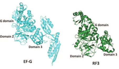
1.13 Analogy between termination and transpeptidation ... 62
1.14 Eukaryotic translation termination ... 64
1.15 Structure of eRF1 ... 64
1.16 Structure of eRF3 ... 65
1.17 Human eRF1-eRF3 complex and inter-dependability ... 68
1.18 Interactions with mammalian ribosome ... 69
1.19 Recycling and ribosome rescue... 72
1.20 Viral protein association with ribosomes ... 77
1.21 Project Outline ... 80
R@#b&+21/)aaaaaaaaaaaaaaaaaaaaaaaaaaaaaaaaaaaaaaaa.aaaa@@aaa@a82 2.1 Materials ... 83
2.2.1 Modifications tested for standardization of purification protocol ... 85
2.3 Ribosome characterization ... 85
2.3.1 Analytical Ultracentrifugation (AUC) ... 85
2.3.2 Size Exclusion Chromatography Multi-angle Laser Light Scattering (SEC MALLS) ... 86
2.4 Single particle cryo electron microscopy (cryo-EM) ... 87
2.5 Crystallization of human 80S ribosomes ... 92
2.6 Release factors (eRF1 and eRF3) purification ... 96
2.6.1 Cloning ... 96
2.6.2 Protein expression ... 96
2.6.3 Protein purification ... 97
2.6.4 eRF1-eRF3 complex purification ... 98
2.7 Biophysical characterization of purified release factors ... 98
Mass spectrometry (MS) ... 98
Dynamic Light Scattering (DLS) ... 99
Native PAGE ... 99
Microscale Thermophoresis Technology (MST) ... 99
2.8 Crystallization of eRF1-eRF3 protein complex ... 100
2.8.1 Cryoprotectant optimisation for the crystals ... 101
2.8.2 Crystal Data collection and processing ... 101
2.9 Post termination complex ... 102
2.10 HIV-1 Gag interactions with ribosomal proteins... 104
2.10.1 Polysome profile analysis ... 104
2.10.2 Linear sucrose density gradient analysis NCp7 ... 105
V@#W&)-8+)aaaaaaaaaaaaaaaaaaaaaaaaaaaaaaaaaaaaaaaaaaaaaa.aa@a106 3.1 Human 80S ribosome ... 107
3.1.1 High-resolution cryo-EM structure of the human 80S ... 109
3.2 Termination factors... 111
3.2.1 Cloning into bacterial expression vectors ... 111
3.2.2 Protein expression and purification ... 112
3.2.3 eRF1-eRF3 complex formation and biophysical characterization ... 115
Mass spectrometry ... 116
Dynamic light scattering (DLS) ... 116
Native gel analysis ... 117
3.2.4 Crystallization of the eRF1-eRF3 complex ... 119 3.3 Pre-termination complex ... 128 3.4 HIV-1 Gag interactions with ribosome ... 132
G@#S()*-))(13aaaaaaaaaaaaaaaaaaaaaaaaaaaaaaaaaaaaaaaaaaaaaaaaUVG K@#!99&3/(.aaaaaaaaaaaaaaaaaaaaaaaaaaaaaaaaaaaaaaaaaaaaaaa@aUV4
5.1 Plasmid purification ... 139 5.2 Expression and purification of TEV Protease ... 140 5.3 Autoinducible media ... 141 5.4 Plasmid pnEAvHX vector map ... 142 5.5 Plasmid pCoGWA vector map ... 143
Table of Figures
Figure 1: Scheme depicting translation cycle. ... 27 Figure 2:. Pictorial representation of mRNa and tRNA ... 29 Figure 3: Structural landmarks of the two subunits ... 32 Figure 4: 2D RNA map for the 16S rRNA ... 33 Figure 5: Decoding by the ribosome ... 35 Figure 6: Peptidyl transferase reaction ... 37 Figure 7: Intersubunit movements. ... 40 Figure 8: Ribosome biogenesis.. ... 43 Figure 9: Conservation of the ribosome core ... 45 Figure 10: Structure-function correlation in 80S ... 49 Figure 11: Structure of RF2 bound to the ribosome. ... 53 Figure 12: Watson-crick and Hoogsteen interactions between nucleotides ... 54 Figure 13: Interactions of the first 2 nucleotides of the stop codons... 55 Figure 14: Interactions of the third nucleotide of the stop codon ... 56 Figure 15: Postulated mechanism for peptidyl tRNA ester bond hydrolysis ... 58 Figure 16: Class II RF ... 60 Figure 17: Proposed model for translation termination ... 61 Figure 18: Structural similarity between EF-G and RF3. ... 62 Figure 19: tRNA superposed on RF2. ... 63 Figure 20: eRF3 and eRF1 structure ... 65 Figure 21: Structural similarity between EF-Tu and eRF3 ... 66 Figure 22: ClustalW sequence alignment of eRF3 ... 67 Figure 23: SAXS model for eRF1-eRF3-GTP complex ... 69 Figure 24: Structural comparison of known eRF3 structures ... 70 Figure 25: Pre-termination complex overview ... 71 Figure 26: NMD pathway ... 73 Figure 27: NGD pathway ... 74 Figure 28: Proposed scheme for translation termination and ribosome recycling ... 75 Figure 29: Data collection for velocity sedimentation ... 86 Figure 30: CTF amplitude at different defocus values ... 88 Figure 31: Comparison between capillary counter diffusion and conventional vapour diffusion ... 93 Figure 32: Bragg's law ... 94 Figure 33: Pipeline for data processing in cryo-EM. ... 103 Figure 34: 80S particle distribution ... 108 Figure 35: High resolution structure of 80S. ... 109 Figure 36: 80S cryo-EM structure ... 110 Figure 37: Agarose gels for eRF3 cloning in pnEAvHX vector. ... 111 Figure 38: eRF3 expression tests ... 112 Figure 39: Nickel-affinity purification of eRF1 and eRF3. ... 113 Figure 40: Ion exchange step for eRF1 and eRF3 purification ... 114 Figure 41: Size exclusion chromatography of eRF1 and eRF1-eRF3 complex ... 115 Figure 42: Mass spectrometry analysis of eRF1-eRF3 complex ... 116
Figure 43: DLS profiles for eRF1-eRF3 complex ... 117 Figure 44: Native gel analysis of eRF1-eRF3 complex. ... 118 Figure 45: MST analysis of eRF1-eRF3 complex ... 118 Figure 46: eRF1-eRF3 crystal structure ... 123 Figure 47: Interactions within the eRF1-eRF3 protein complex ... 125 Figure 48: Conformational flexibility in eRF1 structure ... 126 Figure 49: Scheme for splitting the data set with short mRNA ... 129 Figure 50: The termination complex reconstituted in-vitro ... 130 Figure 51: Polysome profile for Gag and EGFP co-transfected cells ... 132 Figure 52: NCp7 co-sedimentation with 80S ribosomes. ... 133 Figure 53 pnEAvHX vector used for eRF3 cloning in E. Coli UhPi%6'9&#$7') ... 142 Figure 54 pCoGWA vector used for eRF1 cloning in E. Coli UhPi%6'9&#$7') ... 143
Table 1: Canonical protein factors involved in translation. ... 30 Table 2: Composition of prokaryotic and mammalian ribosomes. ... 42 Table 3: ClustalW sequence alignment score for eRF3 sequences. ... 67 Table 4: Proteins known to interact with NC-p7 and their cellular functions. ... 78 Table 5: Data collection parameters on F-30 Polara in-house electron microscope. ... 91 Table 6: Buffer solutions used for every step of purification of eRF1 and eRF3 ... 97 Table 7: Commercial screens tested for eRF1-eRF3 protein complex. ... 100 Table 8: DLS analysis of the complex ... 117 Table 9: The crystallisation conditions for eRF1-eRF3 protein complex... 120 Table 10: The crystallisation conditions for eRF1-eRF3 protein complex in different buffer ... 121 Table 11: Data collection and refinement statistics ... 122 Table 12: Interacting residues in eRF1-eRF3 structure ... 127 Table 13: Cryo-EM data collection parameters for termination complex. ... 128
Abbreviations
Amp AmpicillinATP Adenosine triphosphate cryo-EM Cryo electron microscopy CV Column Volume
DLS Dynamic light scattering DTT Dithiothreitol
EDTA Ethylene diamine tetra acetate
eRF1 eukaryotic Release Factor 1, termination factor Class I eRF3 eucaryotic Release Factor 3, termination factor Class II ESI-TOF Electrospray ionization time-of-flight
FPLC Fast Pressure liquid Chromatography g gram
GDP Guanosine-diphosphate
IW00T0%P8-Guanosyl-methylene-triphosphate GTP Guanosine- triphosphate
HEPES 4-(2-hydroxyethyl)-1-piperazineethanesulfonic acid Y0JI%Y153$53=(%j-D-1-thiogalactopyranoside
ITC Isothermal Titration Calorimetry kDa Kilodaltons
LB Luria Bertani mM Millimolar
MST Microscale thermophoresis NMD Nonsense Mediated-mRNA Decay Ni- NTA Nickel-Nitriloacetic acid PABP PolyA binding protein PI Protease inhibitor
SDS-PAGE Sodium dodecyl sulfate polyacrylamide gel electrophoresis pI Isoleectric point of protein
1.1 Overview of translation
The genetic information in a cell is stored in the DNA and it must be a$#'<b to synthesise proteins. The double stranded DNA acts as a template, during transcription, to generate RNA. The RNA, synthesised by RNA polymerase (RNA pol), is required for the next step of translation to manufacture proteins. These two processes of transcription and translation comprise the central dogma in molecular biology. Three types of RNA are synthesised by their respective RNA polymerases (RNA pol). RNA pol II synthesises messenger RNA (mRNA). This mRNA dictates polypeptide sequence to be created by ribosome, as it is coded so that every three nucleotides of mRNA correspond to an amino acid. Transfer RNA (tRNA), produced by RNA pol III, carries specific amino acids to be incorporated in the growing peptide chain. And lastly, ribosomal RNA (rRNA), transcribed by RNA pol I, forms the catalytic component of the massive macromolecular machine called ribosomes.
Ribosomes are composed of two subunits, each possessing rRNA and protein components. Both subunits harbour specific centres to achieve this humongous and highly regulated task of polypeptide formation. The peptidyl transferase centre (PTC), where the peptide bond is formed; GTPase associated centre (GAC) where several GTP-binding factors hydrolyse GTP to provide a kinetic control mechanism for this process and the peptide exit tunnel which forms a conduit for nascent peptide chain passage; are present in the large subunit (60S/50S). The small (40S/30S) subunit possesses the mRNA channel for associating with mRNA and the decoding site where mRNA is recognised by tRNA.
The tRNA binding on the ribosome is compartmentalised into three (aminoacyl (A), peptidyl (P), and exit (E)) sites depending upon the state of the tRNA bound. The A-site binds the tRNA charged with an amino acid while the P-site tRNA carries the growing peptide chain and the E-site has the uncharged tRNA, ready to exit from the translation machinery. Each of these sites is partly present in both the subunits, which build upon assembly of the full ribosome. The presence of tRNA at each of these sites is annotated as A/A, P/P and E/E, with the first symbol denoting to the contact with the small subunit and second referring to the large subunit.
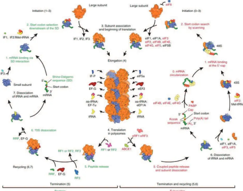
Figure 1: Scheme depicting translation cycle. In bacteria (on the left) and in eukaryotes (on the right). Adapted from Melnikov et. al., 2012.
Protein synthesis is an intricate process that combines high speed with high fidelity (Green & Noller, 1997). It is broadly categorised into four stages, initiation, elongation, termination and recycling. Initiation is the rate-limiting step as it requires start codon recognition by the small subunit (Fig. 1). Once the ribosome has assembled at the start site with the AUG start codon in the P-site, the charged aminoacylated tRNA is delivered by an elongation factor and there begins the next phase of translational elongation. It involves aminoacylated tRNA selection, peptide bond formation (Rodnina et al., 2006), tRNA-mRNA translocation (Frank et al., 2007; Spirin, 2009) in a repeated fashion until a stop codon is encountered in the A-site. The stop codon (UAG, UGA or UAA) recruits release factor instead
of tRNA which allows the release of the newly synthesised nascent peptide chain (Youngman et al., 2008). Finally, the 2 subunits are dissociated in the recycling step (Franckenberg et al., 2012) making them available for the next translation cycle. This basic mechanism of the protein synthesis is conserved across species, but there are stark differences in regulation and certain steps like initiation, associated with the higher complexity of life.
1.2 Key players in translation
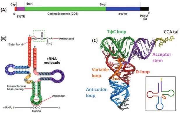
The translation machinery requires RNA and protein components. RNA is synthesised as a single stranded molecule which can form secondary structures by folding over and forming hairpin loops. These structures are stabilised by intramolecular hydrogen bonds formed between the complementary bases (A:U, G:C), as observed for all three types of RNA. mRNA is primarily composed of coding sequences that carry genetic information for sequence of protein to be synthesised. In addition, there are stretches of non-coding or ;!&$'!1('&#<% $#"75!1% '&% &E#% P8- '!<% .8- ends. Prokaryotic mRNA is ready for translation immediately after transcription. However, in eukaryotes its post-transcriptional processing differs from prokaryotes. The eukaryotic mRNA :;1&%;!<#$"5%P8-cap addition, splicing and .8-polyadenylation (Fig. 2A). The P8%9'3%71%'%:5<7A7#<%";'!7!#%D7-methylguanosine) residue, added co-transcriptionally to the first nucleotide with P8-P8-triphosphate bond, which is essential for ribosome recruitment. Furthermore, as soon as mRNA is completely &$'!19$76#<*%'65;&%+P,%'<#!517!#%$#17<;#1% '$#%'<<#<% &5% &E#%.8% #!< to allow export of the mRNA from the nucleus. P8%9'3%'<<7&75!%'!<%35(='<#!=('&75! also ensure that mRNA is not degraded in the cell by nucleolytic enzymes. Finally, it might contain some non-coding stretches (introns) in the open reading frame which are cleaved off from the pre-mRNA, during splicing. The protein-coding sequences are then joined, completing the processing phase.
tRNA acts as an adaptor molecule linking the nucleotide sequence to the amino acid. The single strand of tRNA loops back on itself to form a aclover leafb secondary structure and compacts further to form 3D L-shaped structure (Holley, 1965; Holley et al., 1965) (Fig. 2B). Each of the 3 loops of tRNA has a structure-function association. The anticodon loop
possesses the 3 nucleotides that correspond directly with its specific mRNA codon. The D-(553% '!<% &E#% JkT% (5531% '$#% 17"!7A7#<% 6=% :5<7A7#<% ;$'97(% 6'1#1*% <7E=<$5;$7<7!#% '!<% pseudouridine, respectively (Dudock et al., 1969))%JE#%.8%#!<%E'1%'!%unpaired CCA sequence, which is added by CCA-adding enzymes (tRNA nucleotidyl transferases) during maturation (Deutscher and Ni, 1982). This CCA end is a prerequisite for aminoacylation and is recognised by the enzyme, aminoacyl tRNA synthetase which charges the tRNA with its amino acid (Xiong and Steitz, 2006). For each amino acid to be linked to the tRNA, a single aminoacyl tRNA synthetase exists in a cell.
Figure 2: mRNa and tRNA. (A) Pictorial representation of eukaryotic mRNA. tRNA structure (B) 2D (C) 3D L cshaped, the inset shows the colour coding for different loops.
In prokaryotes, the fMet-tRNA charged with (N-formyl methionine) and in eukaryotes the Met-tRNA charged with methionine are the first residues to be delivered in synthesis of protein coded by AUG start codon on the mRNA (Marcus et al., 1970).
Factors: Translation is regulated by protein factors that bind transiently to the ribosome during the different phases (Table 1). Initiation is regulated by only three factors in prokaryotes (IF1, IF2 and IF3) (Jackson et al., 2010; Myasnikov et al., 2009) as compared to ten factors in eukaryotes. Each of these prokaryotic factors have eukaryotic equivalents, eIF1A, eIF5B and eIF1, performing similar functions (Eiler et al., 2013) (Table 1). eIF1 and
eIF1A are recruited during the recycling phase to the small ribosomal subunit. They allow the appropriate translation initiation site and the start codon selection during scanning on the mRNA (Martin-Marcos et al., 2011). eIF5B accelerates 60S-40S subunit joining. On the other hand, eIF6 is a unique anti association factor, which binds to the 60S and prevents its joining to the 40S. eIF2B promotes GDP-GTP exchange on eIF2. The rest of the eukaryotic initiation factors are majorly involved in either mRNA recruitment (eIF4, eIF3 and PABP) or assist in delivering Met-tRNAi to the 40S subunit (eIF2 and eIF5) apart from performing other important functions. eIF4A, a helicase, forms a component of eIF4F and allows unwinding of &E#%P8%$#"75!%5A%:B> %'!<%'&&'9E:#!&%5A%&E#%3$#-initiation complex to mRNA; eIF4B like eIF4H is an RNA binding protein that enhances the helicase activity of eIF4A (Hinnebusch, 2011). These factors make initiation in eukaryotes a complicated and a highly regulated process.
Table 1: Canonical protein factors involved in translation. The factors performing similar functions are present in the same line.
During elongation eEF1A (EF-Tu in bacteria) delivers the aminoacyl tRNA to the A-site while eEF2 (EF-G in bacteria) provides directionality to the tRNA movement from A to P site
and prevents tRNA from back translocation (Voorhees and Ramakrishnan, 2013). Also, each of these steps has at least one GTPase-associated factor (eIF2, eEF1A, eRF3) which, most likely, fulfils the energy requirement for translation. The role of release and recycling factors is discussed in section III.
Ribosome forms the hub of translation. It is a highly versatile machine which allows recruitment of all these components in a stepwise manner and actively participates in all the steps. In its fully assembled 70S or 80S form it coordinates two major activities of mRNA decoding and peptide bond formation. Below, its structure is discussed in detail.
1.3 The prokaryotic ribosomal structure
Ribosomes are about 20nm in diameter and range from 2-4 MDa in molecular weight, with protein and rRNA components present in 2:1 ratio in prokaryotes as well as eukaryotes. The two subunits have disproportionate component distribution. The small ribosomal subunit possesses a single rRNA chain while the large subunit possesses two rRNA chains in prokaryotes and three rRNA chains in eukaryotes. The overall composition of ribosomes was determined by biochemical studies. However, providing insights into the structural aspects and how these components organise themselves to carry out translation, the two major structural techniques were used cryo-electron microscopy and crystallography.
Like biochemical studies, structural studies on ribosome complexes also involve several technical hurdles and challenges that needed to be overcome. In the context of crystallography, starting from obtaining a homogenous sample to solving the structure, every step had to be optimised to obtain structure of this huge MDa machine. Due to the presence of several flexible components and rRNA that is highly prone to degradation, obtaining crystals of this huge complex was and continues to be a major challenge. Moreover, the lifetime of crystals in an X-ray beam and collecting useful data sets are another issue. The advent of cryo-crystallography technique and synchrotron facilities for X-rays facilitated the acquisition of complete datasets. Interpreting this diffraction data represents another challenge in terms of model building.
For solving any structure by X-ray diffraction a a3E'1#%3$56(#:b%71%encountered. The detectors record the intensity of diffracting x-rays, but miss out on recording the phases. Adding heavy atoms to the crystal, allows determining the phases with respect to the relative position of the atoms in the crystal. But the diffraction power of single heavy atoms is too low compared to the size of the ribosome. Special heavy metal clusters like W18 containing about 2000 atoms came to the rescue as they could be detected in Patterson difference maps (Ban et al., 1998; Ban et al., 2000). In addition, the low resolution electron density maps from electron microscopy were then used to obtain phases at low resolution and further, to localise heavy atom sites and perform phase extensions towards high resolution (Cate et al., 1999; Yusupov et al., 2001).
Figure 3: Structural landmarks of the two subunits. The 50S subunit is coloured grey and 30S yellow. The A- P- and E- site tRNAs are coloured in green, blue and yellow respectively. PDB codes: 2WDL, 2WDK.
Using these technological advances, in the first 2 years of the 21st century, several ribosome structures (Ban et al., 2000; Clemons et al., 2001; Nissen et al., 2000; Noller et al., 2001; Yusupov et al., 2001; Yusupova et al., 2001) were published. They revealed that rRNA dominates the functionally important regions such as the PTC, GAC, decoding site for mRNA and the subunit interface. The rRNA is the major workhorse, and is crucial for polypeptide synthesis while the proteins form the scaffold, justifying application of the term ribozyme (RNA based enzyme) to ribosome (Steitz and Moore, 2003).
The two subunits of this ribozyme have characteristic features. 50S has 3 signature points (Fig. 3); central protuberance (CP), the flexible L1 and L7/L12 stalks (Ban et al., 2000) and its rRNA can be divided into seven domains (including the 5S rRNA as domain VII).
Figure 4: 2D RNA map for the 16S rRNA. The domains are marked in the 16S rRNA.
While the 16S rRNA of the small subunit can be divided into four domains (Fig. 4) and is characterised by the following structural landmarks (Wimberly et al., 2000):
65<=%D'!<%13;$%5$%A55&2%95:351#<%5A%P8%'!<%.8%:7!5$%DEQQ2%<5:'7!1%g7&E%3$5!1% rpS4, rpS5, rpS12, rpS16, rpS17 and rpS20;
&E#%E#'<%95:351#<%5A%&E#%.8%:'K5$%<5:'7!%'!<%$3e+*%$3e.*%$3eO*%$pS9, rpS10, rpS13, rpS14 and rpS19;
The platform made by interaction of central domain with rpS1, rpS6, rpS8, rpS11, rpS15 and rpS18.
Both subunits need to act in a synchronised manner for translation. Several interface contact points exist between these subunits called bridges which convey the amessageb from one subunit to the other. These bridges are highly dynamic to allow conformational rearrangements to the components of this huge machinery, without breaking the two subunits apart.
1.3.1 Unravelling the ribosome
Crystal and cryo-EM structures of ribosomes have been determined in a variety of states with mRNA, tRNA, factors and antibiotics (Beckmann et al., 2001b; Chandramouli et al., 2008; Gao et al., 2003; Halic et al., 2004; Klaholz et al., 2004a; Klaholz et al., 2003; Nilsson et al., 2004; Simonetti et al., 2008; Spahn et al., 2001; Spahn et al., 2004a; Wilson et al., 2005; Yusupova et al., 2001). They have led to a deeper understanding of this machinery and mechanism of translation, down to the residues involved in every step. Below is a domain-by-domain description of the ribosome catalysis.
1.3.2 Decoding centre
At the interface of the small subunit is the decoding centre where the codon-anticodon interactions take place. The binding of the correct aminoacyl tRNA to A site, as dictated by the mRNA, followed by GTP hydrolysis by EF-Tu, dissociation of the factor and the final movement of the tRNA into PTC is called accommodation and is the rate-limiting step in elongation. There are three universally conserved residues in 16S rRNA, A1492, A1493, G530 (E.coli numbering) and protein S12 involved in the recognition of these interactions (Ogle et al., 2001).
In the vacant ribosome A1492 and A1493 appear to be stacked in h44 of the 16S rRNA. It was initially observed that on encountering a cognate tRNA these residues undergo conformational changes. A1492 and A1493 flip out from h44, but retain stacking interaction
with each other while contacting the minor groove of the first two basepairs (codon- anticodon minihelix) while at the third (wobble) position the interactions are weaker, in accord with genetic code degeneracy (Ogle et al., 2001). G530 undergoes a change of conformation from syn to anti, allowing it to probe minor groove of the second and third base pairs. Residue A1913 of 23S rRNA interacts with the codonlanticodon helix (Selmer et al., 2006).
But lately, new structural insights have been obtained wherein, these conformational changes in the decoding centre with the A1492, A1493 and G530 residues were induced not only upon cognate or near-cognate tRNA binding (Demeshkina et al., 2012). Instead, it was shown that these residues react in an exact similar way to near-cognate tRNA as well.
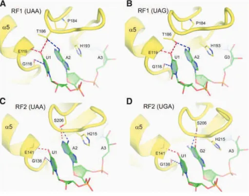
Figure 5: Decoding by the ribosome. (A) and (B) show the conformation of the universally conserved residues at the decoding centre, in the absence and presence of a cognate or near-cognate tRNA. (C) Depicts the formed or closed state of the centre. The discrimination of near-cognate, near-cognate tRNAs and non-cognate tRNAs is suggested to be based on the energy cost imposed by a rigid decoding centre, stressing on Watson-crick (WC) geometry. Adapted from (Demeshkina et al., 2013).
Moreover, the resulting rigid conformation of decoding centre is enforcing Watson-Crick geometry upon the codon-anticodon duplex. Therefore, in the enhancement of the previous induced-fit hypothesis (Schmeing et al., 2005b), the ribosome undergoes conformational changes upon tRNA binding and these very changes are needed to restrain codon-anticodon duplex geometry to force discrimination between cognate, near-cognate or non-cognate tRNA (Fig. 5) (Demeshkina et al., 2013).
Furthermore, the tRNA at the A-site adopts conformation that features a bend at the anticodon stem, thus facilitating interaction between the decoding centre and the factor binding site where EF-Tu is bound. This allows GTP hydrolysis and release of EF-Tu on correct codon-anticodon recognition. In turn, the tRNA relaxes to its open conformation into the PTC (Schmeing et al., 2009).
The energy of these interactions with cognate as well as near-cognate tRNA induces 30S domain closure, moving the shoulder domain of the 30S towards the neck by ~3Å in 70S ribosome complex with tRNA in A, P and E sites (Jenner et al., 2010). This adomain closureb was also observed in studies with isolated 30S complexes but to a larger extent with rotation of head towards the shoulder of the small subunit. Nevertheless, this rate of a<5:'7!%9(51;$#b%tends to dictate the accuracy of the peptide incorporation, as mutations that facilitate this movement have been observed to decrease its accuracy whereas mutations that slow down a<5:'7!%9(51;$#b%increase the accuracy (Ogle et al., 2002).
In case, a non-cognate tRNA is delivered to the A-site, it will not undergo accommodation due to its weaker interactions with the decoding centre. This will prevent hydrolysis of GTP associated with its EF-Tu and thus, will result in tRNA release. This proofreading step of accommodation and domain closure contributes to the high fidelity of ribosomal protein synthesis (Wohlgemuth et al., 2010).
1.3.3 The peptidyl transferase centre (PTC)
This centre in the large ribosomal subunit is formed by 23S rRNA residues U2506, G2583, U2584, and U2585 (E.coli numbering). No metal ion or ribosomal protein is directly involved in the catalysis of the peptide bond formation. The first requirement to proceed towards peptide bond formation is the proper positioning of the two substrates; the incoming aminoacyl tRNA and the tRNA in the P-site harbouring the growing peptide chain.
With tRNA present in the P-site, the +8-hydroxyl group of the A76 residue functions as a vital proton shuttle in substrate-assisted catalysis (Weinger et al., 2004). TE#%i-amino group from the incoming charged tRNA in A-site gives the proton to the +8-hydroxyl group of Om%gE79E%7!%&;$!*%3$5?7<#1%'%3$5&5!%&5%&E#%(#'?7!"%.8-hydroxyl group of tRNA on completion of peptide bond formation (Fig. 6). The tetrahedral transition state stabilization is possible by hydrogen from the polar water molecule, coordinated by rRNA bases. The details, however, are subject to debate.
Also, some proteins have been implicated in aiding the process of peptide bond formation. The N-terminal tail of RPL27 is ordered in the PTC where it interacts with the tRNA substrates. And RPL16 interacts with the acceptor arm of A-site tRNA and becomes ordered in structure.
In order to prevent erroneous peptide hydrolysis, the ester linked carbonyl carbon of the peptidyl tRNA is protected from nucleophilic attack via water molecules by U2585, A2451 and C2063 bases of 23S rRNA. Also, to prevent intramolecular transesterification A$5:% .8- &5% +8-oxygen, 2'-hydroxyl group of A2451 may be essential (Lang et al., 2008). However, no rRNA residue has been shown to be directly involved in this catalysis.
Figure 6T#$&9+(/=8#+%,3)B&%,)&#%&,*+(13@#X-*8&192(8(*#,++,*d#1B#+2&#e-amino group of the aminoacyl +WX!# ,+# +2&# !f)(+&# 13# +2&# *,%013=8# *,%013# 1B# +2&# 9&9+(/=8f+WX!# ,+# +2&# $f)(+&@# !/,9+&/# B%1'# (Carrasco et al., 2011).
1.3.4 Peptide exit tunnel
The growing peptide chain passes through a 80 Å long and 10l20 Å wide, universally conserved tunnel (Nissen et al., 2000) in the large ribosomal subunit. The tunnel, like other active sites of ribosome, is predominantly composed of rRNA core and some protein components. RPL4, RPL22 along with a bacteria-specific extension of RPL23 and the 23SrRNA segments form the tunnel wall in bacteria. RPL39e replaces the RPL23 near the
tunnel exit in eukaryotes (Harms et al., 2001). RPL4 and RPL22 form a constriction of the tunnel in both 50S and 60S, about 30 Å downstream from the PTC.
Upon emerging on the solvent exposed side, the nascent polypeptide chain interacts with several proteins like chaperones and post-translational modification enzymes (Berndt et al., 2009). In bacteria, a deformylase enzyme binds at the N-terminus of the emerging peptide to cleave off the formyl group bound to the first Met residue. It is recruited to the ribosome by RPL32, present on the exit side of the peptide channel. A trigger factor (a bacterial protein chaperone) is known to interact with RPL23 and RPL29 at the end of the tunnel to assist in proper folding. In eukaryotes, L31e is present instead of RPL32 which interacts with protein Zuotin in yeast (Ben-Shem et al., 2011). Zuotin is a part of eukaryote-specific chaperone complex and is involved in co-translational folding.
Moreover, the ribosome-nascent chain complex, as a whole, can be targeted to protein conducting channel in the membrane of cellular components like mitochondria and the endoplasmic reticulum. This happens only if the protein being synthesised expresses a signal sequence in the nascent peptide chain. This signal sequence is recognised by SRP (Signal recognition particle) thus, directing the targeting of the complex to its destination.
Functionally, the tunnel was initially considered as an inert conduit for the peptide. Lately, it has been shown that the tunnel might be involved in protein folding (Lu and Deutsch, 2005) and translational regulation (Tenson and Ehrenberg, 2002). It has an overall electronegative potential that might allow stalling or reducing translation by interacting with long stretches of positively charged residues, such as arginine or lysine. Eukaryote-specific insertion of RPL4 and RPL17 allow contact with the fungal arginine attenuator peptide (AAP) and the human cytomegalovirus (hCMV) peptide chains in 80S affecting their rate of translation (Bhushan et al., 2010), while in prokaryotes RPL22 mediates translation stalling interactions (Seidelt et al., 2009). This translation pausing could be a method for regulating translation, ensuring efficient membrane targeting of certain proteins and in some cases in prokaryotes, confirming the correct splice variant to be translated (due to coupling of transcription and translation).
1.4 Structural dynamics and coordinated ribosome movements
During translation, this giant ribonucleoprotein complex needs to be flexible and dynamic enough to allow binding of factors, tRNA and mRNA translocation. The biochemical and footprinting experiments, in the late 19d,81, first suggested that intersubunit movements allow translocation in two steps. The first step involves movement of tRNA to form a hybrid state where the anticodon stems of the two tRNAs are still in A- and P- sites, while their peptide terminals have moved to the P- and E-sites, depicted as A/P and P/E, respectively. These hybrid intermediate configurations serve in lowering the activation barrier for translocation (Dorner et al., 2006). Subsequently, the second step, catalysed by an elongation factor, allows their movement completely to P-and E-sites, coupled to mRNA movement on the 30S (Moazed and Noller, 1989; Spirin et al., 1987).
The two ribosomal subunits act in a coordinated fashion to allow rotation and swivelling of the small subunit, relative to the large one, thereby ensuring translocation of mRNA and tRNAs. Using cryo-EM studies and solution FRET analysis, the small subunit was observed in a different conformation on binding EF-G (Chen et al., 2013). Frank and Agrawal showed that the small subunit rotates counter-clockwise with respect to the large subunit during translocation, giving rise to the ratchet like mechanism of translocation (Frank and Agrawal, 2000). On ratcheting, the L1 stalk moves by 40 Å, the small subunit body/platform rotates by 5-8° and the head undergoes 12°-15° swivel-like rotation (Fig. 7) (Connell et al., 2007; Spahn et al., 2004a).
In order to allow this intersubunit movement, intersubunit bridges at the extremities are disrupted while those located near the rotation axis (B2a-c, B3, B5, B7a) are essentially maintained (Gao et al., 2003). Later spontaneous fluctuation between this hybrid and classical states was observed for ribosomes containing deacylated tRNA in the P-site, even in the absence of EF-G (Cornish et al., 2008). Binding of the factor and movement of the acceptor stem of deacylated tRNA into E-site seems to stabilise the rotated, hybrid state. This rotation has been seen not only for EF-G, also other GTP binding factors (IF2, EF-G, RF3 and RRF and their eukaryotic orthologs), which makes it a universal mechanism for all the steps of translation (Allen et al., 2005; Gao et al., 2005; Klaholz et al., 2004b).
Intersubunit movement is synchronised with the rotation of the head of the small subunit (Connell et al., 2007; Gao et al., 2003). During the first step of translocation, the head rotates by 12°, in order to prevent breaking apart of the contacts between tRNA and ribosome. Interestingly, an additional movement of the head is predicted for the second step of translocation to create about 20 Å space for anticodon stem loop of tRNA to pass from P- to E-site (Schuwirth et al., 2005). This movement is reversed, on binding of mRNA and tRNA to the P-site, leading to classical pre-translocation state (Berk et al., 2006).
Figure 7: Intersubunit movements. The arrows indicate major conformational rearrangements in ribosome including rotation of 30S body and swivelling of the head. On the 50S the L1 proteins form the dynamic cluster. 50S and 30S PDB codes: 2WDL, 2WDK.
On the large subunit, L1 and L11 stalks are the most flexible regions. L1 stalk comprises RPL1, helices H76, H77 and H78 of 23S rRNA and has been observed in closed! and an open! conformation. The L1 stalk moves 30-40 Å inwards as compared to an open conformation in the presence of E/E site tRNA (classical state). In the P/E hybrid state it moves 15-20 Å further closer to the small subunit (closed) conformation. To allow the release of the deacylated tRNA from the E site, it moves to an open conformation. L11 stalk is associated with the GAC, on the opposite side of the 50S from L1 stalk. It is composed of helices 42, 43 and 44 of 23S rRNA and protein L11"#$%%#&'(#)**+#,)(*-.*/#0+#'+# inward! and an outward! position (Stark et al., 1997; Stark et al., 2000) in ribosome structures with tRNA.
Apart from these global and easily noticeable movements, there exist local rearrangements like in the PTC and the decoding centre, making ribosome a highly adaptable entity. Moreover, these conformational changes during translation and the various translational active sites composing PTC, decoding centre, sites for tRNA binding, GAC and the peptide tunnel are similar and conserved in ribosomes from all domains of life as elaborated with eukaryotic ribosome structure in the next section.