Sabine Schmidt Christian Felley Jean-Yves Meuwly Pierre Schnyder Alban Denys Received: 29 January 2005 Revised: 15 July 2005 Accepted: 18 August 2005 Published online: 12 October 2005 # Springer-Verlag 2005
CT enteroclysis: technique and clinical
applications
Abstract CT enteroclysis (CTE) has been gradually evolving with techni-cal developments of spiral and multi-detector row CT technology. It has nowadays become a well-defined im-aging modality for the evaluation of various small bowel disorders. Vol-ume challenge of 2L of enteral con-trast agent administrated to the small bowel via a nasojejunal catheter ensures luminal distension, the pre-requisite for the detection of mural abnormalities, also facilitating the accurate visualization of intraluminal lesions. CT acquisition is centered on small bowel loops, reconstructed in thin axial slices and completed by multiplanar views. Image analysis is essentially done in cine-mode on work-stations. CTE is of particular diagnostic value in intermediate or advanced stages of Cohn’s disease, including the depiction of extraintes-tinal complications. It has become the
imaging modality of choice for the localization and characterization of small bowel tumors. The cause and degree of low-grade small bowel ob-struction is more readily analyzed with the technique of CTE than conven-tional CT. Limitations of CTE concern the assessment of pure intestinal mo-tility disorders, superficial mucosal lesions and arteriovenous malforma-tions of the small bowel, which are not consistently visualized. CTE should be selectively used to answer specific questions of the small bowel. It essentially contributes to the diagnos-tic quality of modern small bowel imaging, and therefore deserves an established, well-defined place among the other available techniques. Keywords Intestines, CT . Enteroclysis . Crohn’s disease . Intestinal neoplasms . Intestines, stenosis or obstruction
Introduction
The investigation of the small bowel nowadays still rep-resents a diagnostic challenge, since it remains the only intestinal organ not fully accessible by endoscopy. CT enteroclysis (CTE) has been defined as small bowel dis-tension by administration of a high volume of contrast medium via a nasojejunal catheter followed by axial CT acquisition [1–6].
Before the advent of spiral CT, conventional double-contrast barium enteroclysis was the widely recognized gold standard for the assessment of small bowel disorders
[7–15]. Spiral CTE now combines the inherent advantages of conventional barium enteroclysis, i.e., homogenous lu-minal distension resulting from volume challenge [10,12], with those of cross-sectional imaging, which is the simul-taneous detection of intraluminal, mural and extraintestinal pathologies [1–5].
The development of multidetector row technology in 1998 [16] has allowed data acquisition over the entire abdomen in thin slices within one breath-hold, thus re-ducing peristaltic and breathing artifacts. This shorter ac-quisition time and better spatial resolution regarding the z-axis has improved the quality of multiplanar reconstruc-S. Schmidt (*) . J.-Y. Meuwly .
P. Schnyder . A. Denys
Department of Diagnostic and Interventional Radiology, CHUV, rue du Bugnon, 1011 Lausanne, Switzerland e-mail: [email protected] Tel.: +41-21-314-4607 Fax: +41-21-314-4443 C. Felley
Department of Gastroenterology, CHUV, Lausanne, Switzerland
tions, being of particular advantage for the investigation of intestinal pathologies [17]. Therefore, we observe an extension of indications, such as intestinal ischemia with simultaneous CT angiography [18, 19] and multidetector CT enteroclysis [20–23]. There is still a relative paucity of comparative data concerning notably the clinical results of large study populations investigated by CT enteroclysis (CTE). This might partly result from the known low in-cidence of small bowel disorders [14,15,24]. But it might also indicate that CTE has not yet become a successfully adapted and widely used routine modality for small bowel investigation. This review will analyze its diagnostic yield according to the main indications and discuss the technical aspects of CTE as well as alternative, competitive imaging modalities.
Technical aspects of CTE Patient’s preparation
In agreement with other authors [11,12,25,26], we think that fasting for 8–12 h prior to CTE is the adequate pa-tient’s preparation. Rollandi et al. stress the need for sup-plementary rigorous cleansing of the colon [27]. Indeed, a full caecum and ascendant colon might slow the flow of enteral contrast medium through the ileum [28] and there-fore prolong the investigation. We have however rarely encountered this delay in our clinical experience and think therefore that overnight fasting is enough. In certain cir-cumstances, for example Crohn’s disease, a colonic enema with water might additionally be performed immediately before CT data acquisition, thus improving the detection of disease extension.
Intubation technique
Bypassing the stomach is mandatory for rapid enteral ad-ministration of a high volume of liquid, i.e., 2 l of enteral contrast medium, the prerequisite of homogeneous small bowel distension [5,10,25,27,29]. Patients’ tolerance for nasal intubation is generally better than for the oral way [27,29].
It is definitely the need for nasojejunal intubation that has prevented CTE from becoming a widely accepted and performed technique. Patient’s discomfort is related to variable operator experiences in combination with me-chanical difficulties in catheter placement. Some recent improvements of material have increased patient’s com-pliance: In our department like in others [23,25], we use a very thin, small intestinal catheter of 8 F with a plastic olive and Teflon guide (length: 150 cm, 8 French, external/ internal diameter: 2.8 mm/2.1 mm, Fresenius Kabi, Bad Homburg, Germany). This small size mostly allows for straightforward insertion and good handling of the probe
inside the stomach, even if performed by less experienced radiologists. Local anesthetics are applied to the patient’s nasal and buccal mucosa beforehand; we do not use any oral or intravenous drugs. Other authors stress the in-creased patient’s comfort achieved by intravenous admin-istration of sedatives and analgesics given at a dose that still allows patients to follow commands during nasojejunal intubation [30]. However, these authors use a larger naso-jejunal tube (13 F) than our department (8 F), which might be more difficult to swallow and less tolerated by the patients.
An inflatable balloon attached at the catheter’s tip is recommended by several authors to prevent gastroduo-denal reflux during contrast medium administration, which may cause nausea or vomiting [27,29]. In our experience, this shortcoming can mostly be avoided by adequate cath-eter position with the tip well situated distal to the ligament of Treitz. However, whenever the catheter’s tip remains placed in the descending duodenum, especially for diag-nostic purposes, namely assessment of the duodenal wall, inflation with a 30-ml balloon must then be performed in order to prevent gastric reflux [29].
In patients suspected of having ileus or high-grade small bowel obstruction, Maglint et al. recommend to advance the intestinal tube (three-way with sump valve) into the prox-imal jejunum and leave it on overnight suction to reduce luminal distension either therapeutically or for subsequent enteroclysis [29].
Enteral contrast medium administration
Whatever enteral contrast medium is used, it should be heated to 37°C before the administration to increase the patient’s comfort [25].
At our department, after nasojejunal intubation under fluoroscopy guidance, we routinely transfer the patient to the CT unit. Enteral contrast medium is then administrated while the patient is lying on the CT table.
Using the small catheter size described above, a power injector infusion pump is strongly recommended for con-trast medium administration [23,29]. Manual handling of 50-ml syringes filled with contrast agent requires strength from the operator and is a time-consuming procedure. In consensus with other authors [23,25], we agree on a high enteral injection rate of up to 200 ml/min, which imme-diately induces a reflex hypotonia of the bowel loops [2,7]. This contributes to rapid and equal small bowel disten-sion, otherwise obtained by intravenous administration of an antiperistaltic agent. We routinely use 20 mg of sco-polaminbutylbromid (Buscopan, Boehringer Ingelheim, Basel, Switzerland) i.v. or, if contraindicated, 1 mg of glu-cagon (GlucaGen, Novo Nordisk, Bagsvaerd, Denmark), which is ideally injected shortly before data acquisition. In case of suspected or known small bowel strictures, this high flow rate must be reduced. Generally, any significant
patient’s discomfort during enteral contrast administration immediately becomes clinically evident, and the injection speed can then be individually adapted followed by early i. v. injection of the antiperistaltic drugs. The overall feasibil-ity of the above-described method of administration has been confirmed by our nearly daily and longstanding use of CTE.
Choice of enteral contrast medium
The choice of the enteral contrast medium mainly depends on the clinical indication: positive (hyperdense) or neutral (hypodense) small bowel opacification. We think that air is routinely not indicated, unlike for the stomach and duodenum [31], because of technical difficulties in admin-istration associated with patient discomfort and late elimi-nation after CTE. Furthermore, the intraluminal air would induce numerous reconstructions artifacts on CT images. However, conventional air double-contrast enteroclysis has occasionally been proven useful for the detection of early Crohn’s disease [29].
The inherent advantage of neutral contrast agents is the good delineation of the contrast-enhanced small bowel mucosa contrasting well with the hypodense lumen. It allows detection of very small mural lesions, such as pol-yps, tumors or vascular malformations, which might be dissimulated by positive enteral contrast agents [25,32]. Furthermore, opacified mesenteric vessels can thus be ex-actly analyzed on multiplanar reformations without being obscured by the high-density bowel content. Since various image reconstructions and simultaneous CT angiography have nowadays become part of routine small bowel inves-tigation, neutral small bowel opacification is gaining wide-spread acceptance [19,33].
There is a large choice of neutral contrast agents. Pure water is inexpensive and readily available, but is subopti-mal since it is absorbed by the intestinal mucosa. There-fore, this method is relatively contraindicated in patients with renal or cardiac failure [5,19]. Secondly, its capacity for luminal distension is inferior to that of macromolecular substances, such as polyethylenglycol [PEG] [35] or 5% methylcellulose [5,20,22,26,34].
Neutral luminal opacification is indicated in most cases, such as inflammation and tumors. However, when in search of enteral leakages or fistulae, we consider positive opac-ification with iodinated hydrosoluble contrast agent and without i.v. contrast medium injection as the technique of choice. Its hyperdense character allows straightforward vi-sualization of even a small volume of extraluminal contrast accumulation. Dilute barium solutions [1,2,36], the alter-native of positive opacification, are contraindicated if there is any risk of leakage.
In order to increase the attenuation coefficient between hypodense bowel lumen and contrast-enhanced wall, some authors [21, 37] use a paraffin-methylcellose suspension, which leads to better demarcation of small bowel mucosa than that obtained with watery contrast medium alone. Nevertheless, the risk of aspiration during administration of this large volume of fatty emulsion with subsequent chemical pneumonia should be kept in mind. Phase sep-aration between the fatty contrast medium and intestinal residual liquid might furthermore dissimulate small bowel lesions [21].
Acquisition and reconstruction parameters of CTE The use of intravenous iodinated contrast medium is man-datory for the detection of inflammatory, neoplastic or vas-cular small bowel disease. We routinely inject the nonionic contrast medium iopentol (Imagopaque, Nycomed Imag-ing. Oslo, Norway, 300 mg iodine/ml, volume in millili-ters=patient’s body weight plus 30 ml) at a rate of 3 ml/s. Higher injection rates up to 5 ml/s are useful for vascular pathologies [18].
Volumetric acquisition parameters depend on the hard-and software equipment available. With 4- to 16-multide-tector row CT scanners, a collimation beam of 1.25 mm or thinner is recommended. These data provide the same high spatial resolution of multiplanar reconstructions, which should be done with an overlap of at least 50%. Coronal views are particularly suited to display the physiological course of small bowel loops including the ileocoecal valve [20,22]. They should now routinely be part of small bowel imaging since they have been known to increase the radiol-ogists’ diagnostic confidence in image interpretation, even if they generally do not reveal any further abnormalities [17,19,20,23,33,37–39]. Multiplanar views are espe-cially well perceived by the referring surgeons, improving preoperative planning. With modern 16- and 64-channel CT scanners allowing for isotropic image acquisition with-in a few seconds, automatic axial, coronal and sagittal post-processing might become the standard technique of choice. We routinely use the following acquisition parameters: tube voltage 120 kV, tube current 150 mAs, collimation 16×1.25 mm, pitch 1.75, table feed 70 mm/s and dual phase acquisition with a delay of 30 and 70 s. Dual phase acqui-sition, routinely performed in most of our patients, in-creases the chance to visualize all small bowel segments in a distended state since during one of the two phases some loops might be collapsed due to a transitory spasm or a residual peristaltic wave, thus mimicking an organic steno-sis [3]. Assessment of Crohn’s disease activity is best done in the portal venous phase [37,40], while the arterial phase allows the possible detection of vascular pathologies, such as arteriovenous malformations of the bowel wall.
Radiation dose
Over the last 10 years, serious concern about the elevated radiation dose of spiral CT or notably multidetector spiral CT has been raised. The mean effective dose equivalent of multidetector CTE in our department, like in others [26], is 6.2 mSv for each acquisition phase, with an additional 0.5–1 mSv for nasojejunal intubation guided by fluoros-copy; this means 13–13.5 mSv altogether using dual-phase acquisition. Some authors report on radiation doses up to 13 mSv for one acquisition by multidetector row CTE depending on the CT manufacturer and patient’s gender [37,40]. A high patient radiation dose had frequently been quoted as one of the disadvantages of conventional barium double-contrast enteroclysis [14]. However, quite impor-tant discrepancies of published data exist varying from an average dose of 1.5 mSv [41] to a mean effective dose equivalent of 13.99±7.57 mSv [42]. The major reason for this striking standard variation is the varying fluroscopy time, reflecting large differences in operator experience, as is also true with fluroscopy-guided nasojejunal intubation. The radiation dose of CTE is otherwise operator indepen-dent; it only depends on the chosen acquisition parameters, mainly varying with the patient’s body weight. Further-more, we observe a more efficient dose profile using the most recent multidetector CT: Due to the relative reduction of penumbra radiation with an increasing number of chan-nels of CT scanners, the radiation dose necessary for the same slice width and with the same image noise decreases [43].
Clinical applications and diagnostic yield Normal small bowel appearance on CTE
The well-distended and transaxially imaged normal small bowel wall measures 1–2 mm (Fig.1) [2,36,44]. A wall thickness >4 mm in any well-distended segment is abnor-mal. After i.v. contrast medium bolus injection, homoge-neous mural enhancement occurs, even in healthy subjects, predominantly seen at the inner, mucosal side [35,42]. Pathological small bowel wall attenuation is defined by comparison of its value with those of adjacent, healthy loops. During the portal phase, the average CT density of the normal duodenal, jejunal and ileal wall varies between 107–111 (+/−4) Hounsfield units (HU), and between 118– 120 (+/−5) HU during the arterial phase. However, these values have been calculated on small bowel loops orally opacified with only 500 ml of water, and are therefore less distended than with the enteral administration of 2 l of liquid [45].
Provided that adequate luminal distension had been ob-tained, the diameter of the small bowel measures beween 21–29 mm (Fig. 1) [2], and a diameter >3 cm means dilatation.
Crohn’s disease
The sensitivity of conventional double-contrast barium en-teroclysis for Crohn’s disease ranges between 98.2–100%, as was demonstrated on 168 [8] and 143 patients [13], re-spectively, whereby Maglinte et al. had included 43 pa-tients [31%] with early lesions. The diagnostic accuracy of cross-sectional imaging modalities, including CTE, re-mains controversial for the initial stage of Crohn’s disease [36,40,46,47]. Their spatial resolution is still inferior to that of conventional double contrast enteroclysis [13] and does not allow detailed analysis of the small bowel mucosa. Therefore, early mucosal inflammation, histopathologi-cally characterized as aphtoid ulcerations and enlarged lymphoid follicles, is not constantly visualized by CTE, even with the given improvements of multidetector row technology [26,36].
At intermediate or advanced stages of Crohn’s disease, however, the value of CTE is well established [34,36]. The degree of small bowel wall thickening and intensity of Fig. 1 a Axial and b coronal plane of CTE demonstrates the normal small bowel anatomy, centered on the ileocoecal valve (arrow) and the terminal ileum, which is well distended
mucosal contrast enhancement correlate with clinical dis-ease activity at the acute or quiescent phase [36,48]. Due to homogeneous luminal distension, CTE allows a better analysis of the small bowel wall than conventional CT, and thus a more precise and comprehensive recognition of the different pathological enhancement patterns. Acute inflam-mation is characterized by mural stratification (Fig. 2), mainly explained by the strongly contrast enhanced mu-cosa surrounded by the low-density submumu-cosa [5,36,48]. Chronic involvement seen during clinical quiescence is associated with diffuse parietal contrast enhancement and mild thickening, indicating irreversible transmural fibrosis [36,48].
Extraintestinal features of Crohn’s disease, such as in-flammatory extension into the adjacent mesentery, asso-ciated abscesses, enteric fistulae or involvement of other organs, are simultaneously and readily demonstrated by CTE (Fig.3a), and not by conventional barium studies. The latter only inconstantly show enteric fistulous tracts, which can precisely be demonstrated by multiplanar reformatting views [23]. Finally, the enteral volume challenge of CTE also enables the visualization of even moderate segmentary luminal narrowing due to active Crohn’s disease, which might only appear as collapsed or spasmed small bowel loops on traditional CT. The length of these strictures might then be more accurately demonstrated on multiplanar re-constructions, often best appreciated on the coronal plane (Fig.3b).
Furthermore, CTE features allow accurate classification of Crohn’s disease into subtypes that include active in-flammatory, fibrostenosing and fistulizing/perforating cat-egories [49]. Its clinical utility and influence on further patient’s management is certain: broad antiinflammatory medical treatment is indicated for edematous luminal nar-rowing due to active Crohn’s disease, while fistuluos and perforating complications might successfully be managed
by specific medical treatment (Infliximab). In case of re-fractory fistulizing disease (Fig. 4), surgical resection is indicated, which might also be the treatment of choice for the symptomatic fibrostenosing subtype whenever mini-mally invasive procedures, such as stricturoplasty or endo-scopic balloon dilatation, fail or are not possible [49].
The unpredictable and insidious character of Crohn’s disease might be especially harmful in the acute postopera-tive period when early complications occur. Patients might present with unexplained persistent inflammatory syn-drome or complain of mild intermittent, recurrent abdom-Fig. 2 CTE in a 23-year-old woman with acute Crohn’s disease:
discontinous parietal thickening involving several distal ileal seg-ments with pathological contrast enhancement characterized by mural stratification (arrows) and irregular luminal narrowing
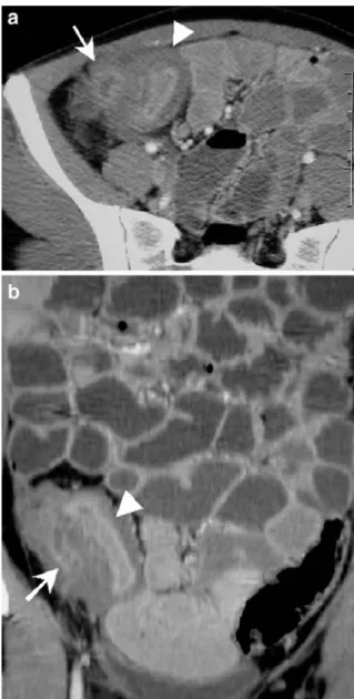
Fig. 3 CTE in a 22-year-old woman with complicated Crohn’s disease: diffuse parietal thickening of the distal ileum (arrowhead) and surrounding inflammation causes luminal narrowing compli-cated by an intraperitoneal abscess (arrow), both seen on a axial and b coronal view; however, coronal reconstructions better demonstrate the exact length of segmentary stenosis as well as the anatomical abscess extension
inal pain, however, without evident physical findings or the clear pathological features seen on conventional CT, which is usually the first diagnostic procedure to be per-formed. In these instances, CTE performed with positive enteral opacification and without i.v. contrast medium is the technique of choice to detect small anastomotic leak-ages or intestinal fistulae. Due to its enteral volume chal-lenge, CTE is superior to conventional CT in this regard [32]. The fistuluous tract is shown by opacification with the positive enterally administered contrast medium (Fig. 5); sometimes it might only become evident on late phase images.
Small bowel neoplasms
Small bowel tumors are very rare, representing less than 2% of all the primary gastrointestinal malignancies, and are often small and generally present with nonspecific abdom-inal symptoms responsible for a significant diagnostic delay in the past [10,24].
The well-distended and i.v. contrast-enhanced small bowel wall provided by CTE with neutral enteral opacification allows detection and differentiation between extra-, intra-mural and luminal lesions, which is of particular impor-tance for neoplastic diseases (Figs.6,7,8). Tumor staging by detecting local extension, adenopathy or distant metas-tasis is simultaneously realized. [33].
Fig. 4 A 73-year-old patient with active Crohn’s disease (fistulizing subtype): conventional CT a with oral and rectal administration of positive contrast medium shows acute inflammation of the ileocaecal region (arrrow) extending into the mesenteric fat of the right iliac fossa (arrowhead), however, without demonstration of the fistulous tract to the iliac muscle, which is only seen on CTE b due to its volume challenge and positive enteral opacification
Fig. 5 A 43-year-old patient after colic resection for inflammatory bowel disease and temporary ileostomy. Conventional CT a with rectal administration of positive contrast medium was performed during the acute postoperative period for persistent inflammatory syndrome. It shows leakage of the rectal stump with consecutive contrast accumulation within a large abscess formation situated in the left pararenal space (short white arrow). The presence of contrast medium in the adjacent jejunal loop (long, thin, white arrow) with-out previous oral contrast administration suggests an underlying fistula. Only CTE b performed with positive enteral opacification some days later is able to demonstrate two small fistulous tracts (short black arrows) extending from the proximal jejunal loop to the abscess cavity, which has meanwhile been drained. Notice the accumulation of the enteral contrast medium in the former abscess cavity (black arrowhead)
Direct comparison of the technique of conventional en-teroclysis, traditional CT and CTE performed on 63 pa-tients revealed 15 cases with neoplastic involvement of the small bowel, whereby CTE provided important supple-mentary diagnostic features [6].
Orjollet et al. and Boudiaf et al. report on a 100% sen-sitivity of CTE for the detection of small bowel tumors performed with the monodetector and multidetector row technique, respectively [23,25]. Using a beam collimation of 5 mm, a pitch of 1 and reconstructed slices of 3 mm, Orjollet et al. revealed 25 small bowel tumors in 48 pa-tients, among them 22 histologically proven, measuring between 0.8 and 6 cm, the mean tumor size being 2.3 cm [25]. Multidetector row CTE performed on 107 patients by Boudiaf et al. (4×2.5-mm collimation and images recon-structed at 3-mm intervals) demonstrated 21 surgically confirmed small intestinal masses of an axial diameter from 5 to 35 mm and a mean diameter of 21 mm [23]. Neutral luminal opacification (2 l) in association with multiplanar reconstructions allowed for confident visualization of even small, centimetric lesions. The diagnostic certitude, how-ever, depends on their degree of hypervascularization and delineation. Detection of little enhancing and sessile adeno-mas mainly seen in familial intestinal polyposis was con-sidered to be the most difficult [25].
Unfortunately, other comparative studies with traditional CT date back to the incremental mode and thick slices of 8–10 mm [50,51], reaching a sensitivity of 73% and 80%, respectively, and including 63 and 35 patients, respectively. In a pictorial essay, Horton et al. highlight the advan-tages of multidetector-row and 3-dimensional [3D] CT with enteral opacification by oral neutral contrast for imaging of small bowel neoplasms, however without taking into
account the technique of CTE. The added value of 3D imaging of small bowel tumors has not been proven in a prospective trial. It probably does not improve tumor de-tection itself, but the 3D images are nowadays rapidly generated. They are well received by referring clinicians since they better demonstrate complicated anatomical rela-tionships than 2D slices, thus especially serving for surgi-cal planning [33].
Fig. 6 CTE in a 40-year-old woman with Peutz-Jeghers disease detected after recurrent episodes of intestinal intussusceptions: mul-tiple pedunculated jejunal and ileal hamartomas (arrows) are nicely seen within the distended bowel loops, 34 among them were resected
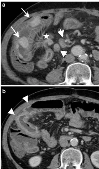
Fig. 7 A 57-year-old man presenting with diarrhea for 3 months (eight times/day): conventional CT (not shown) had only revealed non-specific thickening of the distal ileal wall. CTE shows several intensely i.v. contrast-enhanced nodules a situated in a distal ileal loop, the two largest ones measuring 2–2.5 cm (long arrows). There is adjacent retractile mesenteric fat stranding (star) and associated metastatic lymph nodes (big, short arrows). b nicely demonstrates tethered appearance of adjacent ileal loops showing angulation and kinking as well as mural thickening with pathological i.v. contrast-enhanced mucosa and submucosal edema (arrowheads). Surgical and histopathological findings confirmed the diagnosis of multiple carcinoid tumors of the ileum a with desmoplastic reaction of the mesentery and secondary ischemia of the adjacent ileal wall due to perivascular tumor infiltration (b)
Chronic obscure GI bleeding
Arteriovenous malformations are the most frequent source of chronic obscure GI bleeding occurring in 39–80% of patients, followed by neoplasms (6–15%) and rare causes, such as jejunal, duodenal or Meckel’s diverticula, segmen-tary ischemia, Crohn’s’ disease or radiation enteritis [10, 46,52].
CTE performed for chronic obscure gastrointestinal bleeding or unexplained anemia tends to show a high rate of negative findings similar to conventional enteroclysis, with a reported diagnostic yield as low as 10–20% [52,53]. Up to now, these figures have been confirmed only by one prospective comparison of CTE with wireless cap-sule endoscopy (WCE): CTE performed on eight patients
with obscure gastrointestinal bleeding only resulted in one positive finding, which was suspected jejunal angioma. However, this feature was not confirmed by WCE, which revealed five positive findings, such as angiodysplasia and small bowel wall erosions, allowing a final diagnosis in four patients (50%) [26].
Direct visualization of parietal intestinal arteriovenous malformations has otherwise only been described in iso-lated case reports using traditional CT so far [54, 55]. Published data of spiral CTE prospectively performed on populations with various small bowel disorders, among them 22–50% of patients addressed for unexplained gas-trointestinal bleeding or iron deficiency anemia, did not reveal any arteriovenous malformation of the small bowel [22,23,25,26].
Searching for the source of chronic obscure GI bleeding requires“angio CTE,” which is data acquisition in the dual phase after i.v. bolus injection with a flow rate of 4–5 ml/s and neutral enteral opacification (Fig. 9). This technique provides the highest chances for detecting any etiology, either of tumoral, vascular or even congenital origin. Espe-cially underlying Meckel’s diverticula has been reported to be confidently detected by CTE [56].
Mechanical small bowel obstruction (SBO)
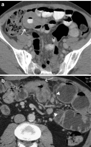
Conventional abdominal CT has been shown to be very sensitive for the detection of high-grade SBO [57], includ-ing its origin, grade and localization [47, 58–60]. False-negative findings occurring with conventional abdominal CT are mainly due to low-grade, partial occlusion for which a transition zone between dilated proximal and collapsed distal small bowel loops is not clearly individualized Fig. 8 Two different CTE features of small bowel lymphoma:
primary distal ileal lymphoma a seen in a 50-year-old woman is characterized by long segmentary and circumferential parietal thickening with aneurysmal luminal dilatation (arrow). Secondary lymphomatous transformation of celiac disease b in a 60-year-old man is demonstrated as excentric jejunal wall thickening extending to the adjacent mesenteric fat (arrow) with multiple associated adenopathies (arrowheads). Note diffuse parietal contrast enhance-ment in both patients without mural stratification in contrast to inflammatory lesions, typically indicating tumoral involvement of the thickened small bowel wall
Fig. 9 CTE in a 79-year-old patient presenting with melena of unknown origin: arterial phase nicely demonstrates the intensely contrast-enhanced infracentrimetric polyp situated in a distal jejunal loop (arrow), exactly depicted thanks to optimal luminal distension with neutral contrast medium
[47, 61]. Therefore, the value of conventional abdominal CT in the diagnosis of SBO relates to the severity of the obstruction. In a retrospective review including 36 patients, CTE has proven to be more sensitive than traditional CT for low-grade SBO (89 versus 50%), especially in ex-cluding malignant disease [62]. Thanks to the enteral vol-ume challenge provided by CTE, even non-obstructive bowel adhesions might be detected whenever kinking of the well-distended bowel loops is observed. This may be responsible for patients’ abdominal pain and can result in future obstructions. CTE prospectively performed on 48 patients showed a sensitivity and specificity of 82.1 and 87.5%, respectively, for the localization and severity of low-grade partial SBO [3]. Therefore, CTE seems to be a valuable tool for any grade of SBO (Figs. 10,11) com-bining the advantages of traditional CT and conventional
enteroclysis; the latter has been the imaging modality of choice for low-grade SBO so far [14,58].
In a recent prospective study, multidetector row CTE revealed a sensitivity of 100% for the site, level and cause of low-grade SBO in 12 out of 31 patients, among them 4 with negative conventional barium follow-through studies [23]. Filmless reading of the whole small bowel in cine-mode and on multiplanar reconstructions provided by the multidetector helical row technique is certainly the decisive advantage of CTE in this regard instead of dealing with
Fig. 10 A 66-year-old man presenting with recurrent episodes of crampy abdominal pain after appendectomy and ileal resection in the past. Conventional CT (not shown) had been normal. CTE shows complete distension of the jejunal loops by the enteral contrast medium methylcellulose (arrowheads) while the ileal loops remain collapsed (long arrows). This feature strongly suggests a fibrotic adhesion at the jejuno-ileal junction causing low-grade SBO, which was confirmed by surgery
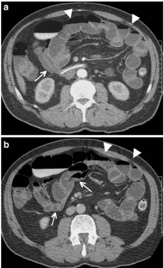
Fig. 11 A 48-year-old woman with long-standing Crohn’s disease and high-grade ileal stenosis impossible to overcome by endoscopy: axial image a of CTE shows one of two pronounced ileal stenoses (arrow) with important prestenotic dilatation (fibrostenosing sub-type). However, exact anatomical relationship of the two narrowed ileal loops (arrow) with adhesions to the equally involved ascendant colon (arrowhead) is far better demonstrated on b coronal view
overlapping small bowel loops on conventional barium studies.
Furthermore, the patient’s immediate operability remains maintained after CTE since the enteral opacification with barium can be avoided, unlike in conventional enteroclysis. However, CTE should not become a substitute for standard CT in the emergency patient presenting with acute SBO [46], but it should be used to investigate low grade partial and intermittent SBO, which is its main advantage over conventional CT [59].
Limitations of CTE in comparison with other diagnostic tools
CTE versus conventional small bowel studies
CTE, as a projection modality, provides precise analysis of anatomical details without superimposition of small bowel loops as often seen at fluoroscopy. Nevertheless, the assessment of motility disorders remains difficult in con-trast to conventional small bowel studies. Patients with predominantly functional disorders should therefore con-tinue to be investigated with conventional enteroclysis [7]. However, many functional small bowel disorders will sooner or later also cause organic changes and therefore become evident on CTE, such as prestenotic luminal dila-tation when segmentary small bowel narrowing is becom-ing significant. Furthermore, CTE is much less operator dependent than conventional enteroclysis; therefore, results are far more standardized and reproducible than those of conventional enteroclysis.
Spatial resolution of CTE still remains suboptimal com-pared to conventional double contrast barium enteroclysis, as it does not provide detailed analysis of the small bowel mucosa [36]; the exclusion of early Crohn’s disease in particular is not reliable.
CTE versus “oral CT enterography”
Oral administration of enteral contrast medium before CT, even using a large volume, is always associated with less optimal small bowel distension than obtained after naso-jejunal intubation [40]. This drawback might not prevent . the detection of many small bowel lesions significantly [40], but it causes diagnostic problems in a small number of difficult cases. Intermittent and low-grade SBO might be impossible to detect [62]. The lack of complete small bowel distention may simulate segmentary mural thickening and mask small intraluminal lesions of the small bowel [36,44, 45,64,65].
According to Wold et al., peroral CT enterography does not show any statistically significant difference concerning the assessment of Crohn’s disease activity as compared to CTE. However, the small number of eight patients
in-cluded in the CTE group, among them only four with active inflammation of the terminal ileum, hampers these conclu-sions [40].
CTE versus MR enteroclysis (MRE)
Thanks to the multiplanar imaging capabilities and the excellent soft tissue contrast resolution inherent in MR imaging, as well as technical progress concerning fast im-aging sequences and the absence of ionizing radiation, MRE has become a very promising tool for the evaluation of small bowel disorders [35,62,65,66]. The mural con-trast enhancement seen in MRE is far more pronounced than that seen with iodinated contrast medium, and is there-fore especially useful for the detection of inflamed bowel segments in Crohn’s disease [35,65,66] or tumors [66]. Dynamic image acquisition, MR fluoroscopy, is possible, provided that the infusion pump is placed in the MR unit [65], while CTE always reflects a“snapshot” of the small intestine.
Persistent limitations of MRE are the limited availability of MR scanners, and the still long acquisition times asso-ciated with a few persistent artifacts due to magnetic sus-ceptibility, chemical shifts or residual peristaltism. The spatial resolution of MR imaging is far inferior to that of CT; small bowel mucosa can therefore not be analyzed in detail like on CTE. The only direct comparison of MRE and multidetector CTE performed on 50 patients resulted in better sensitivity and interobserver agreement of CTE for the detection of various small bowel lesions [22].
CTE versus wireless capsule endoscopy (WCE)
Has the advent of wireless capsule endoscopy (WCE) changed the investigational approach of small bowel dis-orders? Its clinical value is difficult to evaluate because of the lack of a gold standard despite the proven high sen-sitivity and good diagnostic yield [26,67,68]. The exact localization of lesions can also be difficult with WCE [67]. The high incidence of abnormal findings (33%) classified as“others” among the 49 sources for small bowel hemor-rhage revealed by Scarpa et al. shows the problematic interpretation of nonspecific findings and renders treatment options difficult [68]. Up to now, WCE has revealed better sensitivity than barium follow-through [67], conventional enteroclysis, traditional CT [63] and especially CTE [26]. The latter demonstrated fewer positive findings than WCE for obscure gastrointestinal bleeding and Crohn’s disease, whereby WCE is contraindicated in case of intestinal ste-noses. They should therefore be excluded beforehand, which is done best by CTE. WCE seems therefore an ideal diagnostic modality for early Crohn’s disease whenever the localization is beyond the reach of the endoscope and lesions are superficial without associated luminal
narrow-ing. WCE is certainly the method of choice for obscure gastrointestinal bleeding for which CTE is most likely as-sociated with the same low diagnostic performance as con-ventional double contrast enteroclysis [7,26].
Conclusion
In summary, CTE represents a problem-solving modality for well-selected patients concerning the detection and char-acterization of small bowel pathology: It is highly recom-mended for advanced and complicated Crohn’s disease or the preoperative assessment of the inflammatory extent. CTE should be the modality of choice for the detection and localization of small bowel tumors [32] as well as in the clinical setting of recurrent, low-grade obstructive symp-toms of SBO, especially to differentiate between malignant and adhesive origin [23,62]. CTE should not be used as
screening or as the first line investigation, in particular not to search for superficial mucosal alteration due to early inflammatory bowel disease or responsible for obscure gastrointestinal bleeding. It also is not indicated for emer-gency patients with suspected high-grade SBO. CTE should be selectively used to answer specific questions about the small bowel, and it is usually preceded by another imaging modality [32], which is nowadays often abdominal CT without small bowel preparation.
The successful adoption of CTE by the clinical envi-ronment depends on how radiologists can educate sur-geons, gastroenterologists and other specialists. Using a very small intestinal catheter can show them that this tech-nique does not only increase patients’ discomfort because of the necessary nasojejunal intubation. CTE also essen-tially contributes to the diagnostic quality of modern small bowel imaging, and therefore deserves an established place among the other available techniques.
References
1. Klöppel R, Thiele J, Bosse J (1992) The sellink CT method. RöFo 156:291–292
2. Thiele J, Klöppel R, Schulz HG (1993) CT-Sellink-eine neue Methode der Darmwandbeurteilung. RöFo 159 (3):213–2217
3. Bender GN, Timmons JH, Williard WC, Carter J (1996) Computed to-mographic enteroclysis—one method-ology. Invest Radiol 31(1):39–43 4. Schober E, Turetschek K, Schima W,
Mostbeck GH (1997) Methylcellulose spiral CT in the preoperative assess-ment of Crohn’s disease: radiologic and pathologic correlation (abstract). Radiology 205(P):717
5. Rollandi GA, Curone PF, Biscalde E, Nardi F, Bonifacio E, Conzi R, Derchi LE (1999) Spiral CT of the abdomen after distention of small bowel loops with transparent enema in patients with Crohn’s disease. Abdom Imaging 24:544–549
6. Gaffke G, Stroszczynski C, Schlecht I, Jost D, Ludwig WD, Schlag P, Felix R (2001) Diagnostik von Dünndarmtu-moren mit Hilfe der CT-Sellink Meth-ode. Röntgenpraxis 54(6):214–219 7. Antes G, Eggeman F (1986)
Dünn-darmradiologie-Einführung und Atlas. Springer, Berlin Heidelberg New York
8. Cirillo LC, Camera L, Della Noce M, Castiglione F, Mazzacca G, Salvatore M (2000) Accuracy of enteroclysis in Crohn’s disease of the small bowel: a retrospective study. Eur Radiol 10 (12):1894–1898
9. Gourtsoyiannis NC, Bays D,
Papaioannou N, Theotokas J, Barouxis G, Karabelas T (1993) Bengin tumors of the small intestine: preoperative evaluation with a barium infusion technique. Eur J Radiol 16:115–125 10. Gourtsoyiannis N, Mako E (1997)
Imaging of primary small intestinal tumours by enteroclysis and CT with pathological correlation. Eur Radiol 7:625–642
11. Miller RE, Sellink JL (1979) Entero-clysis: the small bowel enema. How to succeed and how to fail. Gastrointest Radiol 4:269–283
12. Maglinte DDT, Herlinger H (1989) Single contrast and biphasic enterocly-sis. In: Herlinger H, Maglinte D (eds) Clincial radiology of the small intes-tine. Philadelphia: Saunders, pp 107– 118
13. Maglinte DDT, Chernish SM, Kelvin FM, O’Connor KW, Hage JP (1992) Crohn’s disease of the small intestine: accuracy and relevance of enteroclysis. Radiology 184:541–545
14. Nolan DJ (1997a) The true yield of the small-intestinal barium study.
Endoscopy 29:447–453
15. Nolan DJ, Traill ZC (1997b) The current role of the barium examination of the small intestine. Clin Radiol 52:809–820
16. Silverman PM, Kalender WA, Hazle JD (2001) Common terminology for single and multislice helical CT. Am J Roentgenol 165(5):1135–1136 17. Raptopoulos V, Schwartz RK,
McNicholas MMJ, Movson J, Pearlma J, Joffe N (1997) Multiplanar helical CT enterography in patients with Crohn’s disease. AJR 169:1545–1550 18. Horton KM, Fishman EK (2001)
Multi-detector row CT of mesenteric ische-mia: can it be done? Radiographics 21:1463–1473
19. Horton KM, Fishman EK (2003) The current status of multidetector row CT and three-dimensional imaging of the small bowel. Radiol Clin North Am 41 (2):199–212
20. Rust GF, Holzknecht N, Olbrich D, Schöpf U, Brüning R, Reiser M (1999) Mehrschicht-Computertomographie des Dünndarms—vorläufige Ergebnisse. Radiologe 39:965–970
21. Rust GF, Spiekermann A, Daum F, Schoepf UJ, Holzknecht N, Matz C, Staebler A, Reiser MF (2001) New developments in imaging the small bowel with multislice computed tomography and negtive contrast me-dium, In: Reiser MF (ed) Multislice CT. Springer, Berlin Heidelberg New York, pp 49–60
22. Schmidt S, Lepori D, Meuwly JY, Duvoisin B, Meuli R, Michetti P et al (2002) Prospective Comparison of MR-Enteroclysis (MRE) with Multidetec-tor-Spiral-CT-Enteroclysis (MSCTE). Eur Radiol 13:1303–1311
23. Boudiaf M, Jaff A, Soyer P, Bouhnik Y, Hamzi L, Rymer R (2004) Small-bowel disease: prospective evaluation of multi-detector row helical CT entero-clysis in 107 consecutive patients. Radiology 233:338–344
24. Gupta S (1982) Primary tumors of the small bowel: a clinico-pathological study of 58 cases. J Surg Oncol 20:161–167
25. Orjollet-Lecoanet C, Menard Y, Martins A, Crombé-Ternamian A, Cotton F, Valette PJ (2000) L’Enter-oscanner: Une nouvelle méthode d’ex-ploration du grêle. J Radiol 81:618–627 26. Voderholzer WA, Ortner M, Rogalla P, Beinhölzl J, Lochs H (2003) Diagnostic yield of wireless capsule enteroscopy in comparison with computed tomography enteroclysis. Endoscopy 35(12): 1009–1014
27. Rollandi GA, Biscalde E (2002) CT Enteroclysis. In: Terrier F, Grossholz M, Becker CD (eds) Spiral CT of the abdomen. Springer, Berlin, Heidelberg New York, pp 369–384
28. Maglinte DDT, Lappas JC, Kelvin FM et al (1987) Small bowel radiography: how, when and why? Radiology 163:297–304
29. Maglinte DDT, Lappa JC, Heitkamp DE, Bender GN, Kelvin FM (2003) Technical refinements in enteroclysis. Radiol Clin North Am 41:213–229 30. Maglinte DDT, Lappa JC, Chernish SM
(1988) Improved tolerance of entero-clysis by use of sedation. Am J Roentgenol 151:951–952
31. Pochaczevsky R (2000) Carbondioxide as a low-attenuation oral contrast agent. Radiology 214:918
32. Maglinte DD, Bender Gn, Heitkamp De, Lappas JC, Kelvin FM (2003) Multidetector-row helical CT entero-clysis. Radiol Clin North Am 41 (2):249–262
33. Horton KM, Fishman EK (2004) Multidetector-row computed tomogra-phy and 3-dimensional computed tomography imaging of small bowel neoplasms. Current concept in diagno-sis. J Comput Assist Tomogr 28: 106–116
34. Turetschek K, Schober E, Wunderbaldinger P, Bernhard C, Schima W, Puespoek A, Vogelsang H, Moeschl P , Mostbeck G (2002) Find-ings at helical CT-enteroclysis in symptomatic patients with Crohn’s disease: correlation with endoscopic and surgical findings. J Comput Ass Tomogr: 26(4):488–492
35. Gourtsoyiannis N, Papanikolaou N, Grammatikakis J, Prassopoulos (2002) MR enteroclysis: technical consider-ations and clinical applicconsider-ations. Eur Radiol 12:2651–2658
36. Gore RM, Balthazar EJ, Ghahremani GG, Miller FH (1996) CT features of ulcerative colitis and Crohn’s disease. AJR 167:3–15
37. Bitterling H, Rock C, Reiser M (2003) Die Computertomographie in der Di-agnostik entzündlicher Darmerkran-kungen: Methodik der MSCT und klinische Ergebnisse. Radiologe 43: 17–25
38. Schmidt S, Chevallier C, Chalaron M, Bessoud B, Verdun FR, Frascarolo P, Schnyder P, Denys A (2005) Multi-detector CT enteroclysis: comparison of the reading performance for axial and coronal views. Eur Radiol 15(2): 238–246
39. Reittner P, Goritschnig T, Petritsch W, Doerfler O, Preidler KW, Hinterleitner T, Szolar DH (2002) Multiplanar spiral CT enterography in patients with Crohn’s disease using a negative oral contrast material: initial result nonin-vasive imaging approach. Eur Radiol 12(9):2253–2257
40. Wold PB, Fletcher JG, Johnson CD, Sandborn WJ (2003) Assessment of small bowel Crohn’s disease: noninva-sive peroral CT enterography compared with other imaging methods and en-doscopy-feasibility study. Radiology 229:275–281
41. Hart D, Haggett PJ, Boardman P, Nolan DJ, Wall BF (1994) Patient radiation doses from enteroclysis examinations. Br J Radiol 67:997–1000
42. Ruiz-Cruces R, Ruiz F, Perez-Martinez M, Lopez J, Tot Ausina I, de los Rios AD (2000) Patient dose from barium procedures. Br J Radiol 73:752–761 43. Flohr TG, Schaller S, Stierstorfer K,
Bruder H, Ohnesorge BM, Schoepf UJ (2005) Multi-detector row CT systems and image-reconstruction techniques. Radiology 203:756–773
44. Macari M, Balthazar EJ (2001) CT of bowel wall thickening: significance and pitfalls of interpretation. AJR
176:1105–1116
45. Horton KM, Eng J, Fishman EK (2000) Normal enhancement of the small bowel: evaluation with spiral CT. J Comp Ass Tomogr 24(1):67–71 46. Bender GN, Maglinte DD, Klöppel R,
Timmons JH (1999) CT-enteroclysis: a superfluous diagnostic procedure or valuable when investigation small-bowel disease? AJR 172:373–378 47. Makanjuola D (1998) Computed
tomography compared with small bowel enema in clinically equivocal intestinal obstruction. Clin Radiol 53:203–208
48. Choi D, Lee SJ, Cho YA, Lim HK, Kim SH, Lee WJ et al (2003) Bowel wall thickening in patients with Crohn’s disease: CT patterns and correlation with inflammatory activity. Clin Radiol 58:68–74
49. Maglinte DDT, Gourtsoyiannis N, Rex D, Howard TH, Kelvin FM (2003) Classification of small bowel Crohn’s subtypes based on multimodality im-aging. Radiol Clin North Am 41: 285–303
50. Dudiak KM, Johnson CD, Stephens DH (1989) Primary tumors of the small intestine: CT evaluation. AJR 152: 995–998
51. Laurent F, Raynaud M, Biset JM, Boisserie-Latroix M, Grelet P, Droullard J (1991) Diagnosis and categorization of small bowel neo-plasms: role of computed tomography. Gastrointest Radiol 16:115–119 52. Antes G, Neher M, Hiemeyer V, Burger
A (1996) Gastrointestinal bleeding of obscure origin: role of enteroclysis. Eur Radiol 6:851–854
53. Moch A, Herlinger H, Kochman ML, Levine MS, Rubesin SE, Laufer I (1994) Enteroclysis in the evaluation of obscure gastrointestinal bleeding. AJR 163:1381–1384
54. Mindelzun RE, Beaulieu CF (1997) Using biphasic CT to reveal gastroin-testinal arteriovenous malformations. AJR 168:437–438
55. Grassi R, di Mizio R, Romano S, Cappabianca S, del Vecchio W, Severini S (2000) Multiple jejunal angiodysplasia detected by enema-he-lical CT. J Clin Imaging 24:61–63 56. Mathon G, Frampas E, D’Alincourt A,
Lerat F, Letessier E, Masliah C, Madoz A, Dupas B (2003) Meckel’s divertic-ulum diagnosed by CT-Enteroclysis. J Radiol 84(6):712–714
57. Shrake PD, Rex DK, Lappas JC, Maglinte DDT (1991) Radiographic evaluation of suspected small bowel obstruction. Am J Gastroenterol 86:175–178
58. Balthazar EJ (1994) CT of small-bowel obstruction. AJR 162:255–261 59. Maglinte DDT, Balthazar EJ, Kelvin
FM, Megibow AJ (1997) The role of radiology in the diagnosis of small-bowel obstruction. AJR 168: 1171–1180
60. Megibow AJ, Balthazar EJ, Cho KC, Medwid SW, Birnbaum BA, Noz ME (1991) Bowel obstruction: evaluation with CT. Radiology 180:313–318 61. Maglinte DT, Scott NG, Harmon BH,
Kelvin FM, Hage JP, Chua GT, Ng AC, Graffis RF, Chernish SM (1993) Ob-struction of the small intestine: accuracy and role of CT in diagnosis. Radiology 188:61–64
62. Walsh DW, Bender GN, Timmons JH (1998) Comparison of computed tomography-enteroclysis and traditional computed tomography in the setting of suspected partial small bowel obstruc-tion. Emerg Radiol 5:29–37
63. Hara AK, Leighton JA, Sharma VK, Fleischer DE (2004) Small bowel: preliminary comparison of capsule en-doscopy with barium study and CT. Radiolgy 230:260–265
64. Mako EK, Mester AR, Tarjan Zs, Kalringer K, Toth G (2000) Enterocly-sis and spiral CT examination in diag-nosis and evaluation of small bowel Crohn’s disease. Eur J Radiol 35: 168–175
65. Umschaden HW, Szolar D, Gaser J, Umschaden M, Haselbach H (2000) Small bowel disease: comparison of MR enteroclysis images with conven-tional enteroclysis and surgical find-ings. Radiology 215:717–725 66. Rieber A, Aschoff A, Nüssle K et al
(2000) MRI in the diagnosis of small bowel disease: use of positive and negative oral contrast media in combi-nation with enteroclysis. Eur Radiol 10:1377–1382
67. Costamagna G, Shah SK, Riccioni ME et al (2002) A prospective trial com-paring small bowel radiographs and video capsule endoscopy for suspected small bowel disease. Gastroenterology 123:999–1005
68. Scapa E, Jacob H, Lewkowocz S et al (2002) Initial experience of wireless-capsule endoscopy for evaluating occult gastrointestinal bleeding and suspected small bowel pathology. Am J Gastroenterol 97:2776–2779