HAL Id: hal-02515051
https://hal.archives-ouvertes.fr/hal-02515051
Submitted on 23 Mar 2020
HAL is a multi-disciplinary open access
archive for the deposit and dissemination of
sci-entific research documents, whether they are
pub-lished or not. The documents may come from
teaching and research institutions in France or
abroad, or from public or private research centers.
L’archive ouverte pluridisciplinaire HAL, est
destinée au dépôt et à la diffusion de documents
scientifiques de niveau recherche, publiés ou non,
émanant des établissements d’enseignement et de
recherche français ou étrangers, des laboratoires
publics ou privés.
Atypical Clinical Manifestations of Loiasis and Their
Relevance for Endemic Populations
Kevin G.Buell, Charles Whittaker, Cédric B Chesnais, Paul D.Jewell,
Sébastien D.S. Pion, Martin Walker, Maria-Gloria Basáñez, Michel Boussinesq
To cite this version:
Kevin G.Buell, Charles Whittaker, Cédric B Chesnais, Paul D.Jewell, Sébastien D.S. Pion, et al..
Atypical Clinical Manifestations of Loiasis and Their Relevance for Endemic Populations. Open
Forum Infectious Diseases, Oxford University Press, 2019, 6 (11), pp.ofz417. �10.1093/ofid/ofz417�.
�hal-02515051�
M A J O R A R T I C L E
Open Forum Infectious Diseases
Received 30 May 2019; editorial decision 11 September 2019; accepted 8 October 2019. *Equal contribution
¶Senior authors
aPresent affiliation: Department of Medicine, Vanderbilt University Medical Center, Nashville,
Tennessee, USA.
Correspondence: María-Gloria Basáñez, MSc, PhD, Department of Infectious Disease Epidemiology, London Centre for NTD Research, Imperial College London, St Mary’s Campus, Norfolk Place, London, W2 1PG, UK (m.basanez@imperial.ac.uk).
Open Forum Infectious Diseases®
© The Author(s) 2019. Published by Oxford University Press on behalf of Infectious Diseases Society of America. This is an Open Access article distributed under the terms of the Creative Commons Attribution License (http://creativecommons.org/licenses/by/4.0/), which permits unrestricted reuse, distribution, and reproduction in any medium, provided the original work is properly cited. DOI: 10.1093/ofid/ofz417
Atypical Clinical Manifestations of Loiasis and Their
Relevance for Endemic Populations
Kevin G. Buell,1,*,a Charles Whittaker,1,* Cédric B. Chesnais,2 Paul D. Jewell,1 Sébastien D. S. Pion,2 Martin Walker,3 Maria-Gloria Basáñez,1,¶, and
Michel Boussinesq2,
1Department of Infectious Disease Epidemiology, London Centre for Neglected Tropical Disease Research and MRC Centre for Global Infectious Disease Analysis, Faculty of Medicine (St Mary’s
Campus), Imperial College London, London, UK, 2 Institut de Recherche pour le Développement (IRD), UMI 233-INSERM U1175-Montpellier University, Montpellier, France, and 3 Department of
Pathobiology and Population Sciences, London Centre for Neglected Tropical Disease Research, Royal Veterinary College, Hatfield, UK
Background. Loiasis is mostly considered a relatively benign infection when compared with other filarial and parasitic diseases,
with Calabar swellings and eyeworm being the most common signs. Yet, there are numerous reports in the literature of more serious sequelae. Establishing the relationship between infection and disease is a crucial first step toward estimating the burden of loiasis.
Methods. We conducted a systematic review of case reports containing 329 individuals and detailing clinical manifestations of
loiasis with a focus on nonclassical, atypical presentations.
Results. Results indicate a high proportion (47%) of atypical presentations in the case reports identified, encompassing a wide
range of cardiac, respiratory, gastrointestinal, renal, neurological, ophthalmological, and dermatological pathologies. Individuals with high microfilarial densities and residing in an endemic country were at greater risk of suffering from atypical manifestations.
Conclusions. Our findings have important implications for understanding the clinical spectrum of conditions associated with Loa loa infection, which extends well beyond the classical eyeworm and Calabar swellings. As case reports may overestimate the true
rate of atypical manifestations in endemic populations, large-scale, longitudinal clinico-epidemiological studies will be required to refine our estimates and demonstrate causality between loiasis and the breadth of clinical manifestations reported. Even if the rates of atypical presentations were found to be lower, given that residents of loiasis-endemic areas are both numerous and the group most at risk of severe atypical manifestations, our conclusions support the recognition of loiasis as a significant public health burden across Central Africa.
Keywords. atypical clinical manifestations; case reports; Loa loa; microfilaremia; systematic review. The filarial nematode Loa loa, transmitted between humans
by Chrysops (tabanid) flies, causes loiasis, a disease endemic to forested areas of Central Africa [1]. An estimated 14 mil-lion people currently reside in high-risk areas (where L. loa microfilaremia prevalence is ≥20%), for example, in Cameroon, Gabon, and Democratic Republic of the Congo [2]. Loiasis is known for 2 hallmark signs, namely “Calabar” swellings (local-ized transient subcutaneous swellings) and adult worm migra-tion under the bulbar conjunctiva (“eyeworm”). Despite being highly prevalent across parts of Central Africa, and estimated as the third most common reason for medical consultation
in heavily affected areas, loiasis is perceived as a relatively be-nign condition [3]. In contrast to other filariases such as on-chocerciasis and lymphatic filariasis, it remains absent from the World Health Organization’s list of prioritized neglected trop-ical diseases [4]. A recently demonstrated association between heavy microfilarial carriage and increased human mortality [5] has led to calls for recognition of loiasis as a significant public health problem [6].
Research on loiasis has been primarily concerned with the impediment it poses to mass treatment with ivermectin for the control of onchocerciasis in Central Africa [7]; individ-uals with high levels of circulating microfilariae (mf; the adult worm’s progeny) in the blood have an increased risk of devel-oping severe adverse events (SAEs) after microfilaricidal treat-ment [8, 9]. Research on clinical manifestations has focused on Calabar swellings and eyeworm migration, including work from endemic areas [10, 11] and retrospective syntheses of cases presenting to clinical consultation in nonendemic coun-tries [12–18]. However, the full spectrum of clinical manifest-ations remains poorly defined, with an array of both benign and severe cardiac, respiratory, renal, gastrointestinal, ophthalmic, neurological, and other manifestations having been observed in individual case reports.
Aiming to characterize the full spectrum of disease associ-ated with loiasis and explore its determinants, we undertook a systematic review and analysis of published case reports con-taining individual patient data (IPD) on clinical manifestations. We present an overview of the atypical disease manifestations of loiasis (defined as presentations featuring signs other than Calabar swellings and/or eyeworm) and assess the influence of different factors on the propensity of individuals to present typically (Calabar swellings and/or eyeworm) or atypically (all other manifestations attributed to loiasis by the reviewed pa-pers’ authors).
METHODS Systematic Review
We conducted a systematic review of published case reports and case series containing IPD on loiasis, without any restric-tions on publication dates. The search, carried out in Embase, Medline (PubMed), Global Health, Web of Science, and Scopus on December 22, 2017, used the following medical subject head-ings: “Loa loa” or “loiasis” or “loaiasis” or “filaria* loa” or “loa filaria*” or “eyeworm*” or “eye worm*” or “calabar swelling*.”
Three of the 5 databases could be searched using predefined subject headings. In Embase and Global Health, “Loa,” “Loa
loa,” and “loiasis” were used. For Medline (Pubmed), “Loa” and
“loiasis” were selected. For these, subject headings were com-bined with medical subject headings using the “OR” function.
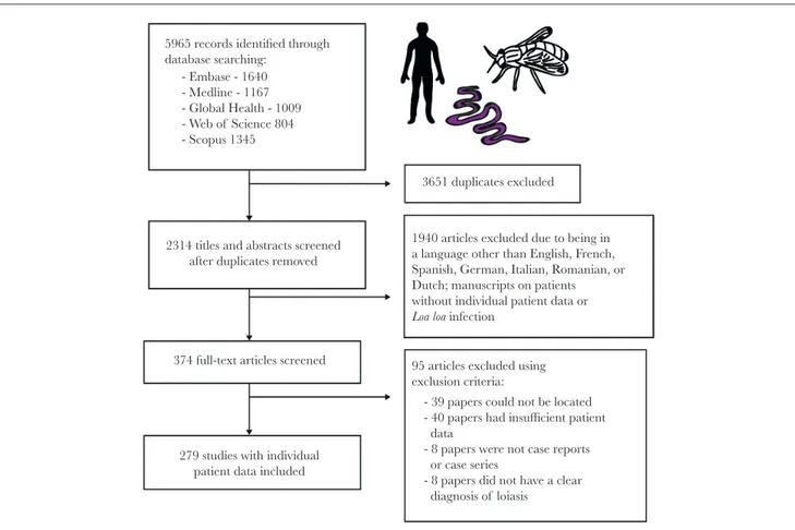
The search yielded 5965 articles; 3651 duplicates were re-moved. Case reports and case series containing IPD written in English, French, Spanish, German, Italian, Romanian, and Dutch were independently reviewed for eligibility by 2 au-thors. Articles were excluded if the patient did not show evi-dence of Loa loa infection (defined as the presence/history of subconjunctival or subcutaneous adult worm migration, Calabar swellings, microfilaremia, or parasite identification following worm extraction, if undertaken) or when the manu-script contained insufficient IPD metadata (eg, no information on cause of consultation, clinical presentation, or diagnostic methods used). Disagreements were resolved by discussion and, if required, arbitration by a third author. A total of 279 ar-ticles were retained for data extraction and analysis (Figure 1). The full list of included papers is given in the Supplementary Data (“Literature Review”). The protocol was registered pro-spectively with PROSPERO [International Prospective Register of Systematic Reviews] (March 4, 2018, CRD42018092232) and followed the guidelines set out by PRISMA [Preferred Reporting Items for Systematic Reviews and Meta-Analyses] for system-atic reviews of IPD.
5965 records identified through database searching: - Embase - 1640 - Medline - 1167 - Global Health - 1009 - Web of Science 804 - Scopus 1345
2314 titles and abstracts screened after duplicates removed
374 full-text articles screened
279 studies with individual patient data included
3651 duplicates excluded
1940 articles excluded due to being in a language other than English, French, Spanish, German, Italian, Romanian, or Dutch; manuscripts on patients without individual patient data or
Loa loa infection
95 articles excluded using exclusion criteria:
- 39 papers could not be located - 40 papers had insufficient patient data
- 8 papers were not case reports or case series
- 8 papers did not have a clear diagnosis of loiasis
Figure 1. Systematic review methodology. Flow diagram showing the number of papers identified, screened, assessed for eligibility, and included in the systematic review of individual patient data from loiasis case reports.
Patient Characteristics
Three authors independently extracted de-identified IPD: pa-tient demographics (age, sex, residency status), physical ex-amination, manifestations attributed to loiasis, blood tests (including eosinophilia and assessment of microfilaremia), di-agnostic imaging, histopathology, and treatment. Antifilarial treatment offered to patients included diethylcarbamazine (DEC; most frequently), ivermectin, albendazole, mebendazole, and imatinib. Where particular data were not available for a given patient, manifestations not reported by the paper’s author(s) were considered to be absent. Co-infections were also recorded.
The residency status of patients was assigned to 3 categories: locals residing in an endemic country (born and living in an en-demic region), locals residing in a nonenen-demic country (born in an endemic region but living in a nonendemic region at the time of consultation), and expatriates (born in a nonendemic region but having spent a period of time in an endemic region). Manifestations were categorized into typical (Calabar swelling and eyeworm) and atypical (all other manifestations attrib-uted to loiasis, including nonclassical ocular signs). When microfilarial densities were available, individuals were assigned to 1 of the following 3 categories: zero (amicrofilaremic individ-uals, with no detectable mf), low (>0 but <8000 mf/mL blood), or high (≥8000 mf/mL blood). The threshold of 8000 mf/mL was chosen, as individuals with higher densities have an increased risk of marked adverse events (ie, functional impairment for several days) after ivermectin treatment [8]. Eosinophilia was defined as a peripheral eosinophil blood count >0.5×10⁹ cells or
a peripheral blood eosinophil count ≥6% [19]. Data Analysis
Descriptive statistical analyses (% of cases with each condi-tion by organ system) were undertaken to characterize the full spectrum of clinical conditions associated with L. loa infection. Pearson’s chi-square test was used to assess whether the propor-tion of patients presenting atypically varied across the different microfilarial density categories. Multivariable logistic regres-sion models were used to investigate the existence of associ-ations between demographic factors (age, sex, residency status), microfilarial density, and type of presentation (atypical or typ-ical), yielding odds ratio (OR) values and their associated 95% confidence intervals (CIs). The statistical methodology is de-scribed in the Supplementary Data (“Statistical Analyses”). All analyses were conducted using R, version 3.4.4 ( https://www.r-project.org/).
RESULTS Patient Demographics
Data from 329 patients were obtained. A total of 169 patients (51.4%) were classified as locals, 156 (47.4%) as expatriates, and 4 (1.2%) could not be assigned due to missing information.
From the local patients, 61 (36.1%) were living in an endemic country, 99 (58.6%) were not living in an endemic country at the time of presenting, and 9 (5.3%) could not be classified due to missing information. A total of 148 patients (45.0%) were female, 170 (51.7%) were male, and there were 11 (3.3%) in-stances in which the patient’s sex was not stated. The patient’s age was reported in 302 (91.8%) cases, with a median (inter-quartile range [IQR]) of 32 (24–41) years.
Clinical Manifestations
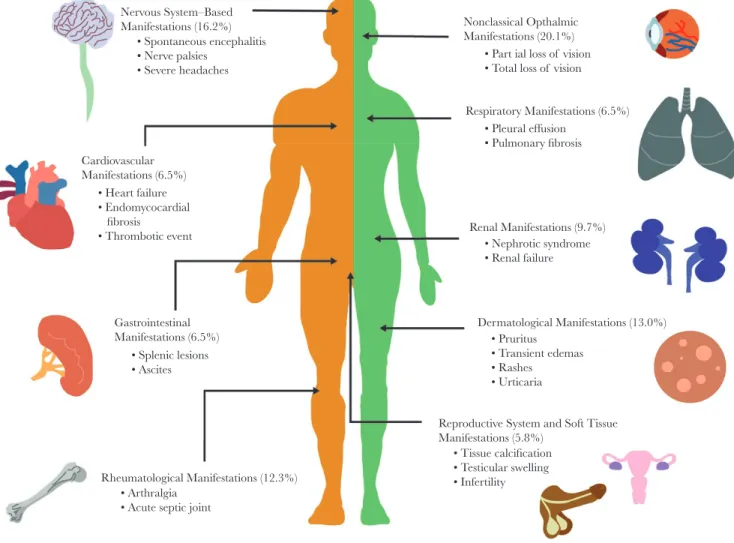
A total of 175 (53.2%) patients had only typical manifestations: 47 patients with Calabar swellings, 77 with eyeworm, and 51 patients with both Calabar swellings and eyeworm. In com-parison, 154 (46.8%) had at least 1 atypical manifestation, of which 29 patients had manifestations spanning 2 different body systems (Figure 2). No patients presented with atypical mani-festations spanning 3 or more organ systems. (In the following, citations preceded by S refer to Supplementary References in the Supplementary Data, “Literature Review.”)
Eight patients (5.2%) had heart failure signs [S11, S97, S155, S184, S208, S217, S221], of which 4 were caused by endomyocardial fibrosis [S97, S155, S184, S221]. Ten patients (6.5%) had respiratory system manifestations. Six were caused by pleural effusion [S39, S53, S74, S80, S228, S229]; 5 contained mf in the pleural fluid [S39, S53, S74, S228, S229], and 1 dem-onstrated mf on broncho-alveolar lavage (BAL) [S80]. Five of 6 patients had resolution of the pleural effusion after receiving antifilarial treatment [S39, S74, S228, S229]. Three patients had signs caused by pulmonary fibrosis [S72, S137, S186], with 1 patient demonstrating mf on BAL [S137]. One patient suffered from acute respiratory distress syndrome, although it was un-clear if loiasis was responsible [S44].
Ten patients (6.5%) had gastrointestinal system manifest-ations [S6, S35, S49, S51, S70, S74, S100, S124]. Two patients had ascites [S74, S79]; 1 contained mf in the ascitic fluid, which resolved with antifilarial treatment [S79]. Microfilariae were in-cidentally discovered in 2 patients during a gastric [S6] and per-itoneal lavage [S51]. Three patients had hypo-echogenic splenic lesions [S35, S100], of which 2 were associated with spleno-megaly [S35]. One subsequent splenectomy demonstrated mf on spleen histology. One patient had bowel obstruction caused by constricting, thickened, and fibrotic lesion of the descending colon, containing mf [S124]. One patient had polyps containing mf upon resection [S70]. Finally, 1 patient suffered from acute hepatitis that improved with antifilarial therapy [S79].
Fifteen patients (9.7%) had renal system manifestations [S12, S62, S77, S89, S116, S149, S169, S192, S201, S224, S227, S234, S256, S258, S268], ranging in severity from asymptomatic pro-teinuria to severe nephrotic syndrome and end-stage renal failure. In the 8 cases where a renal biopsy was performed for the investigation of nephrotic syndrome [S77, S89, S116, S149, S224, S227, S234], membranous glomerulonephropathy was
shown in all but 2 cases, in which focal interstitial inflamma-tion [S227] and focal segmental glomerulosclerosis [S234] were shown. One patient suffered from renal failure secondary to in-terstitial fibrosis and sclerosis of the glomeruli [S89]. Four pa-tients had microfilaruria (mf in urine [S12, S89, S256, S258]), and 3 had mf on histopathology analysis of their renal biopsy
[S62, S224, S234].
Nine patients (5.8%) had reproductive system manifestations
[S22, S38, S124, S131, S164, S220, S226]. Microfilariae were isolated from a sample of follicular fluid in 2 infertile women
[S22, S226] and incidentally found in 3 women during a cervical smear [S38, S164, S220]. Three men were found to have an adult worm in their testicles (tunica vaginalis or spermatid cord) after orchidectomy due to testicular swelling [S124]. Seven (4.5%) women had mammogram results that were highly suggestive of the presence of a L. loa worm, reporting linear, straight, or curved calcifications 0.3–2.5 cm in length [S92, S117, S140, S166, S210, S216]. The indication for mammogram varied, with 4 undergoing routine screening, 2 for a painful breast lump, and
1 for bilaterally painful breasts. Two women had excisions per-formed [S117, S166], and in 1 an adult L. loa was isolated [S117].
Nineteen patients (12.3%) had joint-related manifestations
[S8, S34, S37, S43, S103, S107, S120, S128, S135, S148, S150, S151, S174, S183, S193, S205, S232]. Five patients were assessed for an acute septic joint [S8, S34, S107, S128, S151]. Of the 4 patients whose joints were aspirated for articular fluid, 2 dem-onstrated mf in the synovial fluid [S107, S128], and the other 2 other showed eosinophilia between 60% and 80% [S34, S151].
Five patients demonstrated dead calcified worms on x-ray in painful wrist and finger joints [S37, S103, S120, S150]. Four patients experienced severe arthralgic joint pains that resolved with antifilarial treatment [S35, S43, S193, S232]. Two patients had an acute thrombotic event in the ulnar vein [S148] and popliteal artery [S205].
There were 25 cases (16.2%) related to the nervous system
[S11, S14, S28, S32, S55, S57, S66, S90, S100, S158, S159, S167, S223, S225, S257, S260, S261, S262, S266, S268, S272]. There were 9 cases presenting with spontaneous encephalitis (before Nervous System–Based Manifestations (16.2%) • Spontaneous encephalitis • Nerve palsies • Severe headaches Cardiovascular Manifestations (6.5%) • Heart failure • Endomycocardial fibrosis • Thrombotic event • Splenic lesions • Ascites • Arthralgia • Acute septic joint Gastrointestinal Manifestations (6.5%)
Rheumatological Manifestations (12.3%)
Nonclassical Opthalmic Manifestations (20.1%)
• Part ial loss of vision • Total loss of vision
• Pleural effusion • Pulmonary fibrosis • Nephrotic syndrome • Renal failure • Tissue calcification • Testicular swelling • Infertility Respiratory Manifestations (6.5%) Renal Manifestations (9.7%) Dermatological Manifestations (13.0%)
Reproductive System and Soft Tissue Manifestations (5.8%)
• Pruritus • Transient edemas • Rashes
• Urticaria
Figure 2. The spectrum of atypical clinical manifestations of loiasis. Schematic visualization of some of the atypical presentations of loiasis identified through the system-atic literature review, highlighting the key vital organs and systems involved (confirmed by presence of adult Loa loa and/or microfilarial stages), the percentage of patients presenting with manifestations pertaining to that system, and some of the most common atypical presentations observed for that system.
any antifilarial treatment) [S158, S167, S223, S257, S260, S261, S268], of which 8 cases had mf in the cerebrospinal fluid (CSF)
[S158, S167, S223, S257, S261, S268]. Two patients had severe headaches refractory to medical treatment that resolved with antifilarial therapy [S14, S90]. There were 4 cases of ulnar nerve palsies [S55, S57, S100, S159] and 1 median nerve palsy whose neurological deficit resolved after antifilarial treatment [S57].
One patient had neck pain caused by cervical nerve root irri-tation with associated worms erupting from the overlying skin
[S32]. One patient was diagnosed with an acute change in per-sonality and psychotic polydipsia; the altered behavior resolved with antifilarial treatment [S272]. Three cases presented with dizziness, a change in hearing, or a change in fine motor skills, but there was no clear unifying diagnosis [S11, S28, S272].
There were 32 cases (20.8%) of ophthalmic manifestations outside the classical adult worm migration under the conjunc-tiva [S26, S42, S73, S78, S93, S95, S108, S115, S123, S130, S145, S169, S182, S187, S190, S211, S236, S250, S259, S266]. Twenty-one cases reported reduced/partial loss of vision [S73, S93, S123, S145, S169, S187, S190, S236, S259, S266], and 5 reported total (unilateral/bilateral) loss of vision [S26, S42, S78, S182, S211]. In the cases for which follow-up information was avail-able, 9 patients had improvement in the loss of vision after treat-ment and 7 did not. There were 5 cases of reduced/blurred/loss of vision in which an adult worm was identified in the anterior chamber of the eye [S42, S78, S93, S145, S190]; 2 of these dem-onstrated an inflammatory membrane adherent to the anterior iris. An adult worm was seen in the posterior chamber in 1 case with loss of vision [S169]. Microfilariae were found in the ante-rior and posteante-rior segments of the eye in 5 cases [S169, S259]. In the remaining cases with loss of vision where the worm was not successfully visualized by the physician, a variety of patholo-gies were described. These were chorioretinitis, corneal edema, posterior chamber opacification, vitreous hemorrhage, hem-orrhagic retinopathy, retinal detachment, neovascularization, retinal artery occlusion, and uveitis. From the 32 patients with atypical ocular symptoms, there was no report of co-infections with other agents causing ocular manifestations such as
Onchocerca volvulus and/or Chlamydia trachomatis, nor were
onchocerciasis or trachoma mentioned as possible causes of the manifestations observed (Supplementary Data, “Atypical Ocular Manifestations,” Supplementary Table 1).
There were 20 cases (13.0%) of dermatological signs, with 8 cases of L. loa worms perforating through the skin (without any preceding antifilarial treatment) [S26, S95, S96, S98, S141, S154, S156, S222]. Two patients experienced skin swellings, 1 nasal
[S162] that was excised to identify a worm. The remaining cases were skin rashes of varying nature: transient urticarial rashes, widespread exanthema rashes, and multiple pruritic lesions. One patient suffered from a vasculitis-like syndrome in which a biopsy of the purpuric rash demonstrated infiltrative eosino-phils [S189].
Of the 199 cases of eosinophilia, 182 (91.5%) did not have any other co-infecting helminth reported. Of the 16 cases with (helminth) co-infections (8.5%), 8 presented with Mansonella
perstans [S192, S217, S218, S227, S254, S263, S273, S274], 1 with
O. volvulus [S225], 6 with soil-transmitted helminths
(round-worm, whip(round-worm, hookworm and threadworm) [S19, S198, S229, S260, S262, S272], and 2 with schistosomiasis [S232, S233] (Supplementary Data, “Eosinophilia and Co-Infections”). Factors Associated With Atypical Loiasis Manifestations
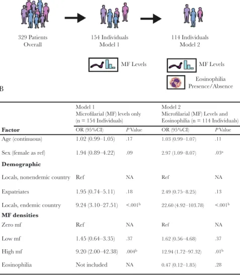
Information on microfilarial densities and typical/atyp-ical manifestations was available for 172 patients (informa-tion on presence/absence of mf, but not on densities, was available for 276 patients). There was a statistically signifi-cant positive association between blood microfilarial density categories (amicrofilaremic, low and high) and atypical presen-tation (atypical or typical, P = .001, Pearson’s chi-square test) (Supplementary Data, “Statistical Analysis,” Supplementary
Table 2). Based on this, 2 multivariate logistic regression models (Models 1 and 2, Figure 3A) were constructed to quantify the impact of microfilarial load on atypical presentation, control-ling for demographic variables.
Model 1 had presentation status (typical/atypical) as the out-come variable and age, sex, residency status, and microfilarial density categories as covariates (n = 154 individuals). Model 2, a more restricted model, included individuals for whom the above information was available but who also had their eosin-ophilia status determined (n = 114 individuals). This second model was considered to control for the potential influence of eosinophilia status, suggesting, in light of recent findings, a role of eosinophils in pathologies arising from microfilarial infec-tion in other filariae [20], as well as accounting for potential differences in eosinophil-associated processes between individ-uals indigenous to L. loa–endemic areas and those temporarily residing in such areas [21].
The results of both models demonstrated a positive associ-ation between blood microfilarial levels and atypical presenta-tion (Figure 3B). Individuals with high microfilarial densities were more likely to present with atypical manifestations com-pared with amicrofilaremic individuals (Model 1: OR, 9.20; 95% CI, 2.00–42.38; P = .004; Model 2: OR, 12.94; 95% CI, 1.72– 97.32; P = .01). Individuals with low microfilarial loads were not significantly more likely to present atypically compared with amicrofilaremic individuals (Model 1: P = .37; Model 2:
P = .37). Analysis of Model 2, including eosinophilia status,
in-dicated that eosinophilia was not significantly associated with the risk of presenting with atypical manifestations (OR, 0.47; 95% CI, 0.12–1.85; P = .28), although, interestingly, most indi-viduals analyzed (86%) were eosinophilic (98/114).
Both models revealed differences in the propensity for dif-ferent residency status groups to present atypically. Locals res-iding in endemic countries were significantly more likely than
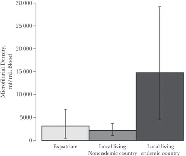
locals residing in nonendemic countries and expatriates to have atypical manifestations (Figure 3B). This effect is in addition to and independent of microfilarial load, although it should be noted that locals residing in endemic countries also had the highest average microfilarial densities (Figure 4), further com-pounding their risk.
DISCUSSION
Although our work corroborates findings from previous reviews regarding the variation in loiasis clinical profiles across dif-ferent residency groups [14, 22], its size and focus on nontypical presentations provide novel insight into poorly resolved aspects of loiasis clinical epidemiology. Specifically, it highlights that
the range of pathologies associated with L. loa infection is far broader than just Calabar swellings and eyeworm passage [23]. Although this may be due to a bias toward atypical manifest-ations in the reports analyzed here (well-known manifestmanifest-ations not meriting case reports) [24, 25], their breadth is striking, and our work provides an important summary of such pres-entations. The review identified numerous cases of vital organ involvement, such as heart, liver, respiratory, and renal sys-tems associated with loiasis. In addition, 25/32 (78.1%) cases with atypical visual manifestations were associated with partial or total vision loss (without onchocerciasis and/or trachoma). Although we were unable to prove causality, the legitimacy of the association is supported by corroboration of mf presence in tissues other than blood, such as pleural and ascitic fluid, eye Model: Atypical Presentation ~Age + Sex + Residency Status
+ MF Levels (+ Eosinophilia Presence/Absence)
329 Patients
Overall 154 IndividualsModel 1 114 IndividualsModel 2
MF Levels MF Levels Eosinophilia Presence/Absence A B Model 1
Microfilarial (MF) levels only (n = 154 Individuals)
Model 2
Microfilarial (MF) Levels and Eosinophilia (n = 114 Individuals) Factor Demographic MF densities Age (continuous) 1.02 (0.99–1.05) 1.94 (0.89–4.22) Ref Ref 1.45 (0.64–3.35) 9.20 (2.00–42.38) Not included 1.95 (0.74–5.11) 9.24 (3.10–27.51) Sex (female as ref)
Locals, nonendemic country Expatriates
Locals, endemic country Zero mf Low mf High mf Eosinophilia OR (95%CI) P Value .17 1.03 (0.99–1.07) .11 2.97 (1.09–8.07) Ref Ref 1.62 (0.56–4.68) 12.94 (1.72–97.32) 0.47 (0.12–1.85) 2.49 (0.75–8.25) 22.60 (4.92–103.78) .18 .37 <.001b <.001b .004b .09 .03a .13 NA NA NA NA .37 .01b NA .28 OR (95%CI) P Value
Figure 3. Association between individual-level factors and atypical loiasis presentation. Schematic illustration of the 2 multivariate logistic regression models used to quantify the influence of various factors on atypical loiasis presentation (A) and the output of these models (B). Microfilarial (MF) density levels were categorised as zero (0 mf/mL blood recorded), low (>0 but <8000 mf/mL blood), and high (≥8000 mf/mL blood). aIndicates statistical significance or near significance (P ≤ .05). bIndicates statistical
chambers, etc., and improvement or successful resolution of pathology upon antifilarial treatment (which would not have taken place were the mf found in these tissues simply the re-sult of their migration to sites of abnormal anatomy). However, we acknowledge we do not, at present, fulfill all the criteria to support causality, particularly because of the scarcity of longi-tudinal studies [5]. The proportion of cases with dermatolog-ical presentations (13%) may seem somewhat small; however, as the review focused on signs rather than symptoms, it likely excluded cases of itching (not systematically recorded in the reports).
These atypical conditions are more common than previ-ously perceived, with 47% of examined patients presenting with a manifestation(s) outside the classical Calabar swellings and eyeworm. Individuals residing in endemic countries were at the greatest risk of developing atypical manifestations. The results presented here likely represent an overestimation of the true prevalence of atypical manifestations due to (various sources of) bias (eg, locals familiar with classical signs may only consult if suffering from other symptoms, whereas expatriates/travelers may consult at the first sign of more typical manifestations). However, our aim was not to evaluate the true proportion of atypical manifestations of loiasis but to describe and summarize the breadth of these presentations and, importantly, to evaluate whether they were related to L. loa microfilarial density. Thus, although we acknowledge recruitment bias in the case reports, this does not impact the analyses performed, as patients did not
know their microfilarial density at the time of consultation. We advocate that large-scale surveys (eg, [10]) be conducted to es-timate rigorously the rates of atypical presentations in endemic areas. Notwithstanding these limitations, even a smaller per-centage of individuals experiencing atypical manifestations of loiasis would represent a substantial number of people given that 14 million reside in areas of high loiasis prevalence and in-tensity in Central Africa [2].
The analyses presented here also highlight that individuals with high microfilarial densities are at the greatest risk of suf-fering atypical manifestations. Although an association between infection intensity and disease is typically assumed in parasitic infections, its demonstration is essential to link infection to morbidity and mortality. Further work (with larger sample sizes and greater statistical power) is needed to corroborate this as-sociation (and to identify any dose–response relationship with microfilarial density). Heavy infection intensity was identified as a crucial factor in a recent study examining the influence of L. loa microfilarial infection on increased risk of mortality [5]. Although the mechanism underpinning this increased risk remains unclear, possibilities include obstructive processes as-sociated with parasite carriage [26] and/or more general inflam-matory responses inducing pathogenic processes in various vital organs. León et al. [27] identified endomyocardial fibrosis in a patient from an endemic area who had L. loa microfilaremia and marked eosinophilia.
As high microfilarial densities also increase the probability of SAEs after ivermectin treatment [8], targeted treatment of individuals most at risk may require drugs that are not strongly and/or fast-acting microfilaricidal. Evidence supports the use of albendazole in instances where other anthelmintics would be unsafe [28, 29], although questions remain surrounding the ef-ficacy and optimal dosing regimen [30].
For those with lower microfilarial densities, ivermectin is highly efficacious at clearing L. loa mf [31]. Recent studies in areas co-endemic for loiasis and onchocerciasis evaluating the impact of mass ivermectin treatment on microfilarial preva-lence and intensity of loiasis have yielded promising results in terms of reductions in the prevalence of heavy microfilaremia [32] (although others have reported less clear-cut results on en-tomological indicators of transmission [33]). However, not all areas endemic for loiasis are also co-endemic with onchocer-ciasis and/or lymphatic filariasis [34, 35], and thus would not receive ivermectin or ivermectin plus albendazole treatment for these filariases. Hence, perhaps with the exception of those loiasis-only areas in which control programs for soil-transmitted helminthiases are being implemented using benzimidazole an-thelmintics (to which albendazole belongs), our results suggest that substantial reductions in the burden of loiasis in these com-munities could be achieved through campaigns targeting loi-asis directly (through chemotherapeutic and/or tabanid control interventions) [1, 28–32, 36]. 0 5000 10 000 15 000 20 000 25 000 30 000 Microfilarial Density, mf/mL Blood
Expatriate Local living Nonendemic country
Local living endemic country
Figure 4. Average microfilarial densities for each of the residency categories examined in the analyses. The mean microfilarial densities for each residency group, locals residing in endemic countries (n = 40), locals residing in nonendemic countries (n = 44), and expatriates (n = 81) were calculated as the arithmetic means of the reported microfilarial densities, quantified by microscope blood smear. Error bars represent the 95% confidence intervals calculated using bootstrapping (see the Supplementary Data, “Confidence Interval Calculation: Non-Parametric Bootstrap”). Abbreviation: mf, microfilariae.
CONCLUSIONS
The work presented here suggests that conceptions of loiasis as a relatively benign infection are likely misplaced and em-phasizes the public health importance of loiasis. Our results highlight the wide spectrum of clinical conditions associated with L. loa carriage, including visual impairment and blind-ness (typically disregarded as sequelae of loiasis). Although es-timates of the burden of disease attributable to loiasis remain outstanding, these will likely be significantly higher than an-ticipated. This will be especially true in communities bearing high microfilarial burdens, as severe manifestations of loiasis occur at higher frequencies in heavily infected individuals. We advocate that large-scale, community-wide (and ideally longi-tudinal) studies be conducted in endemic regions to ascertain the true prevalence of atypical loiasis manifestations and the association between infection intensity and morbimortality to better understand the clinical and public health burden of lo-iasis in its own right and not merely as an impediment to the control of other filarial diseases [37].
Supplementary Data
Supplementary materials are available at Open Forum Infectious Diseases online (https://academic.oup.com/ofid). Consisting of data provided by the authors to benefit the reader, the posted materials are not copyedited and are the sole responsibility of the authors, so questions or comments should be addressed to the corresponding author.
Acknowledgments
Financial support. K.G.B. and P.D.J. were funded as part of their
Foundation Year 2 (FY2) postgraduate medical training program, and C.W. was funded by the Medical Research Council (MRC) doctoral training (nonclinical) program at the Faculty of Medicine, Imperial College London. M.W. and M.G.B. acknowledge funding from the Neglected Tropical Disease Modelling Consortium (https://www.ntdmodelling.org/), funded by the Bill & Melinda Gates Foundation in partnership with the Task Force for Global Health (grant No. OPP1053230); the Bill & Melinda Gates Foundation Projections on Eliminating NTDs (Integrating Mapping with Modelling; grant No. OPP1186851); and the UNICEF/UNDP/World Bank WHO Special Programme for Research and Training in Tropical Disease (TDR; grant No. 2015/540029-0). M.G.B. acknowledges joint center funding from the UK Medical Research Council and Department for International Development (grant No. MR/R015600/1). The funders had no influence on study design; collection, analysis, or interpretation of the data; or the deci-sion to submit the paper for publication.
Disclaimer. The views, opinions, assumptions, or any other information
set out in this article are solely those of the authors and should not be attrib-uted to the funders or any person connected with the funders. The funders had no role in writing the manuscript or in the decision to submit it for publication.
Potential conflicts of interest. All authors: no reported conflicts of
in-terest. All authors have submitted the ICMJE Form for Disclosure of Potential Conflicts of Interest. Conflicts that the editors consider relevant to the content of the manuscript have been disclosed.
Author contributions. K.G.B. and C.W. contributed equally as first
authors. M.G.B. and M.B. contributed equally as last and senior authors. Conceived the study: K.G.B., C.W., M.G.B., M.B. Performed the litera-ture review: K.G.B., P.D.J. Assessed the papers for inclusion and quality: K.G.B., C.B.C., S.D.S.P., M.B. Conducted the statistical analysis: C.W., M.W. Drafted the first version of the paper: K.G.B., C.W. Edited the manuscript
for intellectual input: C.B.C., S.D.S.P., M.W. M.G.B., M.B. All authors read and approved the final version.
References
1. Whittaker C, Walker M, Pion SDS, et al. The population biology and transmission dynamics of Loa loa. Trends Parasitol 2018; 34:335–50.
2. Zouré HG, Wanji S, Noma M, et al. The geographic distribution of Loa loa in Africa: results of large-scale implementation of the Rapid Assessment Procedure for Loiasis (RAPLOA). PLoS Negl Trop Dis 2011; 5:e1210.
3. Pinder M. Loa loa - a neglected filaria. Parasitol Today 1988; 4:279–84. 4. World Health Organization. Neglected tropical diseases 2018. Available at: http://
www.who.int/neglected_diseases/diseases/en/. Accessed 30 November 2018. 5. Chesnais CB, Takougang I, Paguélé M, et al. Excess mortality associated with
loiasis: a retrospective population-based cohort study. Lancet Infect Dis 2017; 17:108–16.
6. Fischer PU. Filarial infection deserves attention as neglected tropical disease. Lancet Infect Dis 2017; 17:12–3.
7. Hoerauf A, Pfarr K, Mand S, et al. Filariasis in Africa—treatment challenges and prospects. Clin Microbiol Infect 2011; 17:977–85.
8. Gardon J, Gardon-Wendel N, Demanga-Ngangue, et al. Serious reactions after mass treatment of onchocerciasis with ivermectin in an area endemic for Loa loa infection. Lancet 1997; 350:18–22.
9. Boussinesq M, Gardon J, Gardon-Wendel N, et al. Three probable cases of Loa
loa encephalopathy following ivermectin treatment for onchocerciasis. Am J Trop
Med Hyg 1998; 58:461–9.
10. Noireau F, Apembet JD, Nzoulani A, Carme B. Clinical manifestations of loiasis in an endemic area in the Congo. Trop Med Parasitol 1990; 41:37–9.
11. Akue JP, Nkoghe D, Padilla C, et al. Epidemiology of concomitant infection due to
Loa loa and Mansonella perstans in Gabon. PLoS Negl Trop Dis 2011; 5:e1329.
12. Gobbi F, Postiglione C, Angheben A, et al. Imported loiasis in Italy: an analysis of 100 cases. Travel Med Infect Dis 2014; 12:713–7.
13. Gantois N, Rapp C, Gautret P, et al. Imported loiasis in France: a retrospective analysis of 47 cases. Travel Med Infect Dis 2013; 11:366–73.
14. Churchill DR, Morris C, Fakoya A, et al. Clinical and laboratory features of pa-tients with loiasis (Loa loa filariasis) in the U.K. J Infect 1996; 33:103–9. 15. Saito M, Armstrong M, Boadi S, et al. Clinical features of imported loiasis: a case
series from the hospital for tropical diseases, London. Am J Trop Med Hyg 2015; 93:607–11.
16. Delabre S, Parola P, Thiberville D, et al. Non-ophthalmological presentation of imported loiasis. Travel Med Infect Dis 2014; 12:406–9.
17. Johann C, Eric C, Sophie M, et al. Imported loiasis in France: a 10 years retrospec-tive review with comparison between African migrants and non-African trav-elers. Trop Med Int Health 2013; 18(Suppl 1):43.
18. Develoux M, Hennequin C, Le Loup G, et al. Imported filariasis in Europe: a series of 31 cases from Metropolitan France. Eur J Intern Med 2017; 37:e37–9. 19. Valent P, Klion AD, Horny HP, et al. Contemporary consensus proposal on
cri-teria and classification of eosinophilic disorders and related syndromes. J Allergy Clin Immunol 2012; 130:607–612.e9.
20. Cadman ET, Thysse KA, Bearder S, et al. Eosinophils are important for protec-tion, immunoregulation and pathology during infection with nematode microfil-ariae. PLoS Pathog 2014; 10:e1003988.
21. Herrick JA, Metenou S, Makiya MA, et al. Eosinophil-associated processes un-derlie differences in clinical presentation of loiasis between temporary residents and those indigenous to Loa-endemic areas. Clin Infect Dis 2015; 60:55–63. 22. Antinori S, Schifanella L, Million M, et al. Imported Loa loa filariasis: three cases
and a review of cases reported in non-endemic countries in the past 25 years. Int J Infect Dis 2012; 16:e649–62.
23. Agbolade OM, Akinboye DO, Ogunkolo OF. Loa loa and Mansonella perstans: neglected human infections that need control in Nigeria. Afr J Biotechnol 2005; 4:1554–8.
24. Carme B, Mamboueni JP, Copin N, Noireau F. Clinical and biological study of Loa
loa filariasis in Congolese. Am J Trop Med Hyg 1989; 41:331–7.
25. Nutman TB, Miller KD, Mulligan M, Ottesen EA. Loa loa infection in temporary residents of endemic regions: recognition of a hyperresponsive syndrome with characteristic clinical manifestations. J Infect Dis 1986; 154:10–8.
26. Corrigan MJ, Hill DW. Retinal artery occlusion in loiasis. Br J Ophthalmol 1968; 52:477–80.
27. León D, Martín M, Corros C, et al. Usefulness of cardiac MRI in the early diag-nosis of endomyocardial fibrosis. Rev Port Cardiol 2012; 31:401–2.
28. Klion AD, Massougbodji A, Horton J, et al. Albendazole in human loiasis: results of a double-blind, placebo-controlled trial. J Infect Dis 1993; 168:202–6. 29. Kamgno J, Boussinesq M. Effect of a single dose (600 mg) of albendazole on Loa
loa microfilaraemia. Parasite 2002; 9:59–63.
30. Kamgno J, Nguipdop-Djomo P, Gounoue R, et al. Effect of two or six doses 800 mg of albendazole every two months on Loa loa microfilaraemia: a double blind, ran-domized, placebo-controlled Trial. PLoS Negl Trop Dis 2016; 10:e0004492. 31. Pion SDS, Tchatchueng-Mbougua JB, Chesnais CB, et al. Effect of a single dose of
ivermectin on Loa loa microfilaremia: systematic review and meta-analysis. Open Forum Infect Dis 2019; 6(X):XXX–XX.
32. Wanji S, Chounna Ndongmo WP, Fombad FF, et al. Impact of repeated annual community directed treatment with ivermectin on loiasis parasitological indica-tors in Cameroon: implications for onchocerciasis and lymphatic filariasis elim-ination in areas co-endemic with Loa loa in Africa. PLoS Negl Trop Dis 2018; 12:e0006750.
33. Kouam MK, Tchatchueng-Mbougua JB, Demanou M, et al. Impact of re-peated ivermectin treatments against onchocerciasis on the transmission
of loiasis: an entomologic evaluation in central Cameroon. Parasit Vectors
2013; 6:283.
34. Cano J, Basáñez MG, O’Hanlon SJ, et al. Identifying co-endemic areas for major fi-larial infections in sub-Saharan Africa: seeking synergies and preventing severe ad-verse events during mass drug administration campaigns. Parasit Vectors 2018; 11:70. 35. Vinkeles Melchers NVS, Coffeng LE, Boussinesq M, et al. Projected number
of people with onchocerciasis-loiasis co-infection in Africa, 1995 to 2025. Clin Infect Dis. in press.
36. Kelly-Hope L, Paulo R, Thomas B, et al. Loa loa vectors Chrysops spp.: perspec-tives on research, distribution, bionomics, and implications for elimination of lymphatic filariasis and onchocerciasis. Parasit Vectors 2017; 10:172.
37. Metzger WG, Mordmüller B. Loa loa—does it deserve to be neglected? Lancet Infect Dis 2014; 14:353–7.