HAL Id: hal-01790581
https://hal.archives-ouvertes.fr/hal-01790581
Submitted on 14 May 2018
HAL is a multi-disciplinary open access archive for the deposit and dissemination of sci-entific research documents, whether they are pub-lished or not. The documents may come from teaching and research institutions in France or abroad, or from public or private research centers.
L’archive ouverte pluridisciplinaire HAL, est destinée au dépôt et à la diffusion de documents scientifiques de niveau recherche, publiés ou non, émanant des établissements d’enseignement et de recherche français ou étrangers, des laboratoires publics ou privés.
Oscarella species (O. carmela and O. pearsei sp. nov)
from California, USA
Alexander Ereskovsky, Daniel Richter, Dennis Lavrov, Klaske Schippers, Scott
Nichols
To cite this version:
Alexander Ereskovsky, Daniel Richter, Dennis Lavrov, Klaske Schippers, Scott Nichols. Transcriptome sequencing and delimitation of sympatric Oscarella species (O. carmela and O. pearsei sp. nov) from California, USA. PLoS ONE, Public Library of Science, 2017, 12 (9), pp.1-25/e0183002. �10.1371/jour-nal.pone.0183002�. �hal-01790581�
Transcriptome sequencing and delimitation
of sympatric Oscarella species (O. carmela and
O. pearsei sp. nov) from California, USA
Alexander V. Ereskovsky1,2, Daniel J. Richter3,4, Dennis V. Lavrov5, Klaske J. Schippers6, Scott A. Nichols6*
1 Institut MeÂditerraneÂen de BiodiversiteÂet d'Ecologie Marine et Continentale (IMBE), CNRS, IRD, Aix
Marseille UniversiteÂ, Avignon UniversiteÂ, Station Marine d'Endoume, Marseille, France,2 Department of
Embryology, Faculty of Biology, Saint-Petersburg State University, 7/9 Universitetskaya emb., St. Petersburg, Russia,3 Department of Molecular and Cell Biology, University of California, Berkeley, CA, United States of
America,4 Sorbonne UniversiteÂs, UPMC UniversiteÂParis 06, CNRS, UMR 7144, Adaptation et DiversiteÂen
Milieu Marin, Equipe EPEP, Station Biologique de Roscoff, Roscoff, France,5 Department of Ecology,
Evolution, and Organismal Biology, Iowa State University, Ames, IA, United States of America,6 Department
of Biological Sciences, SGM 203, University of Denver, Denver, CO, United States of America
*scott.nichols@du.edu
Abstract
The homoscleromorph sponge Oscarella carmela, first described from central California, USA is shown to represent two superficially similar but both morphologically and phyloge-netically distinct species that are co-distributed. We here describe a new species as
Oscar-ella pearsei, sp. nov. and re-describe OscarOscar-ella carmela; the original description was based
upon material from both species. Further, we correct the identification of published genomic/ transcriptomic resources that were originally attributed to O. carmela, and present new Illu-mina-sequenced transcriptome assemblies for each of these species, and the mitochondrial genome sequence for O. pearsei sp. nov. Using SSU and LSU ribosomal DNA and the mito-chondrial genome, we report the phylogenetic relationships of these species relative to other Oscarella species, and find strong support for the placement of O. pearsei sp. nov. in a distinct clade within genus Oscarella defined by the presence of spherulous cells that con-tain paracrystalline inclusions; O. carmela lacks this cell type. Oscarella pearsei sp. nov and
O. carmela can be tentatively distinguished based upon gross morphological differences
such as color, surface texture and extent of mucus production, but can be more reliably identified using mitochondrial and nuclear barcode sequencing, ultrastructural characteris-tics of cells in the mesohyl, and the morphology of the follicle epithelium which surrounds the developing embryo in reproductively active individuals.
Introduction
The homoscleromorph sponge Oscarella carmela Muricy & Pearse, 2004 was described from Carmel, California and was the first record of this genus from the Pacific coast of North Amer-ica [1]. We and others were interested in developing this species as a model for genomic and
a1111111111 a1111111111 a1111111111 a1111111111 a1111111111 OPEN ACCESS
Citation: Ereskovsky AV, Richter DJ, Lavrov DV,
Schippers KJ, Nichols SA (2017) Transcriptome sequencing and delimitation of sympatric Oscarella species (O. carmela and O. pearsei sp. nov) from California, USA. PLoS ONE 12(9): e0183002. https://doi.org/10.1371/journal.pone.0183002
Editor: Ben J. Mans, Onderstepoort Veterinary
Institute, SOUTH AFRICA
Received: April 12, 2017 Accepted: July 21, 2017 Published: September 11, 2017
Copyright:© 2017 Ereskovsky et al. This is an open access article distributed under the terms of theCreative Commons Attribution License, which permits unrestricted use, distribution, and reproduction in any medium, provided the original author and source are credited.
Data Availability Statement: Raw Illumina data are
available from the NCBI SRA (SRX388205 and SRX386257). The O. pearsei mtGenome is available at NCBI (KY682864). Oscarella carmela and Oscarella pearsei ranscriptome assemblies are available atcompagne.org(by name, not accession number).
Funding: The authors received no specific funding
for this work.
Competing interests: The authors have declared
experimental research for several reasons: 1) it is abundant and easily accessible in research aquaria at the Joseph Long Marine Laboratory at the University of California Santa Cruz, 2) embryos of all stages are present year round in the laboratory environment, albeit more abun-dant in late summer and fall, and 3) it is thin and therefore internal cells and tissues can be easily imaged using common microscopy and experimental methods. To facilitate this devel-opment, we sequence expressed sequence tags (ESTs) [2], the mitochondrial genome [3], and a draft nuclear genome [4] for this species.
More recently, we used the Illumina platform to sequence and assemble the transcriptome of O. carmela to improve gene prediction from the draft genome, beyond what was possible using ESTs alone. However, from these data (reported in this article) we noticed that there was considerable sequence divergence at both the nucleotide- and amino acid-levels between the Illumina transcriptome and previously sequenced ESTs and gene predictions from the draft genome. This led us to suspect that there may be two or more cryptic/similar species that are co-distributed. Importantly, the tissues used to create each dataset were each derived from a single individual, otherwise the presence of multiple species may have gone undetected.
The original description of O. carmela reports the existence of color variants ranging from light brown to orange, and morphological variants ranging from thin and smooth to thicker and having a bumpy, microlobate surface [1]. This is consistent with our personal observations in both the lab and in the field. However, without experience collecting these sponges, and without the opportunity to see the various morphotypes side by side, the differences between them appear very subtle and seem to occur along a continuum rather than being discrete (Fig 1). Upon the discovery of such significant disparities between genetic datasets, we were careful to document the morphological differences between individuals and to preserve material for ultrastructural comparison using transmission electron microscopy, and for molecular biology including additional transcriptome sequencing, DNA barcoding and phylogenetic analysis.
Here we report that there are two sympatric species of the genus Oscarella in the Monterey/ Carmel region of central California. Through re-examination of the holotype and paratypes of
O. carmela we have determined that the original description was based on samples derived
from both species. We distinguish between these species, leaving the name O. carmela assigned to holotype specimen, and we describe the new species under the name O. pearsei sp. nov. In addition to morphological and ultrastructural characterization, we present correctly identified transcriptome assemblies corresponding to each species, their mitochondrial genomes, DNA barcoding sequences that can be used to confirm their identity, and we report their phyloge-netic placement relative to each other and other species of Oscarella.
Materials and methods
Sample collection was conducted at the University of California Santa Cruz, Joseph Long Marine Laboratory with permission from Research Coordinator B. Steele.
Transcriptome sequencing and assembly
Oscarella pearsei sp. nov. transcriptome assembly (consisting of quality trimming, error
cor-rection, de novo assembly, identification and removal of cross-contamination, prediction of amino acid sequences, elimination of redundant transcripts, and removal of noise transcripts) was performed according to the protocol described in Peña and colleagues [5]. Within the error correction step, parameter values supplied to the Reptile error correction software v1.1 [6] for the O. pearsei sp. nov. read set were as follows (see methods in [5] for a description of how these values were derived): T_expGoodCnt of 54, T_card of 21, KmerLen of 13, Qthres-hold of 73, Qlb of 60, MaxBadQPerKmer of 8 and Step to 12, with all other settings at their
defaults. The initial de novo assembly using Trinity release 2013-02-25 [7] of O. pearsei sp. nov. contained 122,656 contigs. The decontamination step removed 385 contigs, with 122,271 con-tigs remaining. Initial protein prediction on the decontaminated concon-tigs resulted in 146,347 proteins. Redundancy removal based on these predictions eliminated 67,850 redundant con-tigs and 117,085 proteins, with 54,421 concon-tigs and 29,262 predicted proteins remaining. Subse-quent elimination of noise transcripts below FPKM 0.01 resulted in the further removal of 417 contigs containing 42 proteins, resulting in a final O. pearsei sp. nov. transcriptome data set of 54,004 contigs and 29,220 predicted proteins.
Oscarella carmela transcriptome assembly was performed using essentially the same set of
steps, with the following minor modifications (and associated parameter values and contig/ protein counts): because O. carmela reads were single-end (not paired-end) we performed a single trimming step (equivalent to the second phase of trimming on paired-end data) with Trimmomatic version 0.30 [8], using identical parameter values to those used for O. pearsei sp. nov., with the addition of the directive TOPHRED33, as the reads were produced by an earlier generation sequencing machine. During the error correction step, parameter values supplied to Reptile were as follows: T_expGoodCnt of 10, T_card of 4, KmerLen of 13, Qthreshold of 66, Qlb of 58, MaxBadQPerKmer of 8 and Step to 12, with all other settings at their defaults. For initial de novo transcriptome assembly with Trinity, we used default parameter values for single-end reads, resulting in 66,109 contigs and 61,705 predicted proteins. We did not per-form the removal of cross-contamination step on O. carmela, as it was not sequenced on the
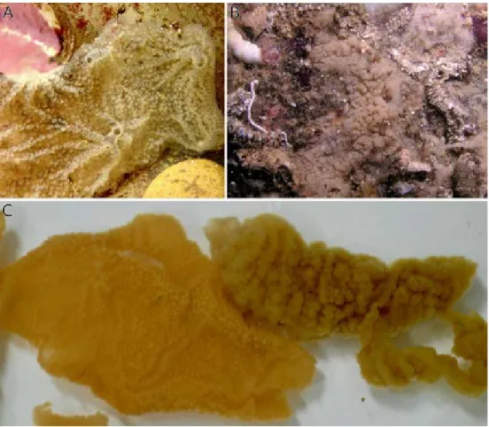
Fig 1. Oscarella pearsei sp. nov. and O. carmela. (A) In situ photographs of O. pearsei sp. nov. and (B) O.
carmela.(C) Upon collection, these species can be distinguished based upon differences in their thickness,
surface texture, and color. Left: O. pearsei sp. nov., Right: O. carmela. (photo credit: J. Mitchell, panel A). https://doi.org/10.1371/journal.pone.0183002.g001
same flow cell as any other sponges. Redundancy removal eliminated 3,378 contigs and 4,616 proteins, with 62,731 non-redundant contigs and 57,089 non-redundant proteins remaining. We subsequently eliminated 2,893 noise contigs and 3,334 noise proteins below the expression threshold of FPKM of 0.01, resulting in a final O. carmela transcriptome data set of 59,838 con-tigs and 53,755 predicted proteins.
Mitochondrial genome assembly
Mitochondrial genome of Oscarella pearsei sp. nov. was assembled using SOAPdenovo version 1.0 [9] from the Illumina genomic sequences collected in the previous study [4] with a mean coverage around ~1500x. The tRNA genes were identified by the tRNAscan-SE program [10]; rRNA and protein genes were identified by similarity searches in local databases using the FASTA program [11]. The presence of inversion was confirmed by mapping individual Illu-mina sequences on mitochondrial genome assembly and by conducting PCR amplifications across the inversion boundaries.
Phylogenetic analyses
Phylogenetic analyses of SSU and LSU data were performed on thehttp://www.phylogeny.fr/ platform [12] and comprised the following steps. Sequences were aligned using CLC Work-bench (v. 6.8) (Qiagen). After alignment, ambiguous regions (i.e., containing gaps and/or poorly aligned) were removed with Gblocks (v0.91b; [13]) using the following parameters:Ð minimum length of a block after gap cleaning: 10, -no gap positions were allowed in the final alignment,Ðall segments with contiguous non-conserved positions bigger than 8 were jected, -minimum number of sequences for a flank position: 85%. Phylogenetic trees were re-constructed using the maximum likelihood method implemented in the PhyML program and support values calculated using the Approximate Likelihood Ratio Test (v3.1/3.0 aLRT) [14]. The default substitution model was selected assuming an estimated proportion of invariant sites (of 0.357) and 4 gamma-distributed rate categories to account for rate heterogeneity ac-ross sites. The gamma shape parameter was estimated directly from the data (gamma = 0.635). Reliability for internal branches was assessed using the aLRT test (SH-Like) [15]. Graphical re-presentation and edition of the phylogenetic trees were performed with TreeDyn (v198.3) [16].
Inferred amino acid sequences of all mtDNA-encoded proteins from homoscleromorph sponges except TatC (absent in Plakinidae) were aligned with Mafft v.6.861b [17]. Conserved blocks within the alignments were selected with Gblocks 0.91 b [13] using relaxed parameters (parameters 1 and 2 = ½, parameter 3 = 8, parameter 4 = 5, all gap positions in parameter 5). Cleaned alignments were concatenated in a dataset 4070 positions in length. Bayesian infer-ences based on amino acid sequinfer-ences were performed with the CAT+GTR+G4 mixture model implemented in the program PhyloBayes MPI version 1.4e [18]. PhyloBayes analysis consisted of four chains over 57,000 generations using CAT+G model (max-diff ~ 0.015) and over 35,000 generations using CAT+GTR+G4 model (max diff. ~0.03). The chains were sampled every tenth generation after the first 100 burn-in cycles and the majority-rule majority consen-sus trees were built to estimate the posterior probabilities of internal branches.
DNA barcoding
Upon collection, an ~1cm x 1cm piece of tissue from each individual was preserved in fixative for Transmission Electron Microscopy (described below) and in 95% EtOH for genomic DNA extraction and DNA barcoding. Genomic DNA for LSU barcoding was isolated using the Gen-Elute Mammalian Genomic DNA Miniprep kit (Sigma-Aldrich) per manufacturer specifica-tions (EtOH was allowed to evaporate before starting procedure). Using 28S-C2-fwd and
28S-D2-rev [19] primers, a fragment of the LSU was amplified by PCR and sequenced by Eurofins Genomics (Germany). Genomic DNA for mitochondrial barcoding was isolated using the standard phenol-chloroform method [20]. Using diplo-cob-f1m and diplo-cob-r1m primers [21], a fragment of cob was amplified by PCR (Invitrogen recombinant Taq DNA polymerase kit), purified with Promega Wizrd SV Gel and PCR Clean-up system, and sequenced by Iowa State University Sequencing facility.
Transmission electron microscopy
For semi-thin sections and for transmission electron microscopy (TEM) investigations, sponges were fixed in a solution composed of one volume of 25% glutaraldehyde, four volumes of 0.2 M cacodylate buffer and five volumes of filtered seawater for at least 2 h before being post-fixed in 2% OsO4in seawater at room temperature for 2 h. After fixation, samples were
washed in 0.2 M cacodylate buffer and distilled water successively, and dehydrated through a graded ethanol series. Specimens were embedded in Araldite resin. Semi-thin sections (1 µm in thickness) were cut on a Reichert Jung ultramicrotome equipped with a "Micro Star" 45Ê diamond knife before being stained with toluidine blue, and observed under a WILD M20 microscope. Digital photos were taken with a Leica DMLB microscope using the Evolution LC color photo capture system. Ultrathin sections (60±80 nm) were cut with a Leica UCT ultrami-crotome equipped with a Drukkert 45Êdiamond knife. Ultrathin sections, contrasted with ura-nyl acetate, were observed under a Zeiss-1000 transmission electron microscope (TEM).
Nomenclatural acts
The electronic edition of this article conforms to the requirements of the amended Interna-tional Code of Zoological Nomenclatures, and hence the new names contained herein are available under that Code from the electronic edition of this article. This published work and the nomenclatural acts it contains have been registered in ZooBank, the online registration sys-tem for the ICZN. The ZooBank LSIDs (Life Science Identifiers) can be resolved and the asso-ciated information viewed through any standard web browser by appending the LSID to the prefixhttp://zoobank.org/. The LSID for this publication is: urn:lsid:zoobank.org:pub: A5472E86-7589-4589-4E78-A2BF-51D73E086CF4. The electronic edition of this work was published in a journal with an ISSN, and has been archived and is available from the following digital repositories: PubMed Central, LOCKSS.
Results
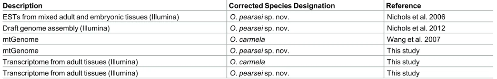
The holotype and paratypes submitted as part of the original species description for Oscarella
car-mela we re-examined and found to include tissue from distinctly separate species (Table 1) based upon distinguishing characteristics defined below. The decision as to which species should retain the name O. carmela was made based upon which corresponded to the holotype deposited at the Museu Nacional Rio de Janeiro (MNRJ), and the other species was described as O. pearsei sp. nov. Thus, previously published studies of the species O. carmela may actually refer to either of these co-distributed species. InTable 2we provide a list of previously published and new genomic and transcriptomic resources, and clarify the species from which they were derived.
Table 1. Original O. carmela holotype and paratypes submitted to MNRJ.
Accession number Re-examined and identified as:
Holotype B-6618 #2 O. carmela
Paratype B-6617 #1 O. carmela
Paratype B-6616 O. pearsei sp. nov.
Transcriptome sequencing
Transcriptome sequencing was performed on adult somatic tissues from a single individual of the species O. pearsei sp. nov., and on tissues and brooded embryos derived from a single indi-vidual of O. carmela. Assembly of the O. carmela transcriptome resulted in 59,838 contigs encoding 53,755 predicted peptides of >50aa in length. Assembly of the O. pearsei sp. nov. transcriptome resulted in 54,004 contigs encoding 29,220 predicted peptides of >50aa in length. The difference in number of peptides predicted from these datasets was explored using CD-HIT [22] to create peptide clusters with high sequence identity. O. carmela peptides formed 37,887 unique clusters whereas O. pearsei sp. nov. peptides formed 29,209 unique clus-ters. These results are consistent with the fact that the O. carmela transcriptome was assembled from fewer reads than the O. pearsei sp. nov. assembly, and differences in transcript number may result from assembly errors due to low sequencing coverage that mimic alternative splice variants. Raw sequence data are available at the NCBI Sequence Read Archive under the acces-sion numbers SRX388205 and SRX386257. Assembled transcriptomes and predicted peptides are available atcompagen.org[23] and as supplemental files (S5andS6Figs).
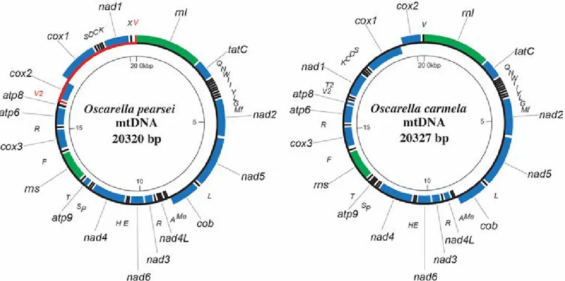
Mitochondrial genome
The mitochondrial genome for O. carmela was previously published [3], and the species desig-nation for this dataset remains unchanged. Here, we report and describe the sequence of the mitochondrial genome for O. pearsei sp. nov. (Fig 2), which was assembled from Illumina DNAseq data and deposited to GenBank (accession number KY682864). The mitochondrial genome of O. pearsei sp. nov. is a circular mapping molecule 20,320 bp in size and 65.6% A+T that fits well with the description of a typical mitochondrial genomes in the family Oscarellidae [24,25]. It contains 42 genes, organized in two clusters with opposite transcriptional polarities. The genes include the unusual tatC, for subunit C of the twin arginine translocase [26] as well as a duplicated trnV(uac). However, mitochondrial gene order in O. pearsei sp. nov. is unique among other representatives of the family Oscarellidae due to an inversion of a mtDNA frag-ment between the two copies of the trnV gene. This inversion allows an easy PCR-based identi-fication of two sympatric species of Oscarella. In addition, the second duplicated tRNA gene (most likely trnT) encodes an unusual tRNA-like structure with well conserved aminoacyl acceptor, TYC and DHU arms, but with the anticodon arm replaced by a 9-nt loop. An identi-cal structure with a very similar sequence (two substitutions) is also encoded in the mtDNA of the closely related O. balibaloi (accession number KY682865). Interestingly, in the latter spe-cies the copy of trnV between atp8 and nad1 encodes a similar structure with identically reduced anticodon arm. The mt-coding sequences of O. carmela and O. pearsei sp. nov. are on average 89.4% identical with the rate of synonymous substitutions estimated between 0.31 and 2.75 (mean = 0.76) and the rate of non-synonymous substitutions estimated between 0.01 and
Table 2. Material source for genomic and transcriptomic data.
Description Corrected Species Designation Reference
ESTs from mixed adult and embryonic tissues (Illumina) O. pearsei sp. nov. Nichols et al. 2006
Draft genome assembly (Illumina) O. pearsei sp. nov. Nichols et al. 2012
mtGenome O. carmela Wang et al. 2007
mtGenome O. pearsei sp. nov. This study
Transcriptome from adult tissues (Illumina) O. carmela This study
Transcriptome from adult tissues (Illumina) O. pearsei sp. nov. This study https://doi.org/10.1371/journal.pone.0183002.t002
0.21 (mean = 0.04). Both synonymous and non-synonymous substitution rates were highest in
tatC, an observation consistent with our previous study [26].
Phylogenetic analysis
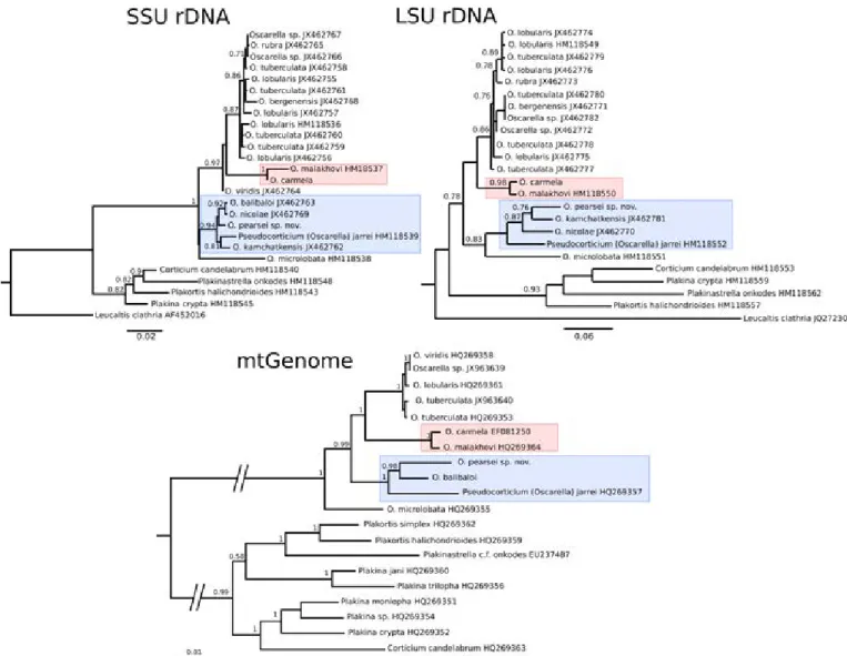
To assess the relationship of O. carmela and O. pearsei sp. nov., to each other and to other known Oscarella species globally, phylogenetic analyses were performed using three datasets: small subunit (SSU; 1,781 aligned bp), large subunit (LSU; 1,211 aligned bp) rDNA and a supermatrix of inferred amino-acid sequences from 14 mitochondrial protein-coding genes (all but tatC). Maximum likelihood and Bayesian analyses of each dataset were highly concor-dant and collectively supported the close relationship of O. carmela to O. malakhovi, and the placement of O. pearsei sp. nov. in a subclade containing O. kamchatkensis, O. nicolae, O.
bali-baloi, and Pseudocorticium jarrei (Fig 3).
DNA barcoding
All museum-deposited samples used for the re-description of O. carmela and the new descrip-tion of O. pearsei sp. nov. were identified using a short, rapidly evolving region of the LSU that has been shown to be useful for DNA barcoding [19,27] as well as a fragment of mitochondrial Cytochrome b gene [21]. Both LSU and cob fragments were found to reliably distinguish be-tween the two species, which were detected living side by side in the laboratory (UCSC and at the type locality in the intertidal zone of Carmel, CA). The O. carmela LSU barcode sequence was deposited to GenBank under accession KY513287 and the O. pearsei sp. nov. barcode
Fig 2. Mitochondrial genome architecture. Mitochondrial genome maps of Oscarella pearsei sp. nov. and O. carmela. Protein (green) and ribosomal (blue)
genes are atp6, atp8-9: subunites 6, 8, and 9 of F0 adenosine triphosphatase (ATP) synthase; cob: apocytochrome b; cox 1±3: cytochrome c oxidase subunites 1±3; nad1-6 and nad4L: NADH dehydrogenase subunits 1±6 and 4L; rns and rnl: small and large subunit rRNAs; tatC: twin-arginine translocase component C. tRNA genes (black) are identified by the one-letter code for their corresponding amino acid. Genes outside the main circle are transcribed clock-wise, insideÐc ounter clock-wise. Large inversion between two copies of trnV in O. pearsei sp. nov. mtDNA is shown in red.
sequence was deposited to GenBank under accession KY513286. The barcode sequence is 458bp in O. pearsei sp. nov. and 446bp in O. carmela. Cob barcoding sequences were found to be identical to the fragments of cob in complete mitochondrial genomes of the two species. No polymorphisms were detected between individuals sampled from within each species (n = 5) in either nuclear or mitochondrial barcodes and the sequence identity for these markers between the two species was 77% and 87%, respectively.
Morphological description
Genus Oscarella Vosmaer, 1884
Type species
Halisarca lobularis Schmidt, 1862 (by monotypy). [Oscaria] Vosmaer, 1881: 163 (preocc. by Oscaria Gray, 1873 ±Reptilia); Oscarella Vosmaer, 1884: pl. 8 (explanation); 1887: 326 (nom.
nov. for Oscaria Vosmaer). Octavella Tuzet and Paris, 1964: 88.
Fig 3. Homoscleromorph phylogenetic relationships inferred from ribosomal DNA and mitochondrial data. Maximum likelihood analyses of
SSU/LSU rDNA and Bayesian analysis of a mitochondrial supermatrix (4070 amino-acid positions derived from 14 mitochondrial protein-coding genes) for Oscarella and outgroup species. Support values reflect Approximate Likelihood Ratio Test and posterior probability values, respectively. The consistently supported clades to which O. carmela and O. pearsei belong are highlighted by pink and blue boxes, respectively.
Diagnosis (modified from Muricy & Diaz 2002)
Homoscleromorpha without skeleton, with a variable degree of ectosome development. The aquiferous system has a sylleibid-like or leuconoid organization, with eurypylous or diplodal choanocyte chambers. Mitochondrial genomes that have been sequenced so far encode a gene absent in other animal mitochondrial genomes: tatC.
Oscarella pearsei sp. nov.
urn:lsid:zoobank.org:act:8431B41C-B08E-4205-8FE6-CCCFFB6EB574
Holotype:. ZIN RAS № 11798, Ethanol.
urn:lsid:zoobank.org:act:8431B41C-B08E-4205-8FE6-CCCFFB6EB574, sea tables at Long Marine Lab. Santa Cruz, California, USA, Collected by Scott Nichols 01.08.2015.
ZIN RAS № 11800, Glutaraldehyde. urn:lsid:zoobank.org:act:8431B41C-B08E-4205-8FE6-CCCFFB6EB574, sea tables at Long Marine Lab. Santa Cruz, California, USA, Collected by Scott Nichols 01.08.2015.
Paratypes:. ZIN RAS № 11799, Ethanol, sea tables at Long Marine Lab. Santa Cruz,
Cali-fornia, USA, Collected by Scott Nichols 01.08.2015.
ZIN RAS № 11801, Glutaraldehyde, sea tables at Long Marine Lab. Santa Cruz, California, USA, Collected by Scott Nichols 01.08.2015.
RBINS-IG 33439-POR.0122, Ethanol, sea tables at Long Marine Lab. Santa Cruz, California, USA, Collected by Scott Nichols 01.08.2015.
RBINS-IG 33439-POR.0123, Glutaraldehyde, sea tables at Long Marine Lab. Santa Cruz, California, USA, Collected by Scott Nichols 01.08.2015.
RBINS-IG 33439-POR.0124 Glutaraldehyde, Ethanol, sea tables at Long Marine Lab. Santa Cruz, California, USA, Collected by Scott Nichols 01.08.2015.
Comparative material examined. Oscarella carmela, Holotype MNRJ 8033 (slide B-6618
#2), Carmel Point, Carmel Bay, intertidal, Collected by J. Pearse, 08. 05. 2003 [1]. Oscarella
car-mela Paratype, CASIZ 168925 (slide B-6617 #1), Carmel Point, Carmel Bay, central California,
intertidal, Collected by J. Pearse. 08. 05. 2003 [1]. Oscarella carmela Muricy, Pearse, 2004. Syn-type: ZIN RAS № 11802, Ethanol (this study), sea tables at Long Marine Lab. Santa Cruz, Cali-fornia, USA, S. Collected by Scott Nichols 01.08.2015. Oscarella carmela Muricy, Pearse, 2004. Syntype, ZIN RAS № 11803, Ethanol (this study), sea tables at Long Marine Lab. Santa Cruz, California, USA, S. Collected by Scott Nichols 01.08.2015. Oscarella carmela Muricy, Pearse, 2004. Syntype ZIN RAS № 11804, Glutaraldehyde (this study), sea tables at Long Marine Lab. Santa Cruz, California, USA, S. Collected by Scott Nichols 01.08.2015. Oscarella carmela Muricy, Pearse, 2004. Syntype ZIN RAS № 11805, Glutaraldehyde (this study), sea tables at Long Marine Lab. Santa Cruz, California, USA, S. Collected by Scott Nichols
01.08.2015.Oscar-ella carmela Muricy, Pearse, 2004. Syntype RBINS-IG 33439-POR.0125, Ethanol (this study),
sea tables at Long Marine Lab. Santa Cruz, California, USA, S. Collected by Scott Nichols 01.08.2015. Oscarella carmela Muricy, Pearse, 2004. Syntype RBINS-IG 33439-POR.0126, Eth-anol (this study), sea tables at Long Marine Lab. Santa Cruz, California, USA, S. Collected by Scott Nichols 01.08.2015. Oscarella carmela Muricy, Pearse, 2004. Syntype RBINS-IG 33439-POR.0127, Glutaraldehyde (this study), sea tables at Long Marine Lab. Santa Cruz, California, USA, S. Collected by Scott Nichols 01.08.2015. Oscarella carmela Muricy, Pearse, 2004. Syn-type, RBINS-IG 33439-POR.0128, Glutaraldehyde (this study), sea tables at Long Marine Lab. Santa Cruz, California, USA, S. Collected by Scott Nichols 01.08.2015. Oscarella malakhovi Ereskovsky, 2006 (ZIN RAS 10697 ZIN RAS 10698: Japan Sea) [28]. Oscarella kamchatkensis Ereskovsky, Sanamyan & Vishnyakov, 2009 (ZIN RAS 11058, ZIN RAS 11059 and ZIN RAS 11060: North Pacific, Avacha Gulf) [29]. Oscarella lobularis (Schmidt, 1862) and Oscarella
tuberculata (Schmidt, 1868). NE Mediterranean Sea (Marseille region), underwater cave of
Maire Island. Oscarella microlobata Muricy, Boury-Esnault, BeÂzac, Vacelet, 1996 and Oscarella
viridis Muricy, Boury-Esnault, BeÂzac, Vacelet, 1996. NE Mediterranean Sea (Marseille region),
underwater cave of Jarre Island [30]. Oscarella balibaloi, PeÂrez, IvanisÏević, Dubois, Pedel, Thomas, Tokina, Ereskovsky, 2011. NE Mediterranean Sea (Marseille region), underwater cave of Maire Island [31]. Oscarella nicolae Gazave, Lavrov, Cabrol, Renard, Rocher, Vacelet, Adamska, Borchiellini, Ereskovsky, 2013 (MNHN DJV155, LSID urn:lsid:zoobank.org:act: DFFAD94B-9CAD-4F99-994D-BDEF75EF98A1, MNHN DJV156) North Sea, Norway (Ber-gen Fjords, Skarvoysundet) [25]. Oscarella ber(Ber-genensis Gazave, Lavrov, Cabrol, Renard, Rocher, Vacelet, Adamska, Borchiellini, Ereskovsky, 2013 (MNHN DJV153, LSID:urn:lsid: zoobank.org:act:2D44BCFA-2163-47C7-9E70-EF6C13E0E4A4, MNHN DJV154) North Sea, Norway (Bergen Fjords, Skarvoysundet) [25].
Diagnosis. Found on the sides and undersides of rocks in the intertidal zone in Carmel,
California, USA. Encrusting Oscarella, tan to orange in color, soft, slimy but delicate consis-tency with two particular mesohylar cell types with inclusions: abundant granular cells and spherulous cells with paracrystalline inclusions. Nucleus in choanocyte has apical position. Embryo follicle layer comprised of multilayer cubic cells. It has only one morphotype of endo-biotic bacteria.
Description. Sponge habit is very thinly encrusting. Size can vary, with most sponges
being less than 10 cm in diameter, but sometimes extending to 30 cm or greater. Color in vivo is light tan to orange (Fig 1A). Preserved fragments are lighter in color, sometimes approaching white. The surface is finely and uniformly lobate with small hemispherical lobes, and adorned with few delicate oscula 3±5 mm in height; oscula are only visible in submerged specimens that are relaxed (i.e., not recently contracted). Sponges are not uniformly attached to the substrate; rather, outgrowths extend from the basopinacoderm and attach to the substrate at their tip. Thus, the sponge can be easily dislodged from the substrate and maintains its integrity.
Soft tissue organization. Spicule and fiber skeleton absent. Ectosome is between 8 to
15 µm thick. Inhalant canals run perpendicular to the surface. Choanocyte chambers ovoid to spherical, eurypilous, from 55 to 70 µm in diameter. Exhalant canals run towards a well devel-oped system of basal cavities leading to the oscula.
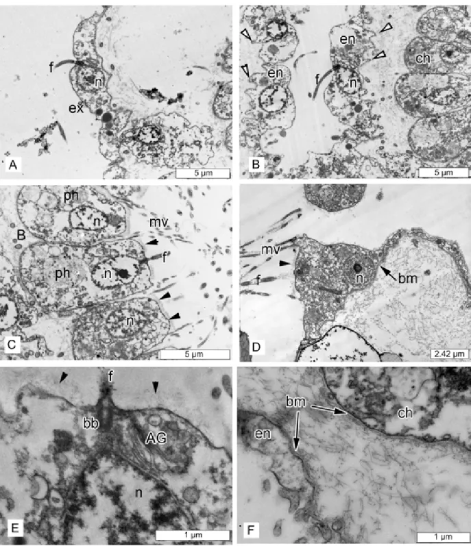
Cytology. Exopinacocytes (Fig 4A) flagellated with flat to oval shape, 13.5 µm wide by 4.9 µm high (N = 10). Cytoplasm containing electron dense granules from 0.6 to 1.2 µm in diameter and electron translucent vacuoles 0.2±0.7 µm in diameter. Nucleus about 2.8 µm in diameter with central position. External surface of exopinacocytes covered by a layer of glyco-calyx 0.15±0.25 µm thick and basal surface has filopodia that extend into the extracellular matrix (S1 Fig).
Endopinacocytes (Fig 4B) flagellated flat cells, about 17.2 µm wide by 3.3 µm high (N = 10). Nucleus ovoid (2.6 µm in diameter). Cytoplasm includes electron dense granules 0.3±0.8 µm in diameter and electron translucent vacuoles 0.3±0.8 µm in diameter. Basal surface has pseu-dopodia that extend into the extracellular matrix.
Choanocytes (Fig 4B, 4C and 4E) ovoid to trapezoidal, about 4.2 µm wide by 7.7 µm high (N = 20). Nucleus (~2.4µm in diameter) has apical position and contacts with flagellar basal apparatus. Cytoplasm includes from one to eight phagosomes 1.2±3.4 µm in diameter.
Apopylar cells (Fig 4D) roughly triangular or oval in section, 6.3 µm wide by 4.7 µm high (N = 5). Nucleus up to 2.9 µm in diameter. Cytoplasm contains mitochondria, digestive vacu-oles, and small osmiophilic inclusions.
Surface of endopinacocytes, choanocytes and apopylar cells covered by thin, irregular gly-cocalyx, choanoderm and pinacoderm underlined by continuous basement membrane (Fig 4D±4F).
Two types of cells with inclusions occur within the mesohyl. Granular cells (Fig 5A) ovoid to elongate, approximately 5.4 x 7.1 µm, sometimes amoeboid-like or irregular (N = 8).
Fig 4. Oscarella pearsei sp. nov. TEM of cells and symbiotic bacteria. (A) exopinacocyte; (B) endopinacocyte; (C) choanocytes; (D) apopylar cell; (E) apical part of choanocyte with glycocalyx layer; (F) basal parts of choanocyte and endopinacocyte with basement
membrane. (Abbreviations: AG±Golgi apparatus, ap±apopylar cell, B±symbiotic bacteria, bb±basal body of flagellum, bm±basement membrane, ch±choanocytes, en±endopinacocyte, ex±exopinacocyte, f±flagellum, mv±microvilli, n±nucleus, ph±phagosome. Arrowheads±white: basal surface has pseudopodia that extend into the extracellular matrix. black: glycocalyx).
Cytoplasm filled with oval granules. Some granular cells contain only osmiophilic granules that have a homogenous, opaque (gray) appearance, whereas others have fewer of this variety (or none) in combination with vacuoles (1.3±2.8 µm in diameter) that contain filamentous content. This variation seems to reflect a dynamic process of vacuolar content maturation. At the very least, intermediate phenotypes are apparent, thus we presume that these represent a single type of granular cell (S2 Fig). Nucleus regular, about 2.2 µm in diameter without nucleo-lus. During reproduction, these cells concentrate around, and incorporate into the eggs after closing of the follicle (S3 Fig).
Spherulous cells with paracrystalline inclusions (Fig 5B) are ovoid or spherical and 9.2 µm long and 8.2 µm in diameter with nucleolated nucleus 2.2 µm in diameter. Cytoplasm is filled with 6±11 spherical heterogeneous inclusions in section (1.9±2.8 µm in diameter), composed of paracrystalline elements included in a homogenous matrix. Paracrystalline elements are ovoid or cylindrical in longitudinal section and round in transversal section (0.7 µm long and 0.23 µm in diameter). These elements are composed of fibrils arranged in a transverse banding pattern with dark bands alternated by clear bands. In cross sections, the paracrystal-line elements are organized in spiral paracrystal-lines. Cytoplasm can also contain electron transparent vacuoles (0.9±1.6 µm in diameter). These cells also include vacuoles with irregular shape and
Fig 5. Oscarella pearsei sp. nov. mesohylar cells. (A) granular cell type 1; insetÐdeta il of granule, scale bar = 0.5 μm; (B)
spherulous cells with paracrystalline inclusions; insetÐde tail of granule, scale bar = 0.5 μm;(C) archeocyte; (D) symbiotic bacteria.
(Abbreviations: B±symbiotic bacteria, g±granule, gpÐgranul e with paracrystalline inclusions, n±nucleus, v±vacuole). https://doi.org/10.1371/journal.pone.0183002.g005
homogenous, opaque (gray) contents from 0.3 to 1.1 µm, internal periphery of these vacuoles have electron dense thickening. During reproduction, these cells concentrate around the eggs and are located inside them after the closing of follicle (S3 Fig).
Archeocytes (Fig 5C) are amoeboid, 7.3 µm wide by 5.0 µm long, and dispersed in low number in the mesohyl. Their cytoplasm includes small osmiophilic inclusions from 0.2 to 0.6 µm in diameter. The nucleus is spherical or ovoid and ~2.4 µm in diameter and often include a nucleolus.
One morphotype of extracellular endobiotic bacteria (Fig 5D) occurs in the mesohyl: B1. These bacteria are rod-like, slightly curved shape 0.74±0,85–1.0 µm in length and 0.34±0.35– 0.37 µm in diameter (N = 22). The cell wall consists of two membranes, and a tight periplasmic space. The cytoplasm is filled with filamentous materials between the cell wall and nucleoid. Nucleoid filamentous network is irregular, with thick elements in the center and thin filaments closer to the periphery. Surface covered with thin filamentous outgrowths. These endobiotic bacteria are not abundant.
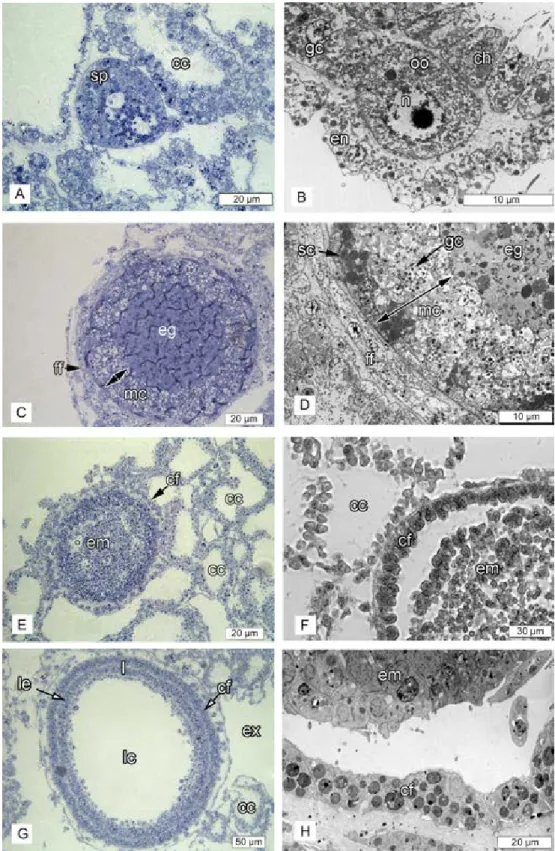
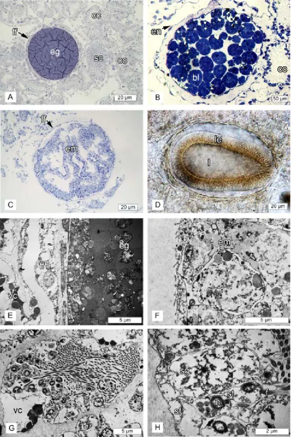
Reproduction. Oscarella pearsei sp. nov. is a viviparous and hermaphroditic species; male
and female reproductive elements are present in the same individuals. The spermatic cysts are oval to spherical in shape, with a diameter of about 35 µm and are randomly distributed in the sponge choanosome (Fig 6A). Spermatogenesis is generally asynchronous inside spermatic cysts.
Oogenesis is asynchronous: all stages from oogonia to egg were observed within the same sponge. Young oocytes have an ovoid or amoeboid shape and are situated between choanocyte chambers and endopinacoderm (Fig 6B). Mature eggs are about 70 µm in diameter, isolecithal and polylecithal, with a cytoplasm full of yolk granules (Fig 6C). Embryogenesis is also asyn-chronous leading to formation of typical cinctoblastula larvae (Fig 6E and 6G). All stages from cleaving embryos to larva were observed from mid-June to September. Eggs and embryos are located in the basal part of the choanosome and are completely surrounded by a follicle. From the stage of vitellogenesis of the oocyte to the embryos/larva stage the follicle transforms from flat monolayer composed of pinacocytes (oogenesis) (Fig 6C and 6D) to cubical one in embryos and larva (Fig 6E±6H). At the late stages of vitellogenesis the area between the follicle and oocyte contains a thick layer (about 20±25 µm) of maternal cells (granular and spherulous cells with paracrystalline inclusions) (Fig 6C and 6D;S3 Fig). Over the course of embryonic development this layer disappears (S3 Fig).
Habitat. Oscarella pearsei sp. nov. is found in Carmel, California (36Ê48' N 121Ê54' W)
in the low intertidal zone, on the underside of rocks. This is the only location where it has been collected in the field, and is found growing alongside Oscarella carmela. Otherwise, both spe-cies are found growing in public aquaria and at research facilities along the central California coast (UCSC Long Marine Lab and the Monterey Bay Aquarium) that maintain flow-through seawater tanks that are supplied with seawater pumped directly from the ocean.
Etymology. The specific name is given in honor of University of California Santa Cruz
Professor Emeritus John Pearse for his support while describing this sponge.
Redescription of Oscarella carmela Muricy, Pearse, 2004
Type material. Holotype: MNRJ 8033 (slide B-6618 #2), Carmel Point, Carmel Bay,
inter-tidal, Collected by J. Pearse, 08. 05. 2003 [1].
Paratypes: CASIZ 168925 (slide B-6617 #1), Carmel Point, Carmel Bay, central California, intertidal, Collected by J. Pearse. 08. 05. 2003 [1].
Syntypes: Oscarella carmela Muricy, Pearse, 2004. ZIN RAS № 11802, Ethanol (this study), sea tables at Long Marine Lab. Santa Cruz, California, USA, S. Collected by Scott Nichols
Fig 6. Oscarella pearsei sp. nov. reproduction. (A) semi-thin section of the spermatocyst; (B) TEM of
young oocyte before vitellogenesis;(C) semi-thin section of the egg with flat monolayer follicle and the thick
layer of maternal cells;(D) TEM of flat monolayer follicle of the egg and the thick layer of maternal cells; (E)
semi-thin section of the embryo with bilayer follicle composed of cubical cells;(F) histological section of
embryos with cubical follicle;(G) semi-thin section of the cinctoblastula larva; (H) TEM of the embryo with
01.08.2015. Oscarella carmela Muricy, Pearse, 2004. ZIN RAS № 11803, Ethanol (this study), sea tables at Long Marine Lab. Santa Cruz, California, USA, S. Collected by Scott Nichols 01.08.2015. Oscarella carmela Muricy, Pearse, 2004. ZIN RAS № 11804, Glutaraldehyde (this study), sea tables at Long Marine Lab. Santa Cruz, California, USA, S. Collected by Scott Nichols 01.08.2015. Oscarella carmela Muricy, Pearse, 2004. ZIN RAS № 11805, Glutaraldehyde (this study), sea tables at Long Marine Lab. Santa Cruz, California, USA, S. Collected by Scott Nichols 01.08.2015. Oscarella carmela Muricy, Pearse, 2004. RBINS-IG 33439-POR.0125, Ethanol (this study), sea tables at Long Marine Lab. Santa Cruz, California, USA, S. Collected by Scott Nichols 01.08.2015. Oscarella carmela Muricy, Pearse, 2004. RBINS-IG 33439-POR.0126, Ethanol (this study), sea tables at Long Marine Lab. Santa Cruz, California, USA, S. Collected by Scott Nichols 01.08.2015. Oscarella carmela Muricy, Pearse, 2004. RBINS-IG 33439-POR.0127, Glutaralde-hyde (this study), sea tables at Long Marine Lab. Santa Cruz, California, USA, S. Collected by Scott Nichols 01.08.2015. Oscarella carmela Muricy, Pearse, 2004, RBINS-IG 33439-POR.0128, Glutaraldehyde (this study), sea tables at Long Marine Lab. Santa Cruz, California, USA, S. Col-lected by Scott Nichols 01.08.2015.
Comparative material examined. Paratype B-6616 as Oscarella carmela specimen from
an aquarium in J. Long Marine Laboratory. After examination it was find that this is Oscarella
pearsei sp.nov.
Oscarella pearsei Ereskovsky, Richter, Lavrov, Schippers, Nichols (this study). ZIN RAS №
11798, Ethanol. urn:lsid:zoobank.org:act:8431B41C-B08E-4205-8FE6-CCCFFB6EB574, sea tables at Long Marine Lab. Santa Cruz, California, USA, Collected by Scott Nichols 01.08.2015; ZIN RAS № 11800, Holotype, Glutaraldehyde. urn:lsid:zoobank.org:act:8431B41C-B08E-4205-8FE6-CCCFFB6EB574, sea tables at Long Marine Lab. Santa Cruz, California, USA, Collected by Scott Nichols 01.08.2015; ZIN RAS № 11799, paratype, Ethanol, sea tables at Long Marine Lab. Santa Cruz, California, USA, Collected by Scott Nichols 01.08.2015; ZIN RAS № 11801, paratype, Glutaraldehyde, sea tables at Long Marine Lab. Santa Cruz, California, USA, Collected by Scott Nichols 01.08.2015; RBINS-IG 33439-POR.0122, paratype, Ethanol, sea tables at Long Marine Lab. Santa Cruz, California, USA, Collected by Scott Nichols 01.08.2015; RBINS-IG 33439-POR.0123, paratype, Glutaraldehyde, sea tables at Long Marine Lab. Santa Cruz, Califor-nia, USA, Collected by Scott Nichols 01.08.2015; RBINS-IG 33439-POR.0124 paratype, Glutar-aldehyde, Ethanol, sea tables at Long Marine Lab. Santa Cruz, California, USA, Collected by Scott Nichols 01.08.2015. Oscarella malakhovi Ereskovsky, 2006 (ZIN RAS 10697 ZIN RAS 10698: Japan Sea) [28]. Oscarella kamchatkensis Ereskovsky, Sanamyan & Vishnyakov, 2009 (ZIN RAS 11058, ZIN RAS 11059 and ZIN RAS 11060: North Pacific, Avacha Gulf) [29].
Oscar-ella lobularis (Schmidt, 1862) and OscarOscar-ella tuberculata (Schmidt, 1868). NE Mediterranean
Sea (Marseille region), underwater cave of Maire Island. Oscarella microlobata Muricy, Boury-Esnault, BeÂzac, Vacelet, 1996 and Oscarella viridis Muricy, Boury-Boury-Esnault, BeÂzac, Vacelet, 1996. NE Mediterranean Sea (Marseille region), underwater cave of Jarre Island [30]. Oscarella
baliba-loi, PeÂrez, IvanisÏević, Dubois, Pedel, Thomas, Tokina, Ereskovsky, 2011. NE Mediterranean Sea
(Marseille region), underwater cave of Maire Island [32]. Oscarella nicolae Gazave, Lavrov, Cab-rol, Renard, Rocher, Vacelet, Adamska, Borchiellini, Ereskovsky, 2013 (MNHN DJV155, LSID urn:lsid:zoobank.org:act:DFFAD94B-9CAD-4F99-994D-BDEF75EF98A1, MNHN DJV156) North Sea, Norway (Bergen Fjords, Skarvoysundet) [25]. Oscarella bergenensis Gazave, Lavrov, Cabrol, Renard, Rocher, Vacelet, Adamska, Borchiellini, Ereskovsky, 2013 (MNHN DJV153,
cubic follicle, ch±choanocyte, eg±egg, em±embryo, en±endopinacocyte, ex±exhalant canal, ff±flat follicle, gc±granular cells; l±larva, lc±larval cavity, le±larval ciliated epithelium, mc±maternal cells; n±nucleus, oo-oocyte, sc±spherulous cells with paracrystalline inclusions, sp±spermatocyst).
LSID:urn:lsid:zoobank.org:act:2D44BCFA-2163-47C7-9E70-EF6C13E0E4A4, MNHN DJV154) North Sea, Norway (Bergen Fjords, Skarvoysundet) [25].
Diagnosis (corrected from [1]). Known from the intertidal and subtidal, in and around
Monterey Bay, California. Brown in color with bumpy, microlobate surface, wavy appearance, soft, slimy consistency. Mesohylar cells of two types with different granular inclusions, and vacuolar cells that also contain granular inclusions. Nucleus in choanocytes has basal position. Embryos follicle consists of one layer of flat cells. It has two morphotypes of endobiotic bacteria.
Description (corrected from [1]). Thinly encrusting, irregular sponges, generally light
brown in color, up to 20±30 cm in diameter, with extremely soft, slimy consistency (Fig 1B and 1C). Thickness, color, and surface are variable. Most specimens <5 mm thick but some-times ranging to ~8mm. The surface is lumpy or wavy in appearance, microlobate with con-spicuous channels and oscula. The sponge is easy to peel off smooth surfaces, as it attaches only at the tips of outgrowths that extend from the basopinacoderm.
Soft tissue organization. Spicule and fiber skeleton absent. Ectosome is 10 to 20 µm
thick. Inhalant canals run perpendicular to the surface. Choanocyte chambers ovoid to spheri-cal, eurypilous, from 45 to 65 µm in diameter, organized around large exhalant canals. Exhal-ant canals run towards a well-developed system of large basal cavities, separated by septa without choanocyte chambers.
Cytology. Exopinacocytes (Fig 7A) flagellated with flat to oval shape 9.5 to 14.3 µm wide by 2.8 µm high (N = 10). Cytoplasm includes electron translucent vacuoles 0.2±0.5 µm in diameter. Nucleus about 2.2 µm in diameter and basal in position. External surface of exopina-cocytes covered by a layer of glycocalyx.
Endopinacocytes (Fig 7B) flagellated, from 8.3 to 13.7 µm wide by 2.9 µm high (N = 10). Nucleus ovoid (1.8 x 3.1 µm in diameter). Cytoplasm includes electron dense granules 0.2± 0.8 µm in diameter.
Choanocytes (Fig 7C) ovoid to trapezoidal, about 4.4 µm wide by 8.2 µm high (N = 20). Nuclei (~2.1 µm diameter) have basal position without any contact with flagellar basal appara-tus. Cytoplasm includes electron transparent vacuoles from 0.5 to 1.9 µm in diameter.
Apopylar cells (Fig 7D) roughly triangular or oval in section, 6.3 µm wide by 4.7 µm high (N = 5). Nucleus spherical, up to 2.9 µm in diameter. Cytoplasm contains mitochondria, diges-tive vacuoles, and small osmiophilic inclusions.
Surface of endopinacocytes, choanocytes and apopylar cells covered by thin irregular layer of glycocalyx. Choanoderm and pinacoderm underlined by continuous basement membrane 0.9±1.7 µm thick.
Three types of cells with inclusions occur within the mesohyl. Granular cells type 1 (Fig 7E) (ªtype 1 cell with inclusionsº described by [1]) are ovoid, 4.5 × 7 µm, irregular, with short pseu-dopodia. Nucleus about 1.9 µm in diameter, ovoid or compressed by the abundant cytoplasmic inclusions. Cytoplasm filled with oval inclusions of one kind, 0.8±1.9 µm wide, with electron dense and homogeneous contents.
Granular cells type 2 (Fig 7F and 7G) (ªarcheocytesº, described by [1]) are irregular to ovoid 5.5 × 7.9 µm, sometimes with short pseudopodia. Cytoplasm filled with abundant spher-ical inclusions with filamentous contents 0.7 µm in diameter and with some phagosomes 0.9 to 1.7 µm in diameter. It appears that granules fuse with each other, resulting in the formation of electron transparent vacuoles from 0.7 to 2.5 µm in diameter. Nucleus is ovoid or irregular, 2.4 µm and sometimes a nucleolus is evident.
Vacuolar cells with granular inclusions (Fig 7H) (type 2 cell with inclusions (ªspherulous vacuolar cellº) described by [1]) have irregular shape, 7.1 × 13.0 µm. Cytoplasm includes 0.8± 1.8 µm oval inclusions with homogenous osmiophilic contents, however central, or lateral part
of this inclusion are clear (light-gray). There are larger vacuoles from 2.1 to 7.1 µm in diameter, with clear and filamentous contents. Nucleus 1.6±1.9 µm in diameter without nucleolus, ovoid or compressed by abundant cytoplasmic inclusions. This cell type may be involved in the secretion of extracellular matrix, because at the end of the ontogeny of these cells, vacuoles appear to fuse into one or two large vacuoles, their membrane seems to merge with cell mem-brane and vacuolar content is presumably released in the mesohyl (Fig 7H). This appears to be the maturation sequence of granules (S4 Fig).
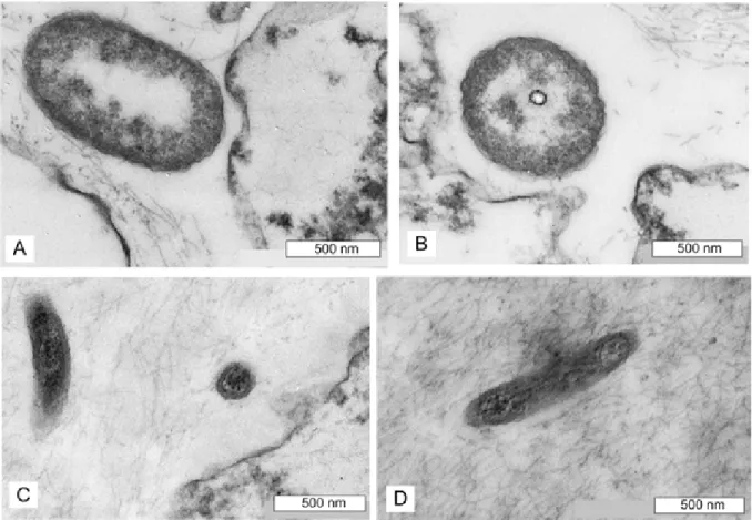
Two morphological types of extracellular, endobiotic bacteria occur in the mesohyl. Type B1 (Fig 8A and 8B) are abundant, ovoid 1.1–1.5 µm in length and 0.7 µm in diameter (N = 22). Cell wall consists of two membranes. Cytoplasm filled with loose filamentous materials con-centrated between the cell wall and central clear part (0.17 µm). Nucleoid is loose filamentous network and with thin filaments closer to the periphery. Surface is even or slightly wavy. Type B2 (Fig 8C and 8D) is rod-like, about 1.1 µm length and about 0.24 µm in diameter. Cell wall consists of two membranes. Cytoplasm filled with dark filamentous materials, and without a clear distinction between nuclear and cytoplasmic regions.
Fig 7. Oscarella carmela TEM of cells. (A) exopinacocyte; (B) endopinacocyte; (C) choanocytes; (D) apopylar cell; (E) granular cell type 1; (F) differentiated granular cell type 2, Inset: granules with filamentous inclusions; (G) granular
cell type 2 with small number of inclusions;(H) vacuolar cell with granular inclusions, InsetÐgr anule. (Abbreviations:
ap±apopylar cell, B1 ±bacteria type 1, en±endopinacocyte, ex±exopinacocyte, f±flagellum, g±granule, mv±microvilli, n± nucleus, v±vacuole).
https://doi.org/10.1371/journal.pone.0183002.g007
Fig 8. Oscarella carmela TEM of symbiotic bacteria. (A, B) associated bacteria type 1; (C, D) associated bacteria type 2.
Reproduction. Oscarella carmela is viviparous and simultaneously hermaphroditic: male
and female reproductive elements are present in the same individuals (Fig 9A). Oogenesis and embryogenesis are asynchronous; all stages from oogonia to egg were observed within the same specimen. Mature eggs are isolecithal and polylecithal, with a cytoplasm full of yolk gran-ules with diameter about 48±58 µm (Fig 9A). Embryogenesis is also asynchronous, resulting in formation of cinctoblastula larvae (Fig 9D). Eggs and embryos (about 80 µm in diameter) are located in the basal part of the choanosome and are surrounded by a follicle (Fig 9A±9D). Fol-licle is simple from oocyte vitellogenesis stage (Fig 9A and 9E) to embryos and larvae (Fig 9B± 9D and 9F) and consists of only one layer of flat cells. The spermatic cysts are oval and have different dimension (from 28±52 x 21±48 µm) and are randomly distributed in the sponge mesohyl (Fig 9G). Spermatogenesis is generally asynchronous inside spermatic cysts (Fig 9G and 9H).
Taxonomic remarks
The absence of spherulous cells with paracrystalline inclusions is consistent with the phyloge-netic placement of Oscarella carmela in the clade that includes O. lobularis, O. tuberculata, O.
viridis, O. rubra, O. malakhovi, O. bergenensis [1,25,28,30,32]. The slimy consistency and abundant mucus of Oscarella carmela resembles O. nicolae [25] and O. balibaloi [31] but differs significantly from O. pearsei sp. nov.
Discussion
Prior to this study there were 19 valid species of the genus Oscarella described, worldwide [33]. Only five of these were found in the Pacific: O. nigraviolacea, O. stillans [34] in the Southern Hemisphere and O. carmela [1], O. malakhovi [28] and O. kamtchakensis [35] in the Northern Hemisphere. Characteristics of Oscarella that are observable in the field, such as coloration, surface morphology and consistency are difficult to use for accurate species discrimination because they can vary within a single species. For example, in the Mediterranean, O. lobularis and O. tuberculata may appear in several different colors: white, yellow, green, red, violet, blue [32,35,36]. Likewise, O. balibaloi [31] varies in color from white to yellow, orange and violet. Characteristics such as tissue consistency and surface morphology are potentially less variable within each species, but they are also more subjective to describe.
The initial clue that the species Oscarella carmela may represent two distinct species was that the EST dataset published under the name O. carmela [2] was significantly divergent from the Illumina transcriptome that was later sequenced from a separate individual (this study). Here, we correct the attribution of the published ESTs and draft genome from O. carmela to O.
pearsei sp. nov., and we report correctly attributed Illumina-sequenced transcriptomes for
each of these species. The mitochondrial genome previously attributed to O. carmela [3] remains unchanged.
O. carmela and O. pearsei sp. nov. are co-distributed along the central California coast,
albeit their distributions are incompletely known. We sampled tissue from individuals of each species (verified both morphologically and by DNA barcoding), growing side by side on the same rock at the O. carmela type locality in Carmel, California. We also found O. carmela, but not O. pearsei sp. nov., in the shallow subtidal zone of Pacific Grove, California on the under-side of rocks. Both species can be found growing in sea tables at the Joseph Long Marine Labo-ratory, University of California Santa Cruz. Specimens of Oscarella have also been found growing in the intertidal in the Santa Barbara region to the south, and in Coos Bay Oregon to the north (Jeff Goddard, pers. comm.). The identity of these species is unknown, but we obtained a sample from Santa Barbara (gift from Desmond Ramirez to K.J.S.), performed
Fig 9. Oscarella carmela follicle and reproduction. (A) semi-thin section of the egg with flat monolayer follicle; (B) semi-thin section of cleaving embryo with flat monolayer follicle; (C) semi-thin section of the embryo with flat
DNA barcoding and found it to represent a third species, distinct from either O. carmela or O.
pearsei sp. nov. It is possible that multiple, undescribed species exist in the Eastern Pacific.
This situation resembles the case of Oscarella systematics in the Mediterranean, where a single species, O. lobularis, was initially recognized but eventually many co-distributed species were described on the basis of their cytological differences and later, using DNA evidence (see [37] [35]).
Oscarella pearsei sp. nov is readily distinguished from O. carmela using LSU and mt-cob
barcodes, and phylogenetic analyses of SSU, LSU and mitochondrial genomic data. Interest-ingly, these two species represent two major branches in the family Oscarellidae and are only distantly related to each other. Furthermore, O. pearsei sp. nov is currently the only species in the family with a distinct mitochondrial gene order, as a result of an inversion encompassing three protein-coding and four tRNA genes.
In addition to genetic evidence, careful re-analysis of the morphology of O. carmela and O.
pearsei sp. nov. also reveals characteristics useful for identification in the field and in the lab.
With respect to gross morphology, O. pearsei sp. nov. has a light tan to orange color while O.
carmela is almost exclusively light-brown. Additionally, the surface of O. pearsei sp. nov. is
finely and uniformly lobate with small hemispherical lobes that are unique for the genus. In contrast, the surface of O. carmela is lumpy or wavy in appearance and microlobate; similar to
O. microlobata, O. kamchatkensis, O. malakhovi. and O. balibaloi [28,30,31,35]. Both O.
car-mela and O. pearsei sp. nov. are soft and delicate to the touch, but O. carcar-mela is generally
thicker, the less delicate of the two, and produces much more mucus.
Because Oscarella lacks skeletal elements (inorganic and organic) the best morphological characters for reliable species discrimination are ultrastructural: each species has unique cell type combinations in the mesohyl, and unique and consistently associated bacterial morpho-types. Specifically, O. pearsei sp. nov. differs from all other described Oscarella species, includ-ing O. carmela (Table 3) in that it lacks vacuolar cells, but contains archeocytes, abundant granular cells of a single type, and spherulous cells with paracrystalline inclusions. In contrast,
O. carmela contains vacuolar cells, has two discrete types of granular cells and lacks both
archeocytes and spherulous cells with paracrystalline inclusions. The latter cell type is a
with flat monolayer follicle of the embryo;(G) spermatocyst; (H) detail of the spermatocyst with surrounding follicle
epithelium, spermatogonia and the spermatids. (Abbreviations: B1 ±bacteria type 1, B2 ±bacteria type 2, bl± blastomeres; ccÐchoan ocyte chamber, eg±egg, em±embryon, f±flagella, ff±flat follicle, l±larva, le±larval epithelium, n±nucleus, sfÐfollicle of a spermatocyst, sgÐsper matogonia, st±spermatid, vc±vacuolar cell with granular inclusions).
https://doi.org/10.1371/journal.pone.0183002.g009
Table 3. The cytological characters of Oscarellidae species with cells that contain paracrystalline inclusions.
Species Spherulous cells with
paracrystalline inclusions Granularcells Micro-granularcells Vacuolarcells Archeocytes Symbiotic bacteria(number)
Pseudocorticium
jarrei Yes Yes Yes No No 5 (HMA)
Oscarella
kamchatkensis Yes Yes Yes No No 3 (LMA)
O. imperialis Yes Yes No Yes No 3 (LMA)
O. microlobata Yes Yes No Yes No 4 (HMA)
O. balibaloi Yes Yes No Yes No 2 (LMA)
O. nicolae Yes Yes No No Yes 1 (LMA)
O. pearsei
sp. nov. Yes Yes No No Yes 1 (LMA)
HMA±high microbes abundance, LMA±low microbes abundance. https://doi.org/10.1371/journal.pone.0183002.t003
synapomorphy for the clade to which O. pearsei sp. nov. belongs, and unites this species with
O. microlobata, O. imperialis, O. kamchatkensis, O. balibaloi, O. nicolae and Pseudocorticium jarrei [25,30,31,35,38].
The granular cells of O. pearsei sp. nov. also differ from granular cells of other Oscarella spe-cies due to the presence of an unusually osmiophilic central region within vacuoles, which is surrounded by opaque (gray) peripheral content. In comparison, in O. balibaloi, granular cells have entire homogeneous, electron-dense inclusions with a crystalline texture that is visible under high magnification [31]. In O. nicolae and O. kamchatkensis granular cells, the granules are also entirely homogeneous and electron dense [25,29]. In O. microlobata granular cells (type I), the cytoplasm is almost completely filled with heterogeneous inclusions, containing 10±30 osmiophilic granules embedded in a dense matrix [30]. Finally, granular cells (type 1) of
O. imperialis are almost completely filled by 2±5 µm spherical vacuoles, and 5±20 µm partly
paracrystalline inclusions (i.e., the inclusions have a partly paracrystalline organization and are partly amorphous) [30].
The final cytological characteristic that is useful for distinguishing between O. carmela and
O. pearsei sp. nov. is the position of the nucleus in choanocytes. We find that choanocytes in O. pearsei sp. nov. invariable have an apically positioned nucleus (just below the microvillar
collar) whereas choanocytes in O. carmela have a basally positioned nucleus.
A limitation of using cytological characteristics for species discrimination is that it is labor intensive. For the most part, tissue has to be prepared for examination by transmission elec-tron microscopy. Fortunately, in reproductive individuals of O. carmela and O. pearsei sp. nov. it is possible to determine species affinity by a unique histological characteristic: the morphol-ogy of the embryonic follicle epithelium. Specifically, O. carmela has a flat monolayer follicle epithelium, whereas the follicle epithelium of O. pearsei sp. nov. is prominent and composed of cuboidal cells that persist from the stage of vitellogenesis to the stage of larval release. Although, this is not one of the characters used in the early taxonomic literature on Oscarella, we find that it reliably distinguishes between these species and that it is even visible in thin, liv-ing samples when viewed with phase microscopy. The presence of a cuboidal follicle epithe-lium has previously been described only from O. nicolae [25], which is found in the North sea, Norway, and cannot therefore be confused with O. pearsei sp. nov.
Based upon this study, we caution that previously published references to the species O.
car-mela were incorrect, and that the actual species under study was what we have now described
as O. pearsei sp. nov. The only exception to this is the published mitochondrial genome for O.
carmela and studies that reference those data. Henceforth, we have corrected the species
names associated with all publicly available genetic resources (i.e., ESTs, transcriptomes, genomes, and mitochondrial genomes), including in the NCBI databases, andcompagen.org [23]. Future studies that involve new collection of Oscarella tissue from the Central California coast (or elsewhere) should be diligent to keep samples individually separated and to save voucher material for DNA barcoding and possibly morphological analysis.
Supporting information
S1 Fig. TEM of an exopinacocyte (ex) of Oscarella pearsei sp. nov., showed basal surface with filopodia (arrowheads) that extend into the extracellular matrix (ECM). f±flagellum,
n±nucleus. (TIF)
S2 Fig. Oscarella pearsei sp. nov. TEM of granular cells at different stage of their ontogeny.
g±granule, n±nucleus, v±vacuole. (TIFF)
S3 Fig. Oscarella pearsei sp. nov. TEM of follicle surrounding mature oocyte. (A) Mature
oocyte with two-layer follicle. (B)Ð(D) details of (A) showing granular and spherulous cells, composed internal layer of the follicle. (E) the part of a mature oocyte with follicle. (E)Ð(H) details of (E) showing granular and spherulous cells, composed internal layer of the follicle and external flat cells of a follicle (F), (H). ch±choanocytes, en±endopinacocytes, F±follicle, flat.c±flat cells of follicle, gc±granular cell, on±oocyte's nucleus, oo±oocyte, nÐnucleus, sc-spherulous cells with paracrystalline inclusions, yg±yolk granules.
(TIFF)
S4 Fig. Oscarella carmela TEM of vacuolar cells with granular inclusions at different stages of their ontogeny. Arrowheads show the apparent release of vacuolar content in the mesohyl.
g±granule, v±vacuole. (TIFF)
S5 Fig. O. pearsei transcriptome. FASTA formatted transcriptome assembly.
(TXT)
S6 Fig. O. carmela transcriptome. FASTA formatted transcriptome assembly.
(TXT)
Acknowledgments
S.A.N. would like to acknowledge Nicole King at the University of California Berkeley, where some of this research was conducted, and Betsy Steele and John Pearse for facilitating access to Long Marine Laboratory and for help with field collections. Thanks to Guilherme R. S. Muricy from Museu Nacional Rio de Janeiro (MNRJ) for the opportunity to work with slides of the type material. D.V.L. thanks Walker Pett (Iowa State University) for his help with the analysis of genomic data. A.E. gratefully thanks JoeÈl Courageot and Alexandre AltieÂof Service Com-mun de Microscopie EÂlectronique et Photographie FaculteÂde MeÂdecine La Timone, Aix-Mar-seille UniversiteÂand Daria Tokina and Sandrine Chenesseau for technical support.
Author Contributions
Conceptualization: Scott A. Nichols.
Data curation: Alexander V. Ereskovsky, Daniel J. Richter, Dennis V. Lavrov, Scott A.
Nichols.
Formal analysis: Alexander V. Ereskovsky, Daniel J. Richter, Dennis V. Lavrov, Klaske J.
Schippers, Scott A. Nichols.
Investigation: Alexander V. Ereskovsky, Dennis V. Lavrov, Klaske J. Schippers, Scott A.
Nichols.
Project administration: Scott A. Nichols. Supervision: Scott A. Nichols.
Validation: Daniel J. Richter.
Visualization: Alexander V. Ereskovsky, Dennis V. Lavrov, Scott A. Nichols.
Writing ± original draft: Alexander V. Ereskovsky, Daniel J. Richter, Dennis V. Lavrov,
Writing ± review & editing: Alexander V. Ereskovsky, Daniel J. Richter, Dennis V. Lavrov,
Klaske J. Schippers, Scott A. Nichols.
References
1. Muricy G, Pearse JS. A new species of Oscarella (Demospongiae: Plakinidae) from California.
Pro-ceedings of the California Academy of Sciences. 2004; 55(33):598±612.
2. Nichols SA, Dirks W, Pearse JS, King N. Early evolution of animal cell signaling and adhesion genes.
Proc Natl Acad Sci U S A. 2006; 103(33):12451±6.https://doi.org/10.1073/pnas.0604065103PMID: 16891419; PubMed Central PMCID: PMCPMC1567900.
3. Wang X, Lavrov DV. Mitochondrial genome of the homoscleromorph Oscarella carmela (Porifera,
Demospongiae) reveals unexpected complexity in the common ancestor of sponges and other animals. Mol Biol Evol. 2007; 24(2):363±73.https://doi.org/10.1093/molbev/msl167PMID:17090697.
4. Nichols SA, Roberts BW, Richter DJ, Fairclough SR, King N. Origin of metazoan cadherin diversity and
the antiquity of the classical cadherin/beta-catenin complex. Proc Natl Acad Sci U S A. 2012; 109 (32):13046±51.https://doi.org/10.1073/pnas.1120685109PMID:22837400; PubMed Central PMCID: PMCPMC3420208.
5. Peña JF, AlieÂA, Richter DJ, Wang L, Funayama N, Nichols SA. Conserved expression of vertebrate
microvillar gene homologs in choanocytes of freshwater sponges. EvoDevo. 2016; 7(1).https://doi.org/ 10.1186/s13227-016-0050-xPMID:27413529
6. Yang X, Dorman KS, Aluru S. Reptile: representative tiling for short read error correction.
Bioinformat-ics. 2010; 26(20):2526±33.https://doi.org/10.1093/bioinformatics/btq468PMID:20834037.
7. Grabherr MG, Haas BJ, Yassour M, Levin JZ, Thompson DA, Amit I, et al. Full-length transcriptome
assembly from RNA-Seq data without a reference genome. Nat Biotechnol. 2011; 29(7):644±52. https://doi.org/10.1038/nbt.1883PMID:21572440; PubMed Central PMCID: PMCPMC3571712.
8. Lohse M, Bolger AM, Nagel A, Fernie AR, Lunn JE, Stitt M, et al. RobiNA: a user-friendly, integrated
software solution for RNA-Seq-based transcriptomics. Nucleic Acids Res. 2012; 40(Web Server issue): W622±7.https://doi.org/10.1093/nar/gks540PMID:22684630; PubMed Central PMCID:
PMCPMC3394330.
9. Li R, Zhu H, Ruan J, Qian W, Fang X, Shi Z, et al. De novo assembly of human genomes with massively
parallel short read sequencing. Genome Res. 2010; 20(2):265±72.https://doi.org/10.1101/gr.097261. 109PMID:20019144; PubMed Central PMCID: PMCPMC2813482.
10. Lowe TM, Eddy SR. tRNAscan-SE: A program for improved detection of transfer RNA genes in
geno-mic sequence. Nucleic Acids Research. 1997; 25(5):955±64.https://doi.org/10.1093/nar/25.5.955 PMID:9023104
11. Pearson WR. Using the FASTA Program to Search Protein and DNA Sequence Databases.
1994;Com-puter Analysis of Sequence Data. 241994. p. 307±31.
12. Dereeper A, Audic S, Claverie JM, Blanc G. BLAST-EXPLORER helps you building datasets for
phylo-genetic analysis. BMC Evol Biol. 2010; 10:8.https://doi.org/10.1186/1471-2148-10-8PMID:20067610; PubMed Central PMCID: PMCPMC2821324.
13. Castresana J. Selection of conserved blocks from multiple alignments for their use in phylogenetic
anal-ysis. Molecular Biology and Evolution. 2000; 17(4):540±52. PMID:10742046
14. Guindon S, Gascuel O. A simple, fast, and accurate algorithm to estimate large phylogenies by
maxi-mum likelihood. Syst Biol. 2003; 52(5):696±704.https://doi.org/10.1080/10635150390235520PMID: 14530136.
15. Anisimova M, Gascuel O. Approximate likelihood-ratio test for branches: A fast, accurate, and powerful
alternative. Syst Biol. 2006; 55(4):539±52.https://doi.org/10.1080/10635150600755453PMID: 16785212.
16. Chevenet F, Brun C, Banuls AL, Jacq B, Christen R. TreeDyn: towards dynamic graphics and
annota-tions for analyses of trees. BMC Bioinformatics. 2006; 7:439.https://doi.org/10.1186/1471-2105-7-439 PMID:17032440; PubMed Central PMCID: PMCPMC1615880.
17. Katoh K, Kano Y, Noda Y. Rho-associated kinase-dependent contraction of stress fibres and the
organi-zation of focal adhesions. J R Soc Interface. 2011; 8(56):305±11.https://doi.org/10.1098/rsif.2010. 0419PMID:20826475; PubMed Central PMCID: PMCPMC3030825.
18. Lartillot N, Rodrigue N, Stubbs D, Richer J. PhyloBayes MPI: phylogenetic reconstruction with infinite
mixtures of profiles in a parallel environment. Syst Biol. 2013; 62(4):611±5.https://doi.org/10.1093/ sysbio/syt022PMID:23564032.
19. Chombard C, Boury-Esnault N, Tillier S. Reassessment of Homology of Morphological Characters in