HAL Id: hal-01858775
https://hal.archives-ouvertes.fr/hal-01858775
Submitted on 22 Aug 2018
HAL is a multi-disciplinary open access archive for the deposit and dissemination of sci-entific research documents, whether they are pub-lished or not. The documents may come from teaching and research institutions in France or abroad, or from public or private research centers.
L’archive ouverte pluridisciplinaire HAL, est destinée au dépôt et à la diffusion de documents scientifiques de niveau recherche, publiés ou non, émanant des établissements d’enseignement et de recherche français ou étrangers, des laboratoires publics ou privés.
and cats, Algiers: A prospective study
Sara Zaidi, Amar Bouam, Amina Bessas, Djamila Hezil, Hicham Ghaoui,
Khatima Ait-Oudhia, Michel Drancourt, Idir Bitam
To cite this version:
Sara Zaidi, Amar Bouam, Amina Bessas, Djamila Hezil, Hicham Ghaoui, et al.. Urinary shedding of pathogenic Leptospira in stray dogs and cats, Algiers: A prospective study. PLoS ONE, Public Library of Science, 2018, 13 (5), pp.e0197068. �10.1371/journal.pone.0197068�. �hal-01858775�
Urinary shedding of pathogenic Leptospira in
stray dogs and cats, Algiers: A prospective
study
Sara Zaidi1, Amar Bouam2, Amina Bessas1, Djamila Hezil1, Hicham Ghaoui1,2, Khatima Ait-Oudhia1, Michel Drancourt2*, Idir Bitam3,4
1 Ecole Nationale Supe´rieure Ve´te´rinaire, Alger, Alge´rie, 2 Aix Marseille Univ, IRD, MEPHI, IHU
Me´diterrane´e Infection, Marseille, France, 3 Ecole Supe´rieure en Sciences de l’Aliment et des Industries Agroalimentaires (ESSAIA), El Harrach, Alger, Alge´rie, 4 Aix Marseille Univ, IRD, VITROME, IHU Me´diterrane´e Infection, Marseille, France
*michel.drancourt@univ-amu.fr
Abstract
Background
Leptospirosis is an important worldwide zoonosis. This disease is caused by pathogenic species of the genus Leptospira which are maintained in the environment via chronic renal infection of carrier animals which can be asymptomatic excretors of the organisms in their urines and become a source of infection for humans and other hosts. The prevalence of ani-mal leptospirosis in Algiers, Algeria, is unknown.
Methodology/principal findings
Real-time PCR and standard PCR and sequencing were used to detect pathogenic
Leptos-pira organisms in the urines of stray dogs and cats in Algiers. In the presence of appropriate
controls, none of the 107 cat urine samples were positive while 5/104 (4.8%) canine urine samples (asymptomatic mixed-breed dogs, three females and two males) were positive in two real-time PCR assays targeting the rrs and hsp genes. The positivity of these samples was confirmed by partial PCR-sequencing of the rpoB gene which yielded 100% sequence similarity with Leptospira interrogans reference sequence. In this study, L. interrogans prev-alence was significantly higher in dogs aged<one year (16.46% - 29.41%) than in adults (0%) (P value = 0.0001) and then in the overall dog population (2.68% - 4.8%) (P = 0.0007).
Conclusions/significance
These results suggest that dogs are maintenance hosts for zoonotic leptospirosis in Algiers, Algeria. To face this situation, effective canine vaccination strategies and raising public health awareness are mandatory. Further investigations incorporating a larger sample in more localities will be undertaken to document the epidemiology of urban animal leptospiro-sis in Algeria at large.
a1111111111 a1111111111 a1111111111 a1111111111 a1111111111 OPEN ACCESS
Citation: Zaidi S, Bouam A, Bessas A, Hezil D,
Ghaoui H, Ait-Oudhia K, et al. (2018) Urinary shedding of pathogenic Leptospira in stray dogs and cats, Algiers: A prospective study. PLoS ONE 13(5): e0197068.https://doi.org/10.1371/journal. pone.0197068
Editor: Brian Stevenson, University of Kentucky
College of Medicine, UNITED STATES
Received: February 12, 2018 Accepted: April 25, 2018 Published: May 16, 2018
Copyright:© 2018 Zaidi et al. This is an open access article distributed under the terms of the
Creative Commons Attribution License, which permits unrestricted use, distribution, and reproduction in any medium, provided the original author and source are credited.
Data Availability Statement: All relevant data are
within the paper.
Funding: AB benefits from a PhD grant from the
Fondation Me´diterrane´e Infection, Marseille, France. This work was supported by the French Government under the « Investissements d’avenir » (Investments for the Future) program managed by the Agence Nationale de la Recherche (ANR, fr: National Agency for Research), (reference: Me´diterrane´e Infection 10-IAHU-03). The funders had no role in study design, data collection and
Introduction
Leptospirosis is a worldwide disease that affects wild and domestic animals and human popu-lations. Affected persons are primarily farmers, fishermen, veterinarians and people working in sewers and slaughterhouses [1]. This zoonosis is caused by pathogenic spirochetes of the genusLeptospira which colonize the renal tubules where they reproduce before being excreted
via the urines [2]. Infected urines or contaminated water are sources of leptospirosis infection andLeptospira can enter the body of mammalian hosts via lacerations in the skin, contacts
with mucosa or conjunctiva and inhalation of aerosols [3–5]. Some host animals such as dogs may have an asymptomatic form or may suffer from a wide range of clinical manifestations, including hepatic and renal failure and severe pulmonary hemorrhage [6]. Asymptomatic and chronic carrier dogs can be maintenance hosts [7] acting as sources of infection and therefore cause a public health problem [8]. Formerly, it was thought that domestic cats were resistant to leptospirosis infection and many practitioners did not consider feline leptospirosis in the dif-ferential diagnosis of other diseases [9]. However, recently published reports on feline leptospi-rosis conclude that cats are exposed toLeptospira and may play a role in the epidemiology of
this disease [10–12].
As a neglected tropical disease, leptospirosis has been increasingly observed in urban settle-ments, especially in slums in developing countries [6]. The prevalence of animal leptospirosis in Algiers, Algeria, is unknown. Only two studies were published about human leptospirosis in Algeria. These two serological investigations were conducted on patients of the Tizi-ouzou Hospital. The first one reported 48 cases of leptospirosis from 2006 to 2007 and the serogroup icterohaemorrhagiae was identified in 60% of cases [13]. In the second prospective study, 175 positive patients were diagnosed from 2005 to 2008, among the serovars identified, icterohae-morrhagiae and grippotyphosa were predominant [14].
The aim of the present work was to detect pathogenicLeptospira organisms in the urines of
stray dogs and cats in Algiers.
Methods
Ethic statement
The study was submitted to and approved by the ethics committee and decision board (num-ber 416/2017) of EPIC- H.U.P.E (EPIC: Entreprise publiqueà caractère industriel et commer-cial; H.U.P.E: Hygiène Urbaine et Protection de l’environnement) of Wilaya of Algiers (Ex: HURBAL). HURBAL was created in 1994 with a new status: EPIC-H.U.P.E under the register number: 16/00-0013132B00. EPIC- H.U.P.E is an institution affiliated with the Algerian Min-istry of the Interior and the Local Government and the Algerian MinMin-istry of Water Resources and Environment. In the context of the National Program for Rabies Control, EPIC- H.U.P.E captures stray dogs and cats in Algiers. Once captured, stray animals were housed in cages and euthanized after expiration of the seven day legal waiting time (in order to allow for owners to claim their pets), in compliance with the Algerian legislation for the protection of animals (Law 01/04/1994), which our protocol respected.
Study design and sampling
This study was designed to screen for the presence ofLeptospira spp. organisms in stray cats
and dogs in the region of Algiers, in the absence of any data available on that topic. Therefore, in this study, we aimed at collecting only the urines of the animals for the molecular detection ofLeptospira spp. DNA. Urine specimens were aseptically collected between April 2017 and
November 2017 were via cystocentesis from 211 stray animals (104 dogs and 107 cats). These analysis, decision to publish, or preparation of the
manuscript.
Competing interests: The authors have declared
animals were captured in the 57 municipalities of the region of Algiers. The sampling was real-ized in animal shelters with an average of seven animals sampled per week. The age of each animal was estimated, based on dentition and physical aspect. Information concerning sex, breed and clinical status was noted. Samples were stored at -20˚C before being transported to the IHU Me´diterrane´e Infection, Marseille, France, for PCR testing and culture was not per-formed. Up to 3 mL of each urine sample was centrifuged at 15,000 g for 20 minutes [15], the supernatant was discarded and the pellet was suspended in 200μL of sterile phosphate-buff-ered saline solution (PBS, pH 7.2) [16].
DNA extraction
A total of 200μL of DNA was extracted using the QIAamp Tissue Kit by QUIAGEN-BioRobot EZ1, according to the manufacturer’s instructions (Qiagen, Hilden, Germany). Extracted DNA was stored at -20˚C under sterile conditions until used in PCR assays.
Real time PCR
Extracted DNA was used in qPCR amplifications to detect pathogenicLeptospira organisms.
The final qPCR reaction mixture consisted of 5μL DNA with 15 μL of mix from the Roche PCR Kit (Roche Diagnostics, Meylan, France). The components of the final reaction mixture of these PCR assays are given inTable 1. A homemade plasmid containing sequences specific toLeptospira spp. was used as a positive control. Three negative controls were incorporated
into each PCR run. Results were recorded as positive when the cycle threshold (Ct) was lower than 33. We performed real-time PCR (qPCR) with two systems (Table 2) in order to confirm the positivity of the samples according to current standards in microbiology. The first system targets a 88-pb fragment of therrs gene coding for 16S rRNA of pathogenic Leptospira: 16S
rRNA Forward (5’-CCCGCGTCCGATTAG-3’), 16S rRNA Reverse (5’-TCCATTGTGGCCG RACAC-3’) and 16S rRNA Probe (5’-CTCACCAAGGCGACGTCGGTAGC-3’) were analyzed as previously described [17]. The second system targets a 103-pb fragment of thehsp gene of L. interrogans: Lint_hsp_MB Forward (5’-CCCGCGTCCGATTAG-3’), Lint_hsp_MB Reverse
(5’-TCCATTGTGGCCGRACAC-3’) and Lint_hsp_MB Probe (5’-CTCACCAAGGCGACG TCGGTAGC-3’) were analyzed as previously described [18]. The PCR cycling parameters for the qPCR were 5 min at 95˚C followed by 39 cycles each consisting of 5 sec of denaturation at 95˚C and 30 sec of annealing at 60˚C.
Standard PCR and sequencing
Samples that tested positive by qPCR were confirmed by standard PCR and sequencing in order to achieve a 100% specificity. The final standard PCR reaction mixture consisted of 5μL of DNA with 15μL of mix from the Roche PCR Kit (Roche Diagnostics). The components of
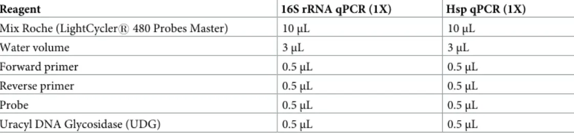
Table 1. Concentration of components in the final reaction mixtures of the real-time polymerase chain reaction (qPCR) assays used in this study.
Reagent 16S rRNA qPCR (1X) Hsp qPCR (1X)
Mix Roche (LightCycler1 480 Probes Master) 10μL 10μL
Water volume 3μL 3μL
Forward primer 0.5μL 0.5μL
Reverse primer 0.5μL 0.5μL
Probe 0.5μL 0.5μL
Uracyl DNA Glycosidase (UDG) 0.5μL 0.5μL
the final reaction mixture of these PCR assays are given inTable 3. Samples were then con-firmed by standard PCR using primers which amplified a 592-pb fragment of therpoB gene:
Lept 1900 Forward CCTCATGGGTTCCAACATGCA-3’) and Lept 2500 Reverse (5’-CGCATCCTCRAAGTTGTAWCCTT-3’), as described by La Scola et al., 2006 [19] (Table 2). The PCR cycling parameters for the standard PCR were 15 min at 95˚C followed by 35 cycles of each consisting of 30 sec denaturation at 95˚C, 30 sec annealing at 51˚C and 6 min exten-sion at 72˚C in an ABI Thermocycler (Applied Biosystems Gene Amp PCR System 2700, Ville-bon sur Yvette, France). Negative controls were incorporated into each PCR run.
The amplified PCR products were separated via gel electrophoresis using a 1.5% agarose gel stained with Sayber Safe (ThermoFisher, Paris, France). The DNA bands were visualized and photographed under ultraviolet light. PCR products were purified and sequenced withrpoB
primers as described previously [19]. All obtained sequences were assembled and edited using ChromasPro (version 1.7.7). The sequences were then analyzed by Basic Local Alignment Search Tool (BLAST) and compared with sequences available in the GenBank database.
Statistical analyses
Statistical analyses were done by MEDCALC1 online softwarehttps://www.medcalc.org/calc/ comparison_of_proportions.phpusing the “N-1” Chi-squared test as recommended by Camp-bell., 2007 [20] and Richardson., 2011 [21]. The confidence interval was calculated according to the recommended method given by Altman et al., 2000 [22].
Results
Sample collection
From April 2017 to November 2017, a total of 104 stray dogs and 107 stray cats captured in Algiers, Algeria were sampled. These animals lived in urban areas, spending most of their time exclusively outdoors and did not receive any vaccine. Of the 104 dogs, 69/104 (66.34%) were males and 35/104 (33.65%) were females. The canine population consisted predominantly of mixed-breed dogs; other dogs belonged to the following races: German shepherd, American
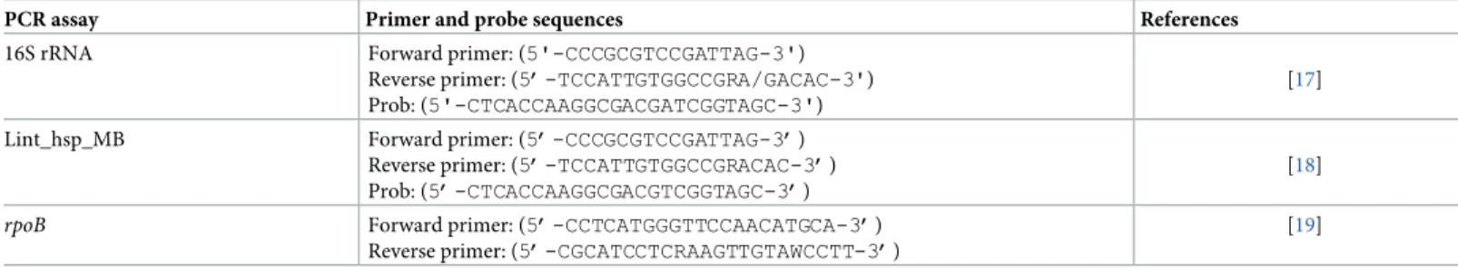
Table 2. Primers and probes used in this study.
PCR assay Primer and probe sequences References
16S rRNA Forward primer: (5'-CCCGCGTCCGATTAG-3') Reverse primer: (5’-TCCATTGTGGCCGRA/GACAC-3') Prob: (5'-CTCACCAAGGCGACGATCGGTAGC-3')
[17] Lint_hsp_MB Forward primer: (5’-CCCGCGTCCGATTAG-3’)
Reverse primer: (5’-TCCATTGTGGCCGRACAC-3’) Prob: (5’-CTCACCAAGGCGACGTCGGTAGC-3’)
[18]
rpoB Forward primer: (5’-CCTCATGGGTTCCAACATGCA-3’) Reverse primer: (5’-CGCATCCTCRAAGTTGTAWCCTT-3’)
[19]
https://doi.org/10.1371/journal.pone.0197068.t002
Table 3. Concentration of components in the final reaction mixtures of the standard polymerase chain reaction assay used in this study.
Reagent rpoB Standard PCR (1X)
Ampli Taq Master Mix 12.5μL
Water volume 6μL
Forward primer 0.75μL
Reverse primer 0.75μL
Staffordshire, shepherd crosses and Pit-bull. The dogs’ age ranged between 2 months and 11 years. Among the 107 cats, 66/107 (61.68%) were males and 41/107 (38.31%) were females. The cats were described as mostly belonging to mixed breeds, some belonging to European or Siamese crossbreeds. The 107 cats sampled were estimated to be under 5 years of age. All sam-pled animals were apparently healthy.
Real time PCR
qPCR targeting the 16S rRNA gene of pathogenicLeptospira and the hsp gene of L. interrogans
revealed that none of the 107 urine samples of cats tested were positive while 5/104 (4.8%) dogs were positive. These five urine specimens were positive in the two qPCR systems (rrs and hsp). Using the Cts obtained from the 16S rRNA qPCR reactions and a calibration curve previ-ously described for this system [23], we extrapolated the number of leptospira genomes per positive reaction (Table 4). Positive dogs were all very young, under one year of age. Three were females and two were males. All positive animals belonged to mixed-breeds (Table 4). In this study,L. interrogans prevalence was significantly higher in dogs aged < one year (5/17;
29.41%) than in adults (0/87; 0%) (P value = 0.0001, 95% CI: 12.73 to 53.13) and than in the overall dog population (5/104; 4.8%) (P = 0.0007, CI: 7.1929 to 48.2975). The sensitivity of our screening test based on the detection of 16S rRNA using qPCR was previously estimated to be of 56% [24], accordingly, the prevalence rate obtained here was estimated to be of 2.68%-4.8%. There was no significant difference regarding prevalence between males (3/69, 2.43% - 4.34%) and females (2/35, 3.19% - 5.71%) (p = 0.758).
Standard PCR and sequencing
All five urine samples detected positive by real-time PCR were confirmed with gel-based PCR assay targeting therpoB and subjected to sequencing analysis. The BLAST (www.ncbi.nlm.nih. gov/blast) analysis of therpoB gene sequence from all samples, once compared with sequences
available in the GenBank database, confirmedLeptospira infection. The BLAST analysis
yielded a 100% sequence homology withL. interrogans homologous gene fragment (GenBank
accession no.CP020414.1).
Discussion
In Algeria, the exact morbidity and the mortality due to leptospirosis are unknown. In 1975, a study based on serology reported seven cases in a military group in Algiers’ suburbs [25]. Two more recent studies reported cases of leptospirosis among hospitalized patients in the region of Tizi-Ouzou, located 100 km east of the capital Algiers. The investigation of 48 patients from 2006 to 2007 in the rural area of Tala-Athmane revealed that they were living in close contact
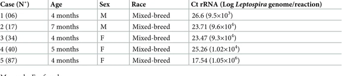
Table 4. Information relative to animals detected positive forL. interrogans DNA in urine samples: Age, sex, race,
number of genomes per positive pPCR reaction. (The values were extrapolated from the calibration curve (21) using
the Ct obtained from the 16S rRNA system).
Case (N˚) Age Sex Race Ct rRNA (LogLeptospira genome/reaction) 1 (06) 4 months M Mixed-breed 26.6 (9.5×10³) 2 (17) 7 months M Mixed-breed 23.71 (9.6×10⁴) 3 (34) 4 months F Mixed-breed 23.47 (9.3×10⁴) 4 (40) 5 months F Mixed-breed 25.26 (1.02×10⁴) 5 (87) 4 months F Mixed-breed 17.54 (1.05×10⁶) M = male; F = female. https://doi.org/10.1371/journal.pone.0197068.t004
to two garbage dumps invaded by rodents, the cases were confirmed serologically by the microagglutination test (MAT) and more than 60% (n = 29) were from the serogroup ictero-haemorrhagiae [13]. A second prospective study conducted from 2005 to 2008 in the same region reported 173 cases among hospitalized patients, the cases were confirmed serologically with a predominance the of serovars icterohaemorrhagiae and grippotyphosa [14].
However, the prevalence of this zoonosis in reservoirs is totally unknown in Algeria as the only report of it is the observation ofLeptospira organisms in histological sections of the liver
in dogs presenting with severe jaundice, subcutaneous hemorrhages and acute nephritis [26]. For this pioneering study of animal leptospirosis in Algeria, we used urines in which lepto-spiral DNA can be found much longer than in blood [27–29]. The need for a rapid diagnosis of leptospirosis has led to the development of numerous PCR assays, which appeared to have more applicability in determining zoonotic risks [30]. This method is rapid, sensitive, specific and robust and many PCR assays were developed to detect universalLeptospira genes such as gyrB, rrs and secY genes or genes restricted to pathogenic species such as lipL32, lfb1, ligA and ligB2 [5]. In this study we aimed at retrospectively confirming the positive detection of Leptos-pira spp. DNA by targeting two different molecular targets. We chose the rrs and hsp as
genus-level targets of identification andrpoB sequencing for the identification at the species level of
pathogenic leptospira.
We confirmed the presence ofL. interrogans in the urines of stray dogs in Algiers using
qPCR and standard PCR-sequencing targeting universal and pathogen-related genes. We observed an overall prevalence of 2.68% - 4.8% and a high prevalence of 29.41% in the specific population of young dogs aged < one year. All animals were apparently healthy, indicating asymptomatic carriers. These dogs were always outside, in contact with garbage and small rodents which were likely sources of infection. Crowding animals in unsanitary quarters is associated with a high prevalence of infection since animals may acquire the disease through contact with urines from infected dogs or infected rodents [31]. Despite the fact that we did not attempt to isolateLeptospira spp. to confirm the role of stray dogs as reservoirs, present
data indicate that stray dogs would indeed be good sentinels to know which serovars/groups are circulating in the rodent populations.
Subclinical, latent leptospirosis in dogs has regularly been reported and can also be observed in unsteady vaccinated animals [32]. In addition, there are data suggesting that clinically asymp-tomatic dogs can be chronic carriers, sheddingLeptospira via urines into the environment [30, 33,34]. Many epidemiological studies were conducted worldwide on the renal carriage of lepto-spirosis in dogs using molecular tools [8,14,34–43], showing a prevalence between 0.2% and 22% worldwideFig 1). Some other studies have not found the presence of pathogenicLeptospira
species in dogs in the USA (0/100) and in Egypt (0/25), but only the presence of antibodies as an evidence of the exposure to the disease [44] [45]. This may be due to the minute amount of Lep-tospira DNA in the blood and urine specimens of infected dogs. Therefore, a highly sensitive
PCR platform is required to obtain an accurate diagnosis and an innovative approach must be adopted to maximize sample DNA input in the PCR or by increasing the volume of urine for DNA extraction. This could be achieved by high-speed sedimentation of a milliliter volume of urines and performing DNA extraction from the complete sediment, as performed in the pres-ent study [46]. Positive dogs in our study were very young dogs (under one year of age). How-ever, it has been shown in the USA that dogs aged between 4 and 6.9 years and between 7 and 10 years faced a significantly higher risk of being infected than dogs under one year of age [47]. In a study conducted in Reunion Island, only adult dogs were positive for leptospirosis [48]. The dif-ference in age range can be explained by the fact that the young stray dogs of our study were not vaccinated against leptospirosis and exposed at an early age to the bacterium in their
environment. Furthermore, the canine population of two previous studies [47,48] was com-posed of domestic dogs with different risk factors and exposure to other maintenance host species.
In the present work, all sampled cats were qPCR-negative forL. interrogans. Some studies
yielded a seroprevalence between 4% and 30% in different countries including Australia [49], Scotland [50], Greece [51], Iran [52], Spain [53], Canada [54], Taiwan [11], Chile [10], Colombia [55] and Brasil [56]. Some other studies found DNA sequences ofLeptospira in urine samples
indicating a renal carriage of leptospirosis in cats. In Taiwan, DNA of pathogenicLeptospira was
detected in 67.8% (80/118) of the urine samples of cats including 71 stray cats and nine house-hold cats [11]. In Canada, DNA ofLeptospira was detected in the urines of sick cats (6/113) and
healthy cats (2/125), corresponding to a prevalence of 8/238 (3.3%) [12]. In Germany, urine sam-ples from 7/215 (3.2%) cats were PCR-positive [57]. In Quebec, PCRs on urines detected urinary excretion in 3.2% of the 250 cats sampled [58]. These results suggest that cats may have a role in the transmission of leptospirosis, as a reservoir or as an accidental host. The role of cats in the transmission of leptospirosis should be reevaluated, as it might in fact be underestimated.
In conclusion, this study demonstrates the high number of leptospiral carriers among asymp-tomatic young dogs. Improving the awareness of dog owners and the prevention of canine lepto-spirosis could be a valuable asset for human leptolepto-spirosis prevention [59]. The prevalence of leptospirosis in countries where dogs are correctly vaccinated remains high, this is mainly due to the difference in epidemiology between the different serovars and the absence of cross-protection in a vaccine, thus, a more effective vaccine needs to be developed [60]. The implementation of a
Fig 1. Molecular detection ofLeptospira spp. DNA in the urines of dogs, worldwide. Germany, 3/200 (1.5%) [8], the USA, 41/500 (8.2%) [14], Teheran (Iran), 33/150 (22%) [34], Canada, 11% (11/100) [35], Sicily (Italy), 5/64 (7.8%) [36], Ireland, 37/525 (7.05%) [37], Porto Alegre City (Brazil), 36/253 (14.2%) [38], Switzerland, 1/20 (5%) [39], Colombia 2/54 (3.7%)[40], New Caledonia, 1/13 (7.6%) [41], Brazil, 26/131 (19.8%) [42], Switzerland, 1/408 (0.2%) [43], Algiers (Algeria), 5/104 (2.68% - 4.8%) [Present work].
surveillance system for canine leptospirosis, using dogs as sentinels for human risk assessment, could also provide a valuable tool for estimating and in turn minimize the risk for humans [61]. Further studies on leptospirosis in other animals and other regions in Algeria should be consid-ered to clarify the status of this disease in our country. Also, further studies in the region of Algiers and other regions of Algeria will have to determine the serotypes of circulating leptospira in order to refine the epidemiology of leptospirosis in Algeria.
Acknowledgments
The authors would like to thank Mr. Schiff Lyès and veterinarians of HURBAL for their partic-ipation in providing samples. AB benefits a PhD grant from the Fondation Me´diterrane´e Infection, Marseille, France. This work was supported by the French Government under the «Investissements d’avenir» (Investments for the Future) program managed by the Agence Nationale de la Recherche (ANR, fr: National Agency for Research), (reference: Me´diterrane´e Infection 10-IAHU-03). The authors acknowledge Magdalen Lardière and Olga Cusack for their expert assistance in editing the manuscript.
Author Contributions
Conceptualization: Sara Zaidi, Idir Bitam. Data curation: Amar Bouam, Amina Bessas. Formal analysis: Amar Bouam, Amina Bessas.
Investigation: Sara Zaidi, Amina Bessas, Djamila Hezil, Hicham Ghaoui, Khatima
Ait-Oudhia.
Supervision: Idir Bitam. Validation: Idir Bitam.
Writing – original draft: Sara Zaidi, Amar Bouam, Amina Bessas, Michel Drancourt, Idir
Bitam.
Writing – review & editing: Michel Drancourt.
References
1. Brown PD, McKenzie M, Pinnock M, McGrowder D. Environmental risk factors associated with leptospi-rosis among butchers and their associates in Jamaica. Int J Occup Environ Med. 2011; 2(1):47–57. PMID:23022818
2. Levett PN. Leptospirosis. Clin Microbiol. 2001; 14(2):296–326.
3. Baron S. Medical microbiology. University of Texas Medical Branch at Galveston; 1996. 1273 p.
4. Monahan AM, Miller IS, Nally JE. Leptospirosis: risks during recreational activities. J Appl Microbiol. 2009; 107(3):707–16.https://doi.org/10.1111/j.1365-2672.2009.04220.xPMID:19302325
5. Musso D, La Scola B. Laboratory diagnosis of leptospirosis: A challenge. J Microbiol Immunol Infect. 2013; 46(4):245–52.https://doi.org/10.1016/j.jmii.2013.03.001PMID:23639380
6. Ko AI, Goarant C, Picardeau M. Leptospira: the dawn of the molecular genetics era for an emerging zoonotic pathogen. Nat Rev Microbiol. 2009; 7(10):736–47.https://doi.org/10.1038/nrmicro2208
PMID:19756012
7. Muñoz-Zanzi C, Mason MR, Encina C, Astroza A, Romero A. Leptospira contamination in household and environmental water in rural communities in southern Chile. Int J Environ Res Public Health. 2014; 11(7):6666–80.https://doi.org/10.3390/ijerph110706666PMID:24972030
8. Llewellyn J-R, Krupka-Dyachenko I, Rettinger AL, Dyachenko V, Stamm I, Kopp PA, et al. Urinary shed-ding of leptospires and presence of Leptospira antibodies in healthy dogs from Upper Bavaria. Berl Munch Tierarztl Wochenschr. 2016; 129(5–6):251–7. PMID:27344919
9. Arbour J, Blais M-C, Carioto L, Sylvestre D. Clinical leptospirosis in three cats (2001–2009). J Am Anim Hosp Assoc. 2012; 48(4):256–60.https://doi.org/10.5326/JAAHA-MS-5748PMID:22611217 10. Azo´car-Aedo L, Monti G, Jara R. Leptospira spp. in Domestic Cats from Different Environments:
Preva-lence of Antibodies and Risk Factors Associated with the Seropositivity. Anim an open access J from MDPI. 2014; 4(4):612–26.
11. Chan K-W, Hsu Y-H, Hu W-L, Pan M-J, Lai J-M, Huang K-C, et al. Serological and PCR detection of feline leptospira in southern Taiwan. Vector Borne Zoonotic Dis. 2014; 14(2):118–23.https://doi.org/10. 1089/vbz.2013.1324PMID:24359421
12. Rodriguez J, Blais M-C, Lapointe C, Arsenault J, Carioto L, Harel J. Serologic and urinary PCR survey of leptospirosis in healthy cats and in cats with kidney disease. J Vet Intern Med. 2014; 28(2):284–93.
https://doi.org/10.1111/jvim.12287PMID:24417764
13. Afiri M, Amara Khorba A, Ait Kaid D. Renal manifestations of leptospirosis: 88 cases. Med Sante Trop. 2013; 23(2):234–5.
14. Afiri M, Toudeft F, Ait KD. Leptospirosis epidemic: 48 cases. Med Sante Trop. 2013; 23(2):234.
15. Branger C, Blanchard B, Fillonneau C, Suard I, Aviat F, Chevallier B, et al. Polymerase chain reaction assay specific for pathogenic Leptospira based on the gene hap1 encoding the hemolysis-associated protein-1. FEMS Microbiol Lett. 2005; 243(2):437–45.https://doi.org/10.1016/j.femsle.2005.01.007
PMID:15686847
16. Harkin KR, Roshto YM, Sullivan JT. Clinical application of a polymerase chain reaction assay for diag-nosis of leptospirosis in dogs. J Am Vet Med Assoc. 2003; 222(9):1224–9. PMID:12725309 17. Smythe LD, Smith IL, Smith GA, Dohnt MF, Symonds ML, Barnett LJ, et al. A quantitative PCR
(Taq-Man) assay for pathogenic Leptospira spp. BMC Infect Dis. BioMed Central; 2002; 2:13.
18. Appelt S, Armougom F, Le Bailly M, Robert C, Drancourt M. Polyphasic analysis of a middle ages cop-rolite microbiota, Belgium. PLoS One. Public Library of Science; 2014; 9(2):e88376.https://doi.org/10. 1371/journal.pone.0088376PMID:24586319
19. La Scola B La, Bui LTM, Baranton G, Khamis A, Raoult D. Partial rpoB gene sequencing for identifica-tion of Leptospira species. FEMS Microbiol Lett. 2006; 263(2):142–7. https://doi.org/10.1111/j.1574-6968.2006.00377.xPMID:16978348
20. Campbell I. Chi-squared and Fisher–Irwin tests of two-by-two tables with small sample recommenda-tions. Stat Med. 2007; 26(19):3661–75.https://doi.org/10.1002/sim.2832PMID:17315184
21. Richardson JTE. The analysis of 2×2 contingency tables-Yet again. Stat Med. John Wiley & Sons, Ltd.; 2011; 30(8):890–890.https://doi.org/10.1002/sim.4116PMID:21432882
22. Altman DG, Machin D, Bryant TN, Gardner MJ (Eds) (2000) Statistics with confidence, 2nd ed. BMJ Books. (p. 49).
23. Emily J. Viau, Alexandria B. Boehm. Quantitative PCR-based detection of pathogenic Leptospira in Hawai’ian coastal streams. Journal of Water and Health. 2011; 9(4):637–646.https://doi.org/10.2166/ wh.2011.064
24. Thaipadunpanit J, Chierakul W, Wuthiekanun V, et al. Diagnostic Accuracy of Real-Time PCR Assays Targeting 16S rRNA and lipl32 Genes for Human Leptospirosis in Thailand: A Case-Control Study. PLoS ONE. 2011; 6(1):e16236.https://doi.org/10.1371/journal.pone.0016236PMID:21283633 25. Aubry P, Bordahandy R, Ferah T, Mailloux M, Thomas J. An anademy of ictero-hemorrhagic
leptospiro-sis in a military group in Algeria. Bull Soc Pathol Exot Filiales. 1975; 68(4):370–6. PMID:1243751 26. Donatien A, Gayot G, Poul J. Leptospirosis of the dog in Algeria. Arch l’Institut Pasteur d’Algerie Inst
Pasteur d’Algerie. 1951; 29(1):20–4.
27. Bal AE, Gravekamp C, Hartskeerl RA, De Meza-Brewster J, Korver H, Terpstra WJ. Detection of lepto-spires in urine by PCR for early diagnosis of leptospirosis. J Clin Microbiol. 1994; 32(8):1894–8. PMID:
7989538
28. Brown PD, Gravekamp C, Carrington DG, van de Kemp H, Hartskeerl RA, Edwards CN, et al. Evalua-tion of the polymerase chain reacEvalua-tion for early diagnosis of leptospirosis. J Med Microbiol. 1995; 43 (2):110–4.https://doi.org/10.1099/00222615-43-2-110PMID:7629850
29. Villumsen S, Pedersen R, Borre MB, Ahrens P, Jensen JS, Krogfelt KA. Novel TaqMan®PCR for detection of Leptospira species in urine and blood: pit-falls of in silico validation. J Microbiol Methods. 2012; 91(1):184–90https://doi.org/10.1016/j.mimet.2012.06.009PMID:22750039
30. Harkin KR, Roshto YM, Sullivan JT, Purvis TJ, Chengappa MM. Comparison of polymerase chain reac-tion assay, bacteriologic culture, and serologic testing in assessment of prevalence of urinary shedding of leptospires in dogs. J Am Vet Med Assoc. 2003; 222(9):1230–3. PMID:12725310
31. Scanziani E, Origgi F, Giusti AM, Iacchia G, Vasino A, Pirovano G, et al. Serological survey of lepto-spiral infection in kennelled dogs in Italy. J Small Anim Pract. 2002; 43(4):154–7. PMID:11996391
32. Klopfleisch R, Kohn B, Plog S, Weingart C, No¨ckler K, Mayer-Scholl A, et al. An emerging pulmonary haemorrhagic syndrome in dogs: similar to the human leptospiral pulmonary haemorrhagic syndrome Vet Med Int. Hindawi Limited;2010; 2010:928541.https://doi.org/10.4061/2010/928541PMID:
21274452
33. Brown CA, Roberts AW, Miller MA, Davis DA, Brown SA, Bolin CA, et al. Leptospira interrogans serovar grippotyphosa infection in dogs. J Am Vet Med Assoc. 1996; 209(7):1265–7. PMID:8837647
34. Zakeri S, Khorami N, Ganji ZF, Sepahian N, Malmasi A-A, Gouya MM, et al. Leptospira wolffii, a poten-tial new pathogenic Leptospira species detected in human, sheep and dog. Infect Genet Evol. 2010; 10 (2):273–7.https://doi.org/10.1016/j.meegid.2010.01.001PMID:20074666
35. Cai HY, Hornby G, Key DW, Osuch MR, Maxie MG. Preliminary study on differentiation of Leptospira grippotyphosa and Leptospira sejroe from other common pathogenic leptospiral serovars in canine urine by polymerase chain reaction assay. J Vet Diagn Invest. 2002; 14(2):164–8.https://doi.org/10. 1177/104063870201400214PMID:11939341
36. Vicari D, Percipalle M, Concetta LM, Li Vecchi L, Curro V, Vitale M, et al. Evidence of canine leptospiro-sis in kennels in Sicily, by PCR method. Rev Cubana Med Trop. 2007; 59(1):61–2. PMID:23427421 37. Rojas P, Monahan AM, Schuller S, Miller IS, Markey BK, Nally JE. Detection and quantification of
lepto-spires in urine of dogs: a maintenance host for the zoonotic disease leptospirosis. Eur J Clin Microbiol Infect Dis. 2010; 29(10):1305–9.https://doi.org/10.1007/s10096-010-0991-2PMID:20559675 38. Oliveira ST, Messick JB, Welker Biondo A, Pires A, Santos D, Stedile R, et al. Exposure to Leptospira
spp. in Sick Dogs, Shelter Dogs and Dogs from an Endemic Area: Points to Consider. Acta Sci Vet. 2012; 40(403):1056–1056.
39. Fraune CK, Schweighauser A, Francey T. Evaluation of the diagnostic value of serologic microaggluti-nation testing and a polymerase chain reaction assay for diagnosis of acute leptospirosis in dogs in a referral center. J Am Vet Med Assoc. 2013; 242(10):1373–80.https://doi.org/10.2460/javma.242.10. 1373PMID:23634681
40. Caldero´n A, Rodrı´guez V, Ma´ttar S, Arrieta G. Leptospirosis in pigs, dogs, rodents, humans, and water in an area of the Colombian tropics. Trop Anim Health Prod. 2014; 46(2):427–32.https://doi.org/10. 1007/s11250-013-0508-yPMID:24254419
41. Gay N, Soupe´-Gilbert M-E, Goarant C. Though not Reservoirs, Dogs might Transmit Leptospira in New Caledonia. Int J Environ Res Public Health. 2014; 11(4):4316–25.https://doi.org/10.3390/
ijerph110404316PMID:24747539
42. Sant’anna R, Vieira AS, Grapiglia J, Lilenbaum W. High number of asymptomatic dogs as leptospiral carriers in an endemic area indicates a serious public health concern. Epidemiol Infect. 2017; 145 (9):1852–4.https://doi.org/10.1017/S0950268817000632PMID:28367783
43. Delaude A, Rodriguez-Campos S, Dreyfus A, Counotte MJ, Francey T, Schweighauser A, et al. Canine leptospirosis in Switzerland-A prospective cross-sectional study examining seroprevalence, risk factors and urinary shedding of pathogenic leptospires. Prev Vet Med. 2017; 141:48–60.https://doi.org/10. 1016/j.prevetmed.2017.04.008PMID:28532993
44. Fink JM, Moore GE, Landau R, Vemulapalli R. Evaluation of three 5’ exonuclease-based real-time poly-merase chain reaction assays for detection of pathogenic Leptospira species in canine urine. J Vet Diagn Invest. 2015; 27(2):159–66.https://doi.org/10.1177/1040638715571360PMID:25776541 45. Felt SA, Wasfy MO, El-Tras WF, Samir A, Rahaman BA, Boshra M, et al. Cross-species surveillance of
Leptospira in domestic and peri-domestic animals in Mahalla City, Gharbeya Governorate, Egypt. Am J Trop Med Hyg. 2011; 84(3):420–5.https://doi.org/10.4269/ajtmh.2011.10-0393PMID:21363980 46. Xu C, Loftis A, Ahluwalia SK, Gao D, Verma A, Wang C, et al. Diagnosis of canine leptospirosis by a
highly sensitive FRET-PCR targeting the lig genes. Dellagostin OA, editor. PLoS One. 2014; 9(2): e89507.https://doi.org/10.1371/journal.pone.0089507PMID:24586833
47. Ward MP, Glickman LT, Guptill LE. Prevalence of and risk factors for leptospirosis among dogs in the United States and Canada: 677 cases (1970–1998). J Am Vet Med Assoc. 2002:53–8.
48. Desvars A, Naze F, Benneveau A, Cardinale E, Michault A. Endemicity of leptospirosis in domestic and wild animal species from Reunion Island (Indian Ocean). Epidemiol Infect. 2013; 141(6):1154–65.
https://doi.org/10.1017/S0950268812002075PMID:22998941
49. Dickeson D, Love DN. A serological survey of dogs, cats and horses in south-eastern Australia for lepto-spiral antibodies. Aust Vet J. 1993; 70(10):389–90. PMID:8257320
50. Agunloye CA, Nash AS. Investigation of possible leptospiral infection in cats in Scotland. J Small Anim Pract. 1996; 37(3):126–9. PMID:8683955
51. Mylonakis ME, Bourtzi-Hatzopoulou E, Koutinas AF, Petridou E, Saridomichelakis MN, Leontides L, et al. Leptospiral seroepidemiology in a feline hospital population in Greece. Vet Rec. 2005; 156 (19):615–6. PMID:15879545
52. Jamshidi S, Akhavizadegan M, Maazi N, Ali AG, Bokaie S. Serologic study of feline leptospirosis in Teh-ran, Iran J Microbiol 2009; 1:32–36.
53. Milla´n J, Candela MG, Lo´pez-Bao JV, Pereira M, Jime´nez MA, Leo´n-Vizcaı´no L. Leptospirosis in wild and domestic carnivores in natural areas in Andalusia, Spain. Vector Borne Zoonotic Dis. 2009; 9 (5):549–54.https://doi.org/10.1089/vbz.2008.0081PMID:18973450
54. Lapointe C, Plamondon I, Dunn M. Feline leptospirosis serosurvey from a Quebec referral hospital. La Rev Vet Can. 2013; 54(5):497–9.
55. Ramı´rez-Ramı´rez MM, Leo´n-Castañeda OM, Rodriguez-Morales AJ. Leptospirosis in an urban setting: cases diagnosed at a private medical center of Western Colombia, 2008–2012. Recent Pat Antiinfect Drug Discov. 2015; 10(1):59–63. PMID:25873099
56. Dos Santos LF, Guimarães MF, de Souza GO, da Silva IWG, Santos JR, Azevedo SS, et al. Seroepide-miological survey on Leptospira spp. infection in wild and domestic mammals in two distinct areas of the semi-arid region of northeastern Brazil. Trop Anim Health Prod. 2017; 49(8):1715–22.https://doi.org/ 10.1007/s11250-017-1382-9PMID:28861677
57. Weis S, Rettinger A, Bergmann M, Llewellyn JR, Pantchev N, Straubinger RK, et al. Detection of Lep-tospira DNA in urine and presence of specific antibodies in outdoor cats in Germany. J Feline Med Surg. 2017; 19(4):470–6.https://doi.org/10.1177/1098612X16634389PMID:26927819
58. Davis MA, Evermann JF, Petersen CR, VancerSchalie J, Besser TE, Huckabee J, et al. Serological sur-vey for antibodies to Leptospira in dogs and raccoons in Washington State. Zoonoses Public Health. 2008; 55(8–10):436–42.https://doi.org/10.1111/j.1863-2378.2008.01137.xPMID:18631236 59. Renaud C, Andrews S, Djelouadji Z, Lecheval S, Corrao-Revol N, Buff S, et al. Prevalence of the
Lep-tospira serovars bratislava, grippotyphosa, mozdok and pomona in French dogs. Vet J. 2013; 196 (1):126–7.https://doi.org/10.1016/j.tvjl.2012.10.002PMID:23141966
60. Hartmann K, Egberink H, Pennisi MG, Lloret A, Addie D, Bela´k S, et al. Leptospira species infection in cats: ABCD guidelines on prevention and management. J Feline Med Surg. 2013:576–81.https://doi. org/10.1177/1098612X13489217PMID:23813819
61. Langston CE, Heuter KJ. Leptospirosis. A re-emerging zoonotic disease. Vet Clin North Am Small Anim Pract. 2003; 33(4):791–807. PMID:12910744
![Fig 1. Molecular detection of Leptospira spp. DNA in the urines of dogs, worldwide. Germany, 3/200 (1.5%) [8], the USA, 41/500 (8.2%) [14], Teheran (Iran), 33/150 (22%) [34], Canada, 11% (11/100) [35], Sicily (Italy), 5/64 (7.8%) [36], Ireland, 37/525 (7.0](https://thumb-eu.123doks.com/thumbv2/123doknet/13781315.439648/8.918.57.866.118.542/molecular-detection-leptospira-worldwide-germany-teheran-canada-ireland.webp)
