Biochemically informed modeling of miRNA
targeting efficacy
by
Kathy S. Lin
B.A. Chemical and Physical Biology, Secondary Field Computer Science (2014) Harvard University
Submitted to the Program of Computational and Systems Biology in Partial Fulfillment of the Requirements for the Degree of
Doctor of Philosophy in Computational and Systems Biology at the
Massachusetts Institute of Technology February 2020
© 2020 Massachusetts Institute of Technology. All rights reserved.
Signature of Author... Kathy S. Lin Computational and Systems Biology Graduate Program October 21, 2019 Certified by... David P. Bartel Professor of Biology Thesis supervisor Accepted by... Christopher Burge Professor of Biology
Biochemically informed modeling of miRNA targeting efficacy by
Kathy S. Lin
Submitted to the Program of Computational and Systems Biology on October 21, 2019 in Partial Fulfillment of the Requirements for the
Degree of Doctor of Philosophy in Computational and Systems Biology
Abstract
In metazoans, microRNAs (miRNAs) are short pieces of RNA that load into Argonaute (AGO) proteins and base-pair to complementary sequences in mRNAs. Upon binding an mRNA, AGO– miRNA complexes recruit machinery that translationally represses and degrades the mRNAs. Mammalian genomes encode hundreds of miRNAs, and most mRNAs in mammals have
evolutionarily conserved target sites to at least one of these miRNAs. Because of the widespread and varied roles of miRNAs in regulating gene expression, there have been many efforts over the past decade to predict the extent of targeting between a miRNA and an mRNA from their
sequences alone. This targeting relationship between a miRNA and an mRNA depends on the binding affinities for the AGO–miRNA complex to target sites on the mRNA, which are poorly predicted by nearest-neighbor rules used for predicting RNA–RNA duplex stabilities. This is presumably because AGO modulates the energetics of duplexes formed between its loaded miRNA and mRNA target sites.
The recent development of a high-throughput method of measuring RNA-binding affinities, RNA bind-n-seq (RBNS), has allowed us to determine the relative KD values for AGO–miRNA complexes binding to hundreds of thousands of potential target sites. In this work, we use these biochemical parameters to build a quantitative model of miRNA targeting that predicts mRNA repression by a miRNA in cells better than existing in silico models. We then expand this approach to all miRNAs, including those for which we have not measured binding affinities for, by training a convolutional neural network (CNN) to predict the binding affinity between arbitrary miRNA and target sequences. We show that CNN-predicted KD values parallel the utility of experimentally determined KD values in predicting the repression of mRNAs in cells. By measuring the binding affinities between miRNAs and their targets, we can also estimate how much binding affinity contributes to miRNA-mediated targeting. Although the majority of the variance in targeting is attributable to binding affinity, about 40% of the variance remains unexplained, motivating future efforts to expand the deep learning framework to learn important features of mRNAs outside of target sites that influence miRNA activity.
Thesis Advisor: David P. Bartel Title: Professor of Biology
Acknowledgments
I have been incredibly lucky to have both amazing scientific and personal support networks throughout my graduate school career. My mentor, Dave, has taught me how to be thoughtful and rigorous in my science and precise in my writing. He generously devotes as much time as I need for advice and mentorship and has created a supportive lab environment for his trainees. I hope that, in my time in his lab, I have absorbed some amount of his work ethic, patience, and ability to think deeply about biology.
I would also like to thank my thesis advisory committee members Phil Sharp and Chris Burge. Phil always keeps me grounded in thinking about the physical interactors in biological processes. He is also an expert on RNA-binding proteins and always urges me to consider the entire
cytoplasmic milieu when I become too focused on the activity of miRNAs. Chris has been a long-time collaborator of the Bartel lab, and I cannot imagine working on miRNA target prediction or RNA bind-n-seq data without his advice and input.
Chris has also advised me academically and professionally in his role as the director of the Computational and Systems Biology program at MIT, which has been a great source of support over the years. I’d especially like to thank Cassandra, Grace, Jacob, and Tristan for their
friendship and for introducing me to so many different card and board games! I also want to thank Maria, Joy, and Max for their mentorship, especially when I was just starting my PhD and Jacquie, for making every milestone throughout graduate school go smoothly.
The Bartel lab has been a nurturing and intellectually stimulating environment, and I could not have asked for better lab-mates to spend graduate school with. Jamie, Stephen, Namita, Sean, Jeff, and Tim have all mentored me, gave me advice on project proposals, and helped me make challenging decisions. Vikram has been indispensable as an expert on miRNA target prediction and has continued to advise me after he left the Bartel lab. I worked especially closely with Sean on my project, as well as Charlie, Namita, Thy, and Gina, and I could not have accomplished anything in my PhD without them. The same could be said about Laura and Asia, who both work to keep the Bartel lab running smoothly. I’ve also had the good fortune of collaborating with Sahin from the Page lab, who is a great resource for advice on how to analyze sequencing data. Lastly, I want to thank my family for their bottomless support and love throughout my life. My brother Milo has always been my role model, both as a scientist and as a person, and I’m excited to finally meet my new niece Terra soon! Finally, I owe a tremendous amount to my partner, Renzo, for being my rock and best friend for the past 6 years. I look forward the next chapter of our lives and whatever adventures come with it.
Table of Contents
Abstract ... 3
Acknowledgments ... 5
Chapter 1. Introduction ... 9
Complexity through gene regulation ... 9
The role of miRNAs in modulating mRNAs ... 10
Evolutionary history of miRNAs ... 12
Biological roles of miRNAs ... 13
Mechanisms of miRNA biogenesis and targeting ... 14
miRNA target prediction ... 18
A high-throughput method for measuring binding affinities for RNA-binding proteins ... 21
Recent applications of neural networks for learning sequence binding preferences ... 22
References ... 24
Chapter 2. The biochemical basis of microRNA targeting efficacy ... 31
Abstract ... 32
Introduction ... 32
Results ... 34
The site-affinity profile of miR-1 ... 34
Distinct canonical and noncanonical binding of different miRNAs ... 39
The energetics of canonical binding ... 42
Correspondence with repression observed in the cell ... 43
The strong influence of flanking dinucleotide sequences ... 44
A biochemical model predictive of miRNA-mediated repression ... 47
Convolutional neural network for predicting site KD values from sequence ... 51
Insights into miRNA targeting ... 55
Figures and figure legends ... 59
Methods ... 72
Experimental methods ... 72
Computational and mathematical methods ... 82
Supplementary figures and legends ... 117
Tables ... 136
Acknowledgements ... 138
References ... 139
Chapter 3. Future directions ... 143
Expanding the deep learning approach for predicting miRNA targets ... 143
References ... 146
Appendix 1. Conserved microRNA targeting reveals preexisting gene dosage sensitivities that shaped amniote sex chromosome evolution ... 147
Appendix 2. The Dynamics of Cytoplasmic mRNA Metabolism ... 159
Appendix 3. Effects of cooperativity on miRNA targeting ... 249
References ... 251
Chapter 1. Introduction
Complexity through gene regulation
Across all known domains of life, organisms encode the necessary instructions for living and growing in their genomes. While larger genomes can contain more information and allow for more complex and potentially beneficial activities, they in turn require more physical storage space and longer copying times. Organisms have therefore evolved multilayered gene regulatory networks that combinatorially expand the space of possible gene expression states from a
relatively small number of genes. The human genome, for example, only contains around 20,000 genes, and yet it can specify countless numbers of molecular, cellular, and organismal processes. Extensive regulation occurs at all levels of gene expression and can affect the production rate, degradation rate, localization, and structural conformation of each intermediate gene product.
In eukaryotes, many of the key regulatory steps occur at the messenger RNA level, as splicing largely determines the ultimate protein sequence that will be produced (Nilsen and Graveley 2010), and the levels of mature mRNAs in the cytoplasm are major determinants of protein levels (Gygi et al. 1999; Schwanhüusser et al. 2011). While steady-state mRNA levels in cells are mostly set by transcription rates (Schwanhüusser et al. 2011), degradation rates are important for determining how quickly mRNA levels respond to shifts in gene regulation, with rapidly-degraded mRNAs being more sensitive than more stable mRNAs (Yang et al. 2003). Unlike the transcription rate of mRNAs, which can be encoded in promotor or enhancer sequences (Kwak et al. 2013; Core et al. 2014), much of the information dictating the
degradation rate of an mRNA needs to be written on the mRNA molecule itself so that it remains with the mRNA after exiting the nucleus.
The role of miRNAs in modulating mRNAs
For plant and animal mRNAs, one such degradation signal is recognized by a short (~22 nucleotides in length) RNA called a microRNA (miRNA) that is loaded into an Argonaute (AGO) protein to form an RNA-induced silencing complex (RISC) (Bartel 2009). In plants, the majority of the interactions between miRNAs and their target mRNAs involve Watson-Crick base-pairing along the entire length of the miRNA and result in AGO-catalyzed cleavage of the mRNA molecules (Jones-Rhoades, Bartel, and Bartel 2006). While this mode of mRNA
silencing can happen in animals (Bartel 2018), the predominant way animal miRNAs lead to the decay of their targets, particularly in mammals, is by pairing with positions 2–7 of the miRNA (Doench and Sharp 2004; Lewis, Burge, and Bartel 2005), known as the “seed region,” and recruiting deadenylation and decapping proteins (Rehwinkel et al. 2005; Wu, Fan, and Belasco 2006; C. Y. A. Chen et al. 2009). In humans, there are four AGO proteins (AGO 1–4), with AGO2 generally being the most abundant and the only one capable of cleaving mRNA targets (Liu et al. 2004), although all four are capable of recruiting TNRC6, causing the downstream deadenylation of their targets (C. Y. A. Chen et al. 2009).
Because of the short pairing requirement for being targeted by a miRNA and the fact that animals can express hundreds of distinct miRNA species, virtually all mRNAs are potential targets of a miRNA. The number of possible miRNA–target interactions decreases when considering that many miRNAs and mRNAs have tissue-specific expression or are only
expressed during certain developmental windows (Bartel 2018). However, it has been estimated that the majority of mammalian mRNAs harbor evolutionarily conserved target sites for
endogenous miRNAs (Friedman et al. 2009) and are thus likely to be functional targets of miRNAs in the organism.
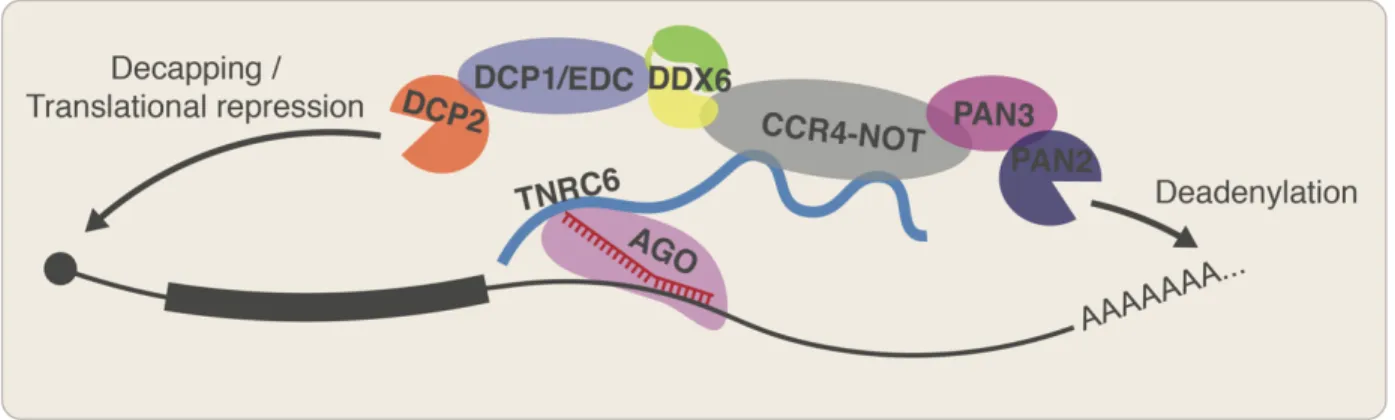
Upon stably binding their target mRNAs, animal miRNAs recruit degradation enzymes through the intermediate protein TNRC6 (Jonas and Izaurralde 2015) (Figure 1), which contains a long, largely unstructured domain with glycine–tryptophan (GW) repeats that can engage the tryptophan binding pockets found on AGO (Pfaff et al. 2013). TNRC6, in turn, recruits the deadenylases CCR4-NOT and the PAN2/PAN3 complex (Jonas and Izaurralde 2015) and
decapping proteins DCP1 and DCP2 (Rehwinkel et al. 2005). In addition to causing the decay of mRNAs, TNRC6 also recruits DDX6, which further recruits factors that block translation
initiation, causing the mRNAs to be translationally repressed as well (Jonas and Izaurralde 2015; Kamenska et al. 2016). In post-embryonic cells, this period of translational repression is shortly followed by mRNA decay, such that the mRNA decay process dominates the total effect of miRNAs on protein production (Eichhorn et al. 2014). However, during embryogenesis (at least in the context of zebrafish embryos) deadenylation of mRNAs, including by miRNAs, leads to further translational repression, rather than degradation (Subtelny et al. 2014).
Figure 1. RISC complexes target mRNAs for translational repression and degradation.
A miRNA (red) loaded into AGO (pink) recognizes target sequences complementary to positions 2–7 of the miRNA on an mRNA (black) and recruits TNRC6 (blue line), which recruits a combination of proteins that deadenylate, decap, and translationally repress the mRNA.
Evolutionary history of miRNAs
MicroRNAs arose early in animal evolution and have been found in almost all metazoan species so far examined (Grimson et al. 2008). Over time, gene duplication events have resulted in groups of miRNAs arising from the same ancestral gene. These miRNAs often retain the same seed sequence as the ancestral gene, perhaps due to the evolutionary pressure for a functional miRNA to keep its seed sequence as changing the seed sequence by just one nucleotide can drastically alter its cohort of transcript targets. miRNAs with the same seed sequence are grouped into families, and although family members are usually paralogs, a few have independently converged to the same seed sequence (Bartel 2018). This categorization of miRNAs into families is useful because targeting specificity relies so much on the miRNA seed region.
Of the miRNA families found in humans, about 27 have been conserved since the emergence of bilaterian animals (Bartel 2018) more than 550 million years ago (Martin et al. 2000) and miRNAs as a class of RNAs have been found in as distant of a relative to humans as
Amphimedon queenslandica, a sea sponge (Grimson et al. 2008), though no miRNAs are
conserved between humans and sponges. miRNA evolution in general seems to have been quite dynamic, with no miRNAs shared between poriferans, cnidarians, and bilaterians (Grimson et al. 2008). Thus while the conserved miRNAs in humans were mostly discovered computationally (Lim et al. 2003), poorly conserved miRNAs in humans are continuously being annotated with the advent of high-throughput small-RNA sequencing techniques (Kozomara, Birgaoanu, and Griffiths-Jones 2019).
Biological roles of miRNAs
As modulators of mRNA decay rates, miRNAs have roles in maintaining and tuning mRNA levels in spatially and temporally specific ways. For example, miR-122, which constitutes the majority of the expressed miRNAs in hepatocytes, is thought to be responsible for keeping these cells terminally differentiated (Hsu et al. 2012), and deletion of miR-122 in mice leads to a number of liver diseases, including hepatitis and liver cancer (Hsu et al. 2012; Tsai et al. 2012). A number of other miRNAs have similarly crucial roles in the development and function of various tissues in model organisms, including the heart (Heidersbach et al. 2013; Wei et al. 2014), brain (Sanuki et al. 2011), immune cells (Lovat et al. 2015; Lu et al. 2010, 2015), and pancreas (Latreille et al. 2014). Perhaps the most dramatic effects of miRNAs are observed in early developmental contexts. For example, during the maternal-to-zygotic transition in zebrafish embryogenesis, miR-430 is one of the factors responsible for turning over maternally-deposited transcripts, clearing the way for the zygotic transcriptome (Giraldez et al. 2006). Knocking out both maternal and zygotic Dicer, which are critical for miRNA biogenesis, causes gastrulation and brain development defects in zebrafish during embryogenesis which can be rescued by injecting mature miR-430 into the developing embryos (Giraldez et al. 2006).
However, large-scale miRNA knockout experiments in various model organisms have revealed that while many miRNAs are crucial for normal development and function in their respective organisms, the majority of miRNAs in worms (Alvarez-Saavedra and Horvitz 2010; Miska et al. 2007), about 20% of miRNAs in flies (Y. W. Chen et al. 2014), and the majority of miRNAs in mice (C. Y. Park et al. 2012) seem to have unappreciable phenotypes when deleted. This is partly due to incomplete phenotypic studies; later studies have identified knockout phenotypes for most conserved miRNAs in mice (Bartel 2018). Some miRNAs may also appear
to not have a phenotype when knocked out because they play a role in important processes that are not usually observed in lab settings. For example, miR-143 and miR-145 are co-transcribed miRNAs conserved throughout vertebrates, and yet mice lacking these two miRNAs appear to develop and function normally. A defect was only observed upon intestinal injury, after which mice lacking miR-143/145 were unable to regenerate their intestinal epithelia and died from the resulting complications whereas wild-type mice were able to recover fully in 9 days (Chivukula et al. 2014). MicroRNA families may also function redundantly with each other, which is supported by the finding that a majority of worm miRNAs surveyed do have knockout phenotypes when the knockouts are performed in sensitized backgrounds where the miRNA biogenesis pathway activity is reduced (Brenner et al. 2010).
Aside from helping to set mRNA levels in the cell, miRNAs have also been reported to play a role in reducing gene expression noise by driving higher transcription rates, primarily for lowly expressed mRNAs (Schmiedel et al. 2015). Lowly expressed mRNAs are the most impacted by stochasticity in gene expression, and miRNAs, which preferentially target lowly expressed mRNAs (Sood et al. 2006; Farh et al. 2005), may provide a mechanism for cells to ensure the steady production of these mRNAs.
Mechanisms of miRNA biogenesis and targeting
The modularity of the process of loading miRNAs into AGO makes miRNA-mediated mRNA repression versatile for both the cell and for researchers attempting to modulate gene expression. While some miRNAs are loaded better than others (Schwarz et al. 2003; Frank, Sonenberg, and Nagar 2010; Suzuki et al. 2015), AGO proteins can load any piece of RNA with any primary sequence as long as it is between 21 and 25 nucleotides in the length, contains a
5′-monophosphate, and is paired with a complementary or nearly complementary companion sequence (known as a passenger strand or miRNA* strand) with ~2 nucleotides of 3′ overhang on each side (Bartel 2018). The passenger strand is usually the strand with the weaker base-pairing at its 3′-end (Khvorova, Reynolds, and Jayasena 2003; Schwarz et al. 2003), and is ejected upon successful loading of the miRNA (Kawamata and Tomari 2010) (Figure 2). Even though any such sequence, endogenous or otherwise, can load into AGO, pair to mRNAs, and recruit degradation machinery, most endogenous miRNAs arise from RNA hairpins within genomically-encoded transcripts and are processed via the canonical miRNA biogenesis pathway (Figure 2). This process starts in the nucleus, where the RNA molecule containing the hairpin (called a pri-miRNA) is recognized and cleaved by the enzyme DROSHA (Lee et al. 2003) to
Figure 2. Canonical miRNA biogenesis pathway. After transcription, the
pri-miRNA is recognized and cleaved (grey triangles) by Microprocessor in the nucleus, which consists of DROSHA (blue) and two copies of DGCR8 (green). Microprocessor measures and cleaves the pri-miRNA approximately 11 base-pairs away from the basal junction. The resulting pre-miRNA is then exported to the cytoplasm, where it is recognized and cleaved by Dicer (orange) near the single-stranded loop into a mature miRNA duplex. One strand of this duplex is loaded into AGO (pink), while the other strand is ejected and degraded.
produce a short hairpin with the first 3′ overhang. DROSHA is assisted by two copies of DGCR8, uses a combination of structural and sequence motifs to specify which RNA hairpins are eventually processed into miRNAs (Fang and Bartel 2015), and cuts them approximately 11 base-pairs away from the basal junction of the pri-miRNA stem-loop (Nguyen et al. 2015). The cleavage product, termed the pre-miRNA, is exported to the cytoplasm where it is further
processed by the enzyme Dicer near the loop to produce the mature miRNA duplex (Zhang et al. 2004), which is competent to load into AGO (Figure 2).
Once a miRNA is loaded into AGO, structural work has revealed that nucleotides 2–5 of the miRNA are pre-formed into a helical structure by AGO, anticipating the conformation it would adopt upon binding its mRNA target and facilitating rapid searching of potential targets in the sea of mRNA sequences in the cytoplasm (Klum et al. 2018). Canonically, there are six types of target sites (Bartel 2009), the top four of which have been shown to robustly lead to
downstream repression of their host mRNAs (Figure 3). These all involve some amount of contiguous Watson-Crick base-pairing to nucleotides 2–7 of the miRNA, with the best sites also base-pairing to position 8 of the miRNA and containing an A nucleotide opposite the first nucleotide of the miRNA (Lewis, Burge, and Bartel 2005). This preference for an A across from the 5′-most nucleotide of the miRNA, regardless of the sequence of the miRNA at this position, is conferred by a binding pocket within AGO,
Figure 3. Canonical site-types. miR-1 loaded
into AGO (pink) base-pairing to an 8mer site embedded in an mRNA. The other 5 canonical site types are listed below the 8mer site in descending order of efficacy.
rather than base-pairing to the miRNA itself (Schirle et al. 2015). Although a small handful of functional noncanonical site types have been reported (Chi, Hannon, and Darnell 2012; Kim et al. 2016), the AGO–miRNA complex is generally intolerant of mismatches, bulges, and wobble pairings to the seed region of its miRNA (Doench and Sharp 2004), which greatly enhances the specificity a miRNA can have for its targets. Indeed, single-molecule studies have shown that while miRNAs have similar on-rates of binding to different target sequences, there is a sharp increase in off-rate when comparing miRNAs binding a “seed-matched” target and the same miRNA binding other sequences (Chandradoss et al. 2015).
In some cases, nonoptimal pairing to the seed region can be rescued by extensive
complementarity (often at least five nucleotides) to the 3′-end of the miRNA (Bartel 2009). Due to their greater pairing constraints, these so-called compensatory sites are much more rare than canonical sites and harder to retain evolutionarily. However, they offer a way for mRNAs to be targeted specifically by a miRNA while avoiding cross-targeting by another miRNA with the same seed sequence. Two of the first examples discovered of a miRNA and target interaction of any kind was a pair of compensatory sites for the miRNA let-7 in the 3′ UTR sequence of the
lin-41 mRNA in C. elegans (Reinhart et al. 2000; Brennecke et al. 2005). Seed-matched sites can
also benefit from pairing to the 3′-end of the miRNA, and such “supplemental sites” constitute around 5% of the seed-matched sites in the human genome (Bartel 2009). In some cases, particularly when the 3′-pairing extends through to the very 3′-end of the miRNA, a target sequence can trigger the decay of the miRNA (Ameres et al. 2010). The mechanisms of this process of target RNA-directed miRNA degradation (TDMD) are currently unknown, and only a few endogenous examples have been found (Ameres et al. 2010; De La Mata and Großhans 2018; Bitetti et al. 2018; Kleaveland et al. 2018). However, the effects can be substantial,
decreasing the half-life of a miRNA from days (which is typical of the average miRNA) to hours (Kingston and Bartel 2019).
Regardless of the mode of pairing, target sites to miRNAs are the most effective in the 3′ UTR of an mRNA, starting about 15 nucleotides downstream of the stop codon (Grimson et al. 2007). This is most likely due to scanning and translating ribosomes precluding AGO from binding 5′ UTR and ORF sequences and/or dislodging AGO complexes that have bound in these regions. During translation termination, this ribosome protection extends about 15 nucleotides into the 3′ UTR such that miRNA-binding sites can only avoid ribosome interference when they are 15 nucleotides downstream of the stop codon. In fact, an ineffective ORF site directly upstream from a stop codon can be converted to an effective site by moving the stop codon upstream of the site (Grimson et al. 2007), showing that the ribosome, rather than sequence context, is responsible for the reduced efficacy of sites outside the 3′ UTR. Within the 3′ UTRs of mRNAs, bioinformatic analyses have shown that sites closer to either the stop codon or the very 3′-end of the mRNA are more effective than sites in the center of the 3′ UTR (Grimson et al. 2007), although the mechanisms causing this phenomenon are currently unknown.
miRNA target prediction
Because the human genome encodes upwards of 300 different conserved miRNAs, and each one can interact with hundreds to thousands of targets, which can shift depending on the expression profiles of different cell-types, experimentally measuring each miRNA–target interaction would be a monumental feat. On top of the endogenous miRNAs, a virtually countless number of possible synthetic duplexes are competent for loading into AGO. These are mostly used by researchers to knock down transcripts through the AGO-mediated slicing pathway, and they
would ideally be designed to reduce repression of off-targets. For all these reasons, there has been a long-standing interest in developing algorithms that can quantitatively predict the miRNA targeting efficacy for any arbitrary miRNA and mRNA.
One of the first successful approaches for predicting miRNA targeting involved looking for mRNAs with potential target sites in their 3′ UTRs that were complementary to positions 2–8 of a miRNA and evolutionarily conserved (Lewis et al. 2003). Because a single mismatch to the seed region of a miRNA can abrogate miRNA affinity, there was a need for a method to score the conservation level of an entire site, rather than the individual nucleotides in a site. This led to the development of the probability of conserved targeting (PCT) metric (Friedman et al. 2009), which calculates the branch-length score of a target site sequence relative to the background level of conservation of the entire 3′ UTR of the mRNA. This value is further normalized to the same metric calculated for control sequences in order to control for the differing amounts of conservation that may be conferred by dinucleotide content, rather than miRNA targeting.
Other methods have been developed to score potential target sites to a miRNA that incorporate features such as local sequence context of the site, the degree of possible
supplementary pairing to the 3′-end of the miRNA, the predicted structural accessibility of the site in its 3′ UTR context, and the predicted RNA duplex stability between the miRNA and a site (Kiriakidou et al. 2004; Krek et al. 2005; Grimson et al. 2007; Garcia et al. 2011; Gumienny and Zavolan 2015; Agarwal et al. 2015). These features are regressed against some measurement of miRNA-dependent repression of the mRNA housing the site. One of the most straight-forward ways to measure miRNA activity in cells is to transfect a miRNA of interest into a cell-line that does not normally express that miRNA and measure the mRNA abundance fold-changes between transfected and mock-transfected populations either using microarrays or RNA-seq.
Because transfection datasets evaluated using microarrays are the most widely available, most miRNA target prediction models are trained on these transfection datasets and often validated on orthogonal datasets.
In addition to models that simply combine a list of features that may correlate with miRNA activity, some efforts to predict miRNA targeting efficacy attempt to construct a biochemical model of miRNA occupancy (Krek et al. 2005; Khorshid et al. 2013).
Unfortunately, these models require knowledge of the binding affinities between AGO–miRNA complexes and their target mRNAs. Because only a few of these values have been determined experimentally (Wee et al. 2012; Salomon et al. 2015; Schirle, Sheu-Gruttadauria, and MacRae 2014; Schirle et al. 2015; Jo et al. 2015; Klum et al. 2018; Chandradoss et al. 2015), they must be estimated computationally. A popular method of estimating the affinity between a miRNA and a target sequence is by using nearest-neighbor (NN) rules for estimating RNA–RNA duplex stabilities (Xia et al. 1998). However, the handful of experimentally-measured binding affinities between miRNAs and their targets have shown that AGO substantially alters the energetics of binding between its loaded miRNA and a potential target (Salomon et al. 2015).
Others have attempted to use data from cross-linking and immunoprecipitation (CLIP) experiments to learn the energetics of pairing between miRNAs and their targets (Khorshid et al. 2013). In these experiments, RISC bound to RNA is cross-linked to that RNA using ultraviolet light. The resulting complexes are then immunoprecipitated, and the bound RNA is isolated and sequenced (Chi et al. 2009). These experiments provide a much more direct read-out of miRNA binding than transfection experiments because they reflect the engagement of RISC complexes on individual targets, rather than repression of entire mRNAs. However, CLIP data are subject to cross-linking biases (Lambert et al. 2014), CLIP data often contain large amounts of background
binding events (Jaskiewicz et al. 2012), and it is impossible to determine fundamental binding constants from CLIP enrichments without knowledge of the concentrations of RISC in the CLIP experiments. As a result, miRNA target prediction algorithms that use CLIP data to learn
miRNA–target binding affinities (Khorshid et al. 2013; Gumienny and Zavolan 2015) perform no better than the best miRNA prediction algorithms that use NN rules and site-type information (Agarwal et al. 2015) in predicting the repression of mRNAs in cells.
A high-throughput method for measuring binding affinities for RNA-binding proteins
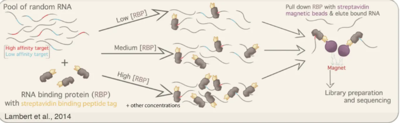
Recently, Lambert et al. have developed a technique called RNA bind-n-seq (RBNS) for measuring the affinities of RNA-binding proteins to a random library of potential RNA binding partners in vitro (Lambert et al. 2014). In this technique, the RNA-binding protein of interest is tagged with an epitope that binds streptavidin, purified, and incubated with a random library of RNA molecules flanked by sequencing primers. The RNA-binding proteins are then pulled down using streptavidin beads, along with any bound RNA, the bound RNA is isolated and sequenced, and the enriched sequences are compared to those obtained by sequencing the input pool of RNA
Figure 4. RNA bind-n-seq protocol. RNA-binding proteins tagged with a streptavidin binding
tag are purified and incubated with a pool of random RNA. The RNA-binding proteins are then pulled down with streptavidin beads and their associated RNA molecules are reverse transcribed to make a cDNA library and sequenced. A range of different protein concentrations are used to capture a wide range of binding affinities. Figure adapted from Figure 1A of
molecules (Figure 4). Sequence motifs that interact with the RNA-binding protein become enriched over other sequences in the pull-down library, and enrichments for individual k-mers can be determined (with k being dependent on how deeply the libraries are sequenced). This process is repeated with several different concentrations of the RNA-binding protein such that both high- and low-affinity binding interactions can be captured and quantified. This method has revealed the binding preferences for 78 human RNA-binding proteins (Dominguez et al. 2018), proving its utility and adaptability to any RNA-binding protein. RBNS is therefore an attractive method for measuring the binding affinities between RISC and its targets. The data obtained from RBNS are also theoretically sufficient for fitting the binding constants between RNA-binding proteins and k-mers, in addition to enrichments of k-mers.
Recent applications of neural networks for learning sequence binding preferences
In addition to experimental advancements that may aid miRNA target prediction, the large amounts of high-throughput sequencing data collected in recent years have fueled the
development of more data-driven models of nucleic acid binding preferences. Alipahani et al. developed a deep learning model, DeepBind, that learns the binding specificities of DNA- and RNA-binding proteins and showed their model could learn from a wide range of different nucleic acid binding assays. DeepBind was also shown to outperform all other existing methods for predicting binding partners for transcription factors and RNA-binding proteins (Alipanahi et al. 2015). These types of models can pick up on hierarchically-dependent features and learn nonlinear interactions between features and can therefore be especially helpful for learning the binding specificities of RNA-binding proteins, which often have single nucleotide, di-nucleotide, and structural preferences (Y. Park and Kellis 2015). However, DeepBind and other similar
models do not train on any features of the DNA- or RNA-binding proteins themselves, which means that a new model must be trained for each new DNA- or RNA-binding protein of interest. This would have to be overcome if deep learning were applied to the problem of predicting miRNA targets because each new AGO–miRNA complex is essentially a new RNA-binding protein with its own unique binding profile. Given that there are hundreds of conserved endogenous miRNAs in humans and 47 = 16,384 unique miRNA seed sequences possible, miRNA target prediction algorithms need to be able to generalize to arbitrary miRNA sequences without acquiring more data for each new miRNA sequence.
References
Agarwal, Vikram, George W. Bell, Jin Wu Nam, and David P. Bartel. 2015. “Predicting Effective MicroRNA Target Sites in Mammalian MRNAs.” ELife 4 (AUGUST2015). https://doi.org/10.7554/eLife.05005.
Alipanahi, Babak, Andrew Delong, Matthew T. Weirauch, and Brendan J. Frey. 2015. “Predicting the Sequence Specificities of DNA- and RNA-Binding Proteins by Deep Learning.” Nature Biotechnology. https://doi.org/10.1038/nbt.3300.
Alvarez-Saavedra, Ezequiel, and H. Robert Horvitz. 2010. “Many Families of C. Elegans MicroRNAs Are Not Essential for Development or Viability.” Current Biology. https://doi.org/10.1016/j.cub.2009.12.051.
Ameres, Stefan L., Michael D. Horwich, Jui Hung Hung, Jia Xu, Megha Ghildiyal, Zhiping Weng, and Phillip D. Zamore. 2010. “Target RNA-Directed Trimming and Tailing of Small Silencing RNAs.” Science. https://doi.org/10.1126/science.1187058.
Bartel, David P. 2009. “MicroRNAs: Target Recognition and Regulatory Functions.” Cell 136 (2): 215–33. https://doi.org/10.1016/j.cell.2009.01.002.
Bartel, David P. 2018. “Metazoan MicroRNAs.” Cell. https://doi.org/10.1016/j.cell.2018.03.006. Bitetti, Angelo, Allison C. Mallory, Elisabetta Golini, Claudia Carrieri, Héctor Carreño
Gutiérrez, Emerald Perlas, Yuvia A. Pérez-Rico, et al. 2018. “MicroRNA Degradation by a Conserved Target RNA Regulates Animal Behavior.” Nature Structural and Molecular
Biology. https://doi.org/10.1038/s41594-018-0032-x.
Brennecke, Julius, Alexander Stark, Robert B. Russell, and Stephen M. Cohen. 2005. “Principles of MicroRNA-Target Recognition.” In PLoS Biology.
https://doi.org/10.1371/journal.pbio.0030085.
Brenner, John L., Kristen L. Jasiewicz, Alisha F. Fahley, Benedict J. Kemp, and Allison L. Abbott. 2010. “Loss of Individual MicroRNAs Causes Mutant Phenotypes in Sensitized Genetic Backgrounds in C. Elegans.” Current Biology.
https://doi.org/10.1016/j.cub.2010.05.062.
Chandradoss, Stanley D., Nicole T. Schirle, Malwina Szczepaniak, Ian J. Macrae, and Chirlmin Joo. 2015. “A Dynamic Search Process Underlies MicroRNA Targeting.” Cell.
https://doi.org/10.1016/j.cell.2015.06.032.
Chen, Chyi Ying A., Dinghai Zheng, Zhenfang Xia, and Ann Bin Shyu. 2009. “Ago-TNRC6 Triggers MicroRNA-Mediated Decay by Promoting Two Deadenylation Steps.” Nature
Structural and Molecular Biology. https://doi.org/10.1038/nsmb.1709.
Chen, Ya Wen, Shilin Song, Ruifen Weng, Pushpa Verma, Jan Michael Kugler, Marita Buescher, Sigrid Rouam, and Stephen M. Cohen. 2014. “Systematic Study of Drosophila MicroRNA Functions Using a Collection of Targeted Knockout Mutations.” Developmental
Cell. https://doi.org/10.1016/j.devcel.2014.11.029.
Chi, Sung Wook, Gregory J. Hannon, and Robert B. Darnell. 2012. “An Alternative Mode of MicroRNA Target Recognition.” Nature Structural and Molecular Biology.
https://doi.org/10.1038/nsmb.2230.
Chi, Sung Wook, Julie B Zang, Aldo Mele, and Robert B Darnell. 2009. “Ago HITS-CLIP Decodes MiRNA-MRNA Interaction Maps.” Nature 460 (7254): 479–86.
Chivukula, Raghu R., Guanglu Shi, Asha Acharya, Eric W. Mills, Lauren R. Zeitels, Joselin L. Anandam, Abier A. Abdelnaby, et al. 2014. “An Essential Mesenchymal Function for MiR-143/145 in Intestinal Epithelial Regeneration.” Cell.
https://doi.org/10.1016/j.cell.2014.03.055.
Core, Leighton J., André L. Martins, Charles G. Danko, Colin T. Waters, Adam Siepel, and John T. Lis. 2014. “Analysis of Nascent RNA Identifies a Unified Architecture of Initiation Regions at Mammalian Promoters and Enhancers.” Nature Genetics.
https://doi.org/10.1038/ng.3142.
Doench, John G., and Phillip A. Sharp. 2004. “Specificity of MicroRNA Target Selection in Translational Repression.” Genes & Development 18 (5): 504–11.
https://doi.org/10.1101/gad.1184404.
Dominguez, Daniel, Peter Freese, Maria S. Alexis, Amanda Su, Myles Hochman, Tsultrim Palden, Cassandra Bazile, et al. 2018. “Sequence, Structure, and Context Preferences of Human RNA Binding Proteins.” Molecular Cell.
https://doi.org/10.1016/j.molcel.2018.05.001.
Eichhorn, Stephen W., Huili Guo, Sean E. McGeary, Ricard A. Rodriguez-Mias, Chanseok Shin, Daehyun Baek, Shu hao Hsu, Kalpana Ghoshal, Judit Villén, and David P. Bartel. 2014. “MRNA Destabilization Is the Dominant Effect of Mammalian MicroRNAs by the Time Substantial Repression Ensues.” Molecular Cell 56 (1): 104–15.
https://doi.org/10.1016/j.molcel.2014.08.028.
Fang, Wenwen, and David P. Bartel. 2015. “The Menu of Features That Define Primary MicroRNAs and Enable De Novo Design of MicroRNA Genes.” Molecular Cell. https://doi.org/10.1016/j.molcel.2015.08.015.
Farh, Kyte Kai How, Andrew Grimson, Calvin Jan, Benjamin P. Lewis, Wendy K. Johnston, Lee P. Lim, Christopher B. Burge, and David P. Bartel. 2005. “Biochemistry: The Widespread Impact of Mammalian MicroRNAs on MRNA Repression and Evolution.” Science. https://doi.org/10.1126/science.1121158.
Frank, Filipp, Nahum Sonenberg, and Bhushan Nagar. 2010. “Structural Basis for 5′-Nucleotide Base-Specific Recognition of Guide RNA by Human AGO2.” Nature.
https://doi.org/10.1038/nature09039.
Friedman, Robin C., Kyle Kai How Farh, Christopher B. Burge, and David P. Bartel. 2009. “Most Mammalian MRNAs Are Conserved Targets of MicroRNAs.” Genome Research 19 (1): 92–105. https://doi.org/10.1101/gr.082701.108.
Garcia, David M, Daehyun Baek, Chanseok Shin, George W Bell, Andrew Grimson, and David P Bartel. 2011. “Weak Seed-Pairing Stability and High Target-Site Abundance Decrease the Proficiency of Lsy-6 and Other MicroRNAs.” Nature Structural & Molecular Biology 18 (10): 1139–46. https://doi.org/10.1038/nsmb.2115.
Giraldez, Antonio J., Yuichiro Mishima, Jason Rihel, Russell J. Grocock, Stijn Van Dongen, Kunio Inoue, Anton J. Enright, and Alexander F. Schier. 2006. “Zebrafish MiR-430 Promotes Deadenylation and Clearance of Maternal MRNAs.” Science.
https://doi.org/10.1126/science.1122689.
Grimson, Andrew, Kyle Kai How Farh, Wendy K. Johnston, Philip Garrett-Engele, Lee P. Lim, and David P. Bartel. 2007. “MicroRNA Targeting Specificity in Mammals: Determinants beyond Seed Pairing.” Molecular Cell 27 (1): 91–105.
Grimson, Andrew, Mansi Srivastava, Bryony Fahey, Ben J. Woodcroft, H. Rosaria Chiang, Nicole King, Bernard M. Degnan, Daniel S. Rokhsar, and David P. Bartel. 2008. “Early Origins and Evolution of MicroRNAs and Piwi-Interacting RNAs in Animals.” Nature. https://doi.org/10.1038/nature07415.
Gumienny, Rafal, and Mihaela Zavolan. 2015. “Accurate Transcriptome-Wide Prediction of MicroRNA Targets and Small Interfering RNA off-Targets with MIRZA-G.” Nucleic Acids
Research 43 (3): 1380–91. https://doi.org/10.1093/nar/gkv050.
Gygi, Steven P., Yvan Rochon, B. Robert Franza, and Ruedi Aebersold. 1999. “Correlation between Protein and MRNA Abundance in Yeast.” Molecular and Cellular Biology. https://doi.org/10.1128/mcb.19.3.1720.
Heidersbach, Amy, Chris Saxby, Karen Carver-Moore, Yu Huang, Yen Sin Ang, Pieter J. de Jong, Kathryn N. Ivey, and Deepak Srivastava. 2013. “MicroRNA-1 Regulates Sarcomere Formation and Suppresses Smooth Muscle Gene Expression in the Mammalian Heart.”
ELife. https://doi.org/10.7554/eLife.01323.001.
Hsu, Shu Hao, Bo Wang, Janaiah Kota, Jianhua Yu, Stefan Costinean, Huban Kutay, Lianbo Yu, et al. 2012. “Essential Metabolic, Anti-Inflammatory, and Anti-Tumorigenic Functions of MiR-122 in Liver.” Journal of Clinical Investigation. https://doi.org/10.1172/JCI63539. Jaskiewicz, Lukasz, Biter Bilen, Jean Hausser, and Mihaela Zavolan. 2012. “Argonaute CLIP -
A Method to Identify in Vivo Targets of MiRNAs.” Methods. https://doi.org/10.1016/j.ymeth.2012.09.006.
Jo, Myung Hyun, Soochul Shin, Seung Ryoung Jung, Eunji Kim, Ji Joon Song, and Sungchul Hohng. 2015. “Human Argonaute 2 Has Diverse Reaction Pathways on Target RNAs.”
Molecular Cell. https://doi.org/10.1016/j.molcel.2015.04.027.
Jonas, Stefanie, and Elisa Izaurralde. 2015. “Towards a Molecular Understanding of MicroRNA-Mediated Gene Silencing.” Nature Reviews Genetics. https://doi.org/10.1038/nrg3965. Jones-Rhoades, Matthew W., David P. Bartel, and Bonnie Bartel. 2006. “MicroRNAs and Their
Regulatory Roles in Plants.” Annual Review of Plant Biology. https://doi.org/10.1146/annurev.arplant.57.032905.105218.
Kamenska, Anastasiia, Clare Simpson, Caroline Vindry, Helen Broomhead, Marianne Bénard, Michèle Ernoult-Lange, Benjamin P. Lee, Lorna W. Harries, Dominique Weil, and Nancy Standart. 2016. “The DDX6-4E-T Interaction Mediates Translational Repression and P-Body Assembly.” Nucleic Acids Research. https://doi.org/10.1093/nar/gkw565.
Kawamata, Tomoko, and Yukihide Tomari. 2010. “Making RISC.” Trends in Biochemical
Sciences. https://doi.org/10.1016/j.tibs.2010.03.009.
Khorshid, Mohsen, Jean Hausser, Mihaela Zavolan, and Erik Van Nimwegen. 2013. “A Biophysical MiRNA-MRNA Interaction Model Infers Canonical and Noncanonical Targets.” Nature Methods. https://doi.org/10.1038/nmeth.2341.
Khvorova, Anastasia, Angela Reynolds, and Sumedha D. Jayasena. 2003. “Functional SiRNAs and MiRNAs Exhibit Strand Bias.” Cell 115 (2): 209–16. https://doi.org/10.1016/S0092-8674(03)00801-8.
Kim, Doyeon, You Me Sung, Jinman Park, Sukjun Kim, Jongkyu Kim, Junhee Park, Haeok Ha, Jung Yoon Bae, Sohui Kim, and Daehyun Baek. 2016. “General Rules for Functional MicroRNA Targeting.” Nature Genetics. https://doi.org/10.1038/ng.3694.
Mammalian MicroRNA Metabolism.” BioRxiv. https://doi.org/10.1101/607150.
Kiriakidou, M., Peter T. Nelson, Andrei Kouranov, Petko Fitziev, Costas Bouyioukos, Zissimos Mourelatos, and Artemis Hatzigeorgiou. 2004. “A Combined Computational-Experimental Approach Predicts Human MicroRNA Targets.” Genes & Development 18 (10): 1165–78. https://doi.org/10.1101/gad.1184704.
Kleaveland, Benjamin, Charlie Y. Shi, Joanna Stefano, and David P. Bartel. 2018. “A Network of Noncoding Regulatory RNAs Acts in the Mammalian Brain.” Cell.
https://doi.org/10.1016/j.cell.2018.05.022.
Klum, Shannon M, Stanley D Chandradoss, Nicole T Schirle, Chirlmin Joo, and Ian J MacRae. 2018. “Helix‐7 in Argonaute2 Shapes the MicroRNA Seed Region for Rapid Target Recognition.” The EMBO Journal. https://doi.org/10.15252/embj.201796474.
Kozomara, Ana, Maria Birgaoanu, and Sam Griffiths-Jones. 2019. “MiRBase: From MicroRNA Sequences to Function.” Nucleic Acids Research. https://doi.org/10.1093/nar/gky1141. Krek, Azra, Dominic Grün, Matthew N Poy, Rachel Wolf, Lauren Rosenberg, Eric J Epstein,
Philip MacMenamin, et al. 2005. “Combinatorial MicroRNA Target Predictions.” Nature
Genetics 37 (5): 495–500. https://doi.org/10.1038/ng1536.
Kwak, Hojoong, Nicholas J. Fuda, Leighton J. Core, and John T. Lis. 2013. “Precise Maps of RNA Polymerase Reveal How Promoters Direct Initiation and Pausing.” Science. https://doi.org/10.1126/science.1229386.
La Mata, Manuel De, and Helge Großhans. 2018. “Turning the Table on MiRNAs.” Nature
Structural and Molecular Biology. https://doi.org/10.1038/s41594-018-0040-x.
Lambert, Nicole, Alex Robertson, Mohini Jangi, Sean McGeary, Phillip A. Sharp, and
Christopher B. Burge. 2014. “RNA Bind-n-Seq: Quantitative Assessment of the Sequence and Structural Binding Specificity of RNA Binding Proteins.” Molecular Cell 54 (5): 887– 900. https://doi.org/10.1016/j.molcel.2014.04.016.
Latreille, Mathieu, Jean Hausser, Ina Stützer, Quan Zhang, Benoit Hastoy, Sofia Gargani, Julie Kerr-Conte, et al. 2014. “MicroRNA-7a Regulates Pancreatic β Cell Function.” Journal of
Clinical Investigation. https://doi.org/10.1172/JCI73066.
Lee, Yoontae, Chiyoung Ahn, Jinju Han, Hyounjeong Choi, Jaekwang Kim, Jeongbin Yim, Junho Lee, et al. 2003. “The Nuclear RNase III Drosha Initiates MicroRNA Processing.”
Nature. https://doi.org/10.1038/nature01957.
Lewis, Benjamin P., Christopher B. Burge, and David P. Bartel. 2005. “Conserved Seed Pairing, Often Flanked by Adenosines, Indicates That Thousands of Human Genes Are MicroRNA Targets.” Cell 120 (1): 15–20. https://doi.org/10.1016/j.cell.2004.12.035.
Lewis, Benjamin P., I. Hung Shih, Matthew W. Jones-Rhoades, David P. Bartel, and Christopher B. Burge. 2003. “Prediction of Mammalian MicroRNA Targets.” Cell 115 (7): 787–98. https://doi.org/10.1016/S0092-8674(03)01018-3.
Lim, Lee P., Margaret E. Glasner, Soraya Yekta, Christopher B. Burge, and David P. Bartel. 2003. “Vertebrate MicroRNA Genes.” Science. https://doi.org/10.1126/science.1080372. Liu, Jidong, Michelle A. Carmell, Fabiola V. Rivas, Carolyn G. Marsden, J. Michael Thomson,
Ji Joon Song, Scott M. Hammond, Leemor Joshua-Tor, and Gregory J. Hannon. 2004. “Argonaute2 Is the Catalytic Engine of Mammalian RNAi.” Science.
Pizzi, Caterina Vicentini, et al. 2015. “MiR-15b/16-2 Deletion Promotes B-Cell
Malignancies.” Proceedings of the National Academy of Sciences of the United States of
America. https://doi.org/10.1073/pnas.1514954112.
Lu, Li Fan, Mark P. Boldin, Ashutosh Chaudhry, Ling Li Lin, Konstantin D. Taganov, Toshikatsu Hanada, Akihiko Yoshimura, David Baltimore, and Alexander Y. Rudensky. 2010. “Function of MiR-146a in Controlling Treg Cell-Mediated Regulation of Th1 Responses.” Cell. https://doi.org/10.1016/j.cell.2010.08.012.
Lu, Li Fan, Georg Gasteiger, I. Shing Yu, Ashutosh Chaudhry, Jing Ping Hsin, Yuheng Lu, Paula D. Bos, et al. 2015. “A Single Mirna-Mrna Interaction Affects The Immune Response In A Context- And Cell-Type-Specific Manner.” Immunity.
https://doi.org/10.1016/j.immuni.2015.04.022.
Martin, M. W., D. V. Grazhdankin, S. A. Bowring, D. A.D. Evans, M. A. Fedonkin, and J. L. Kirschvink. 2000. “Age of Neoproterozoic Bilatarian Body and Trace Fossils, White Sea, Russia: Implications for Metazoan Evolution.” Science.
https://doi.org/10.1126/science.288.5467.841.
Miska, Eric A., Ezequiel Alvarez-Saavedra, Allison L. Abbott, Nelson C. Lau, Andrew B. Hellman, Shannon M. McGonagle, David P. Bartel, Victor R. Ambros, and H. Robert Horvitz. 2007. “Most Caenorhabditis Elegans MicroRNAs Are Individually Not Essential for Development or Viability.” PLoS Genetics.
https://doi.org/10.1371/journal.pgen.0030215.
Nguyen, Tuan Anh, Myung Hyun Jo, Yeon Gil Choi, Joha Park, S. Chul Kwon, Sungchul Hohng, V. Narry Kim, and Jae Sung Woo. 2015. “Functional Anatomy of the Human Microprocessor.” Cell. https://doi.org/10.1016/j.cell.2015.05.010.
Nilsen, Timothy W., and Brenton R. Graveley. 2010. “Expansion of the Eukaryotic Proteome by Alternative Splicing.” Nature. https://doi.org/10.1038/nature08909.
Park, Chong Yon, Lukas T. Jeker, Karen Carver-Moore, Alyssia Oh, Huey Jiin Liu, Rachel Cameron, Hunter Richards, et al. 2012. “A Resource for the Conditional Ablation of MicroRNAs in the Mouse.” Cell Reports. https://doi.org/10.1016/j.celrep.2012.02.008. Park, Yongjin, and Manolis Kellis. 2015. “Deep Learning for Regulatory Genomics.” Nature
Biotechnology. https://doi.org/10.1038/nbt.3313.
Pfaff, Janina, Janosch Hennig, Franz Herzog, Ruedi Aebersold, Michael Sattler, Dierk Niessing, and Gunter Meister. 2013. “Structural Features of Argonaute-GW182 Protein Interactions.”
Proceedings of the National Academy of Sciences of the United States of America.
https://doi.org/10.1073/pnas.1308510110.
Rehwinkel, Jan, Isabelle Behm-Ansmant, David Gatfield, and Elisa Izaurralde. 2005. “A Crucial Role for GW182 and the DCP1:DCP2 Decapping Complex in MiRNA-Mediated Gene Silencing.” RNA. https://doi.org/10.1261/rna.2191905.
Reinhart, Brenda J., Frank J. Slack, Michael Basson, Amy E. Pasquienelll, Jill C. Bettlnger, Ann E. Rougvle, H. Robert Horvitz, and Gary Ruvkun. 2000. “The 21-Nucleotide Let-7 RNA Regulates Developmental Timing in Caenorhabditis Elegans.” Nature.
https://doi.org/10.1038/35002607.
Salomon, William E, Samson M Jolly, Melissa J Moore, Phillip D Zamore, William E Salomon, Samson M Jolly, Melissa J Moore, Phillip D Zamore, and Victor Serebrov. 2015.
“Single-Properties of Its Nucleic Acid Guides.” Cell. https://doi.org/10.1016/j.cell.2015.06.029. Sanuki, Rikako, Akishi Onishi, Chieko Koike, Rieko Muramatsu, Satoshi Watanabe, Yuki
Muranishi, Shoichi Irie, et al. 2011. “MiR-124a Is Required for Hippocampal Axogenesis and Retinal Cone Survival through Lhx2 Suppression.” Nature Neuroscience.
https://doi.org/10.1038/nn.2897.
Schirle, Nicole T., Jessica Sheu-Gruttadauria, Stanley D. Chandradoss, Chirlmin Joo, and Ian J. MacRae. 2015. “Water-Mediated Recognition of T1-Adenosine Anchors Argonaute2 to MicroRNA Targets.” ELife. https://doi.org/10.7554/eLife.07646.
Schirle, Nicole T., Jessica Sheu-Gruttadauria, and Ian J. MacRae. 2014. “Structural Basis for MicroRNA Targeting.” Science. https://doi.org/10.1126/science.1258040.
Schmiedel, Jörn M., Sandy L. Klemm, Yannan Zheng, Apratim Sahay, Nils Blüthgen, Debora S. Marks, and Alexander Van Oudenaarden. 2015. “MicroRNA Control of Protein Expression Noise.” Science. https://doi.org/10.1126/science.aaa1738.
Schwanhüusser, Björn, Dorothea Busse, Na Li, Gunnar Dittmar, Johannes Schuchhardt, Jana Wolf, Wei Chen, and Matthias Selbach. 2011. “Global Quantification of Mammalian Gene Expression Control.” Nature. https://doi.org/10.1038/nature10098.
Schwarz, Dianne S., György Hutvágner, Tingting Du, Zuoshang Xu, Neil Aronin, and Phillip D. Zamore. 2003. “Asymmetry in the Assembly of the RNAi Enzyme Complex.” Cell.
https://doi.org/10.1016/S0092-8674(03)00759-1.
Sood, Pranidhi, Azra Krek, Mihaela Zavolan, Giuseppe Macino, and Nikolaus Rajewsky. 2006. “Cell-Type-Specific Signatures of MicroRNAs on Target MRNA Expression.” Proceedings
of the National Academy of Sciences of the United States of America.
https://doi.org/10.1073/pnas.0511045103.
Subtelny, Alexander O., Stephen W. Eichhorn, Grace R. Chen, Hazel Sive, and David P. Bartel. 2014. “Poly(A)-Tail Profiling Reveals an Embryonic Switch in Translational Control.”
Nature. https://doi.org/10.1038/nature13007.
Suzuki, Hiroshi I., Akihiro Katsura, Takahiko Yasuda, Toshihide Ueno, Hiroyuki Mano, Koichi Sugimoto, and Kohei Miyazono. 2015. “Small-RNA Asymmetry Is Directly Driven by Mammalian Argonautes.” Nature Structural and Molecular Biology.
https://doi.org/10.1038/nsmb.3050.
Tsai, Wei Chih, Sheng Da Hsu, Chu Sui Hsu, Tsung Ching Lai, Shu Jen Chen, Roger Shen, Yi Huang, et al. 2012. “MicroRNA-122 Plays a Critical Role in Liver Homeostasis and
Hepatocarcinogenesis.” Journal of Clinical Investigation. https://doi.org/10.1172/JCI63455. Wee, Liang Meng, C. Fabián Flores-Jasso, William E. Salomon, and Phillip D. Zamore. 2012.
“Argonaute Divides Its RNA Guide into Domains with Distinct Functions and RNA-Binding Properties.” Cell. https://doi.org/10.1016/j.cell.2012.10.036.
Wei, Yusheng, Siwu Peng, Meng Wu, Ravi Sachidanandam, Zhidong Tu, Shihong Zhang, Christine Falce, Eric A. Sobie, Djamel Lebeche, and Yong Zhao. 2014. “Multifaceted Roles of MiR-1s in Repressing the Fetal Gene Program in the Heart.” Cell Research.
https://doi.org/10.1038/cr.2014.12.
Wu, Ligang, Jihua Fan, and Joel G. Belasco. 2006. “MicroRNAs Direct Rapid Deadenylation of MRNA.” Proceedings of the National Academy of Sciences of the United States of America 103 (11): 4034–39. https://doi.org/10.1073/pnas.0510928103.
Jiao, Christopher Cox, and Douglas H. Turner. 1998. “Thermodynamic Parameters for an Expanded Nearest-Neighbor Model for Formation of RNA Duplexes with Watson - Crick Base Pairs.” Biochemistry. https://doi.org/10.1021/bi9809425.
Yang, Edward, Erik van Nimwegen, Mihaela Zavolan, Nikolaus Rajewsky, Mark Schroeder, Marcelo Magnasco, and James E. Darnell. 2003. “Decay Rates of Human MRNAs: Correlation with Functional Characteristics and Sequence Attributes.” Genome Research. https://doi.org/10.1101/gr.1272403.
Zhang, Haidi, Fabrice A. Kolb, Lukasz Jaskiewicz, Eric Westhof, and Witold Filipowicz. 2004. “Single Processing Center Models for Human Dicer and Bacterial RNase III.” Cell.
Chapter 2. The biochemical basis of microRNA targeting efficacy
Sean E. McGeary1,2,3†, Kathy S. Lin1,2,3,4†, Charlie Y. Shi1,2,3, Thy Pham1,2,3, Namita Bisaria1,2,3, Gina M. Kelley1,2,3, and David P. Bartel1,2,3,4
1Howard Hughes Medical Institute, Cambridge, MA, 02142, USA
2Whitehead Institute for Biomedical Research, Cambridge, MA, 02142, USA
3Department of Biology, Massachusetts Institute of Technology, Cambridge, MA 02139, USA 4Computational and Systems Biology Program, Massachusetts Institute of Technology,
Cambridge, MA, 02139, USA
†These authors contributed equally to this work.
S.E.M. developed AGO-RBNS and associated analyses, which he implemented with help from T.P. and N.B. K.S.L. devised and implemented the biochemical model and CNN. C.Y.S., G.M.K., and T.P. performed transfection and sequencing experiments. C.Y.S. and S.E.M. designed and performed the massively parallel reporter assay. S.E.M., K.S.L., and D.P.B. designed the study and wrote the manuscript with input from other authors.
Abstract
MicroRNAs (miRNAs) act within Argonaute proteins to guide repression of mRNA
targets. Although various approaches have provided insight into target recognition, the sparsity of miRNA–target affinity measurements has limited understanding and prediction of targeting efficacy. Here, we adapted RNA bind-n-seq to enable measurement of relative binding affinities between Argonaute–miRNA complexes and all ≤12-nucleotide sequences. This approach revealed noncanonical target sites unique to each miRNA, miRNA-specific differences in canonical target-site affinities, and a 100-fold impact of dinucleotides flanking each site. These data enabled construction of a biochemical model of miRNA-mediated repression, which was extended to all miRNA sequences using a convolutional neural network. This model
substantially improved prediction of cellular repression, thereby providing a biochemical basis for quantitatively integrating miRNAs into gene-regulatory networks.
Introduction
MicroRNAs (miRNAs) are ~22-nt regulatory RNAs that derive from hairpin regions of precursor transcripts (Bartel 2018). Each miRNA associates with an Argonaute (AGO) protein to form a silencing complex, in which the miRNA pairs to sites within target transcripts and the AGO protein promotes destabilization and/or translational repression of bound target (Jonas and Izaurralde 2015). miRNAs are grouped into families based on the sequence of their extended seed (nucleotides 2–8 of the miRNA), which is the region of the miRNA most important for target recognition (Bartel 2009). The 90 most broadly conserved miRNA families of mammals each have an average of >400 preferentially conserved targets, such that mRNAs from most human genes are conserved targets of at least one miRNA (Friedman et al. 2009). Most of these
90 broadly conserved families are required for normal development or physiology, as shown by knockout studies in mice (Bartel 2018).
Deeper understanding of these numerous biological functions would be facilitated by a better understanding of miRNA targeting efficacy, with the ultimate goal of correctly predicting the effects of each miRNA on the output of each expressed gene. In principle, targeting efficacy should be a function of the affinity between AGO–miRNA complexes and their target sites, in that greater affinity to a target site would cause increased occupancy at that site and thus increased repression of the target mRNA. Until very recently, binding affinities have been known for only a few target sequences of only three miRNAs (Wee et al. 2012; Salomon et al. 2015; Schirle, Sheu-Gruttadauria, and MacRae 2014; Schirle et al. 2015; Jo et al. 2015; Klum et al. 2018; Chandradoss et al. 2015). In a recent study, high-throughput imaging and cleavage analyses provide extensive binding and slicing data for two of these three miRNAs, let-7a and miR-21 (Becker et al. 2019). Although these measurements provide insight and enable a quantitative model that predicts the efficiency of miR-21–directed slicing in cells (Becker et al. 2019), the sparsity of binding-affinity data still limits insight into how targeting might differ between different miRNAs and prevents construction of an informative biochemical model of targeting efficacy relevant to the vastly more prevalent, non-slicing mode of miRNA-mediated repression.
With insufficient affinity measurements, the most informative models of targeting
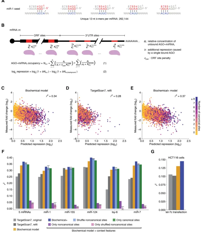
efficacy rely instead on indirect inference through correlative approaches. These models focus on mRNAs with canonical 6–8-nt sites matching the miRNA seed region (Fig. 1A) and train on features known to correlate with targeting efficacy (including the type of site as well as various features of site context, mRNAs, and miRNAs), using datasets that monitor mRNA changes that
occur after introducing a miRNA (Grimson et al. 2007; Agarwal et al. 2015; Gumienny and Zavolan 2015; Paraskevopoulou et al. 2013). Although the correlative model implemented in TargetScan7 performs as well as the best in vivo crosslinking approaches at predicting mRNAs most responsive to miRNA perturbation, it nonetheless explains only a small fraction of the mRNA changes observed upon introducing a miRNA (r2 = 0.14) (Agarwal et al. 2015). This low value indicates that prediction of targeting efficacy has room for improvement, even when accounting for the fact that experimental noise and secondary effects of inhibiting direct targets place a ceiling on the variability attributable to direct targeting. Therefore, we adapted RNA bind-n-seq (RBNS) (Lambert et al. 2014) and a convolutional neural network (CNN) to the study of miRNA–target interactions, with the goal of obtaining the quantity and diversity of affinity measurements needed to better understand and predict miRNA targeting efficacy.
Results
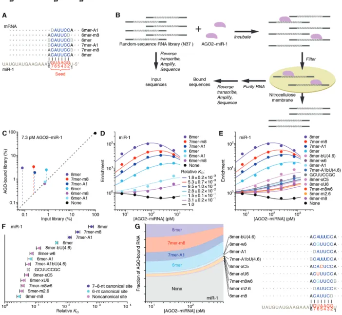
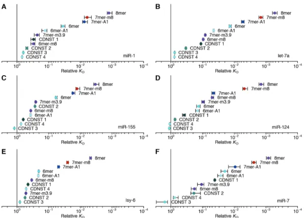
The site-affinity profile of miR-1
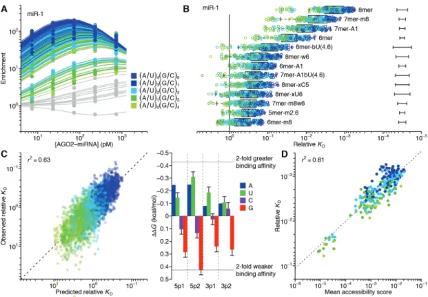
As previously implemented, RBNS provides qualitative relative binding measurements for an RNA-binding protein to a virtually exhaustive list of binding sites (Lambert et al. 2014; Dominguez et al. 2018). A purified RNA-binding protein is incubated with a large library of RNA molecules that each contain a central random-sequence region flanked by constant primer-binding regions. After reaching primer-binding equilibrium, the protein is pulled down and any co-purifying RNA molecules are reverse transcribed, amplified, and sequenced. To extend RBNS to AGO–miRNA complexes (Fig. 1B), we purified human AGO2 loaded with miR-1 (Flores-Jasso, Salomon, and Zamore 2013) (fig. S1A) and set up five binding reactions, each with a different concentration of AGO2–miR-1 (range, 7.3–730 pM, logarithmically spaced) and a constant
concentration of an RNA library with a 37-nt random-sequence region (100 nM). We also modified the protein-isolation step of the RBNS protocol, replacing protein pull-down with nitrocellulose filter binding, reasoning that the rapid wash step of filter binding would improve retention of low-affinity molecules that would otherwise be lost during the wash steps of a pull-down. This modified method was highly reproducible, with high correspondence observed between the 9-nt k-mer enrichments of two independent experiments using different preparations of both AGO2–miR-1 and RNA library (fig. S1B; r2 = 0.86).
When analyzing our AGO-RBNS results, we first examined enrichment of the canonical miR-1 sites, comparing the frequency of these sites in RNA bound in the 7.3 pM AGO2–miR-1 sample with that of the input library. As expected from the site hierarchy observed in meta-analyses of site conservation and endogenous site efficacy (Bartel, 2009), the 8mer site (perfect match to miR-1 nucleotides 2–8 followed by an A) was most enriched (38 fold), followed by the 7mer-m8 site (perfect match to miR-1 nucleotides 2–8, enrichment 14 fold), then the 7mer-A1 site (perfect match to miR-1 nucleotides 2–7 followed by an A, enrichment 7.2 fold), and the 6mer site (perfect match to miR-1 nucleotides 2–7, enrichment 3.0 fold) (Fig. 1, A and C). Little if any enrichment was observed for either the 6mer-A1 site (perfect match to miR-1 nucleotides 2–6 followed by an A) or the 6mer-m8 site (perfect match to miR-1 nucleotides 3–8) at this lowest concentration of 7.3 pM AGO2–miR-1 (Fig. 1, A and C), consistent with their weak signal in previous analyses of conservation and efficacy(Friedman et al. 2009; Agarwal et al. 2015; Kim et al. 2016). Enrichment of sites was quite uniform across the random-sequence region, which indicated minimal influence from either the primer-binding sequences or supplementary pairing to the 3′ region of the miRNA (fig. S1D). Although sites with
Russell, & Cohen, 2005; Wee et al., 2012), the minimal influence of supplementary pairing reflected the rarity of such sites in our library.
Analysis of enrichment of the six canonical sites across all five AGO2–miR-1 concentrations illustrated two hallmarks of this experimental platform (Lambert et al. 2014). First, as the concentration increased from 7.3 pM to 73 pM, enrichment for each of the six site types increased (Fig. 1D), which was attributable to an increase in signal over a constant low background of library molecules isolated even in the absence of AGO2–miR-1. Second, as the AGO2–miR-1 concentration increased beyond 73 pM, 8mer enrichment decreased, and at the highest AGO2–miR-1 concentration, enrichment of the 7mer-m8 and 7mer-A1 site decreased (Fig. 1D). These waning enrichments indicated the onset of saturation for these high-affinity sites (Lambert et al. 2014). These two features, driven by AGO–miRNA-independent
background and partial saturation of the higher-affinity sites, respectively, caused differences in enrichment values for different site types to be highly dependent on the AGO2–miR-1
concentration; the lower AGO2–miR-1 concentrations provided greater discrimination between the higher-affinity site types, the higher AGO2–miR-1 concentrations provided greater
discrimination between the lower-affinity site types, and no single concentration provided results that quantitatively reflected differences in relative binding affinities.
To account for background binding and ligand saturation, we developed a computational strategy that simultaneously incorporated information from all concentrations of an RBNS experiment to calculate relative KD values. Underlying this strategy was an equilibrium-binding model that predicts the observed enrichment of each site type across the concentration series as a function of the KD values for each miRNA site type (including the “no-site” type), as well as the stock concentration of purified AGO2–miR-1 and a constant amount of library recovered as
background in all samples. Using this model, we performed maximum likelihood estimation (MLE) to fit the relative KD values, which explained the observed data well (Fig. 1D). Moreover, these relative KD values were robustly estimated, as indicated by comparing values obtained using results from only four of the five AGO2–miR-1 concentrations (r2 ≥0.994 for each of the
ten pairwise comparisons, fig. S1, F and G). These quantitative binding affinities followed the same hierarchy as observed for site enrichment, but the differences in affinities were of greater magnitude (Fig. 1D and fig. S1C).
Up to this point, our analysis was informed by the wealth of previous computational and experimental data showing the importance of a perfect 6–8-nt match to the seed region (Bartel, 2009). However, the ability to calculate the relative KD of any k-mer of length ≤12 nt (the 12-nt limit imposed by the sparsity of reads with longer k-mers) provided the opportunity for a de novo search for sites, without bias from any previous knowledge. In this search, we 1) calculated the enrichment of all 10-nt k-mers in the bound RNA in the 730 pM AGO2–miR-1 sample, which was the sample with the most sensitivity for detecting low-affinity sites, 2) for the ten most enriched k-mers, determined the extent of complementarity to the miR-1 sequence, 3) assigned a site most consistent with the observed k-mers, and 4) removed all reads containing this newly identified site from both the bound and input libraries. These four steps were iterated until no 10-nt k-mer remained that was enriched ≥10-fold, thereby generating 14 sites for AGO2–miR-1. We then applied our MLE procedure to calculate relative KD values for this expanded list of sites (Fig. 1, E and F).
This unbiased approach demonstrated that the 8mer, 7mer-m8, 7mer-A1, and 6mer sites to miR-1 were the highest-affinity site types of lengths ≤10 nt. It also identified eight novel sites with binding affinities resembling those of the 6mer-m8 and the 6mer-A1 (Fig. 1F). Comparison
of these sites to the sequence of miR-1 revealed that miR-1 can tolerate either a wobble G at position 6 or a bulged U somewhere between positions 4 and 6 and achieve affinity at least 7–11 fold above that of the remaining no-site reads, and that it can tolerate either a mismatched C at position 5 or a mismatched U at position 4 and achieve affinity 4–5 fold above that of the no-site reads. The GCUUCCGC motif also passed our cutoffs, which was more difficult to explain, as it had contiguous complementarity to positions 2–5 of miR-1 flanked by noncomplementary GC dinucleotides on both sides. Nonetheless, among the 1,398,100 possible motifs ≤10 nt, this was the only one that satisfied our criteria yet was difficult to attribute to miRNA pairing.
Our analytical approach and its underlying biochemical model also allowed us to infer the proportion of AGO2–miR-1 bound to each site (Fig. 1G). The 8mer site occupied 3.8–17% of the silencing complex over the concentration course, whereas the 7mer-m8, by virtue of its greater abundance, occupied a somewhat greater fraction of the complex. In aggregate, the marginal sites, including the 6mer-A1, 6mer-m8, and seven noncanonical sites, occupied 6.1– 9.8% of the AGO2–miR-1 complex. Moreover, because of their very high abundance, library molecules with no identified site occupied 32–53% of the complex (Fig. 1G). These results support the inference that the summed contributions of background binding and low-affinity sites to intracellular AGO occupancy is of the same order of magnitude as that of canonical sites, suggesting that an individual AGO–miRNA complex spends about half its time associated with a vast repertoire of background and low-affinity sites (Denzler et al. 2014, 2016). This
phenomenon would help explain why sequences without recognizable sites often crosslink to AGO in cells.
Our results confirmed that AGO2–miR-1 binds the 8mer, 7mer-m8, 7mer-A1, and 6mer sites most effectively and revealed the relative binding affinities and occupancies of these sites.
In addition, our results uncovered weak yet specific affinity to the 6mer-A1 and 6mer-m8 sites plus seven noncanonical sites, all with affinities outside the dynamic range of recent high-throughput imaging experiments (Becker et al. 2019). Although alternative binding sites for miRNAs have been proposed based on high-throughput in vivo crosslinking studies (Chi, Hannon, and Darnell 2012; Loeb et al. 2012; Helwak et al. 2013; Khorshid et al. 2013;
Grosswendt et al. 2014), our approach provided quantification of the relative strength of these sites without the confounding effects of differential crosslinking efficiencies, potentially enabling their incorporation into a quantitative framework of miRNA targeting.
Distinct canonical and noncanonical binding of different miRNAs
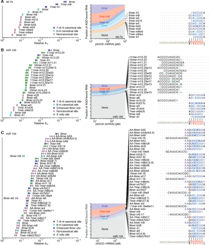
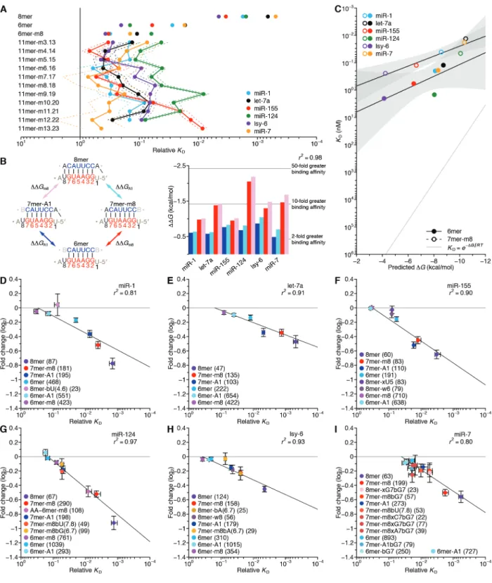
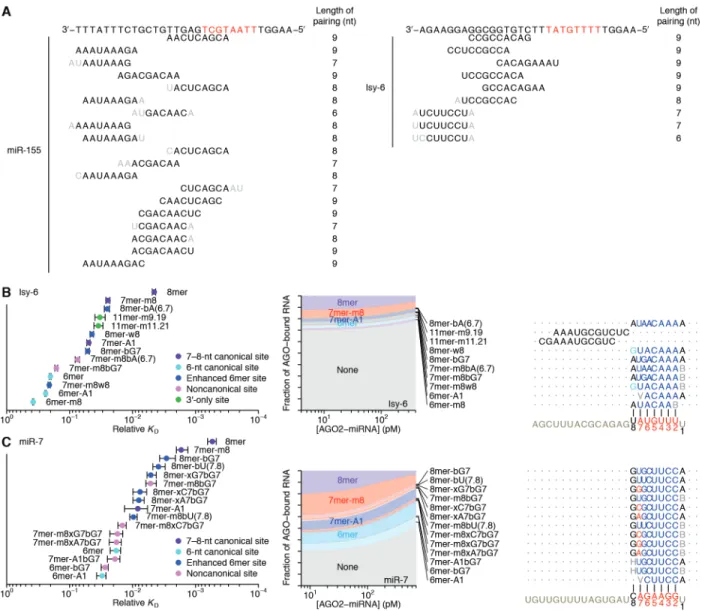
We extended our analysis to five additional miRNAs, including let-7a, miR-7, miR-124, and miR-155 of mammals, chosen for their sequence conservation as well as the availability of data examining their regulatory activities, intracellular binding sites, or in vitro binding affinities (Bartel 2018; Wee et al. 2012; Salomon et al. 2015; Chi, Hannon, and Darnell 2012; Loeb et al. 2012), and lsy-6 of nematodes, which is thought to bind unusually weakly to its canonical sites (Garcia et al. 2011) (Fig. 2 and fig. S2, B and C). In the case of let-7a, previous biochemical analyses have determined the KD values of a few sites (Wee et al. 2012; Salomon et al. 2015), and our values agreed well, which further validated our high-throughput approach (fig. S1H).
The site-affinity profile of let-7a resembled that of miR-1, except the m8 and 6mer-A1 site for let-7a had greater binding affinity than essentially all of the noncanonical sites (Fig. 2A). As with miR-1, the noncanonical sites each paired to the seed region but did so imperfectly, typically with a single wobble, single mismatch, or single-nucleotide bulge, but these