Publisher’s version / Version de l'éditeur:
Journal of Experimental Botany, 66, 7, pp. 1833-1843, 2015-04-01
READ THESE TERMS AND CONDITIONS CAREFULLY BEFORE USING THIS WEBSITE. https://nrc-publications.canada.ca/eng/copyright
Vous avez des questions? Nous pouvons vous aider. Pour communiquer directement avec un auteur, consultez la première page de la revue dans laquelle son article a été publié afin de trouver ses coordonnées. Si vous n’arrivez pas à les repérer, communiquez avec nous à PublicationsArchive-ArchivesPublications@nrc-cnrc.gc.ca.
Questions? Contact the NRC Publications Archive team at
PublicationsArchive-ArchivesPublications@nrc-cnrc.gc.ca. If you wish to email the authors directly, please see the first page of the publication for their contact information.
Archives des publications du CNRC
This publication could be one of several versions: author’s original, accepted manuscript or the publisher’s version. / La version de cette publication peut être l’une des suivantes : la version prépublication de l’auteur, la version acceptée du manuscrit ou la version de l’éditeur.
For the publisher’s version, please access the DOI link below./ Pour consulter la version de l’éditeur, utilisez le lien DOI ci-dessous.
https://doi.org/10.1093/jxb/eru524
Access and use of this website and the material on it are subject to the Terms and Conditions set forth at
The FRK1 mitogen-activated protein kinase kinase kinase (MAPKKK)
from Solanum chacoense is involved in embryo sac and pollen
development
Lafleur, E.; Kapfer, C.; Joly, V.; Liu, Yang; Tebbji, F.; Daigle, C.;
Gray-mitsumune, M.; Cappadocia, M.; Nantel, A.; Matton, D. P.
https://publications-cnrc.canada.ca/fra/droits
L’accès à ce site Web et l’utilisation de son contenu sont assujettis aux conditions présentées dans le site LISEZ CES CONDITIONS ATTENTIVEMENT AVANT D’UTILISER CE SITE WEB.
NRC Publications Record / Notice d'Archives des publications de CNRC:
https://nrc-publications.canada.ca/eng/view/object/?id=c756149d-75b5-4190-b99c-af1fa85c6fd0 https://publications-cnrc.canada.ca/fra/voir/objet/?id=c756149d-75b5-4190-b99c-af1fa85c6fd0
RESEARCH PAPER
The FRK1 mitogen-activated protein kinase kinase kinase
(MAPKKK) from Solanum chacoense is involved in embryo
sac and pollen development
Edith Laleur1,*, Christelle Kapfer1,*, Valentin Joly1, Yang Liu1, Faiza Tebbji1,2,†, Caroline Daigle1,
Madoka Gray-Mitsumune1,‡, Mario Cappadocia1, André Nantel2 and Daniel P. Matton1,§
1 Institut de recherche en biologie végétale, Département de sciences biologiques, Université de Montréal, 4101 rue Sherbrooke est,
Montréal, QC H1X 2B2, Canada
2 Institut de recherche en biotechnologie, Conseil national de recherches du Canada, 6100 Avenue Royalmount, Montréal,
QC H4P 2R2, Canada
*These authors contributed equally to this work.
† Present address: Department of Biology, McGill University, 1205 Avenue Docteur Penield, Montréal, QC H3A 1B1, Canada. ‡ Present address: Department of Biology, Concordia University, 7141 rue Sherbrooke ouest, Montréal, QC H4B 1R6, Canada. § To whom correspondence should be addressed. E-mail: dp.matton@umontreal.ca
Received 30 September 2014; Revised 2 December 2014; Accepted 5 December 2014
Abstract
The fertilization-related kinase 1 (ScFRK1), a nuclear-localized mitogen-activated protein kinase kinase kinase (MAPKKK) from the wild potato species Solanum chacoense, belongs to a small group of pMEKKs that do not pos-sess an extended N- or C-terminal regulatory domain. Initially selected based on its highly speciic expression proile following fertilization, in situ expression analyses revealed that the ScFRK1 gene is also expressed early on during female gametophyte development in the integument and megaspore mother cell and, later, in the synergid and egg cells of the embryo sac. ScFRK1 mRNAs are also detected in pollen mother cells. Transgenic plants with lower or barely detectable levels of ScFRK1 mRNAs lead to the production of small fruits with severely reduced seed set, resulting from a concomitant decline in the number of normal embryo sacs produced. Megagametogenesis and microgametogenesis were affected, as megaspores did not progress beyond the functional megaspore (FG1) stage and the microspore collapsed around the irst pollen mitosis. As for other mutants that affect embryo sac develop-ment, pollen tube guidance was severely affected in the ScFRK1 transgenic lines. Gametophyte to sporophyte com-munication was also affected, as observed from a marked change in the transcriptomic proiles of the sporophytic tissues of the ovule. The ScFRK1 MAPKKK is thus involved in a signalling cascade that regulates both male and female gamete development.
Key words: Embryo sac development, gametophyte to sporophyte communication, MAPKKK, megagametogenesis, microgametogenesis, pollen tube guidance, seed and fruit development, Solanaceae.
Introduction
Flowering plants or angiosperms exhibit a two-staged life cycle, alternating between a short-lived haploid gametophyte generation composed of only a few cells, and a temporally predominant diploid sporophytic generation. The haploid generation begins with specialized diploid cells (mother cells)
of the sporophyte that undergo meiosis to give rise to haploid spores. These spores undergo cell proliferation and differen-tiation to produce multicellular haploid gametophytes. The male gametophyte (pollen grain) develops within the stamen of the anther and consists of two sperm cells encased within
This is an Open Access article distributed under the terms of the Creative Commons Attribution License (http://creativecommons.org/licenses/by/3.0/), which permits unrestricted reuse, distribution, and reproduction in any medium, provided the original work is properly cited.
© The Author 2015. Published by Oxford University Press on behalf of the Society for Experimental Biology. This paper is available online free of all access charges (see http://jxb.oxfordjournals.org/open_access.html for further details)
a vegetative cell. The female gametophyte (embryo sac or megagametophyte) develops within the carpel of the ovary and, in most cases, leads to the formation of an eight-nucle-ate, seven-celled Polygonum-type embryo sac harbouring one egg cell, two synergid cells, three antipodal cells, and one central cell. The major function of gametophyte generation is thus to produce haploid gametes, the egg and sperm cells, which, upon fusion (one sperm cell fusing with the egg cell and the other with the central cell, giving rise to the embryo and endosperm, respectively), will lead to a new sporophytic generation.
Numerous genetic screens have been carried out to iden-tify genes affecting gametophyte development (Feldmann
et al., 1997; Bonhomme et al., 1998; Christensen et al., 1998;
Pagnussat et al., 2005; Muralla et al., 2011). One would expect that, considering the developmental complexity involved in gamete production, genes involved in intracellular signalling and cell–cell communication would be readily found among gametophyte essential genes. From the curated data set of
Arabidopsis genes required for gametophyte function listed in
Muralla et al. (2011), of the 173 genes that displayed a game-tophyte defective phenotype, only 9 (0.05%) are classiied as being involved in signalling pathways, of which three are protein kinases. These include the two-component histidine kinase CKI1 (At2g47430) involved in cytokinin perception that severely affects the female gametophyte but only weakly affects the male gametophyte (Pischke et al., 2002; Hejatko
et al., 2003); the FUSED (FU) Ser/Thr kinase, involved in cytokinesis, that severely affects both male and female game-tophytes (Oh et al., 2005); and the calmodulin-binding recep-tor-like cytoplasmic kinase 2 (CRCK2) that severely affects male gametophyte development but only weakly affects the female gametophyte (Boavida et al., 2009).
With >1000 and 1400 members, respectively, the kinase superfamily from Arabidopsis and rice represents a very large fraction of the proteome in comparison with other eukary-otes. For example, in Arabidopsis, kinases represent 4% of the proteome, compared with ~2% in human, Caenorhabditis
ele-gans, Drosophila melanogaster, and yeast (Shiu and Bleecker, 2003; Shiu et al., 2004; Champion et al., 2004; Dardick et al., 2007). Nonetheless, only a few kinases have been found in gametophyte defective mutant screens, most probably due to the high level of functional redundancy found in major kinase groups, mainly the receptor kinase family (>600 in Arabidopsis and 1200 in rice) and the mitogen-activated protein kinase (MAPK) superfamily (MAPK, MAPKK, MAPKKK, and MAPKKKK) with >100 members in both Arabidopsis and rice (Hamel et al., 2006; Rao et al., 2010). An example of such redundancy in the MAPKKK family is observed with the
Arabidopsis ANP1/2/3 kinases that regulate cell division where
the triple mutant is not transmitted through the male and female gametes, although other phenotypes, such as reduction of plant size (anp2/anp3 double mutants), were also observed (Krysan et al., 2002). The ANP1/2/3 kinases are related to the tobacco NPK1 MAPKKK that is part of a cascade, the NACK–PQR pathway, possibly involved in cellularization/ differentiation which occurs during stage FG5 (Nishihama
et al., 2002; Soyano et al., 2003; Chevalier et al., 2011).
In this study, the isolation and functional characteriza-tion of a new MAPKKK from the pMEKK subfamily in
Solanum chacoense is described. Down-regulation of this
sin-gle MAPKKK named ScFRK1 (fertilization-related kinase) severely affects both embryo sac and pollen development and leads to partial parthenocarpic fruit production upon pollination.
Materials and methods
Plant material and plant transformation
All plant material and growth conditions are as described in Gray-Mitsumune et al. (2006). For sense and antisense constructs, the ScFRK1 cDNA was inserted in a modiied pBin19 transformation vector with a Caulilower mosaic virus (CaMV) 35S double enhancer promoter (Bussière et al., 2003). Sense and antisense constructs were individually transformed in Agrobacterium tumefaciens LBA4404 by electroporation. Solanum chacoense plants were transformed by the leaf disc method as previously described (Matton et al., 1997). DNA and RNA analyses
Nucleic acid isolation, blotting, and hybridization are as described in O’Brien et al. (2007). Sequence analysis and phylogeny are as described previously (Gray-Mitsumune et al., 2006). Accession num-bers are AY427828, KC768863 (ScFRK1), AY427829 (ScFRK2), and KC768864 (ScFRK3).
Protein localization through transient expression
The ScFRK1 coding region was fused in-frame to the N-terminus of green luorescent protein (GFP) in the 35S-driven Gateway vector pMDC83 (Curtis and Grossniklaus, 2003). A 35S::GFP construct was used as a control. Microparticle bombardment was performed as described previously (Germain et al., 2008).
Tissue ixation and electron microscopy observations
For pollen viability estimation through outer structure analysis, fresh pollen was observed with a Hitachi S-3000N variable pressure scanning electron microscope at 30 Pa and 15 kV. For transmission electron microscopy (TEM), samples were ixed in 2.5% glutaral-dehyde in 0.1 M sodium cacodylate buffer pH 7.4, post-ixed in 2% osmium tetroxide (OsO4) in the same buffer, dehydrated in
etha-nol, and embedded in Spurr’s resin. Observations were made on a Hitachi H-7500 microscope. Statistics for pollen defects observed by scanning electron microscopy (SEM) were scored from ≥100 pollen grains per wild-type (WT) or transgenic line.
Cytological analysis of microsporogenesis
Meiosis and microspore development were studied by squash-ing anthers in lacto-acetic orcein (1% orcein), accordsquash-ing to Dyer’s method (Dyer, 1963), modiied by substituting propionic acid with acetic acid. To monitor starch accumulation inside developing pol-len, anthers were also squashed in iodine (I2–KI), according to
Eriksson’s method (Eriksson, 1962). Pollen fertility was estimated after staining freshly collected mature pollen with acetocarmine (1%) or iodine. All observations were made with a Zeiss AxioImager M1 microscope equipped with an AxioCam HRc camera.
Tissue ixation and optical microscopy observations
Pistils were ixed in FAA for 24 h at 4 °C (50% ethanol, 1.35% for-maldehyde, and 5% glacial acetic acid). Samples were then dehy-drated in increasing series of tert-butyl alcohol (from 70% to pure
tert-butyl alcohol). Pistils were iniltrated with Paraplast Plus par-afin at 60 °C. Thin sections (10 μm) were prepared from embed-ded samples and tissue sections were stained in 0.5% Astra Blue and 1% safranine after parafin removal. Alternatively, thin tions (10 μm) were prepared from embedded samples and tissue sec-tions were stained in 0.05% Toluidine Blue. In situ hybridizasec-tions were performed as described previously (O’Brien et al., 2005). For differential interference contrast (DIC) microscopy observations, loral buds were dissected and ovaries were ixed in FAA solution overnight (50% ethanol, 0.5% acetic acid, and 1% formaldehyde). Clearing of ovules was performed with an increasing ratio of etha-nol–methylsalicylate solutions (0:100, 75:25, 50:50, 25:75, 100:0) for 1 h each and left overnight in 100% methylsalicylate. After dissection from the placenta, ovules were observed with a Zeiss AxioImager M1 microscope equipped with an AxioCam HRc camera.
cDNA microarrays analysis
DNA microarrays comprised 7741 expressed sequence tags (ESTs) corresponding to 6374 unigenes derived from fertilized ovary cDNA libraries covering embryo development from zygote to late tor-pedo stages (Germain et al., 2005). Experimental conditions were as described previously (Gray-Mitsumune et al., 2006; Tebbji et al., 2010).
Results
Sequence analysis and cellular localization of the ScFRK1 kinase
Using a subtraction selection screen targeting only genes weakly expressed during fertilization and early embryo-genesis, ive members from the MAPKKK family were iso-lated in S. chacoense, a self-incompatible wild potato species (Germain et al., 2005). Three of these, named ScFRK1–
ScFRK3, were phylogenetically classiied in the pMEKK
subfamily of the MAPKKKs (Gray-Mitsumune et al., 2006), although they differed signiicantly from the majority of the pMEKKs due to their small size, consisting of practically only a kinase domain with little N- or C-terminal putative regulatory domains. In Arabidopsis thaliana, 21 MAPKKKs are classiied as pMEKKs (mean size ~675 amino acids), with ive of those (AtMAPKKK17–AtMAPKKK21) <400 amino acids in length. Three of these (AtMAPKKK19– AtMAPKKK21) are closely related to the ScFRK1–ScFRK3 family in S. chacoense, although ScFRK3 is closer to the MAPKKK19–MAPKKK21 group (Supplementary Fig. S1A available at JXB online). Amino acid sequence identity within this group ranges from 31% to 75% (46–85% simi-larity) (Supplementary Fig. S1B). Functional analysis of this family has only been reported for the ScFRK2 kinase, which has been shown to be involved in ovule and pollen development (Gray-Mitsumune et al., 2006; O’Brien et al., 2007), and AtMAPKKK20, involved in the osmotic stress response (Kim et al., 2012). Here the functional analysis of the ScFRK1 kinase is reported.
The ScFRK1 clone codes for an open reading frame of 323 amino acids with an estimated mol. wt of 37 kDa. A short C-terminal region of 42 amino acids (position 282–323,
Fig. 1A) follows kinase subdomain XI. Analysis of the sequence revealed the presence of a cluster of two short basic
amino acid sequences predicted to form a bipartite nucleo-plasmin-type nuclear localization sequence (NLS) (Brameier
et al., 2007) (Fig. 1A). The algorithm also predicted a higher NLS potential for the irst one (NLS1). To verify this, the ScFRK1 coding region was fused in-frame to the N-terminus of GFP. A 35S::GFP construct was used as a control. As expected for the GFP alone, expression was detected in both the cytoplasm and the nucleus (Fig. 1B; GFP control). In con-trast, luorescence of the ScFRK1–GFP fusion protein was restricted to the nuclei and co-localized with the 4′,6-diamid-ino-2-phenylindole (DAPI) signal (Fig. 1B; ScFRK1 full). To determine if the two basic sequences were used as a bipartite NLS or if they acted redundantly, the individual role of each predicted NLS was analysed. As shown in Fig. 1B, a sharp cytoplasmic luorescence was observed in most of the onion cells bombarded with the ScFRK1Δ1 or ScFRK1Δ1Δ2 constructs. However, ambiguous nucleocytoplasmic locali-zation was obtained in ~45% of bombarded cells with the ScFRK1Δ1 construct. Consistent with the above-mentioned prediction, the deletion of NLS2 did not disrupt the nuclear localization of ScFRK1 to the same extent. Only 25% of the cells bombarded with the Δ2 construct showed cytoplasmic or nucleocytoplasmic luorescence (data not shown). This suggests that although the two C-terminal NLS in ScFRK1 form a bipartite NLS, NLS1 predominates, consistent with the NLS strength prediction.
Pollination and fertilization trigger a stepwise decrease of ScFRK1 mRNA abundance in ovaries
The ScFRK1 expression pattern was determined by RNA gel blot analysis with various vegetative (roots, stems, and leaves), generative (petals), and reproductive tissues (stamens, pol-len, styles, and ovaries). At anthesis, strong ScFRK1 mRNA accumulation was only observed in the ovary (Fig. 2A) and, to a lesser extent, in the style (Fig. 2B). Faint expression Fig. 1. Characterization of the ScFRK1 bipartite nuclear localization signal. (A) Details of the wild type and modiied C-terminal region of ScFRK1 constructs used for protein localization studies. The ScFRK1 coding region was fused in-frame to the N-terminus of GFP. The two short basic amino acid sequences predicted to form the NLS are shown in bold. (B) Visualization of GFP expression (top) and DAPI (nucleus) localization (bottom) in bombarded onion cells expressing the fusion constructs. Scale bar=25 µm.
could also be detected in the leaf (Fig. 2A). Pollination and fertilization had dramatic effects on ScFRK1 accumulation in ovaries. Although pollen tubes only reach the ovules ~36 h after landing on the stigma, ScFRK1 steady-state mRNA levels had already signiicantly declined 12 h after pollina-tion and were barely detectable after fertilizapollina-tion (Fig. 2A). To determine if this stepwise down-regulation of ScFRK1 steady-state mRNA levels was caused by pollination and fertilization, and was not developmentally regulated, non-pollinated pistils were collected from 3 d before anthesis to 3 d after anthesis. As shown in Fig. 2B, peak accumulation of ScFRK1 mRNAs is observed 1 d prior to anthesis and, without pollination, only slightly declines in ovary thereafter. Even 3 d after anthesis, strong ScFRK1 mRNA accumulation is still observed, conirming the stepwise roles of pollination and fertilization in ScFRK1 mRNA accumulation in ovaries. ScFRK1 is expressed in both the sporophyte and the gametophyte
In order to determine the spatial expression pattern of the
ScFRK1 gene, in situ RNA hybridizations were performed
using gynoecia from various developmental stages (Fig. 2C). On the day of anthesis, ScFRK1 mRNA signal was strongly detected in ovules and in the vascular tissue and, to a lesser extent, in the ovary wall (Fig. 2C, i). At medium magniica-tion, the strongest accumulation is detected in the integument of the ovule as well as in the epidermis of the placenta that is in direct continuity with the ovule integument (Fig. 2C, iii). Closer examination of the ScFRK1 expression pattern revealed that the gene is expressed in the synergids and the egg cell of the embryo sac (Fig. 2C, v). The asymmetric stain-ing pattern observed is typical for these cells since the two synergids have their large vacuole located towards the cha-lazal pole, while the vacuole of the egg cell has the reverse orientation. This concentrates the mRNA signal at the micropylar pole for the synergids and towards the chalazal pole for the egg cell. In young lower buds bearing ovules at the megaspore mother cell (MMC) stage, ScFRK1 mRNA signal was already observed in the single ovule integument (Fig. 2C, vii). Solanum chacoense produces unitegmic-ten-uinucellate ovules, a trait that occurs almost universally in the asterid clade (Albach et al., 2001). At this stage, denser staining was consistently observed at the tip of the growing integument as well as in the MMC (Fig. 2C, vii). At the same
Fig. 2. ScFRK1 expression analysis. (A) RNA gel blot analysis of the ScFRK1 gene. All tissues were collected from greenhouse-grown plants. Fertilized ovaries were dissected from pistils 0–96 hours after pollination (HAP). A 10 μg aliquot of total RNA isolated from S. chacoense tissues was blotted and probed using the full-length ScFRK1 cDNA (upper panel). Membranes were stripped and re-probed using a partial 18S rRNA to ensure equal loading of each RNA sample (lower panel). (B) RNA gel blot analysis of the ScFRK1 gene before and after anthesis in unpollinated pistil tissues. S, style; O, ovary. (C) In situ localization of ScFRK1 transcripts in ovules and anthers. (i and ii) Unfertilized mature ovary sections. (iii and iv) Magniication of unfertilized mature ovary sections. (v and vi) ScFRK1
expression in the ovule integument and embryo sac of mature ovules at anthesis. (vii and viii) ScFRK1 expression in young ovules at the megaspore mother cell stage isolated from ~2 mm lower buds. (ix and x) Cross-sections of young anthers isolated from ~2 mm lower buds. i, iii, v, vii, and ix, antisense probe. ii, iv, vi, viii and x, control sense probe. Digoxigenin labelling is visible as red to purple staining. All hybridizations used 10 μm thick sections and an equal amount of either ScFRK1 sense or antisense probe. ec, egg cell; in, integument; mmc, megaspore mother cell; ov, ovule; ow, ovary wall (pericarp); pe, placenta epidermis; pl, placenta; sy, synergid, zy, zygote. Images v–viii were taken under DIC optics in order to better show the cellular structures. Scale bars: 200 μm (i, ii); 50 μm (iii, iv, ix, x); and 20 μm (v–viii).
stage, ScFRK1 mRNA signal was also detected in developing anthers, more prominently observable in pollen mother cells (PMCs; Fig. 2C, ix).
ScFRK1 knock-down transgenic lines show reproductive defects
In order to assign a function to the ScFRK1 gene, transgenic plants carrying an ScFRK1 sense or antisense construct were generated. The ScFRK1 cDNA was placed downstream of a double enhancer CaMV35S promoter in a modiied pBin19 vector in a sense or antisense orientation (Bussière et al., 2003). Kanamycin-resistant plants were grown to maturity in the greenhouse and cross-pollinated to determine if any abnormal phenotype linked to sexual reproduction, based on the ScFRK1 expression proile, could be observed. Numerous plants showed a marked reduction in fruit size, irrespective of the transgenic population from which they were isolated (sense or antisense lines). The ScFRK1 expression level was monitored in transgenic lines showing a decrease in fruit size by RNA gel blot analyses of ovaries collected on the day of anthesis. All the affected lines showed a reduced accumulation of ScFRK1 mRNAs. Three lines expressing variable levels of
ScFRK1, down to almost undetectable levels, were chosen
for further analyses (Fig. 3). Lines S27 and S1 were co-sup-pressed lines retrieved from the ScFRK1 sense overexpression population, while AS13 came from the antisense expressed population. Overall plant growth and vegetative development appeared unaffected in all ScFRK1 transgenic lines. However, the ScFRK1 down-regulated lines exhibited severe defects in seed and fruit development. Fruit volume ranged from 13% (S1) to 35–40% (AS13 and S27) when compared with the WT (Fig. 3A, C) or transgenic plants unaffected in ScFRK1 expression (data not shown). Seed production was also strongly affected, with S1 producing only 2% of the normal seed content of an S. chacoense fruit, while AS13 and S1 pro-duced only 15% of the WT seed count (Fig. 3A). The reduced seed set could thus explain the small fruit size observed. Down-regulation of ScFRK1 affects embryo sac development
Since ScFRK1 is expressed before and after anthesis and with an expression level inluenced by both pollination and fertilization, the reduced seed set observed could result from either aberrant ovule development or post-fertilization seed abortion. To assess this, cleared ovules from all lines were observed prior to pollination. As shown in Table 1, decreas-ing levels of ScFRK1 mRNA led to a concomitant decrease in the number of normal embryo sacs observed. In the most strongly affected transgenic line, Scfrk1-S1, this led to an almost complete absence of normal embyro sacs, explaining the strongly reduced seed set observed. In order to determine when the defect appeared during female gametophyte devel-opment, ovules from different developmental stages were observed in lowers buds of the Scfrk1-S1 line, since almost all of its ovules were affected at anthesis. Table 2 shows the cor-respondence between lower bud length and developmental
stages of the S. chacoense female gametophyte. Observation of cleared ovules revealed that megasporogenesis was unaf-fected. Ovules from the MMC to the functional mega-spore stage could be routinely observed in both WT plants (Fig. 4A, B, E) and the Scfrk1-S1 line (Fig. 4C, D, G). Thus, meiosis of the MMC ultimately producing the functional megaspore appeared normal in the ScFRK1 transgenic line. Afterwards, no cell divisions could be observed in the
Scfrk1-S1 line (Fig. 4H that would correspond to the dyad Fig. 3. Analyses of ScFRK1 transgenic plants. (A) Seed count and fruit volume measurements from WT plants and transgenic lines S1, S27, and AS13. (B) RNA gel blot analyses of ScFRK1 mRNA (upper panel) and 18S rRNA (lower panel) of ovary tissues derived from WT plants and transgenic lines S1, S27, and AS13. (C) Comparison of fruits and fruit slices from WT plants and transgenic lines S1, S27, and AS13. Scale bar=1 cm. (This igure is available in colour at JXB online.)
Table 1. Percentage of normal and abnormal embryo sacs as observed in cleared ovules by DIC microscopy
CC, central cel; EC, egg cell; ES, embryo sac; SY, synergid. Fifty ovules were observed for each plant line in two consecutive generations.
Plant line Normal ES Modiied ES ES absent
WT 96 4 0
frk1-AS13 44 30 26
frk1-S27 32 22 46
frk1-S1 0 6 94
ES with 1 EC, 2 SY, and 1CC
ES with fewer cells
No ES observed
stage in the WT shown in Fig. 4F, and later at anthesis in
Fig. 4J or K). In the WT plants, the surviving megaspore underwent three successive mitotic divisions to produce an eight-nucleate megagametophyte. In WT ovules at anthesis, the three antipodals have already degenerated and only the central cell with its fused polar nuclei, the synergids, and the egg cell are visible (Fig. 4I). In the transgenic lines, affected embryo sacs showed either a clear lack of organization with a shrunken and illed embryo sac (Fig. 4J) or retained a single
cell (Fig. 4K). Thus, down-regulation of the ScFRK1 gene affects megagametogenesis as the functional megaspore never progresses beyond the FG1 stage.
Pollen development is also affected in ScFRK1 transgenic lines
Although no ScFRK1 mRNA signal could be detected in mature pollen, a strong signal was observed in cross-sections of lower buds in the anthers (Fig. 2C, ix). The ~2 mm lower buds corresponded to anthers where the sporogenous cells are differentiating into PMCs. Pollen from the Scfrk1-S27 and Scfrk1-S1 lines was used to pol-linate a fully compatible S. chacoense genotype. When pollen from the WT (S-alleles S12S14, also used as the host
for plant transformation) was used to pollinate this fully compatible genotype (S-alleles S11S13), 100% of the
polli-nated lowers developed fruits (n=20). When pollen from the Scfrk1-S27 line was used, only one in 20 pollinations led to the production of a fruit, while the use of pollen from the Scfrk1-S1 line did not lead to fruit production (n=20). This suggested that pollen development was also affected in transgenic plants down-regulated in ScFRK1 mRNA lev-els. Pollen observation in dehiscent anthers revealed that, in transgenic lines, pollen viability was severely affected, as estimated by acetocarmine staining of >1000 pollen grains scored per line (Fig. 5, upper panels). Compared with the Table 2. Correspondence between lower buds length and
developmental stages of S. chacoense female gametophyte
Flower bud length Female gametophyte development stage 1.0–1.5 mm Ovule primordia
1.5–2.5 mm Megaspore mother cell 2.5–3.0 mm Dyad
3.0–4.0 mm Tetrad and functional megaspore 4.0–5.0 mm Uninucleated and binucleated embryo sac 5.0–6.0 mm Tetranucleated and octanucleated embryo sac Open lower–anthesis Mature embryo sac four nuclei (antipodals have
degenerated)
Abbreviations: FRK, fertilization-related kinase; MAPK, mitogen-activated protein kinase; MAPKK, mitogen-mitogen-activated protein kinase kinase; MAPKKK, mitogen-activated protein kinase kinase kinase; WT, wild type.
Fig. 4. Megasporogenesis and megagametogenesis in WT S. chacoense and in the Scfrk1-S1 transgenic plant line. Cleared ovules were
observed by DIC microscopy. Stages determined following Table 2. (A, C) Megaspore mother cell stage. (B, D) Tetrad stage. (E, G) Functional megaspore stage (FG1). (F, H) Binucleated megagametophyte stage. In F, the arrows point to the two nuclei in the late two-nucleate stage (FG3). In H, embryo sac development is halted at the FG1 stage in the Scfrk1-S1 transgenic line. (I, J, K) Mature embryo sac stage at anthesis (FG8). mmc, megaspore mother cell; fm, functional megaspore; cc, central cell; ccn, central cell nucleus; sy, synergid cell; ec, egg cell. Scale bar=20 μm.
Fig. 5. Pollen viability estimation in WT and two ScFRK1 transgenic plant lines. Pollen viability and phenotype were estimated through viable stain analysis and electron microscopy observations. Upper panels: pollen was stained with 1% acetocarmine. Viable pollen grains are lightly stained in pink, while dead pollen cells are shown as empty and shrivellled shells. Scale bar=50 μm. Middle panels: examination of fresh pollen outer structure through scanning electron microscopy (SEM) analysis under low vacuum conditions. Affected mutants lines produced shrivelled and collapsed pollen grains. Scale bar=20 μm. Lower panels: transmission electron microscopy (TEM) of pollen sections revealed that the collapsed or shrivelled pollen grains are devoid of cytoplasm and organelles. Scale bar=10 μm.
WT, where >98% of the pollen grains are stainable, <20% of the Scfrk1-S27 transgenic pollen and <1% of the Scfrk1-S1 transgenic pollen appeared viable. When fresh pollen is observed by SEM under low vacuum conditions, affected lines produced shrivelled and collapsed pollen (Fig. 5, mid-dle panels). TEM of pollen sections revealed that the col-lapsed or shrivelled pollen grains were devoid of a dense cytoplasm and of organelles, in sharp contrast to WT pol-len (Fig. 5, lower panels).
Cytological analysis of microsporogenesis
In order to determine precisely when the pollen started to collapse, a cytological analysis of developing pollen was conducted. Microscopic observations of PMCs both at late prophase II/metaphase II of meiosis (Fig. 6A, B) and at the tetrad stage (Fig. 6C, D) revealed no differences between the WT and the Scfrk1-S1 line. Similarly, the young mononucle-ate microspores from Scfrk1-S1 and the WT line appeared indistinguishable (Fig. 6E, F), suggesting that the defect occurred at later stages of development. This was indeed the case since at later stages of gametogenesis the two lines started to show substantial differences. Mitosis occurred nor-mally in the microspores of the WT, and was followed by dif-ferentiation of the generative and vegetative nuclei (Fig. 6G).
In contrast, in line Scfrk1-S1, <20% of the microspores underwent the irst pollen mitosis (PMI), but <1% continued their development, leading to differentiation of the generative and vegetative nuclei (Fig. 6I). In S. chacoense, microsporo-genesis proceeds similarly to the microsporomicrosporo-genesis reported in both tomato (de Nettancourt and Eriksson, 1968) and
S. verrucosum (de Nettancourt and Dijstra, 1969). In these species, starch accumulation begins shortly after PMI, while starch hydrolysis begins 2 d before anthesis and is completed by the time the lower opens. In the present study, the iodine test was used to monitor starch accumulation and hydroly-sis in developing pollen. The test revealed that almost all WT young pollen started to accumulate starch just after the pollen mitosis (Fig. 6H), and starch accumulation reached a maximum 3 d before anthesis. At this time, the pollen grains appeared almost black (Fig. 6L), and only the gen-erative nucleus remained visible with the acetocarmine stain (Fig. 6K), the vegetative nucleus being completely hidden by the starch grains. In contrast, in Scfrk1-S1 pollen, starch started to accumulate only in a limited number (<1%) of the pollen grains (Fig. 6J), most probably in those where mitosis had been completed and differentiation of the generative and vegetative nuclei had occurred. Most probably these pollen grains continued their development in a similar way to the WT (Fig 6M, N). At anthesis, almost all WT pollen appeared viable, having completed starch hydrolysis (Fig. 6O). In line
Scfrk1-S1, however, >99% of the pollen appeared shrunken,
with only very few grains showing a normal appearance (Fig. 6P).
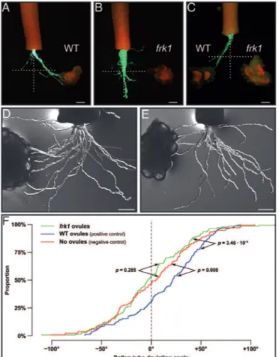
Pollen tube guidance is severely affected in the ScFRK1 transgenic plants
The integrity of the embryo sac, the female gametophyte, has been shown to be a prerequisite for the ability of the ovule to attract pollen tubes. Mutants lacking a mature female gametophyte or affected in the development of its cells are defective in pollen tube guidance (reviewed in Marton and Dresselhaus, 2010; Chevalier et al., 2011;
Takeuchi and Higashiyama, 2011). To determine if the
ScFRK1 transgenic lines are also affected in pollen tube
guidance, a semi in vivo pollen tube guidance system was used. The Scfrk1-S1 line was selected for this analysis as it showed the lowest percentage of functional embryo sacs. WT lowers were hand pollinated with fully compatible pol-len and styles were collected 24 h later. The detached styles are then laid on a microscopic slide covered with pollen tube growth medium with ovules placed at ~650 μm from the cut style end, a distance corresponding to the radius of the ovary. Pollen tubes start to emerge ~30 hours after pollination (HAP). Figure 7 shows the result of two dif-ferent assay systems. First, a single-choice assay was used with ovules from either WT or Scfrk1-S1 plants as shown in Fig. 7A, B, respectively. Attraction was determined from the bulk pattern obtained, with each pollinated style being counted as one assay. An attraction phenotype was scored when there was a clear trend and the majority of the pol-len tubes grew toward the ovules as observed in Fig. 7A Fig. 6. Comparative cytological analyses of WT and Scfrk1-S1 pollen.
Late prophase II/metaphase II showing 12 chromosomes in WT (A) and in transgenics plants (B). Tetrads surrounded by callose from WT plants (C) and transgenics (D). Mononucleate microspores just released from the tetrads of WT plants (E) and transgenics (F). Young binucleate WT pollen stained with lacto-acetic orcein showing generative (dark) and vegetative (pale) nuclei (G), and initiation of starch accumulation, visualized with the iodine test (H). The same developmental stage as G and H in transgenics (I, J). WT pollen grains 3 d before anthesis (K, L). The same developmental stage as K and L in transgenics (M, N). Mature WT (O) and transgenic (P) pollen stained with iodine; by this time, starch hydrolysis has been completed. Note the collapsed pollen grains in the transgenic line surrounding one viable pollen grain (P). Scale bar=20 μm.
with WT ovules, while typical absence of attraction can be observed in Fig. 7B with Scfrk1-S1 ovules. When WT ovules were used, 59% of the assays showed pollen tubes grow-ing toward WT ovules (n=80), while only 10% of the pol-len tubes grew toward the Scfrk1-S1 ovules (n=60), giving a highly signiicant P<0.0001 value in a two-sample bino-mial test. Furthermore, although 10% of the assays with the Scfrk1-S1 ovules showed pollen tubes growing toward them, pollen tubes never reached these ovules, in contrast to what is observed with WT ovules in Fig. 7A. Next, a two-choice assay system was used, with equidistant ovules from WT and Scfrk1-S1 plants (Fig. 7C). In this case, no differ-ence in attraction between WT and Scfrk1-S1 ovules (null hypothesis) would lead to a 50:50 distribution. Out of 65 assays, 83% (54) showed clear attraction to WT ovules while 17% (11) grew toward Scfrk1-S1 ovules, which is highly sig-niicant in a one-sample binomial test (P<0.0001). As in the single-choice system, when pollen tubes grew toward
Scfrk-S1 ovules, none reached the ovules. It is interesting to
note that similar results were obtained in Torenia fournieri, where the investigators used a microluidistic device to chan-nel the pollen tubes toward a targeted ovary or a control
(Horade et al., 2013). To conirm the results of this quick assay system, the two-sample Kolmogorov–Smirnov test, a non-parametric test comparing empirical distribution func-tions in two samples, widely used in axon guidance studies, was also used. In this case, growth angles for all distinguish-able pollen tubes were calculated from their exit to their end point on a total of ~150 pollen tubes from ive semi in vivo single-choice assays (Fig. 7D–F). A mean angle of 2.5 ° was obtained for the negative control (without ovules), – 0.8 ° for assays with Scfrk1-S1 ovules, and 16.3 ° for assays with WT ovules. Attraction was thus observed with WT ovules (P=0.008) but not with Scfrk1-S1 ovules (P=0.285).
Embryo sac-dependent gene expression and gametophytic to sporophytic communication
In order to isolate embryo sac-expressed genes and spo-rophytic genes that would depend on embryo sac gene expression, the Scfrk1-S1 ovule transcriptome was com-pared with the one from WT ovules using a 7.7K DNA microarray made from ovule-derived ESTs (Tebbji et al., 2010). Analysis of variance (ANOVA) testing, along with a Benjamini and Hochberg multiple testing correction algo-rithm, was used to select ESTs that showed a statistically signiicant difference in transcript abundance between the WT and the Scfrk1-S1 ovules. A Welch’s t-test (P<0.05) was initially used to compare the proiles from the Scfrk1-S1 versus control and the control versus control comparisons. They were then further restricted with a ≥1.5 fold variation (1.5 cut-off up or down). Seventy-nine ESTs correspond-ing to 69 unigenes showed statistically lower transcript abundance between the WT and the Scfrk1-S1 ovules (Supplementary Table S1 at JXB online). These genes, iden-tiied as down-regulated in Scfrk1-S1 ovules, are thus most probably embryo sac-expressed genes. On the other hand, 118 ESTs (98 unigenes) were transcribed signiicantly more in Scfrk1-S1 ovules, and may thus be associated with spo-rophytic adaptation to the absence of female gametophyte (Supplementary Table S2).
Blast2GO was used to analyse functional category enrich-ment between up- and down-regulated genes, and a Fisher’s exact test was performed to determine which categories were signiicantly regulated (Supplementary Table S3 at JXB online). Among them, gene ontology (GO) terms related to chromatin remodelling (e.g. DNA packaging, DNA con-formation change, histone exchange, nucleosome organiza-tion, and chromatin assembly and disassembly), cell cycle control (e.g. cell cycle, interphase), intracellular traficking, as well as development of reproductive tissues characterized down-regulated genes. On the other hand, up-regulated genes are mostly associated with response to stress (e.g. defence response, systemic acquired resistance, response to cold and to radiation), senescence (ageing, organ senescence), and amino acid metabolism (e.g. serine family amino acid meta-bolic process).
SignalP 4.1, SecretomeP 1.0, and NetNGlyc 1.0 were also used to predict secretion and glycosylation of pro-teins encoded by regulated ESTs (Supplementary Fig. S3, Fig. 7. Pollen tube guidance is affected in ScFRK1 transgenic plants. (A)
Single-choice pollen tube guidance assay with WT S. chacoense ovules. Pollen tubes express the GFP marker under the control of the tomato Lat52 promoter (A–C). (B) Single-choice pollen tube guidance assay with Scfrk1-S1 ovules. (C) Two-choice pollen tube guidance assay with WT and Scfrk1-S1 ovules. (D, E) Single-choice pollen tube guidance assay with limited pollen load in order to measure pollen tube angles from WT (D) and Scfrk1-S1 ovules (E). (F) Kolmogorov–Smirnov statistical analysis from the angle distribution in D and E. Scale bar=200 μm.
Supplementary Table S4 at JXB online). A characteristic of embryo sac genes is their enrichment for small and secreted proteins (Jones-Rhoades et al., 2007). Interestingly, proteins harbouring a signal peptide are signiicantly more frequent in down- and up-regulated genes than in unregulated genes (P=5.6e–12 and P=3.8e–6, respectively). In contrast, the
pro-portion of unconventionally secreted proteins decreases sig-niicantly in both data sets (P=0.05 and P=0.02, respectively). Furthermore, 30% of secreted peptides potentially encoded in down and up ESTs correspond to small, cysteine-rich pro-teins (CRPs) such as defensins, lipid transfer propro-teins, rapid alkalinization factor (RALF) peptides, Papaver S-like pro-teins, and γ-thionins. Conventionally, up-regulated secreted proteins are signiicantly less glycosylated (P=0.03), which holds true for up-regulated CRPs (P=4.6e–4).
Discussion
Among the protein kinases known to affect key aspects of plant reproductive development only a few have been char-acterized from the MAPK superfamily. These include the
Arabidopsis YODA (MAPKKK4), involved in embryo and
stomata development (Bergmann et al., 2004; Lukowitz
et al., 2004); the S. chacoense FRK2 MAPKKK, involved in ovule (Gray-Mitsumune et al., 2006) and pollen development (O’Brien et al., 2007); the redundant Arabidopsis MAP3Kε1 (MAPKKK7) and MAP3Kε2 (MAPKKK6), involved in pollen viability (Chaiwongsar et al., 2006); the Arabidopsis MPK6, involved in anther, inlorescence as well as in embryo development (Bush and Krysan, 2007); and Arabidopsis MPK3 and MPK6, also involved in defence responses and in ovule development (Wang et al., 2008). Recently, mpk3/mpk6 double mutant pollen tubes were also shown to be defective speciically in the funicular guidance phase (Guan et al., 2014).
In this study, the characterization of ScFRK1, a novel MAPKKK from S. chacoense that affects both male and female gametophyte development, is reported. Although the three FRK genes, ScFRK1, 2, and 3, are expressed in repro-ductive tissues, they are not genetically redundant, since down-regulation of ScFRK1 is not complemented by the presence of the others. They also share limited amino acid sequence identity, <45% (Supplementary Fig. S1 at JXB online), and are not all predicted to be in the same subcel-lular compartment (only ScFRK1 and 2 are predicted to be localized in the nucleus). Furthermore, an RNA interference effect on ScFRK2 and 3 is unlikely considering the low nucle-otide sequence identity between ScFRK1 and the two other kinases (<38%) and the fact that no stretches of >15 and 12 identical nucleotides are found with ScFRK2 and ScFRK3, respectively. Thus, as expected, ScFRK2 and 3 expression levels were not signiicantly down-regulated in the Scfrk1-S1 line (Supplementary Fig. S2), conirming that the pheno-type observed is due to the down-regulation of the ScFRK1 gene. Furthermore, down-regulation of ScFRK2 showed no observable phenotype, nor any reproductive defects, while overexpression lines led to the conversion of ovules into car-pelloid structures (Gray-Mitsumune et al., 2006).
As for ScFRK2, the other member of the family previously examined, ScFRK1 had a complex and peculiar expression pattern. While ScFRK2 expression is weak at anthesis and is fertilization induced, ScFRK1 is strongly expressed at anthe-sis and is pollination repressed. Since ScFRK1 is expressed early on during pollen and ovule development, with pheno-types observed in down-regulated lines affecting the game-tophytes, high expression in the ovary at anthesis and the post-pollination steady-state ScFRK1 mRNA decrease are puzzling and remain to be examined.
In Arabidopsis, the putative orthologues of the Solanum FRK family members are MAPKKK19, 20, and 21. Interestingly, MAPKKK20 was among the genes regulated by the male germline-speciic DUO1 MYB transcription factor (Borg et al., 2011). In contrast to ScFRK1 transgenic lines, DUO1 mutants only affect the male gametophyte.
DUO1 mutants progress normally through PMI but fail to
complete the generative cell cycle (Durbarry et al., 2005;
Rotman et al., 2005). In contrast, in line Scfrk1-S1, <20% of the microspores underwent PMI, and <1% continued their development, leading to differentiation of the generative and vegetative nuclei (Fig. 6I). Consistent with this, starch accumulation that normally begins shortly after PMI in sola-naceous species (Fig. 6J) was only detected in <1% of the
Scfrk1-S1 pollen grains (Fig. 6J), and those were presumably the ones that had progressed through PMI and completed the differentiation of the generative and vegetative nuclei. Thus, in pollen, ScFRK1 most probably does not act in the same pathway or would act upstream of genes such as DUO1.
As ScFRK1 is expressed in both sporophytic and game-tophytic tissues, it is puzzling that only the gametophyte is affected in down-regulated transgenic lines. Considering that the ovule sporophytic tissue in ScFRK1 transgenic lines does not show any evidence of developmental defects at maturity, and that in described gametophytic mutants sporophytic tis-sue develops normally (Bencivenga et al., 2011), this suggests that down-regulation of ScFRK1 mostly affects gametophyte development. Since very weak expression can still be detected in ScFRK1 down-regulated lines, a different threshold effect between the expression observed in the integument and the young ovule at the MMC stage (see Fig. 2, vii for WT expres-sion) could also explain why the sporophyte is not affected. Interestingly, ScFRK1 expression is not equally distributed in the integument, with higher levels at the tip of the integument in young ovules at the MMC stage (Fig. 2, vii) and, in mature ovules, in the cell layers immediately surrounding the embryo sac, the inner epidermis, also called the ‘integumentary tape-tum’ in unitegmic families such ase Solanaceae (Fig. 2iii,
v). The inner epidermis has been endowed with numerous features. During the initial stages of gametophyte develop-ment, the ultrastructure of the inner epidermis cells has been described as akin to that of meristematic cells. By dividing profusely, its cells were considered to provide the necessary conditions to co-ordinate the intensive growth of the embryo sac. Once fully differentiated, the inner epidermis provides nutrition to the embryo sac (Kapil and Tiwari, 1978).
As expected from Arabidopsis mutants that lack a func-tional embryo sac (reviewed in Chevalier et al., 2011;
Takeuchi and Higashiyama, 2011; Dresselhaus and Franklin-Tong, 2013), or for plants where the synergid cells had been physically ablated (Higashiyama et al., 2001), pollen tube guidance was severely compromised in the Scfrk1-S1 trans-genic line (Fig. 7). Absence of the embryo sac led to the isolation of embryo sac-dependent genes (down-regulated genes; Supplementary Table S1 at JXB online) that could be directly involved in pollen tube guidance or other cell–cell interaction functions. Among these, two RALFs, ScRALF4 and 5, were isolated. These are closely related to Arabidopsis RALF27 and 32 and are quite ubiquitously expressed in
S. chacoense (Germain et al., 2005). No functions have yet been ascribed to these RALFs. However, involvement of RALF peptides in plant reproduction has been recently highlighted with the characterization of ScRALF3, involved in sporophyte to gametophyte communication. Although expressed in the sporophytic tissue of the ovule, down-reg-ulation of ScRALF3 expression by RNA interference led to improper embryo sac development through loss of embryo sac nuclei polarization and an increase in asynchronous divi-sions (Chevalier et al., 2013). Absence of the embryo sac also had an impact on the surrounding sporophytic tissue with the isolation of several up-regulated genes in the ovule, sug-gesting interactions between the female gametophyte and the maternal sporophyte, as observed previously in Arabidopsis (Johnston et al., 2007). Furthermore, the majority of pro-teins encoded in up and down ESTs correspond to secreted peptides, 30% of which are small CRPs. These proportions are in line with recent studies in Arabidopsis (Supplementary Figs S4 and S5).
Interestingly, the GO terms associated with down-regulated genes in Scfrk1-S1 (chromatin remodelling, cell cycle control, intracellular traficking, and development of reproductive tis-sues) are consistent with the dynamic nature of the develop-ment of the embryo sac, with its rounds of mitosis, cellular polarization, and positioning, as well as with the expression of elevated amount of secreted proteins, such as from the synergid iliform apparatus. Similarly, GO terms associated with up-regulated genes are predominantly linked to stress responses, as if absence of the female gametophyte would be sensed as a scar and elicit a wound or defence response. The characterization of the other members of this MAPK cas-cade, such as MAPKK and MAPK, as well as downstream nuclear or cellular targets should reveal essential steps in male and female gametophytes development.
Supplementary data
Supplementary data are available at JXB online.
Figure S1. (A) Section of a pMEKK phylogenetic tree showing the most closely related orthologues of ScFRK1–3 in A. thaliana. (B) Percentage sequence identity and similar-ity between S. chacoense FRK1, 2, and 3 and A. thaliana MAPKKK19, 20, and 21, based on a ClustalW multiple pro-tein sequence alignment.
Figure S2. Speciic down-regulation of ScFRK1 in trans-genic plants.
Figure S3. Secretion and glycosylation predictions for pro-teins up-, down- and not regulated in the Scfrk1 mutant.
Figure S4. Conventional and unconvential secretion pre-dictions for proteins regulated in Scfrk1 and other ovule mutants.
Figure S5. CRP content in proteins regulated in Scfrk1 and other ovule mutants.
Table S1. Information about frk1 down-regulated ESTs.
Table S2. Information about frk1 up-regulated ESTs.
Table S3. GO term enrichment in frk1 up- and down-regu-lated ESTs compared with unregudown-regu-lated ESTs.
Table S4. Summary of secretion and glycosylation predic-tions on Scfrk1 up-, down-, and unregulated ESTs.
Acknowledgements
This work was supported by the NSERC (Natural Sciences and Engineering Research Council of Canada), FRQNT (Fonds de recherche du Québec– Nature et technologies), and by the Canada Research Chair program. EL and CK are recipients of MSc fellowships from the NSERC; while FT and CD are recipients of PhD fellowships from FRQNT and the NSERC, respectively.
References
Albach DC, Soltis PS, Soltis DE. 2001. Patterns of embryological and biochemical evolution in the asterids. Systematic Botany 26, 242–262. Bencivenga S, Colombo L, Masiero S. 2011. Cross talk between the sporophyte and the megagametophyte during ovule development. Sexual Plant Reproduction 24, 113–121.
Bergmann DC, Lukowitz W, Somerville CR. 2004. Stomatal development and pattern controlled by a MAPKK kinase. Science 304, 1494–1497.
Boavida LC, Shuai B, Yu HJ, Pagnussat GC, Sundaresan V, McCormick S. 2009. A collection of Ds insertional mutants associated with defects in male gametophyte development and function in Arabidopsis thaliana. Genetics 181, 1369–1385.
Bonhomme S, Horlow C, Vezon D, de Laissardiere S, Guyon A, Ferault M, Marchand M, Bechtold N, Pelletier G. 1998. T-DNA mediated disruption of essential gametophytic genes in Arabidopsis is unexpectedly rare and cannot be inferred from segregation distortion alone. Molecular and General Genetics 260, 444–452.
Borg M, Brownield L, Khatab H, Sidorova A, Lingaya M, Twell D. 2011. The R2R3 MYB transcription factor DUO1 activates a male germline-speciic regulon essential for sperm cell differentiation in Arabidopsis. The Plant Cell 23, 534–549.
Brameier M, Krings A, MacCallum RM. 2007. NucPred—predicting nuclear localization of proteins. Bioinformatics 23, 1159–1160. Bush SM, Krysan PJ. 2007. Mutational evidence that the Arabidopsis MAP kinase MPK6 is involved in anther, inflorescence, and embryo development. Journal of Experimental Botany 58, 2181–2191.
Bussière F, Ledû S, Girard M, Héroux M, Perreault J-P, Matton DP. 2003. Development of an efficient cis–trans–cis ribozyme cassette to inactivate plant genes. Plant Biotechnology Journal 1, 423–435.
Chaiwongsar S, Otegui MS, Jester PJ, Monson SS, Krysan PJ. 2006. The protein kinase genes MAP3K epsilon 1 and MAP3K epsilon 2 are required for pollen viability in Arabidopsis thaliana. The Plant Journal 48, 193–205.
Champion A, Jouannic S, Guillon S, Mockaitis K, Krapp A, Picaud A, Simanis V, Kreis M, Henry Y. 2004. AtSGP1, AtSGP2 and MAP4K alpha are nucleolar plant proteins that can complement ission yeast mutants lacking a functional SIN pathway. Journal of Cell Science 117, 4265–4275.
Chevalier E, Loubert-Hudon A, Matton DP. 2013. ScRALF3, a secreted RALF-like peptide involved in cell–cell communication between
the sporophyte and the female gametophyte in a solanaceous species. The Plant Journal 73, 1019–1033.
Chevalier E, Loubert-Hudon A, Zimmerman EL, Matton DP. 2011. Cell–cell communication and signalling pathways within the ovule: from its inception to fertilization. New Phytologist 192, 13–28.
Christensen CA, Subramanian S, Drews GN. 1998. Identiication of gametophytic mutations affecting female gametophyte development in Arabidopsis. Developmental Biology 202, 136–151.
Curtis MD, Grossniklaus U. 2003. A gateway cloning vector set for high-throughput functional analysis of genes in planta. Plant Physiology 133, 462–469.
Dardick C, Chen J, Richter T, Ouyang S, Ronald P. 2007. The rice kinase database. A phylogenomic database for the rice kinome. Plant Physiology 143, 579–586.
de Nettancourt D, Dijstra M. 1969. Starch accumulation in the microspores of a solanum species and possible implications in mutation breeding. American Potato Journal 46, 239–242.
de Nettancourt D, Eriksson G. 1968. Effects of irradiation upon starch formation and starch hydrolysis in tomato microspores. Hereditas 60, 167–176.
Dresselhaus T, Franklin-Tong N. 2013. Male–female crosstalk during pollen germination, tube growth and guidance, and double fertilization. Molecular Plant 6, 1018–1036.
Durbarry A, Vizir I, Twell D. 2005. Male germ line development in Arabidopsis. duo pollen mutants reveal gametophytic regulators of generative cell cycle progression. Plant Physiology 137, 297–307. Dyer A. 1963. The use of lacto-propionic orcein in rapid squash methods for chromosome preparations. Stain Technology 38, 85–90.
Eriksson G. 1962. Radiation induced reversions of a waxy allele in barley. Radiation Botany 2, 35–39.
Feldmann KA, Coury DA, Christianson ML. 1997. Exceptional segregation of a selectable marker (KanR) in Arabidopsis identiies genes important for gametophytic growth and development. Genetics 147, 1411–1422.
Germain H, Gray-Mitsumune M, Laleur E, Matton DP. 2008. ScORK17, a transmembrane receptor-like kinase predominantly expressed in ovules is involved in seed development. Planta 228, 851–862.
Germain H, Rudd S, Zotti C, Caron S, O’Brien M, Chantha S-C, Lagacé M, Major F, Matton DP. 2005. A 6374 unigene set corresponding to low abundance transcripts expressed following fertilization in Solanum chacoense Bitt., and characterization of 30 receptor-like kinases. Plant Molecular Biology 59, 513–529. Gray-Mitsumune M, O’Brien M, Bertrand C, Tebbji F, Nantel A, Matton DP. 2006. Loss of ovule identity induced by overexpression of the fertilization-related kinase 2 (ScFRK2), a MAPKKK from Solanum chacoense. Journal of Expermental Botany 57, 4171–4187.
Guan Y, Lu J, Xu J, McClure B, Zhang S. 2014. Two mitogen-activated protein kinases, MPK3 and MPK6, are required for funicular guidance of pollen tubes in Arabidopsis. Plant Physiology 165, 528–533.
Hamel LP, Nicole MC, Sritubtim S, et al. 2006. Ancient signals: comparative genomics of plant MAPK and MAPKK gene families. Trends in Plant Science 11, 192–198.
Hejatko J, Pernisova M, Eneva T, Palme K, Brzobohaty B. 2003. The putative sensor histidine kinase CKI1 is involved in female gametophyte development in Arabidopsis. Molecular Genetics and Genomics 269, 443–453.
Higashiyama T, Yabe S, Sasaki N, Nishimura Y, Miyagishima S, Kuroiwa H, Kuroiwa T. 2001. Pollen tube attraction by the synergid cell. Science 293, 1480–1483.
Horade M, Kanaoka MM, Kuzuya M, Higashiyama T, Kaji N. 2013. A microluidic device for quantitative analysis of chemoattraction in plants. RSC Advances 3, 22301–22307.
Johnston AJ, Meier P, Gheyselinck J, Wuest SE, Federer M, Schlagenhauf E, Becker JD, Grossniklaus U. 2007. Genetic
subtraction proiling identiies genes essential for Arabidopsis reproduction and reveals interaction between the female gametophyte and the maternal sporophyte. Genome Biology 8, R204.
Jones-Rhoades MW, Borevitz JO, Preuss D. 2007. Genome-wide expression proiling of the Arabidopsis female gametophyte identiies families of small, secreted proteins. PLoS Genetics 3, 1848–1861.
Kapil RN, Tiwari SC. 1978. The integumentary tapetum. Botanical Review 44, 457–490.
Kim JM, Woo DH, Kim SH, Lee SY, Park HY, Seok HY, Chung WS, Moon YH. 2012. Arabidopsis MKKK20 is involved in osmotic stress response via regulation of MPK6 activity. Plant Cell Reports 31, 217–224.
Krysan PJ, Jester PJ, Gottwald JR, Sussman MR. 2002. An Arabidopsis mitogen-activated protein kinase kinase kinase gene family encodes essential positive regulators of cytokinesis. The Plant Cell 14, 1109–1120.
Lukowitz W, Roeder A, Parmenter D, Somerville C. 2004. A MAPKK kinase gene regulates extra-embryonic cell fate in Arabidopsis. Cell 116, 109–119.
Marton ML, Dresselhaus T. 2010. Female gametophyte-controlled pollen tube guidance. Biochemical Society Transactions 38, 627–630. Matton DP, Maes O, Laublin G, Xike Q, Bertrand C, Morse D, Cappadocia M. 1997. Hypervariable domains of self-incompatibility RNases mediate allele-speciic pollen recognition. The Plant Cell 9, 1757–1766.
Muralla R, Lloyd J, Meinke D. 2011. Molecular foundations of reproductive lethality in Arabidopsis thaliana. PLoS One 6, e28398. Nishihama R, Soyano T, Ishikawa M, et al. 2002. Expansion of the cell plate in plant cytokinesis requires a kinesin-like protein/MAPKKK complex. Cell 109, 87–99.
O’Brien M, Chantha SC, Rahier A, Matton DP. 2005. Lipid signaling in plants. cloning and expression analysis of the obtusifoliol 14α-demethylase from Solanum chacoense Bitt., a pollination- and fertilization-induced gene with both obtusifoliol and lanosterol demethylase activity. Plant Physiol 139, 734–749.
O’Brien M, Gray-Mitsumune M, Kapfer C, Bertrand C, Matton DP. 2007. The ScFRK2 MAP kinase kinase kinase from Solanum chacoense affects pollen development and viability. Planta 225, 1221–1231. Oh SA, Johnson A, Smertenko A, Rahman D, Park SK, Hussey PJ, Twell D. 2005. A divergent cellular role for the FUSED kinase family in the plant-speciic cytokinetic phragmoplast. Current Biology 15,
2107–2111.
Pagnussat GC, Yu HJ, Ngo QA, Rajani S, Mayalagu S, Johnson CS, Capron A, Xie LF, Ye D, Sundaresan V. 2005. Genetic and molecular identiication of genes required for female gametophyte development and function in Arabidopsis. Development 132, 603–614.
Pischke MS, Jones LG, Otsuga D, Fernandez DE, Drews GN, Sussman MR. 2002. An Arabidopsis histidine kinase is essential for megagametogenesis. Proceedings of the National Academy of Sciences, USA 99, 15800–15805.
Rao KP, Richa T, Kumar K, Raghuram B, Sinha AK. 2010. In silico analysis reveals 75 members of mitogen-activated protein kinase kinase kinase gene family in rice. DNA Research 17, 139–153.
Rotman N, Durbarry A, Wardle A, Yang WC, Chaboud A, Faure JE, Berger F, Twell D. 2005. A novel class of MYB factors controls sperm-cell formation in plants. Current Biology 15, 244–248.
Shiu SH, Bleecker AB. 2003. Expansion of the receptor-like kinase/Pelle gene family and receptor-like proteins in Arabidopsis. Plant Physiology 132, 530–543.
Shiu SH, Karlowski WM, Pan R, Tzeng YH, Mayer KF, Li WH. 2004. Comparative analysis of the receptor-like kinase family in Arabidopsis and rice. The Plant Cell 16, 1220–1234.
Soyano T, Nishihama R, Morikiyo K, Ishikawa M, Machida Y. 2003. NQK1/NtMEK1 is a MAPKK that acts in the NPK1 MAPKKK-mediated MAPK cascade and is required for plant cytokinesis. Genes and Development 17, 1055–1067.
Takeuchi H, Higashiyama T. 2011. Attraction of tip-growing pollen tubes by the female gametophyte. Current Opinion in Plant Biology 14, 614–621. Tebbji F, Nantel A, Matton DP. 2010. Transcription proiling of fertilization and early seed development events in a solanaceous species using a 7.7 K cDNA microarray from Solanum chacoense ovules. BMC Plant Biology 10, 174.
Wang H, Liu Y, Bruffett K, Lee J, Hause G, Walker JC, Zhang S. 2008. Haplo-insuficiency of MPK3 in MPK6 mutant background uncovers a novel function of these two MAPKs in Arabidopsis ovule development. The Plant Cell 20, 602–613.