Publisher’s version / Version de l'éditeur:
Proceedings of the Ultrasonics Symposium 2006, 2006-10-03
READ THESE TERMS AND CONDITIONS CAREFULLY BEFORE USING THIS WEBSITE. https://nrc-publications.canada.ca/eng/copyright
Vous avez des questions? Nous pouvons vous aider. Pour communiquer directement avec un auteur, consultez la
première page de la revue dans laquelle son article a été publié afin de trouver ses coordonnées. Si vous n’arrivez pas à les repérer, communiquez avec nous à PublicationsArchive-ArchivesPublications@nrc-cnrc.gc.ca.
Questions? Contact the NRC Publications Archive team at
PublicationsArchive-ArchivesPublications@nrc-cnrc.gc.ca. If you wish to email the authors directly, please see the first page of the publication for their contact information.
NRC Publications Archive
Archives des publications du CNRC
This publication could be one of several versions: author’s original, accepted manuscript or the publisher’s version. / La version de cette publication peut être l’une des suivantes : la version prépublication de l’auteur, la version acceptée du manuscrit ou la version de l’éditeur.
Access and use of this website and the material on it are subject to the Terms and Conditions set forth at
A Piezoelectric Membrane Sensor for Biomedical Monitoring
Ono, Y.; Liu, Q.; Kobayashi, M.; Jen, C.-K.; Blouin, A.
https://publications-cnrc.canada.ca/fra/droits
L’accès à ce site Web et l’utilisation de son contenu sont assujettis aux conditions présentées dans le site LISEZ CES CONDITIONS ATTENTIVEMENT AVANT D’UTILISER CE SITE WEB.
NRC Publications Record / Notice d'Archives des publications de CNRC:
https://nrc-publications.canada.ca/eng/view/object/?id=20277bbc-4c38-431a-8ec9-204661a919dc https://publications-cnrc.canada.ca/fra/voir/objet/?id=20277bbc-4c38-431a-8ec9-204661a919dcA Piezoelectric Membrane Sensor for
Biomedical Monitoring
Y. Ono1, Q. Liu2, M. Kobayashi3, C.-K. Jen3, and A. Blouin3 1
Department of Systems and Computer Engineering, Carleton University, Ottawa, ON, K1S 5B6, CANADA
2
Department of Electrical and Computer Engineering, McGill University, Montreal, QC, H3A 2A7, CANADA
3
Industrial Materials Institute, National Research Council Canada, Boucherville, QC, J4B 6Y4, CANADA (yuuono@sce.carleton.ca)
Abstract—A piezoelectric membrane sensor, consisting
of a metal foil, a piezoelectric ceramic film and a top electrode, has been developed. Thick lead zirconate titanate composite film was coated onto a stainless steel (SS) foil and a top electrode was formed using a sliver paste. The SS foil served as the bottom electrode as well as the substrate. Due to the porosity in the piezoelectric film and the thin metallic membrane substrate, the high flexibility was realized. The porosity was observed by a scanning electron microscopy. This membrane sensor has been worked as a unimorph-type bending sensor as well as an ultrasonic sensor. Biomedical applications using this sensor were demonstrated. The sensor was directly attached onto a wrist and signals corresponding to arterial pulse waves have been successfully obtained. The signals had a signal-to-noise ratio of better than 20dB. Breathing curves were also measured on a human belly. In addition, ultrasonic signal reflected from a finger bone was observed with a pulse-echo technique. Thus, this sensor could be used as a wearable sensor, which does not disturb daily life activities including sleeping, for real-time and continuous monitoring of personal health conditions.
Keywords-piezoelectric membrane sensor; unimorph; ultrasonic probe; biomedical monitoring
I. INTRODUCTION
Due to dietary habits, lack of exercise, and the daily stresses of modern life and society, lifestyle diseases such as diabetes, obesity, hypertension and heart diseases have become serious problems to maintaining a high quality of life. Daily monitoring of physiological activities and parameters, including pulse rate, blood pressure and breathing, would provide valuable information for the diagnosis and treatment of these diseases.
This information would be beneficial not only to health care providers and their patients but also to apparently non-symptomatic persons in order to identify such diseases in the earliest stages or, indeed, to prevent them entirely. For example, the arterial pulse waveform has been shown to contain considerable information for the diagnosis of cardiac conditions [1, 2]. However, current measurement techniques, which make use of photoelectric methods
or pressure meters, may not be suitable for continuous monitoring or may have inadequate sensitivity to measure physiological signals in sufficient detail to provide diagnostic value.
Piezoelectric unimorphs and bimorphs have been used for a variety of sensor applications, such as pressure, impact, acceleration and vibration measurements, as well as actuators [3]. Various kinds of piezoelectric ceramics, polymers and single crystals have been used to develop these sensors.
For biomedical applications such as monitoring of physiological activities and medical diagnosis involving imaging, flexibility of the sensors may be beneficial in order to have a steady contact of the sensors onto the human body which are curved, deformable, movable and unstable [4]. Such flexibility could be also desirable to develop wearable and compact biomedical sensors which do not disturb daily activities.
Piezoelectric polymers such as polyvinylidene fluoride have high flexibility. However, they have low electromechanical coupling coefficients compared to piezoelectric ceramics. Thus, sensitivity of the sensors made with the piezoelectric polymers may be insufficient to detect detailed features of physiological signals.
In this study, piezoelectric membrane sensor, consisting of a metal foil, a piezoelectric ceramic film and a top electrode, has been developed. This membrane sensor has worked as a unimorph-type bending sensor as well as an ultrasonic sensor. Noninvasive monitoring of pulse waves and breathing curves and an ultrasonic pulse-echo measurement of a bone in a finger are demonstrated with the membrane sensor developed.
II. FABRICATION
The fabrication process is based on a sol-gel composite spray method developed previously [5, 6]. The piezoelectric lead zirconate titanate (PZT) powders were purchased with a particle size distribution of 1-3µm. The powders were dispersed into PZT sol-gel solution by a ball milling method to
achieve the paint. An airbrush was then used to spray the PZT sol-gel composite directly onto a stainless steel (SS) square foil of 33mm by 33mm. The thickness of the SS foil was 38µm to assure the flexibility of the device. The foil served as both substrate and bottom electrode. SS was selected because of its high corrosion resistance and harmlessness to a human body.
After each PZT layer spray, thermal treatments such as drying, firing and annealing were carried out. Each layer had a thickness of 5-15µm and eight to twelve layers were made until the film reached the desired thickness. The final film thickness was 70µm measured by a micrometer. The film was then electrically poled using a corona discharging technique. The corona poling method was chosen because it could pole the piezoelectric film over a large area with different curvatures and with ease. A silver paste top electrode of 15mm by 15mm was deposited at room temperature. The thickness of the silver top electrode was about 20µm with fairly good uniformity. This convenient approach makes the selection of electrode size, i.e. the sensor size, simple.
Finally, the entire structure was sandwiched by polyimide films so that it can be waterproof and can operate at temperatures up to 150°C. Copper strips were used for electrical connection. PZT films with a thickness ranging from 10-80µm can operate as ultrasonic sensors having a centre frequency ranging from 5-30MHz. This frequency range is suitable for medical and biomedical applications because of its sufficient spatial resolution and acceptable ultrasonic attenuation. The detailed ultrasonic performance of this membrane sensor has been reported in [6]. Fig. 1 (a) and (b) show photographs of the sensor without and with electrical cables and polyimide protection films, respectively.
(b)
PZT Composite Film
Silver Paste Top Electrode SS Foil Membrane
(a)
Copper Strip Polyimide Film
Figure 1. Photographs of a piezoelectric membrane sensor, consisting of 38µm-thick SS foil, PZT composite film and silver paste top electrode, (a) without and (b) with electrical cables and
polyimide protection films.
III. CHARACTERIZATION OF PZT COMPOSITE FILM
Fig. 2 presents a scanning electron microscopy (SEM) image of the PZT composite film. Fig. 2 shows that the film was not dense but porous. Due to the thin metallic membrane substrate and the porosity
in the piezoelectric film, a highly flexible sensor was obtained.
1µm 1µm
Figure 2. A SEM image of a PZT composite film.
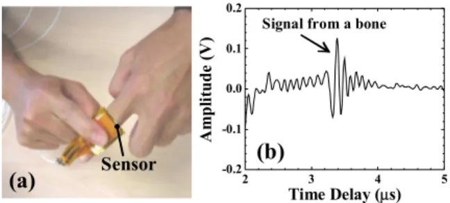
Table I summarizes physical, mechanical and electrical properties of the PZT composite films in comparison to those of the PZT powder grains from which the PZT films were made. The data of the powders were obtained from the data sheet of the manufacturer.
TABLE I. PHYSICAL, MECHANICAL AND ELECTRICAL PROPERTIES OF PZT COMPOSITE FILMS AT ROOM TEMPERATURE, COMPARED TO THOSE OF PZT POWDER FROM WHICH THE FILMS WERE MADE.
Properties Film Powder*
Density ρ (kg/m3) 4.4 x 10³ 7.7 x 10³
Curie temperature Tc (C°) --- 350 Longitudinal wave
velocity VL (m/s) 2200 3580 Shear wave velocity VS (m/s) 1160 1880
Young’s modulus E (N/m2) 1.5 x 10¹⁰ 7.1 x 10¹⁰ Dielectric constant 130 1800 Longitudinal piezoelectric coefficient d33 (m/V) 37 x 10-12 400 x 10-12 Transverse piezoelectric coefficient d31 (m/V) -27 x 10-12 -175 x 10-12
* Data from the manufacturer
The density, ρ, and longitudinal wave ultrasonic velocity, VL, of the film were obtained using a
resonant spectroscopy method [7]. The ρ was about 60% of the raw material of the PZT powder due to the porosity in the film. The VL was 2200m/s. Shear wave
ultrasonic velocity, VS, was estimated 1160m/s using
the measured VL of the film and the ratio of VS/VL of
the powder, assuming that such ratios are the same between the film and powder. Young’s modulus, E, of the film was calculated 1.5 x 10¹⁰ N/m2 with the ρ, VL
and VS of the film in Table I using: 2 S 2 L 2 S 2 L 2 S V V ) V 4 V 3 ( V E − − ρ = (1)
The relative dielectric constant of the PZT composite films was about 130, measured by an impedance analyzer at 1kHz. Longitudinal piezoelectric constant, d33, of the PZT composite film
was obtained using a laser interferometer by
measuring the thickness changes, ∆h, of the PZT film under a 10kHz AC voltage, V, applied to the film. To reduce the measurement error on the film thickness variations induced by the bending motion of the sensor, a sample with the PZT film coated onto a 12mm-thick SS substrate was used for d33
measurement. The d33 of the PZT composite film was
found to be 37 x 10-12 m/V using:
V h
d33 =∆ (2)
Transverse piezoelectric constant, d31, of the PZT
composite film was measured using low coherence interferometry [8]. The PZT film was coated onto the entire surface of a 38µm-thick SS foil of 13mm by 29mm. A silver paste top electrode was deposited onto the entire area of the film to get a unimorph bending sensor. One end of the sensor was firmly clamped. A tip displacement, , of the other end due to its deflection by a 1Hz AC voltage applied to the sensor was measured. A d31 of this PZT composite
film was then evaluated to be -27 x 10-12 m/V using:
V L t ) B 1 ( AB 3 ) B 2 B 3 B 2 ( A 2 B A 1 d 2 P 3 2 4 2 31 δ + + + + + = , (3)
where A = Em/Ep, B = tm/tp , Em and Ep are Young’s
modulus of the SS foil and the PZT composite film respectively, tm and tp are their thickness and L
(=20mm) is the length of the free area of the sensor between the clamping point and the tip [9].
IV. APPLICATIONS
A. Pulse wave monitoring
Arterial pulse waveforms contain significant information for the diagnosis of hypertension and cardiac failure. The membrane sensor in Fig. 1 was directly attached and fixed onto a wrist using fingers. Mechanical displacements of the skin surface of the wrist due to heartbeat caused a bending motion of the sensor which was converted into the electric signals. The electric voltage output of the sensor was observed using a digital oscilloscope. Fig. 3 shows a typical result of pulse wave monitoring. The signals corresponding to the pulse waves have been successfully obtained with a signal-to-noise ratio of better than 20dB. It is noted that the signals in Fig. 3 are raw data without amplification, filtering or averaging.
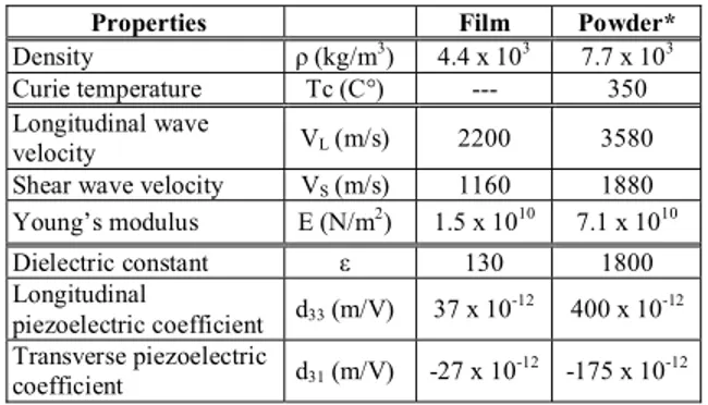
B. Breathing curve monitoring
Monitoring of breathing conditions during sleeping is one of useful information for appropriate diagnosis of sleep disorders such as obstructive sleep apnea syndrome [10, 11]. Polysomnography in a sleep laboratory is the standard technique for the diagnosis of sleep disorders, but the test is expensive and not
widely available. Development of less costly systems available for home sleep study is desirable.
Measurements of breathing curves were attempted using the membrane sensors in Fig. 1. The sensor was attached and fixed on a human belly by a waistband. Fig. 4 presents a typical result of a breathing curve measured. Output signal of the sensor varied corresponding to the motion of the belly due to breathing. Thus, the sensor can be used for monitoring of breathing condition for polysomnography at home due to its flexibility and compactness.
0 1 2 3 4 -10 -5 0 5 10 15
A
m
pl
it
ude (
m
V
)
Time (s)
Figure 3. Output signls of the membrane sensors in Fig. 1 attached onto a wrist, corresponding to pulse waves due to
heartbeats. 0 5 10 15 20 -40 -20 0 20 40
A
m
pl
it
ude (
m
V
)
Time (s)
Figure 4. Output signls of the membrane sensors in Fig. 1 attached on to a belly, corresponding to breathing curve.
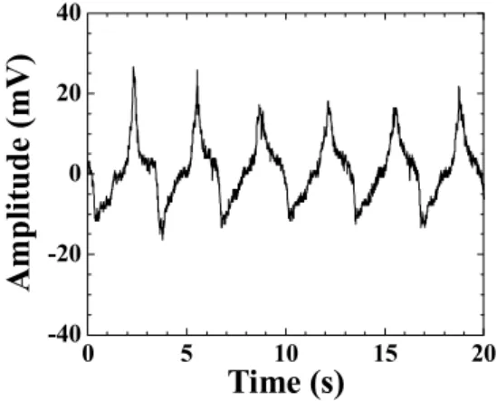
C. Biomedical ultrasonic probe
In order to investigate the capability of the membrane sensors as an ultrasonic probe for biomedical applications, the sensor was attached onto a finger with a gel couplant as shown in Fig. 5 (a). A clear ultrasonic signal reflected from a bone in the finger was observed with a pulse-echo technique as shown in Fig. 5 (b). Ultrasonic center frequency and 6dB bandwidth of the signal were 6MHz, respectively.
2 3 4 5 -0.2 -0.1 0.0 0.1 0.2 A m pl it ude ( V ) Time Delay (µs)
Signal from a bone
Sensor
(a)
(b)
Figure 5. Ultrasonic signal from a bone at a finger using the membrane sensor in a pulse-echo technique: (a) experimental
setup and (b) ultrasnoic signal reflected from a bone.
V. CONCLUSIONS
A piezoelectric membrane sensor, consisting of a metal foil, a piezoelectric ceramic film and a top electrode, has been developed. Thick PZT composite film was fabricated onto the SS foil of 33mm by 33mm by 38µm-thick using an air spray coating technique. The thickness of the film was 70µm. A top electrode of 15mm by 15mm was formed using silver paste. The SS foil served as the bottom electrode as well as the substrate. Piezoelectricity was achieved using a corona discharge poling method.
Due to the porosity in the piezoelectric film and the thin metallic membrane substrate, a highly flexible sensor was realized. The porosity was observed by the SEM. The density of the film was about 4.4 x 10³kg/m3, which is about 60% of the raw PZT material density. The relative dielectric constant was 130 measured by an impedance analyzer. The measured piezoelectric constants d33 and d31 were 37 x
10-12m/V at 10kHz and -27 x 10-12m/V at 1Hz, respectively, using optical interferometric techniques. The entire structure was sandwiched between polyimide films so that it can be waterproof and can operate at temperatures up to 150°C. Copper strips were used for electrical connection. This membrane sensor works as a unimorph-type bending sensor as well as an ultrasonic sensor.
The sensor was tested for biomedical monitoring. The sensor was directly attached onto a wrist and the signals corresponding to the pulse waves have been successfully obtained. The signals had a signal-to-noise ratio of better than 20dB. The breathing curves were also measured on a human belly. Optimal design and configuration of the sensor, a detailed analysis of the signals with comparison to physiological activities, improvement of the SNR of the sensor and measurement reproducibility, and development of reliable methods to attach the sensor on a human body
are planned in future studies. The developed sensor also operated as an ultrasonic sensor and a clear ultrasonic echo from a finger bone was observed with a pulse-echo technique.
Thus, this sensor could be used as a wearable sensor, which does not disturb daily life activities including sleeping, for real-time and continuous monitoring of personal health conditions, and also as an ultrasonic probe for biomedical applications.
ACKNOWLEDGMENT
The authors thank to K.-T. Wu and Y. Souliere for their technical assistance. Financial support from Natural Sciences and Engineering Research Council of Canada is acknowledged.
REFERENCES
[1] M. F. O'Rourke, A. Pauca, and X.-J. Jiang, “Pulse wave analysis,” Br. J. Clin. Pharmacol, vol. 51, pp. 507-522, 2001. [2] B. Wilkinson, I. R. Hall, I. S. MacCallum, C. M. McEniery,
B. J. Van Der Arend, Y. E. Shu, L. S. MacKay, D. J. Webb, and J. R. Cockcroft, “Pulse-wave analysis: clinical evaluation of a noninvasive, widely applicable method for assessing endothelial function,” Arterioscler Thromb. Vasc. Biol. vol. 22, pp. 147-152, Jan. 2002.
[3] K. Uchino, Piezoelectric Actuators and Ultrasonic Motors, Boston, MA: Kulwer Academic, 1997.
[4] M. Akiyama, N. Ueno, K. Nonaka, and H. Tateyama, “Flexible pulse-wave sensors from oriented aluminum nitride nanocolumns,” Appl. Phys. Lett., vol. 82, pp. 1977-1979, March 2003.
[5] D. A. Barrow, T. E. Petroff, R. P. Tandon and M. Sayer, “Characterization of thick lead zirconate titanate films fabricated using a new sol gel based process”, J. Appl. Phys. vol. 81, pp. 876-881, Jan. 1997.
[6] M. Kobayashi, C.-K. Jen, and D. Lévesque, “Flexible ultrasonic transducers,” IEEE Trans. Ultrason. Ferroelect. Freq. Contr., vol. 53, pp. 1478-1486, Aug. 2006.
[7] Y. Zhang, Z. Wang and J.D.N. Cheeke, “Resonant spectrum method to characterize piezoelectric films in composite resonators,” IEEE Trans. Ultrason. Ferro. Freq. Contrl., vol. 50, pp. 321-333, 2003.
[8] M.L. Dufour, G. Lamouche, S. Vergnole, B. Gauthier, C. Padioleau, M. Hewko, S. Levesque, and V. Bartulovic, “Surface inspection of hard to reach industry parts using low coherence interferometory,” Proc. Photonics North, 2006 (in press).
[9] Q.-M. Wang, X.- H. Du, B. Xu, and L. Eric Cross, “Electromechanical coupling and output efficiency of piezoelectric bending actuators,” IEEE Trans. Ultrason. Ferroelect. Freq. Contr., vol. 46, pp. 638-646, May 1999. [10] J.M. Fry, M.A. DiPhillipo, K. Curran, R. Goldberg, and A.S.
Baran, “Full polysomnography in the home,” Sleep, vol. 21, pp. 635-642, 1998.
[11] L.D. Victor, “Obstructive sleep apnea,” Am. Fam. Physician, vol. 60, pp. 2279-2286, Nov. 1999.