HAL Id: tel-03008638
https://tel.archives-ouvertes.fr/tel-03008638
Submitted on 16 Nov 2020HAL is a multi-disciplinary open access archive for the deposit and dissemination of sci-entific research documents, whether they are pub-lished or not. The documents may come from teaching and research institutions in France or abroad, or from public or private research centers.
L’archive ouverte pluridisciplinaire HAL, est destinée au dépôt et à la diffusion de documents scientifiques de niveau recherche, publiés ou non, émanant des établissements d’enseignement et de recherche français ou étrangers, des laboratoires publics ou privés.
The chloride/potassium co-transporter KCC2 in
synaptic plasticity, hippocampal rhythmogenesis and
memory
Clemence Simonnet
To cite this version:
Clemence Simonnet. The chloride/potassium co-transporter KCC2 in synaptic plasticity, hippocampal rhythmogenesis and memory. Neurons and Cognition [q-bio.NC]. Sorbonne Université, 2019. English. �NNT : 2019SORUS374�. �tel-03008638�
Sorbonne Université
Ecole doctorale n°158 Cerveau, Cognition, Comportement
Institut du Fer à Moulin, Equipe « Plasticité des réseaux corticaux et épilepsie »
The chloride/potassium co-transporter KCC2 in synaptic
plasticity, hippocampal rhythmogenesis and memory
Le co-transporteur de chlore et potassium KCC2 dans la plasticité synaptique,
rythmogenèse hippocampique et mémoire
Par Clémence Simonnet
Thèse de doctorat en Neurosciences
Dirigée par Jean-Christophe Poncer
Présentée et soutenue publiquement le 25 septembre 2019 Devant un jury composé de :
Dr Alberto Bacci Président du Jury
Dr Lisa Genzel Rapporteur
Dr Peter Blaesse Rapporteur
Dr Pascale Quilichini Examinateur
Dr Stéphanie Daumas Membre invitée
iii
Abstract
Information transfer, storage and retrieval in the brain rely on an exquisite balance between excitation and inhibition. At the cellular level, memory encoding involves long-term potentiation of excitatory synapses, while at the network level, cortical rhythmogenesis underlies memory encoding as well as consolidation and requires inhibitory, GABAergic signaling to synchronize neuronal ensembles. Therefore, alterations of this balance are observed in most disorders associated with cognitive deficits. In the mature central nervous system, GABA is the main neurotransmitter mediating fast inhibitory transmission and activation of GABAARs usually leads to an influx of chloride ions. Therefore, a tight contr ol of transmembrane chloride gradients is crucial to maintain the efficacy and polarity of GABA transmission and primarily involves the chloride/potassium co-transporter KCC2. Down-regulation of KCC2 expression and subsequent alteration of GABA signaling is therefore thought to be involved in many neurological and psychiatric disorders such as epilepsy, autism or Rett syndrome. However, recent evidence suggests the role of KCC2 extends beyond the mere control of GABAergic signaling in neurons. Thus, through interactions with a variety of molecular partners, KCC2 influences neuronal membrane excitability as well as the function and plasticity of glutamatergic synapses. Altogether, KCC2 therefore appears at the crossroads of excitatory and inhibitory transmission.
During my PhD, I explored the consequences of KCC2 down-regulation in the dorsal hippocampus on learning and memory, and the underlying mechanisms both at the cellular and network levels. My results demonstrate that KCC2 knockdown in principal neurons of the dorsal hippocampus affects both spatial and contextual memory. This effect is associated with deficits in long-term potentiation of hippocampal synapses as well as neuronal hyperexcitability and hippocampal rhythmopathy, including abnormal sharp-wave ripple generation and gamma-band activity during sleep. These alterations likely contribute to impair both memory encoding and consolidation. Importantly, however, KCC2 knockdown in dorsal hippocampus is not sufficient to generate spontaneous, epileptiform activity. Since KCC2 is down-regulated in many disorders associated with cognitive impairment, my results suggest that strategies aiming to restore KCC2 expression may hold therapeutic potential in these disorders. I therefore started testing this hypothesis in experimental models of Rett syndrome.
iv
Résumé
Dans le cerveau, le transfert d’information nécessite un parfait équilibre entre signalisations excitatrice et inhibitrice. Au niveau cellulaire, la formation d’un souvenir dépend de la potentiation à long-terme des synapses excitatrices alors qu’au niveau du réseau neuronal, la rythmogenèse hippocampique contrôle la formation et la consolidation de ce même souvenir et nécessite une transmission GABAergique inhibitrice afin de synchroniser les ensembles neuronaux. Ainsi, une altération de cet équilibre existe dans la plupart des pathologies associées à des troubles cognitifs. Dans le système nerveux central mature, GABA est le principal neurotransmetteur responsable de la transmission inhibitrice et l’activation de récepteurs GABAA entraîne un influx d’ions chlore. l’efficacité et la polarité de la transmission GABAergique dépendent du maintien de la concentration intra-neuronale de chlore, et ce rôle revient principalement au co-transporteur de chlore et potassium KCC2. La diminution d’expression de KCC2 et l’altération de la transmission GABAergique qui en découle sont souvent invoqués pour expliquer les troubles présentés par les patients épileptiques, ou atteints du Syndrome de Rett. Cependant, de récents résultats suggèrent que le rôle de KCC2 ne se limite pas seulement à un contrôle de la signalisation GABAergique. A travers des interactions avec des partenaires protéiques, KCC2 influence l’excitabilité neuronale ainsi que le fonctionnement et la plasticité des synapses glutamatergiques. KCC2 participe ainsi au maintien des transmissions excitatrices et inhibitrices.
Au cours de ma thèse, j’ai donc étudié le rôle d’une diminution d’expression de KCC2 dans l’hippocampe dorsal, sur l’apprentissage et la mémoire, ainsi que les mécanismes sous-jacents au niveau cellulaire et du réseau. Mes résultats démontrent une altération de la mémoire spatiale et contextuelle lorsque l’expression de KCC2 est diminuée dans les neurones principaux. Cet effet est associé à une diminution de la potentiation à long terme des synapses hippocampiques ainsi qu’une hyperexcitabilité neuronale et des déficits de rythmogenèse, plus spécifiquement des sharp-wave ripples et des oscillations gamma anormales lors du sommeil. Ces altérations contribuent probablement à des déficits d’apprentissages et de consolidation. De plus, un point important est l’absence d’activité spontanée épileptique chez ces animaux. Puisque l’expression de KCC2 est diminuée dans des pathologies associées à des troubles cognitifs, mes résultats suggèrent que des stratégies permettant de stabiliser l’expression de KCC2 pourraient être considérées comme de potentielles options thérapeutiques. J’ai donc commencé à tester cette hypothèse dans un modèle murin pour le syndrome de Rett.
v
Aknowledgments
First of all, I would like to thank my reviewers: Lisa Genzel, Peter Blaesse, Pascale Quilichini and Alberto Bacci. I really appreciate that you all agreed on being part of my jury and am looking forward to discuss these 4 years of work with you. I also want to thank the members of my thesis committee who saw my project evolving over the years: Michaël Zugaro and Elise Morice.
Et je passe enfin au français! « La seule page que nous comprendrons » dixit de nombreuses personnes de mon entourage.
Merci à toi Jean-Christophe, pour m’avoir accueilli un peu au dernier moment dans ton équipe. Même si j’ai décidé de me lancer dans cette aventure avec toi via des entretiens téléphoniques, je n’ai jamais regretté mon choix. Merci d’avoir toujours su me laisser la liberté dont j’avais besoin pour mener à bien ce projet, j’ai bien conscience qu’une bonne partie du temps, tu ne savais pas exactement où j’étais, avec mes allers-retours entre différents labos et mon travail au palais de la découverte. Merci pour ta disponibilité, ton écoute et de m’avoir fait grandir scientifiquement. J’ai appris énormément avec toi, et je réalise que je suis fière de ce projet que tu m’as aidé à développer, en me poussant par moment pour que je fasse certaines manips. Enfin, merci du temps passé à corriger cette thèse.
Merci aussi à Stéphanie, pour m’avoir tant appris, ta gentillesse et ta disponibilité chaque fois que j’en ai eu besoin. Merci aussi pour ces rares moments où je n’en pouvais plus et où tu m’as soit rassurée soit conseillé de me reposer.
Un grand merci à toutes les personnes de l’équipe ! Tout d’abord Sabine, merci pour ta gentillesse et de participer à créer un labo dans lequel on se sente si bien. Merci à Sana, pour nos discussions scientifiques, ou non, ta présence dans le bureau participait largement à la bonne humeur générale. Merci à Florian et Etienne, qui vivront bientôt eux aussi l’expérience de l’écriture de thèse (oui Florian, tu as encore le temps), merci à Marion pour nos discussions sur le trajet du retour, merci à Marie pour m’avoir appris à utiliser Matlab. Thank you Manisha, for the time you spent explaining lots of different concepts to me, more generally, I really appreciate knowing you, you were a great addition to the team and I hope you’ll be
vi really happy in your future life. Merci enfin à Xavier et Floriane, aux étudiants que j’ai eu, Agathe et Yousra, et de manière plus globale à tous ceux qui ont participé à la vie de cette équipe quelques mois ou quelques années, Ferran, Martin, Eric, Emmanuel…
Si je n’ai jamais regretté d’avoir accepté de faire ma thèse avec Jean-Christophe, j’ai aussi eu l’excellente surprise d’arriver dans un institut avec une si bonne ambiance. Pouvoir passer n’importe qu’elle porte pour discuter, emprunter du matériel, poser des questions est une chance incroyable. Merci plus particulièrement à Imane pour le temps passé à m’apprendre le clonage, Albert avec lequel je n’aurai pas travaillé longtemps avant qu’il ne parte, et puis bien sûr, tous ceux, doctorants et post-doctorants qui participent largement à la vie de l’IFM : Fanny, Jimmy, Valentina, Giorgia, Vincenzo, Amina, et tous les autres… Merci à tout ceux qui nous permettent de travailler dans de bonnes conditions, nos zootechnicien(ne)s, toutes celles et ceux qui ont travaillé au pôle laverie, particulièrement Géraldine et Dominique avec lesquelles j’ai souvent eu l’occasion de discuter au détour d’un couloir. Merci aussi à Christine et Jocelyne pour votre disponibilité.
Cependant, ma thèse n’a pas été faite qu’à l’IFM, et j’ai passé un certain temps enfermée dans l’animalerie à Jussieu. Merci donc à Dorian et Soumee avec lesquelles j’ai passé un certain temps à coordonner l’utilisation des salles et plus simplement à discuter entre 2 expériences. Merci à Sandrine pour ta bonne humeur contagieuse, et à Fiona avec laquelle j’ai beaucoup discuté lors de cette fin de thèse, ainsi qu’à tout le reste du B4.
Finalement, merci à celles et ceux qui les premiers m’ont encadré lors de mes stages et m’ont donné l’envie de continuer, Afsaneh, Seth, Marcy, Kay et bien sûr Anna.
En parallèle de ma thèse, j’ai eu la chance de pouvoir travailler au palais de la découverte. Quelques jours par an dans un environnement différent, qui bien souvent, me permettait de me rappeler pourquoi j’aimais les sciences. Entre la semaine du cerveau et les semaines des jeunes chercheurs, j’ai pu m’épanouir et m’amuser. Merci à Aurélie, Ludovic et Véronique pour votre investissement. Je suis fière de ce que nous avons pu créer avec votre aide. Merci aussi à Elodie pour tout ce que tu m’as appris sur les fourmis et à tous ceux que j’ai croisé.
Je me suis rendue compte l’année dernière à quel point j’avais un large groupe d’amis autour de moi sur lesquels je pouvais compter à n’importe quel moment. Votre aitié à tous m’a été incroyablement précieuse. Un grand merci à tous mes ami(e)s qui m’ont permis de
vii rester à peu près zen au cours des années. Merci donc à mes vieux amis, Auguste et bien sûr Marie. Marie, nous ne nous serons jamais autant vues que depuis que tu fais ta thèse à Paris come moi. Et même quand tu es à Cuba, je sais que je peux compter sur toi, une telle amitié est rare et j’ai une chance incroyable de t’avoir rencontré quand nous étions petites. Merci à tous ces gens que j’ai rencontré pendant le master ou la thèse. Merci à Thomas pour nos déjeuners hebdomadaires au Jardin des Plantes, à Adriana pour nos discussions au détour d’un couloir du palais, à Célia pour nos sessions piscine (1h de natation, 2h à râler sur nos thèses : un ratio correct), et à tous le reste du groupe de Pasteur pour les soirées raclette : Anne-Claire, Radhia, Noura, Marianne, Anne et Antoine.
Un immense merci aussi au Anges (Fous ?) du Palais pour nos éclats de rire, cette conversation WhatsApp qui n’en finit pas et les feuilles de menthe. Nous avons presque tous écrit nos thèses en même temps, et soyons honnête, il y avait toujours pire que soit dans le groupe, ce qui était plutôt rassurant. Merci donc à Lydie (et les Vincent), Audrey, Anyssa, Antoine, Nicolas, Miléna, Camille, Medhi, Oriane, Greg et Caroline. Un merci peut-être plus particulier à Caroline avec laquelle j’ai fait pas mal de médiation et longuement discuté autour d’un verre.
Parmi toutes les rencontres que j’ai pu faire ces dernières années, je suis heureuse que mon chemin ai croisé le tien, Quentin. Je ne t’ai jamais caché être de nature (un peu) anxieuse et être à quelques mois de ma fin de thèse, mais tu as quand même eu envie de passer ce temps avec moi. Merci pour ta présence, ton calme, ton écoute et de savoir trouver les mots justes quand j’en ai besoin. J’ai hâte de voir ce que l’avenir nous réserve (étape 1 : partir en vacances, étape 2 : trouver du travail ensemble à l’étranger).
Enfin, si j’en suis là aujourd’hui, c’est aussi grâce à ceux qui m’ont toujours vu grandir. Merci donc à toute ma famille. A mes parents d’avoir toujours été présent et de m’avoir laissé suivre mon petit bonhomme de chemin, même quand cela impliquait changer de ville/pays/programme tous les ans. J’ai réalisé un jour que vous ne m’aviez jamais dit que je ne pouvais pas faire quelque chose, et en grandissant, j’ai compris quelle chance ça a été. Maman, jai été tellement heureuse le jour où tu es venue me voir au Palais et que tu m’as dit avoir compris ce que je faisais. Merci bien sûr à mes sœurs et mon frère. Edgar on verra bien ce que tu feras, mais les sciences, c’est vraiment chouette. Enfin, merci à Odette (et Pierre) d’avoir été des troisièmes grands-parents et de votre soutien indefectible.
viii
Table of content
INTRODUCTION ... 3
I. KCC2, a neuronal cation-chloride cotransporter influencing both GABAergic and glutamatergic signaling ...6
1. Expression of KCC2 in the CNS ...6
a. The Cation-Chloride Co-transporters (CCC) family ...6
b. KCC2 structure and regulatory sequences ...7
c. KCC2 isoforms ...10
d. Spatial and temporal expression of KCC2 ...10
2. KCC2 controls neuronal chloride homeostasis ...14
a. KCC2 co-transport potassium and chloride: thermodynamic considerations ...14
b. KCC2 controls the reversal potential of GABAAR mediated currents ...15
c. Osmotic regulation by KCC2 ...17
3. Emerging role of KCC2 in dendritic spines ...18
a. KCC2 is necessary in spinogenesis ...19
b. KCC2 affects LTP expression, independent of its ion transport function ...21
II. KCC2 is down-regulated in neurological and psychiatric disorders ...24
1. KCC2 mutations in human pathology ...25
2. KCC2 down-regulation in the pathology ...27
a. Psychiatric and neurological disorders ...27
b. Epilepsy ...30
c. KCC2 down-regulation in brain insults ...32
3. Rett syndrome and KCC2...33
a. MecP2 gene expression in RTT ...33
b. Circuit dysfunction in RTT ...34
c. KCC2 imbalance in RTT ...37
d. MecP2, a regulator of KCC2 expression? ...38
4. Therapeutic strategies ...40
a. KCC2 over-expression ...41
b. Restoring chloride concentration with bumetanide ...42
ix
III. The hippocampus, a key structure in learning and memory ...47
1. Overview of the hippocampal structure and circuits ...48
a. Anatomy of the hippocampus ...48
b. The hippocampal tri-synaptic loop ...50
c. Hippocampal connectivity, a more complete view ...52
2. Hippocampus-dependent learning and memory ...55
a. Hippocampal networks underlying spatial and contextual memories ...55
b. Spatial and contextual behavioral paradigms ...57
c. Other behaviors related to the hippocampus ...60
3. Cellular and network substrates for memory encoding and consolidation ...61
a. Long-term plasticity mechanism and relevance to memory encoding ...61
b. The role of hippocampal rhythmogenesis in memory encoding and consolidation .69 IV. Rationale and objective of the project ...76
MATERIALS AND METHODS ... 81
1. How to knockdown or rescue KCC2? ...81
2. Animals and surgical procedures ...83
3. Electrophysiology ...84
4. Behavioral experiments ...88
5. Biochemistry and Immunohistochemistry ...91
6. Statistics ...93
RESULTS ... 97
I. Part I : The effect of chronic KCC2 downregulation on learning and memory ...97
1. KCC2 down-regulation in dorsal hippocampal neurons impairs spatial and contextual memory in mice ...98
2. KCC2 down-regulation in principal cells is sufficient to affect contextual memory 100 3. KCC2 down-regulation impairs hippocampal LTP ... 101
4. KCC2 knockdown impairs hippocampal rhythmogenesis ... 102
5. Conclusion ... 104
II. Part 2: Rescuing KCC2 function/expression in a mouse model of Rett syndrome ... 127
x 2. Treatments to restore chloride homeostasis or KCC2 stability in MecP2308/Y mice
... 128
3. Development of a new model of MecP2 deficiency ... 130
4. Conclusion ... 131
DISCUSSION ... 143
I. KCC2 knockdown induces hyperexcitability and hippocampal rhythmopathy 144 1. Mechanisms for abnormal ripple generation ... 144
2. Relevance to pathology and memory deficits ... 145
3. KCC2 knockdown in dorsal hippocampus is not sufficient to trigger epileptiform activities ... 148
II. What mechanism(s) underlies memory impairment upon KCC2 knockdown? . 150 1. Hypothesis 1: KCC2 ion transport-function is required for memory ... 150
2. Hypothesis 2: KCC2 interaction with protein partners is necessary for learning and memory ... 151
III. Rescuing KCC2 expression in a mouse model of RTT ... 153
IV. General conclusion ... 156
ANNEXES ... 159
Annexe 1: Posters and oral presentations ... 160
Annexe 2: Publications ... 165
xi
List of figures and tables
Figure 1. CCCs expression control the chloride ion flux through GABAARs ...4
Figure 2. KCC2 regulation through phosphorylation of specific residues ...9
Figure 3. Spatio-temporal expression of KCC2 ...12
Figure 4. KCC2 is expressed in the vicinity of glutamatergic synapses ...19
Figure 5. KCC2 is necessary for LTP and GluA1 activity-driven insertion at the membrane 21 Figure 6. KCC2 expression controls cofilin activation ...23
Figure 7. KCC2 mutations associated with epilepsy, autism or schizophrenia ...26
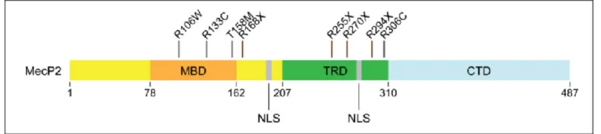
Figure 8. Schema of MecP2 protein and major mutations associated with RTT ...34
Table 1. RTT mouse models and associated behavioral and electrophysiological deficits ...35
Figure 9. Hypotheses on MecP2 role in KCC2 transcription regulation ...40
Figure 10. Different therapeutic options to rescue deficits following KCC2 down-regulation 45 Figure 11. Anatomy of the hippocampus ...48
Figure 12. Intra-hippocampic connectivity ...51
Figure 13. Extra-hippocampic inputs...54
Figure 14. LTP induction and expression mechanisms ...65
Figure 15. Theta oscillations allow spatial memory encoding and consolidation ...71
Figure 16. Hippocampal ripples are necessary for consolidation ...74
Table 2. Information about plasmid constructs ...82
Figure 17. LTP expression depends on the cutting solution used ... 86
Table 3. Infection score and related virus spread in the hippocampus ...92
Figure 18. Infection size with CaMKII-shKCC2 seems to correlate with ability to remember ...93
Figure 19. KCC2 down-regulation in dorsal hippocampal neurons impairs spatial and contextual memory ... 107
Figure 20. KCC2 down-regulation in principal cells is sufficient to alter contextual memory ... 109
xii Figure 22. Effect of KCC2 knockdown on hippocampal rhythmogenesis during REM sleep
... 113
Figure 23. KCC2 knockdown induces hyperexcitability in CA1 and altered SPW-Rs... 115
Figure S1. Locomotor activity is not altered upon KCC2 down-regulation ... 117
Figure S2. Evaluation of anxiety upon KCC2 down-regulation ... 119
Figure S3. KCC2 down-regulation in neurons does not affect spatial memory in Morris water maze ... 121
Figure S4. Compared SPW-R properties before and after learning ... 123
Table 4. Statistical data of project 1... 124
Figure 24. Over-expressing KCC2 in MecP2308 mice rescues LTP ... 133
Figure 25. Bumetanide or CLP-290 do not rescue locomotor activity and grip strength in MecP2308/Y mice ... 134
Figure 26. MecP2308/Y mice in anxiety and fear-conditioning memory tasks ... 135
Figure 27. KCC2 is not down-regulated in MecP2308 mice ... 136
Figure 28. KCC2 expression following MecP2 suppression in dorsal hippocampus ... 136
Figure S5. Effect of chronic treatment with CLP-290 on locomotor activity and grip strength ... 137
xiii
List of abbreviations
4-AP 4 Amino-pyridine
AIS Axon initial segment
AMPA α-Amino-3-hydroxy-5methyl-4-isoxazolepropionic acid ASD Autism spectrum disorder
BBB Blood-brain barrier
BDNF Brain-derived neurotrophic factor CA cornu Ammonis
CaMKII Ca2+/calmodulin-dependent protein kinase II CCC Cation-Chloride Co-transporter
CNS Central nervous system
CREB cAMP response element-binding protein
CTD Carboxy-terminal domain
DBS Deep-brain stimulation
DG Dentate gyrus
dHPC Dorsal hippocampus
DLPFC Dorsolateral prefrontal cortex
Dlx Distal-less homeobox
DMSO Dimethyl sulfoxide
EC Enthorinal cortex
EEG Electroencephalogram
EGABA Reversal potential of GABAAR mediated currents
Egr4 Early growth response transcription factor 4 EPSC Excitatory postsynaptic current
Erk1/2 Extracellular signal-regulated kinases 1 and 2 F/G-actin Microfilament/globular-actin
FERM Four.one, Ezrin, Radixin, Moesin
GABA γ-Amino Butyric Acid
GABAAR Receptor subtype A for GABA GDP Giant depolarizing potentials
HEK Human embryonic kidney
HFS high frequency stimulation HPCD Hydroxypropyl-β-cyclodextrin IGF1 Insulin-Growth Factor 1 iPSC Induced-pluripotent stem cell IPSC Inhibitory postsynaptic current KCC2 K+/Cl- cotransporter type 2 LIMK Lin-11, Isl-1 and Mec-3 kinase
LTD Long-Term Depression
xiv mAChR Muscarinic acethylcholine receptor
MBD Methyl-binding domain
MecP2 Methyl CpG binding protein 2
MF Mossy fiber
MS-DBB Medial septum – diagonal band of broca MUA Multi-unit activity
NEM N-Ethylmaleimide
NKCC1 Na+/K+/2Cl- cotransporter type 1 NLS Nuclear localization signal NMDAR N-methyl-D-Aspartate receptor NOR Novel object recognition
NREM Non Rapid Eye Movement
NTD Animo-Terminal Domain
OR Object recognition
OSR Osmotic stress resistant protein PAK Serine/threonine-protein kinase
PKC Protein kinase C
PLCγ Shc/FRS-2 and phospholipase Cγ
PP Perforant path
PPI Protein phophatase 1
PR Place recognition
PSD Post-synaptic density
PV Parvalbumin
REM Rapid eye movement
REST RE1-silencing transcription factor
RTT Rett syndrome
SC Schaffer collateral
SCI Spinal cord injury
shRNA Short hairpin RNA
SOM Somatostatin
SPAK Ste20p-related proline/alanine-rich kinase SPW-R Sharp-wave ripple
TLE Temporal lobe epilepsy
TRD Transcriptional repression domain TrkB Tropomyosin receptor kinase B USF Upstream stimulating factor VREST Resting membrane potential
WNK With no lysine kinase
3
Introduction
In the central nervous system, information processing relies mostly on a transfer of information through chemical synapses. Following the emission of an action potential, a presynaptic neuron releases its neurotransmitter into the synaptic cleft. The postsynaptic neuron can be either activated or inhibited, depending on the nature of the neurotransmitter and transmembrane ion gradients. In the mature CNS, excitation relies mainly on the release of glutamate while γ-Amino butyric acid (GABA) signaling usually produces neuronal inhibition.
Learning and memory rely on the coordinated action of these synaptic signals. At the cellular level, long-term potentiation of glutamatergic transmission results in changes in synaptic function and morphology. At the network level, network oscillations (such as theta and gamma-band oscillations) in brain regions such as the hippocampus allow information encoding during exploration while other oscillatory activities such as sharp-wave ripples and theta during rapid eye movement (REM) sleep participate in memory consolidation. These rhythms rely on the coordinated activity of excitatory and inhibitory signaling as well as neuronal excitability. Therefore, mechanisms acting to perturb either glutamatergic or GABAergic signaling or neuronal excitability are likely to results in alterations in the ability of an animal to learn and store new information.
Both excitatory and inhibitory transmission require ions flux through the neuronal plasma membrane. At rest, the neuronal membrane is electrically polarized with a net negative charge on the inside of the membrane. An action potential therefore corresponds to an abrupt depolarization of the membrane potential. Indeed, fast excitatory transmission results from glutamate receptor activation, leading to an entrance of cations (Na+, Ca2+) underlying excitatory postsynaptic currents (EPSCs). On the other hand activation of ionotropic GABAA receptors (GABAARs) mediate hyperpolarizing inhibitory postsynaptic currents (IPSCs) through the influx of anions (Cl-, HCO3-) and prevent action potential firing.
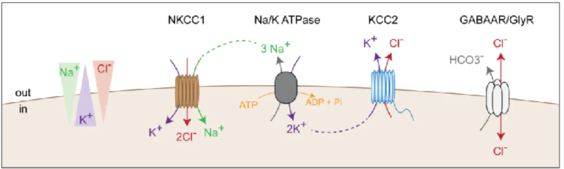
4 Electrochemical gradients are thus essential to synaptic transmission and the control of neuronal excitability. The Na/K ATPase maintains intraneuronal concentration of Na+ and K+ while Cation-Chloride Cotransporters (CCCs) regulate transmembrane Cl- gradients. Among these proteins, two are mainly expressed in the mature CNS: KCC2 usually extrudes chloride ions under isotonic conditions, typically maintaining a low intraneuronal chloride concentration, while NKCC1 participates in the influx of Cl- ions. The subsequent transmembrane Cl- gradient largely determines the polarity of GABAARs (Figure 1). Hence, changes in the activity of these transporters directly impact GABAergic signaling and have been involved in various disorders.
Figure 1. CCCs expression control the chloride ion flux through GABAARs
KCC2 extrude chloride ions using the K+ gradient created by the Na/K ATPase while NKCC1 intrudes Cl-. The resulting chloride gradient determines the direction of Cl- flux through GABAAR.
Nevertheless, modulation of synaptic transmission is also observed under physiological conditions. Indeed, in all brain regions and neuronal subtypes, synaptic efficacy is modulated in response to neuronal activity. This synaptic plasticity allows neurons to strengthen or weaken their connections through long-term potentiation (LTP) and depression (LTD), respectively. LTP mechanism requires the influx of Ca2+ via the N-methyl-D-Aspartate receptor (NMDAR) resulting in the addition of α-Amino-3-hydroxy-5methyl-4-isoxazolepropionic acid receptor (AMPAR) in the postsynaptic density (PSD). Somewhat surprisingly, KCC2 has been shown to influence these forms of plasticity through an ion-transport independent mechanism.
My PhD thesis focus on the role of KCC2 in learning and memory and the impact of KCC2 down-regulation on cellular and network mechanisms underlying memory encoding and consolidation. Before presenting my experimental results, I will introduce the function
5 and regulation of KCC2 in physiological and pathological conditions and explore the different mechanisms involved in learning and memory. My experimental results will then be described and discussed in the second and third parts of the manuscript.
6
I.
KCC2, a neuronal cation-chloride cotransporter influencing both
GABAergic and glutamatergic signaling
1. Expression of KCC2 in the CNS
a. The Cation-Chloride Co-transporters (CCC) family
Fast excitatory and inhibitory signaling rely on transmembrane ion fluxes through primarily AMPA and GABAA receptors, respectively. While AMPA receptors are mainly permeable to sodium and potassium ions (Dingledine et al., 1999), GABAAR are predominantly permeable to chloride and, to a lesser extent, bicarbonate ions (Hübner and Holthoff, 2013). Transmembrane electrochemical gradients of these ions are maintained through the function of various ionic pumps and transporters. In particular, the Na/K ATPase serves as an active ion pump allowing the influx of K+ and efflux of Na+ ions and thereby contributes to maintain the transmembrane gradients of these ions (Schmidt and Dubach, 1971). Intraneuronal chloride concentration on the other hand is under the control of cation-chloride cotransporters (CCCs) that are non-electrogenic, secondary active transporters using Na+ and K+ gradients to transport chloride ions (Blaesse et al., 2009; Gamba, 2005).
All genes encoding CCCs belong to the SLC12 gene family (SLC12A1-9). CCCs are expressed widely in the body and can be classified in 4 families, depending on their stoichiometry and the cations that are co-transported (Gamba, 2005):
- Na+/Cl- co-transporters (NCC): expressed selectively in kidney
- Na+/K+/2Cl- cotransporters (NKCC1-2): NKCC1 is the only one expressed in the CNS
- K+/Cl- (KCC1-4): KCC2 is almost exclusively expressed in the brain
- CCC-interacting protein CIP1 and the polyamine transporter CCC9, identified on the basis of their homology of structure and mRNA sequence with other CCCs. However, their role as co-transporters is not defined yet. CIP1 regulates the transport activity of NKCC1 (Caron et al., 2000) and KCC2 (Wenz et al., 2009).
7 CCCs have multiple physiological roles. They are mainly acting to control intracellular chloride concentration. Another major function is their role in osmotic regulation that I will address later in this introduction.
b. KCC2 structure and regulatory sequences
KCC2 was only identified in the late 90s, by screening a DNA library to identify a gene with homology to the NKCC1 and 2 transporters (Payne et al., 1996). So far, only NKCC1 topology has been characterized (Gerelsaikhan and Turner, 2000) and the secondary structure of other CCCs is expected to be similar. KCC2 three-dimensional structure was predicted using hydropathy profile alignment, i.e. by analyzing the hydrophobic and hydrophilic domains of the protein based on its amino acids composition (Payne et al., 1996). KCC2 is 140 kDa glycoprotein with 12 putative transmembrane domains, flanked with intracellular amino- (NTD, amino acids 1-103) and carboxy-terminal domains (CTD, last 500 amino acids), and a large extracellular loop containing glycosylation sites between the 5th and 6th transmembrane domains. More recently, mass spectrometry experiment confirmed the presence of 6 glycosylation sites and some of the regulatory sites in the CTD (Agez et al., 2017).
The ternary structure of KCC2 is unknown, as the protein has not been crystallized yet. This is probably due to the double difficulty of purifying a transmembrane protein and obtaining a crystal. However, a plausible ternary structure of CCCs is based on the crystallographic model of the CTD of MaCCC, a prokaryotic CCC transporter from the archaea Methanosarcina acetivorans (Warmuth et al., 2009). MaCCC CTD is shorter than that of eukaryotic CCCs, but the difference is mainly due to insertion of loops, which are not predicted to significantly impact the 3D structure.
Finally, the quaternary structure of KCC2 and its role are still a matter of debate. It is known that KCC2 can form multimeric structures with itself as well as other CCCs as bi-, tri- and tetrameric KCC2 complexes can be detected by biochemical assay (Blaesse et al., 2006; Simard et al., 2007; Uvarov et al., 2009). The proteins are interacting together mainly through disulfide bridges, as a majority of the monomeric form can be observed in western blot after treatments with DTT or β-mercaptoethanol (Agez et al., 2017; Blaesse et al., 2006). Moreover, recent experiments suggested CTD is important for the dimerization (Agez et al.,
8 2017), as also demonstrated for KCC1 (Casula et al., 2001) and MaCCC (Warmuth et al., 2009).
KCC2 oligomerization might be necessary for its transport function. Indeed, throughout development, the proportion of oligomer forms increase in all brain regions. In the rat lateral superior olive, KCC2 is mainly in its monomeric form at post-natal day 3 (P3), which correlates with low chloride extrusion capacity. But as neurons mature, KCC2 forms oligomers and by P12, the chloride extrusion capacity increases, reflecting functional KCC2 (Blaesse et al., 2006).
Two groups have also suggested that lipid rafts could influence KCC2 oligomerization and function. However, they disagree on the role of these lipid rafts on KCC2 function. Watanabe and colleagues showed KCC2 lipid raft insertion and oligomerization were facilitated by the phosphorylation of Y1089 residue (Watanabe et al., 2009). In neurons expressing a constitutively dephosphorylated Y1089D residue, KCC2 activity was decreased and its localization within lipid rafts disrupted. As the function of numerous transporters depends on their localization in lipid rafts (Martens et al., 2004), they proposed that KCC2 function may be increased when localized within the lipid rafts. On the other hand, Hartmann and colleagues suggested lipid rafts might decrease KCC2 function, as chemical alteration of lipid rafts increased KCC2 transport function and affected KCC2 clustering. This apparent discrepancy may be explained by the fact that the first study did not investigate the effect of disrupting lipid rafts on KCC2 function but directly acted on its phosphorylation and its redistribution at the membrane. Therefore, the conclusion of this work arise from mostly correlative observations.
KCC2 membrane stability and function rely on phosphorylation and dephosphorylation of KCC2 key tyrosine, threonine or serine, most of them localized in the CTD (Figure 2). I will only concentrate on a few of them that are important in mature neurons but more information can be found in recent reviews (Alessi et al., 2014; Côme et al., 2019).
In our group, GABAAR activation was shown to stabilize KCC2 at the plasma membrane. Indeed, a transient rise in [Cl-]i leads to inhibition of WNK1 kinase. In the absence
of GABAAR activation, WNK1-SPAK-OSR1 pathway phosphorylates KCC2 T906 and T1007 residues and increases KCC2 membrane diffusion and reduces its membrane clustering and function (Heubl et al., 2017; de Los Heros et al., 2018; Rinehart et al., 2009).
9 Figure 2. KCC2 regulation through phosphorylation of specific residues
KCC2 membrane expression is modulated through a variety of intracellular signaling pathways. Its C-terminal domain hosts different residues important for its stability. Indeed, phosphorylation of S940 increases KCC2 membrane stability and function while phosphorylation of T1007 or T906 is associated with opposite effects. Phosphorylation of Y903 and Y1087 may on the other hand lead to increased membrane diffusion/recycling.
Excitatory transmission has also been shown to control KCC2 transport function and stability (Kaila et al., 1997; Wang et al., 2006; Woodin et al., 2003). Indeed, postsynaptic Ca2+ influx through NMDARs induces protein phophatase 1 (PP1) – dependent dephopshorylation of KCC2 S940 residue (Lee et al., 2011) which in turn reduces the stability of KCC2 and increases its membrane diffusion near excitatory but not inhibitory synapses(Chamma et al., 2013). On the opposite, phosphorylation of S940 by the protein kinase C (PKC) increases KCC2 activity and membrane stability (Lee et al., 2007). Finally, in HEK-293 cells, phosphorylation of Y903 and Y1087 decreases the cell surface stability of KCC2. Moreover, activation of the muscarinic acethylcholine receptors (mAChRs) in cultures of hippocampal neurons increases KCC2 tyrosine phosphorylation (Lee et al., 2010).
Interestingly, among the various rodent models of epilepsy, one of them consists in using pilocarpine, a mAChR agonist. Moreover, mouse models with constitutively dephosphorylated S940 residue are associated with increased seizure susceptibility, as I will discuss later (see Introduction II.2.b, Silayeva et al., 2015).
10
c. KCC2 isoforms
The human KCC2 gene (SLC12A5) is predicted to encode different transcripts variants, possibly with different roles. Two full-length KCC2, referred to as KCC2a and KCC2b have been identified (Uvarov et al., 2007), and can interact together (Uvarov et al., 2009). KCC2a has a longer NTD (40 more amino acids) with an extra regulatory site for SPAK. KCC2a and KCC2b form functional transporters, but their expression patterns during development and in the brain differ. While KCC2b is largely expressed throughout the whole adult brain, KCC2a expression is really low in cortex, hippocampus, thalamus and cerebellum. Both isoforms are expressed in the hypothalamus, brain stem and spinal cord (Markkanen et al., 2014).
In mouse neonatal brain, both isoforms are expressed but only KCC2b is up-regulated during postnatal development (Markkanen et al., 2014; Rivera et al., 1999). Importantly, knock-out mice for both isoforms die at birth from severe motor and respiratory deficits, whereas mice expressing KCC2a only survive for almost 2 weeks, then die of spontaneous seizures (Uvarov et al., 2007). This suggests a role of KCC2a in spinal cord and brain stem development. Thus, KCC2a expression in these structures might be sufficient to control for intraneuronal chloride concentration and neuronal function required for normal breathing (Dubois et al., 2018), whereas its low expression in the cortex might rapidly promote hyperactivity and seizures.
Shorter transcripts have also been identified and lack some of the exons of the
SLC12A5 gene (AK294059, AK295096, AK098371, EXON6B) (Tao et al., 2012; Wang et
al., 2008). These transcripts may not encode for functional transporter proteins but may have a regulatory effect still to be characterized. Moreover, these transcripts may be species-specific (Gagnon and Di Fulvio, 2013) or even tissue-specific. For instance, one such transcript was recently identified in the pancreas but not in the brain (Kursan et al., 2017). I will address later how some of these transcripts may be associated to the pathology (see Introduction II.1).
d. Spatial and temporal expression of KCC2
11 Although KCC2 was thought to be expressed only in the CNS, recent evidence revealed KCC2 expression in other tissues such as chicken cardiomyocytes (Antrobus et al., 2012), human osteoblasts (Bräuer et al., 2003) or pancreatic islets β-cells where it regulates insulin secretion (Kursan et al., 2017).
In the CNS, KCC2 is expressed only in neurons, due to the presence of two upstream and intronic RE-1 binding sites for the neuron-restrictive silencing factor (NRSF), also called RE1-silencing transcription factor (REST) (Karadsheh and Delpire, 2001; Yeo et al., 2009). Other binding sites have been described for early growth response transcription factor 4 (Egr4), mainly expressed in postnatal neurons (Uvarov et al., 2006) and upstream stimulating factor (USF1 and 2) via an E-box controlling element (Markkanen et al., 2008).
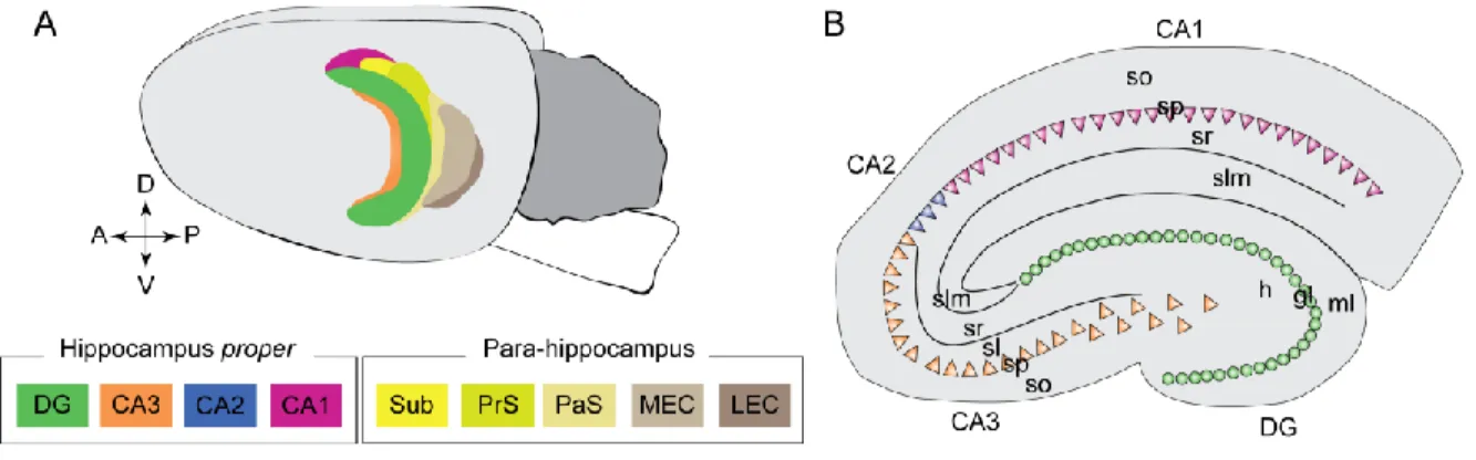
As described before, KCC2 (hereby referring to both isoforms) is expressed throughout the brain (Markkanen et al., 2014), including spinal cord (Hübner et al., 2001), cerebellum (Williams et al., 1999), thalamus (Barthó et al., 2004), auditory brainstem (Blaesse et al., 2006) and cortical structures (Gulyás et al., 2001; Rivera et al., 1999) (Figure 3). KCC2 is expressed in excitatory neurons (Rivera et al., 1999), Purkinje cells (Mikawa et al., 2002) and in, at least, parvalbumin-containing and some calbindin-containing interneurons (Gulyás et al., 2001). Although KCC2 seems ubiquitously expressed, the reversal potential of GABAAR mediated currents (EGABA), which depends in part on transmembrane chloride
gradients, shows differences depending on the structure and neuronal type (Klein et al., 2018; Schmidt et al., 2018). This could be due to differential expression or function of the protein, co-expression with other CCCs such as NKCC1 and/or differences in bicarbonate metabolism.
At the subcellular level, KCC2 shows diffuse expression throughout the somatodendritic membrane of neurons but also forms clusters, in particular in the vicinity of both GABAergic and glutamatergic synapses (Chamma et al., 2013; Gauvain et al., 2011; Gulyás et al., 2001) . However, its expression is strictly excluded from the axon and axon initial segment (AIS) (Williams et al., 1999), except in bipolar retinal cells (Vardi et al., 2000). Notably, the sodium/potassium/chloride cotransporter NKCC1 is, on the contrary, expressed in both the somatodendritic and axonal membrane (Jang et al., 2001; Khirug et al., 2008).
12 The AIS preserves the axonal identity by acting as a gatekeeper through two main mechanisms. Sub-membrane ankyrin-spectrin scaffolding complex forms a surface diffusion barrier impeding lipids and membrane proteins trafficking. The AIS also acts as an intracellular traffic filter. Indeed, specific motor proteins, such as myosin VI, are transporting the vesicles to the axon (Leterrier and Dargent, 2014; Lewis et al., 2011). Recent work in epithelial cells suggests that the subcellular localization of NKCC1 depends on alternative splicing (Carmosino et al., 2008). A similar mechanism can be hypothesized for KCC2. It is also possible that vesicles containing KCC2 are not expressing the cargo proteins for axonal targeting. As a consequence of KCC2 and NKCC1 differential expression in the axon, chloride concentration in axon terminals is higher than in the somatodendritic compartment (Price and Trussell, 2006).
Figure 3. Spatio-temporal expression of KCC2
A. KCC2 expression increases in the rodent brain during post-natal development. B. KCC2 expression in the hippocampus. Scale bar 600 µm. B. Immunostaining of a neuron transfected with recombinant KCC2 (red) and GFP (green). Higher magnification of the region of interest: KCC2 is expressed in the soma and dendrites (arrow) of neurons but is excluded from the axon (white arrowhead). D. KCC2 is localized close to both excitatory synapses (ES) and inhibitory synapses (IS).
(Adapted from Chamma et al., 2013; Gulyás et al., 2001; Rivera et al., 1999)
KCC2 developmental expression
KCC2 expression in the forebrain increases during development (Clayton et al., 1998; Jansen et al., 2010; Rivera et al., 1999). In humans, KCC2 expression increases in utero
13 around 25 weeks post-conception with its maximum after birth (Sedmak et al., 2016), in contrast with mice and rats where KCC2 expression mainly increases after birth (Watanabe and Fukuda, 2015) (Figure 3). However, these results cannot be generalized to all rodents as guinea pig neonates already exhibit a high amount of KCC2 mRNA by embryonic day 42 (E42) without any up-regulation after birth (Rivera et al., 1999).
Since over-expression of brain-derived neurotrophic factor (BDNF) in embryos was shown to increase KCC2 expression at E18 (Aguado et al., 2003), its effect on developmental up-regulation of KCC2 was studied. BDNF binding to tropomyosin receptor kinase B (TrkB) activates the extracellular signal-regulated kinases 1 and 2 (Erk1/2) pathway. This in turn increases KCC2 expression through Egr4-dependent KCC2 transcription (Ludwig et al., 2011a). Another in vitro study also reported the importance of both RE-1 sites to drive the effect of BDNF (Yeo et al., 2009). As KCC2a mRNA expression is relatively constant during development and restricted to specific brain regions (Markkanen et al., 2014), these regulations might only affect KCC2b transcription (Ludwig et al., 2011a; Yeo et al., 2009). However, a recent in vivo experiment reported a normal increase in KCC2 expression in BDNF-/- mice at P5-6 and P13-14 (Puskarjov et al., 2015). This suggests that other mechanisms exist to allow for KCC2 up-regulation during development (Ludwig et al., 2011b) and BDNF expression is not strictly necessary.
Nevertheless, this effect of BDNF on KCC2 expression differs in adult neurons. BDNF incubation for 2 to 3 hours is sufficient to reduce KCC2 expression in acute hippocampal slices and decrease Cl- extrusion capacity of the cell membrane (Rivera et al., 2002). This regulation depends on the activation of both Shc/FRS-2 and phospholipase Cγ (PLCγ) – cAMP response element-binding protein (CREB) signaling (Rivera et al., 2004).
KCC2 expression is stable during aging in both rodents and humans (Ferando et al., 2016; Sedmak et al., 2016). However, KCC2 down-regulation was observed following LTP-induction protocol in aged mice (21 to 28 weeks old) (Ferando et al., 2016). fEPSP amplitude was reduced upon bicuculline application in old but not young mice, suggesting a possible contribution of a depolarizing GABAergic component. Moreover, in these old mice, unstimulated synapses were also potentiated, which did not occur in younger mice. Interestingly, application of the KCC2 enhancer CLP-257 (Gagnon et al., 2013) restored the synapse specificity of LTP in old mice, suggesting an age-dependent role of KCC2 membrane expression in LTP, with a possible impact on learning and memory that, however, was not
14 explored in this study. I will further address this point later in this introduction (see Introduction I.3.b).
2. KCC2 controls neuronal chloride homeostasis
a. KCC2 co-transport potassium and chloride : thermodynamic considerations
Under physiological conditions, KCC2 extrude chloride ions using the potassium gradient maintained by the Na/K ATPase. It is the only KCC working in isotonic conditions, thanks to its ISO domain (Acton et al., 2012). Since the reversal potential of both Cl- and K+ ions are equal, KCC2 operates close to its thermodynamic equilibrium (Payne, 1997). The reversal potential of an ion is calculated according to the Nernst equation:
where R represent the ideal gaz constant, T the temperature in Kelvin, F the Faraday constant and z the valence of the studied ion.
Hence, the equation ECl-= EK+ is equivalent, after simplification, to [Cl-]i*[K+]i = [Cl-]o*[K+]o
Both extracellular chloride concentration and intracellular potassium concentration can be considered as fixed and close to 140 mM. Such approximations suggests that for [Cl-]i
higher than [K+]o, KCC2 extrudes both choride and potassium ions.Conversely, KCC2 will
intrude ions when [K+]o ishigher than [Cl-]i. Physiological [Cl-]i has been estimated to vary
between 7 and 13 mM in a computational model (Doyon et al., 2011) while physiological [K+]o ranges between 2 and 4 mM. However, intense neuronal activation induces a rise in
[K+]o up to 10 mM in cortical neurons (Avoli et al., 1996; Thompson et al., 1988). This would
result in a change of ion transport directionality through KCC2.
Hence, KCC2 is in an interesting position to reduce excitability. In basal conditions, KCC2 will extrude chloride and potassium ions, restoring a low intraneuronal chloride concentration and preserving the inhibitory GABAergic signaling. However, following an
15 intense neuronal activation, reverse transport through KCC2 will cope with the increased potassium concentration in the extracellular compartment.
b. KCC2 controls the reversal potential of GABAAR mediated currents
Developmental switch in the polarity of GABA transmission
A few years before KCC2 was identified (Payne et al., 1996), Ben-Ari and collaborators recorded spontaneous giant depolarizing potentials (GDPs) in rat immature CA3 neurons (Ben-Ari et al., 1989). These GDPs were observed in 85% pyramidal neurons during the first postnatal week, then their occurrence decreased and disappeared by post-natal day 12 (P12). These GDPs were dependent on GABA signaling as application of the GABAAR blocker bicuculline suppressed them, while GABA application increased their frequency and depolarized the membrane. Around P5, a switch in the polarity of GABA responses from depolarizing to hyperpolarizing occurs in hippocampal neurons. The precise timing of the switch was later refined and placed around P13 in the hippocampus (Khazipov et al., 2004).
As described previously, KCC2 up-regulation parallels the shift in the polarity of GABAAR mediated currents in rat forebrain (Rivera et al., 1999). Moreover, inhibition of KCC2 expression by incubation of slices with antisense oligonucleotides against KCC2 mRNA was sufficient to prevent this shift. Thus, GABA transmission depends on KCC2 expression which dynamically controls intraneuronal concentration. Several studies later revealed that the precise timing of KCC2 up-regulation differs depending on the brain region but is highly correlated with the timing of the shift in the polarity of GABAAR-mediated currents in each brain region (see Watanabe and Fukuda, 2015 for review).
As I will discuss later in this introduction, KCC2/NKCC1 expression ratio is altered in different disorders (see Introduction II) and delayed shift in the polarity of G ABA transmission has been observed in two rodent models of autism (Tyzio et al., 2014) and more recently in a mouse model of Rett syndrom (Lozovaya et al., 2019). Hence, alterations of GABA signaling around birth might affect brain function and be responsible for at least some of the symptoms associated with these disorders.
16 Impact of KCC2 knockdown on GABAergic signaling in adulthood
As observed during development, KCC2 expression controls the polarity of GABA signaling (Rivera et al., 1999). This suggests that in disorders associated with KCC2 down-regulation, GABA might become paradoxically excitatory. In line with this hypothesis, several studies reported a depolarized value of EGABA upon KCC2 knockdown (e.g.,
Huberfeld et al., 2007; review in Côme et al., 2019) and computational models revealed that decreasing KCC2 activity may reduce synaptic inhibition (Doyon et al., 2011).
However, KCC2 interacts with protein partners, including some involved in the trafficking or recycling of various transmembrane proteins and receptors (Mahadevan et al., 2017). This suggests that KCC2 down-regulation might also affect other neuronal properties, including intrinsic neuronal properties. Indeed, our team recorded both EGABA and the resting
membrane potential (Vrest) in rat dentate granule cells and CA1 pyramidal neurons and
reported that both values were similarly depolarized in neurons lacking KCC2, leaving the driving force of ion flux through GABAARs almost intact upon KCC2 knockdown (Goutierre et al., 2019). The driving force reflects the difference between Vrest and EGABA and therefore
determines the amplitude and polarity of GABAergic currents. Therefore, KCC2 knockdown has little effect on GABAergic signaling at rest but instead increases neuronal excitability through reduced membrane trafficking of the leak potassium channel Task -3 which normally interacts with KCC2.
Despite the importance of Vrest in setting the efficacy and polarity of GABA signaling,
such a value is rarely reported in studies looking at the effect of KCC2 down-regulation or is not commented. Upon conditional ablation of KCC2, another team reported a depolarized value of Vrest compensating the shift in EGABA in cerebellar granule cells but not Purkinje cells
(Seja et al., 2012). This observation coincides with the fact that Purkinje cells express mainly Task-1 and Task-5 (Karschin et al., 2001) while cerebellar granule cells, like dentate granule cells and pyramidal CA1 neurons, express mainly Task-3 (Marinc et al., 2014). Altogether, these data suggest that KCC2 down-regulation might affect neuronal excitability and GABAergic signaling efficacy differently depending on the neuron types and brain regions.
Activation of GABAAR has been shown to lead to intra-neuronal accumulation of Cl -associated with an increase in KCC2 activity in order to restore chloride homeostasis (Heubl et al., 2017; Viitanen et al., 2010). Hamidi and collaborators therefore used a protocol inducing epileptiform activities in the hippocampus to study the impact of KCC2 blocker
17 VU0240551 (Hamidi et al., 2015). Spontaneous interictal discharges can be induced in hippocampal slices by 4-AP bath application (Voskuyl and Albus, 1985) and two types of epileptiform activities can be recorded: short- and long-lasting events (Perreault and Avoli, 1991). Bath application of VU0240551 reduced the occurrence of short-lasting events and increased the interval of occurrence of long-lasting events (Hamidi et al., 2015), suggesting the mechanisms maintaining these two types of activity might be different. Indeed, blocking KCC2 might prevent the increase in [K+]o resulting from intense GABAAR activation
(Viitanen et al., 2010), thereby reducing cell excitability and long-lasting event frequency. Blocking KCC2 also increased the interval of occurrence of pharmacologically isolated GABAergic events and decreased their duration (Hamidi et al., 2015). This is presumably due to the fact that blocking KCC2 affects the intraneuronal concentration of Cl- and therefore the efficacy of GABAergic signaling.
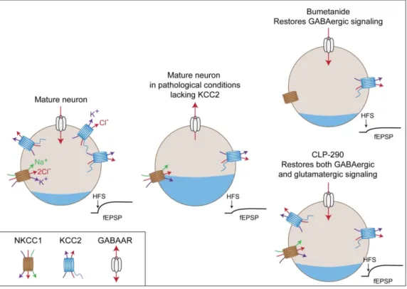
Altogether, these experiments suggest that even though chronic KCC2 down-regulation might not predominantly affect GABAergic signaling at rest, upon intense neuronal activation (for example during LTP induction or during a seizure) the lack of KCC2 might affect the efficacy and polarity of GABA transmission. Understanding whether KCC2 knockdown affects GABAergic signaling in vivo, and to what extent, is necessary to develop and validate therapeutic options for the pathology. Indeed, restoring chloride homeostasis with bumetanide might be of therapeutic interest (see Introduction II.4.b).
c. Osmotic regulation by KCC2
In addition to their critical role in controlling transmembrane ion gradients, CCCs also play an essential role in osmotic regulation. Thus, CCCs possess an important ion efflux or influx capacity and are used by many cells to adjust their internal osmolarity. Notably, KCCs were first described in epithelial and red blood cells as swelling-activated transporters, the activity of which was critical to face osmotic challenges (Dunham and Ellory, 1981; Lauf and Theg, 1980; Zeuthen and MacAulay, 2002). When cells are exposed to increased extracellular osmolarity, water molecules are moving outside of the cell, thus reducing cell volume and conversely. To compensate for osmolarity gradients, mechanisms are activated to reduce the movement of water molecules. As CCCs co-transport K+ and Cl-, ionic movements across the
18 cell membrane are indirectly or directly coupled to water transport (MacAulay and Zeuthen, 2010; Zeuthen and MacAulay, 2002). It has been estimated that up to 500 molecules of water may cross the membrane for each chloride ion transported by NKCC1 (MacAulay and Zeuthen, 2010).
In the CNS, postsynaptic ionotropic receptors lead to net ion influx into the postsynaptic neuron and water influx. Thus, intense neuronal activity directly impacts cell volume and may lead to cell swelling and cell death in extreme cases (Pasantes-Morales and Tuz, 2006). Since neurons lack aquaporins (Amiry-Moghaddam and Ottersen, 2003), other mechanisms may be necessary for coping with synaptic activity-induced swelling. Hence, digital holographic microscopy revealed an important water influx through NKCC1 and NMDARs following glutamate application. Furosemide, but not bumetanide application strongly reduced cell swelling, suggesting a role of KCC2 in water extrusion (Jourdain et al., 2011). In one study, KCC2 function was proven essential to neuronal survival following NMDA-induced excitotoxicity (Pellegrino et al., 2011). Although this latter study did not fully explore the mechanisms underlying such neuroprotective effect, it may involve the role of KCC2 in coping with activity-induced cell swelling. Moreover, other KCCs are activated during osmotic challenges and KCC3 and KCC4 participate to the water extrusion and recovery of cell volume (Boettger et al., 2003; Frenette‐ Cotton et al., 2018; Race et al., 1999).
In order to restore the neuronal osmolarity, the swelling-induced activity of KCC2 is most likely dependent on the inhibition of the WNK/SPAK/OSR1 pathway, which results in dephosphorylation of T906 and T1007 residues (Gagnon et al., 2006; de Los Heros et al., 2018; Rinehart et al., 2009). Conversely, extracellular hyperosmotic challenges activate the WNK/SPAK/OSR1 pathway, thus inhibiting KCC2 membrane expression. Notably, NKCC1 is also regulated by this pathway in the opposite direction (Kahle et al., 2010).
3. Emerging role of KCC2 in dendritic spines
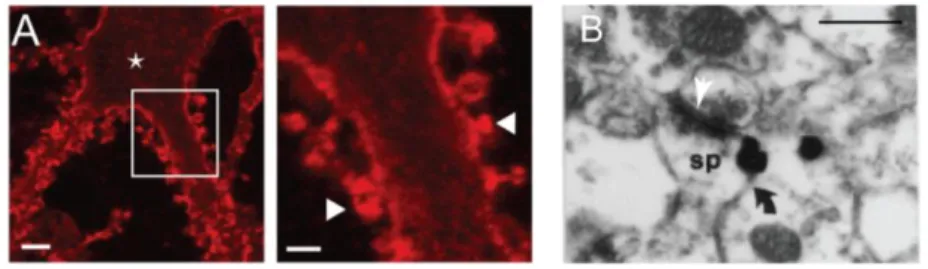
In 2001, Gulyas et al reported for the first time KCC2 expression in dendritic spines, close to glutamatergic synapses in the hippocampus (Gulyás et al., 2001). Interestingly, they observed high level of KCC2 in dendritic spines and lower level in the shafts, hosting
19 GABAergic synapses. This observation suggested that KCC2 aggregates in dendritic spines and was later confirmed by our team (Gauvain et al., 2011b). This protein seems to be excluded from the post-synaptic density (PSD) as KCC2 does not colocalize with PSD-95 (Figure 4). However, the limitations of optical fluorescence imaging currently preclude further analysis of the relation between KCC2 and the PSD.
Figure 4. KCC2 is expressed in the vicinity of glutamatergic synapses
A. Maximum intensity projection of confocal optical sections showing KCC2 expression (red) is localized at the
membrane in dendrites (left) and spines (right). Scale bar left 5 µm, and right 2 µm. B. Silver-intensified gold particle (black arrow) staining KCC2 are localized closed to the post-synaptic density (white arrow) in dendritic spines (sp) of pyramidal cells. Scale 0.6 µm.
(Adapted from Gauvain et al., 2011 and Gulyás et al., 2001)
Since KCC2 controls chloride homeostasis, its role in GABAergic signaling has been extensively studied in both normal and pathological conditions (Hübner et al., 2001; Tyzio et al., 2014). However, what could be the functional role of KCC2 at glutamatergic synapses?
a. KCC2 is necessary in spinogenesis
As we have seen previously, KCC2 expression increases after birth. To understand how this affects brain development, Li et al. took advantage of a KCC2 knockout mouse (Li et al., 2007). As these mice die shortly after birth, they used primary hippocampal neuronal cultures from embryos to study the dendritic morphology and synaptic function of these neurons. They observed long, abnormal dendritic protrusions and a decrease in the number of functional glutamatergic synapses in KCC2-/- neurons. The abnormal dendritic morphology was confirmed in brain slices from P16 KCC2hypo/null mice, a mouse model that still retains around 20% of KCC2 expression compared to WT mice (Li et al., 2007). These effects were independent of GABA signaling as blocking GABAARs had no impact on spine morphology.
20 However, over-expression of a transport-deficient KCC2 in KCC2-/- neurons rescued normal spine morphology, suggesting a transport-independent mechanism. Moreover, transfecting WT neurons with KCC2-CTD, likely acting to disrupt the interaction of endogenous KCC2 with its protein partners, also altered dendritic morphology.
The authors then screened for proteins interacting with KCC2 in immunoprecipitation assays, and identified the actin-related protein 4.1N (Denker and Barber, 2002). KCC2 interacts with the FERM domain of 4.1N through its CTD (Li et al., 2007). Hence, the over-expression of FERM-4.1N to disrupt its interaction with KCC2 in WT neurons was sufficient to mimic the altered morphology observed upon KCC2 knockout. This study was the first to observe a transport-independent function of KCC2, involving an interaction with actin-related protein 4.1N. In this context, it is also striking that KCC2 expression increases concomitantly with the development of dendritic spines (Miller and Peters, 1981).
While KCC2 expression seems to be required for normal spinogenesis, precocious KCC2 over-expression, however, also perturbs dendritic development in a brain-region specific manner. Thus, in the hippocampus, premature KCC2 overexpression induces abnormal dendritic arborization with decreased dendritic length and branching (Cancedda et al., 2007). Instead, neurons from the somatosensory cortex show an increase in dendritic spine density (Fiumelli et al., 2013). In addition, these effects on dendritic spine development are dependent on its chloride transport function in the hippocampus, but not in the somatosensory cortex (Awad et al., 2018). Since KCC2 expression at the membrane is higher in hippocampal neurons compared to somatosensory neurons at P7, we might hypothesize different impact of KCC2 expression and function during development. This suggests caution is necessary before generalizing molecular mechanisms to different neurons and brain regions.
Interestingly, no change in synapse density was observed when KCC2 was suppressed in mature neurons, after the period of synaptogenesis. In these conditions, instead, dendritic spines exhibited larger heads, likely owing to altered water export (Gauvain et al., 2011). Indeed, this effect was due to KCC2 transport function and was mimicked by chronic exposure to the KCC2 antagonist VU0240551. Altogether, these data suggest that KCC2 plays a crucial role in spinogenesis during development but is not required for the structural maintenance of dendritic spines in mature neurons.
21
b. KCC2 affects LTP expression, independent of its ion transport function
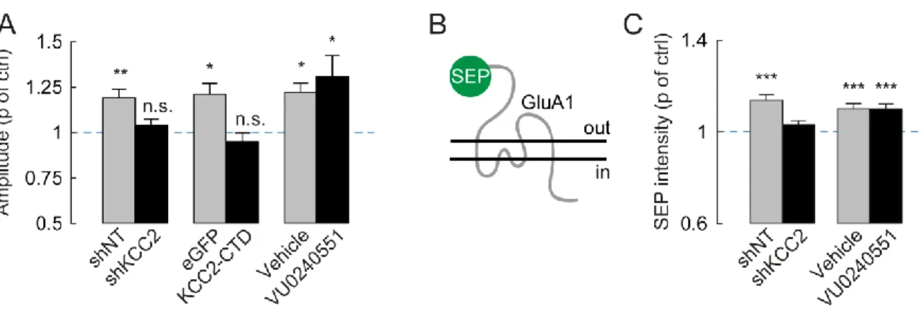
The function of KCC2 in dendritic spines of mature neurons is not, however, limited to osmotic regulation of spine head volume. Although spines head volume usually positively correlates with quantal size (Ashby et al., 2006; Harris et al., 1992), KCC2 knockdown in mature neurons leads to both spine head enlargement and reduced mEPSC amplitude (Gauvain et al., 2011). This effect on quantal size was shown to be independent of KCC2 function but instead involve interaction of its CTD with intracellular partners, as it was mimicked by overexpressing KCC2-CTD. Single particle tracking experiments further revealed that KCC2-actin interaction, likely via 4.1N (Li et al., 2007) forms a molecular barriers that hinders the lateral diffusion of AMPA receptors within dendritic spines. Upon KCC2 extinction, perisynaptic receptors diffuse faster and escape dendritic spines, resulting in reduced postsynaptic clustering and reduced quantal size of glutamatergic currents (Gauvain et al., 2011).
Figure 5. KCC2 is necessary for LTP and GluA1 activity-driven insertion at the membrane
A. Change of mEPSP amplitude upon chemical LTP (cLTP) protocol: neurons lacking KCC2 (shKCC2) or over-expressing its CTD (KCC2CTD) do not express LTP while blocking KCC2 transport function with VU0240551 has no effect. B. Neurons were transfected with GluA1-SEP, SEP fluorescence is only visible when the receptor subunit is expressed at the membrane. C. Upon cLTP induction, GluA1 is not transported to the membrane in the absence of KCC2. Treatment with VU0240551 has no effect on GluA1 trafficking.
(Adapted from Chevy et al., 2015)
Since lateral diffusion of AMPARs is also key to the expression of long term potentiation, promoting their diffusion by suppressing KCC2 expression may be expected to influence LTP expression. This hypothesis was then tested in our lab using RNA interference-based KCC2 knockdown both in vivo and in vitro (Figure 5). Chronic extinction of KCC2