Driving Tissue Morphogenetic Cascades Using Tunable Nanolayered
Surface Coatings
by
Nisarg Jaydeep Shah
B.S., Chemical and Biomolecular Engineering, Johns Hopkins University (2009) S.M., Chemical Engineering Practice, Massachusetts Institute of Technology (2011)
SUBMITTED TO THE DEPARTMENT OF CHEMICAL ENGINEERING IN PARTIAL FULFILLMENT OF THE REQUIREMENTS FOR THE DEGREE OF
DOCTOR OF PHILOSOPHY IN CHEMICAL ENGINEERIN R
AT THE MOFTECHNOLOGY
MASSACHUSETTS INSTITUTE OF TECHNOLOGY
JUN 3
0
201
MAY 2014
LIBRARIES
@ 2014 Massachusetts Institute of Techno ogy. All rights reserved
Signature of Author.
Signature redacted
Nisarg J. Shah Department of Chemical Engineering May 8, 2014
C ertified by...
S ig
...
Paula T. Hammond David H. Koch (1962) Chair Professor in Engineering Thesis SupervisorAccepted by...S
ig n a tu re
...
Patrick S. Doyle Singapore Research Chair Professor of Chemical Engineering Chairman, Committee for Graduate StudentsDriving Tissue Morphogenetic Cascades Using Tunable Nanolayered Surface Coatings
by
Nisarg Jaydeep Shah
Submitted to the Department of Chemical Engineering
on May 8, 2014, in partial fulfillment of the requirements for the degree of Doctor of Philosophy in Chemical Engineering
Abstract
Harnessing the synergy between materials at the nanoscale can be a valuable tool in understanding and probing cellular phenomena and in driving specific processes that lead to tissue and organ regeneration and repair. Biomedical implants attempt to materially replace damaged tissues, restore function, improve mobility, and alleviate pain. However, these implants often fail to recapitulate the original tissue properties. In the context of orthopedic implants, the functional success of an implant critically
depends on its stable interaction and bonding with the host tissue. Yet, aseptic implant loosening accounts for over half of all joint replacement failures. Non-biological implant materials such as metals and plastics are used for their mechanical strength, but these are typically not suitable for direct integration with the host tissue. Conversely, traumatic wounds and congenital defects that result in large bone tissue defects are often
corrected with autograft bone, which results in the matching of native bone tissue properties. However, the limited availability of donor bone coupled with pain at the site renders autografting suboptimal therapy. Permanent implants require multiple rounds of surgery and are often unable to recapitulate complex tissue reconstruction with high fidelity and carry the risk of inducing a foreign body response. Orthogonally, therapies to alleviate diseases in the bone such as osteosarcoma and osteoporosis are of significant interest to improve clinical outcomes. However systemic delivery of these therapies leads to toxic side-effects that limit their clinical use. These significant challenges to improving human health can be overcome by designing and combining materials with complementary biological properties.
This body of work investigates the use of tunable, modular, nanoscale coatings that can prevent premature implant failure, induce targeted tissue repair by recapitulating aspects of the natural wound healing cascade and target therapy to the bone tissue. At the nanoscale, these coatings can impart multifunctionality to nanoparticles that can be used as a therapeutic agent. Central to the polymer-based multilayered coating, was a water-based layer-by-layer (LbL) deposition process, by which each component was
introduced on the surface in nanoscale layers. On surrogate bone implants, coatings containing osteoconductive hydroxyapatite (HAP) and 5-10 pg of osteoinductive bone morphogenetic protein 2 (BMP-2) contained within the nanostructured coating acted synergistically to induce osteoblastic differentiation of endogenous progenitor cells in a rat. The tuned release of BMP-2 over 3-4 weeks, controlled by a hydrolytically
degradable poly(P-amino ester), was essential for tissue regeneration and, in the presence of HAP, the modular coating encouraged the direct deposition of highly
cohesive trabecular bone on the implant surface with an interfacial tensile strength that was 2-3 fold higher than implants coated with bioactive bone cement. Importantly, the failure was as a result of bone fracture, rather than at the bone-implant interface. The coated implants demonstrated long-term stability and mechanical integrity.
To repair large bone defects, two strategies were employed that involved the use of adaptive growth factor delivery from LbL surface coatings. In the first instance, scaffolds made of polycaprolactone and
P-tricalcium
phosphate (p-TCP) were developed and used as osteoconductive scaffolds. The scaffolds were coated with multilayer films that sequestered BMP-2 and angiogenic vascular endothelial growth factor (VEGF). In vivo, at an intramuscular ectopic location, the mineral density of ectopic bone formed de novo by BMP-2NEGF multilayer coated scaffolds resulted in bone formation that wasapproximately 33% higher than when only BMP-2 was delivered. Bone formed
throughout the scaffold when both growth factors were released, which suggests more complete remodeling due to an increased local vascular network. Using another
strategy for orthotopic, craniomaxillofacial bone repair, scaffolds made of a poly(lactic-co-glycolic acid) (50:50) copolymer were used with multilayer coatings containing tunable amounts of BMP-2 and mitogenic platelet derived growth factor (PDGF)-BB from 0.2-2pg. Complete release of the growth factors occurred over readily adapted time scales, which extended from 8-10 days and 22-25 days for PDGF-BB and BMP-2 respectively. Each component of the system was tested for its effect on tissue
formation. The adaptive growth factor release and synergistic effect of BMP-2 and PDGF-BB promoted local bone formation that bridged a critical-size defect in the rat calvaria as early as 2 weeks after implantation. Dual growth factor delivery resulted in mechanically competent bone that restored the properties of the bone and regenerated the native calvaria form. This approach could be a customizable, off-the-shelf, cell-free option for bone tissue repair.
In addition to focusing on multilayer coatings for macroscale implants and scaffolds, an approach for generating polymer coated functional and targeted nanoscale drug carriers specifically for treating bone disease such as osteosarcoma and osteoporosis was developed. The approach utilized the modularity of LbL assembly to generate tissue-specific drug carriers for systemic administration. This was accomplished via surface modification of drug-loaded nanoparticles with an aqueous polyelectrolyte, poly(acrylic acid) (PAA), with 40% of side-chains functionalized with alendronate, a potent clinically used bisphosphonate. Encapsulation of doxorubicin at 5mg/kg in an LbL-targeted liposome demonstrated potent toxicity. Active targeting of 143B
osteosarcoma xenografts in NCR nude mice prolonged tumor accumulation by over 7 days. Tumor burden reduced, and in some cases the entire tumor mass was eliminated within 40 days, with a 3 dose regimen.
Collectively, this work provides insight into probing and modifying cellular interactions to control morphogenetic processes at varying length and time scales, and in different disease states with a polymer-based multilayer coating approach. Towards the
development of next-generation biomedical therapies this approach allows for driving cell morphogenetic phenomena and controlling local microenvironments to understand and engineer specific therapies with the ultimate goal to improve human health.
Thesis Supervisor: Paula T. Hammond
Thesis Supervisor Paula T. Hammond, Ph.D.
David H. Koch (1962) Professor in Engineering Department of Chemical Engineering
The David H. Koch Institute for Integrative Cancer Research Massachusetts Institute of Technology
Thesis Committee Sangeeta N. Bhatia, Ph.D. John and Dorthy Wilson Professor
Harvard-MIT Division of Health Sciences and Technology Department of Electrical Engineering and Computer Science The David H. Koch Institute for Integrative Cancer Research
Massachusetts Institute of Technology
Robert S. Langer, Sc.D.
David H. Koch (1962) Institute Professor Department of Chemical Engineering
Harvard-MIT Division of Health Sciences and Technology The David H. Koch Institute for Integrative Cancer Research
Massachusetts Institute of Technology
Howard J. Seeherman, Ph.D., V.M.D. Restituo LLC.
For my parents, Jaydeep and Jaishri Shah
Acknowledgements
I have been fortunate to have worked with a set of phenomenally talented individuals who have contributed in a myriad of ways towards my graduate school experience. The work described in this thesis has been strongly influenced by my interactions with them. My foremost appreciation is for my mentor and advisor, Prof. Paula Hammond. She has been an inspirational role-model for me and I have learned a tremendous amount from my interactions with her. She provided me with an opportunity to work on a fascinating, cutting-edge research project that challenged me and helped my
professional development. She was open-minded about new research directions and enthusiastic about letting me pursue several independent projects that helped broaden my skills. Her enthusiasm and perpetual optimism was infectious and helped me
overcome scientific challenges. She has all the qualities of an inspirational mentor, a leading scientist, and a truly wonderful person.
I would like to thank my thesis committee, Prof. Sangeeta Bhatia, Prof. Bob Langer and Dr. Howard Seeherman, for their participation. They brought a tremendous amount of experience and expertise between them and always offered reasoned and
constructive feedback that helped overcome scientific challenges. Their support has been highly instrumental in shaping this body of work. I am also grateful to Profs. Myron Spector (VA Boston Healthcare System/Brigham and Women's Hospital), Robert
Padera (Brigham and Women's Hospital) and Myron Nevins (Harvard School of Dental Medicine) who have been tremendously helpful collaborators and from whom I am learned a lot about biomedical research. I would like to acknowledge Prof. Justin Hanes (Johns Hopkins University), who welcomed me into his lab to pursue undergraduate research under the mentorship of Ben Tang. My experience in Justin's lab largely influenced my decision to pursue graduate school.
I am indebted to past and present Hammond Lab members who have been wonderful colleagues and friends and also helped make a positive work environment. Mara was a great mentor during my early days in the group, and helped me get started with my research. Ray was a truly gifted individual, full of ideas and an infectious enthusiasm, and helped me with animal studies in the early days. I am grateful to other members of
the LbL delivery subgroup for their input. In particular, Anita and Jessie, who provided valuable advice and Josh, whose advice and assistance has been invaluable. Zhiyong, Amanda and Abby could not have been better office-mates and I will always remember the breakfast club and the wonderful treats. I enjoyed discussing research and working with Nasim, who is a multitalented individual with tons of good advice. I would like to acknowledge Mohi, Stephen and Jason for their enthusiasm and assistance in lab. I am grateful to Christine and Liz, whose excellent organizational skills and constant
willingness to help with the several important administrative aspects of graduate school so much easier. I have been fortunate to build strong friendships in this lab that will last a lifetime.
The support and administrative staff at MIT have been generous with their time, patient and generous with their advice. Suzanne, Katie and Joel at the student office have been extraordinarily supportive. Drs. Alison Hayward and Alton Swennes are extraordinary veterinarians and tremendously helpful. Catrina and Wayne were incredibly helpful surgery techs and their assistance made the animal studies more manageable.
I would like to dedicate this thesis to my family whose support has helped make this journey possible. To my parents, Jaydeep and Jaishri, who have always believed in me
and provided me with boundless love and unconditional support to pursue opportunities to succeed. They have instilled in me a passion for education, a strong work ethic and a sense of perseverance to pursue my passions and bring them to fruition. I feel truly blessed to have the support of such wonderful parents. Being the only child, I can only imagine the decisions that they had to make so allow me to go far away from home to pursue higher education. To my uncles, aunts and cousins for their love, support and encouragement. To my grandparents, who have always been a supportive presence in my life. This is for all of you.
NISARG JAYDEEP SHAH Massachusetts Institute of Technology May 8, 2014
N.J. Shah is thankful for support from the MITSCEP 1936 Course Xa Fellowship, National Institutes of Health (NIH), the MIT Energy Initiative, the Henry C. & Frances Keany Rickard Health Science Fund at MIT, the MIT Portugal Program and the David H. Koch (1962) Professorship. This work was supported by the NIH (RO1 AG029601, RO1 EB010246) and in part by the David H. Koch Institute for Integrative Cancer Research (supported by the NCI through grant P30 CA014051), the Institute for Soldier
Nanotechnologies (supported by the U.S. Army Research Office under contract W91 1 NF-07-D-0004), the Center for Materials Science and Engineering MRSEC Shared Facilities (supported by the NSF through grant DMR-0213282) at MIT. The use of equipment in the laboratories of Prof. Sangeeta Bhatia and Prof. Robert Langer is gratefully acknowledged.
Contents
Acknow ledgem ents ... 9
C o n te n ts ... 1 3 F ig u re s ... 1 7 Chapter 1 Introduction ... 25
1.1 Biom edical im plants designed for long-term functionality... 25
1.2 Traumatic and congenital defects that result in large tissue loss ... 26
1.3 Tissue-specific delivery of drug in the body ... 26
1.4 Controlled Drug Release... 27
1.5 Layer-by-Layer Assem bly (LbL) ... 28
1.6 Thesis Overview ... 31
1.7 References...33
Chapter 2 Osteophilic Multilayer Coatings for Accelerated Bone Tissue Growth.39 2.1 Introduction ... 39
2.2 M aterials and M ethods... 40
2.2.1. M aterials...40
2.2.2 Preparation of electrostatic film s ... 40
2.3.3 In vitro cellular assays... 40
2.3 Results and Discussion... 41
2.4 Conclusions ... 46
2.5 References...53
Chapter 3 Surface-mediated bone tissue morphogenesis from tunable nanolayered im plant coatings ... 57
3.1 Introduction ... 57
3.2 M aterials and M ethods... 59
3.2.1. Rationale and study design ... 59
3.2.2 Preparation of electrostatic film s. ... 59
3.2.3 Anim al studies... 60
3.2.4 Pull-out tensile testing. ... 60
3.2.5 Histology. ... 60
3.2.6 pCT analysis ... 60
3.2.7 Statistical analysis... 6 1 3 .3 R e s u lts ... 6 1 3.3.1. A two-part m ultilayer osteogenic coating ... 61
3.3.3 Integration of the implant with the bone tissue. ... 65
3.3.4 Host-im plant interactions at the cellular level. ... 66
3.3.5 Quantifying bone deposition... 67
3.4 Discussion...68
3.5 References...81
Chapter 4 Tunable dual growth factor delivery from polyelectrolyte multilayer film s ... 87
4.1 Introduction ... 87
4.2 Materials and Methods... 89
4.2.1. Materials... 89
4.2.2. Preparation of polyelectrolyte solutions... 90
4.2.3. Polyelectrolyte m ultilayer film construction... 90
4.2.4. Release characterization... 90
4.2.5 Cell culture. ... 91
4.3 Results and Discussion... 95
4.3.1. Fabricating a dual growth factor releasing layer-by-layer system...95
4.3.2. In vitro kinetics of dual growth factor releasing film s ... 96
4.3.3. In vitro rhBM P-2 activity assay ... 98
4.3.4. In vitro rhVEGF165 activity assay ... 99
4.3.5 In vivo evaluation of LbL films using intramuscular bone formation model... 100
4.4 Conclusions ... 101
4.5 References...110
Chapter 5 Adaptive growth factor delivery from polyelectrolyte coatings promotes synergistic bone tissue repair and reconstruction ... 117
5.1 Introduction ... 117
5.2 Materials and Methods...120
5.2.1. Materials...120
5.2.2. Alendronate conjugation to membrane...120
5.2.3. PEM deposition, characterization and release ... 120
5.2.4. In vivo critical size defect studies ... 121
5.2.5. pCT analysis. ... 121
5.2.6. Histology analysis...121
5.2.7. Mechanical testing of calvaria ... 121
5.2.8. Statistical analysis...122
5.3. Results ... 122
5.3.1. Tunable growth factor deposition in PEM coatings ... 122
5.3.2. Modification of support PLGA mem brane...122
5.3.4. Histological evaluation of regenerated bone ... 125
5.3.5. Com parison of bone m echanical properties ... 126
5.4 Discussion...127
5.5 References...136
Chapter 6 Osteotropic therapy via targeted Layer-by-Layer nanoparticles ... 139
6.1 Introduction ... 139
6.2 M aterials and Methods...140
6.2.1. PAA-Alendronate conjugation ... 140
6.2.2. LbL on Q Ds ... 141
6.2.3. Liposom e synthesis...141
6.2.4. LbL on liposom es ... 142
6.2.5 Physicochem ical characterization. ... 142
6.2.6. In vitro Binding/Cytotoxicity ... 142
6.2.7 Fluorescence-activated cell sorting analysis. ... 143
6.2.8 Confocal M icroscopy ... 143
6.2.9 Xenograft development/targeting/treatment/monitoring - NCR nude...143
6.2.10 Pharmacokinetics (circulation) - immune-proficient BALB/c. ... 144
6.2.11 Histology ... 144
6.2.12 Statistical Analysis...145
6.3 Results and Discussion...145
6.4 Conclusions ... 150
5.5 References...159
Chapter 7 Conclusions and Future Directions...161
7.1 Thesis Sum m ary ... 161
7.2 Future Directions...163
7.2.1. Sequential release of therapeutics ... 163
7.2.2. Pre-clinical studies in large anim al m odels...164
7.2.3. Dental Implants coated using the LbL approach for rapid osseointegration 164 7.2.4. Therapy with disease-modifying properties for osteoarthritis...164
7.2.5. Translational considerations of LbL coated biomedical products...165
7.3 Concluding Rem arks ... 166
A ppendix A ... 169
Appendix B ... 175
Appendix C ... 197
Figures
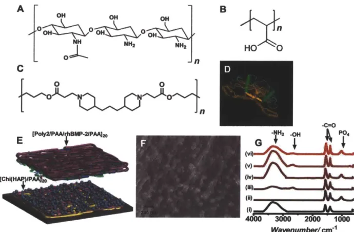
Figure 2.1. Design and fabrication of the osteophilic multilayer architecture.
Components of the film (A) Chitosan (75%-85% deacetylated chitin), a polycation (B) Poly(acrylic acid) (PAA), (C) A hydrolytically degradable poly(P-amino ester) (Poly2), a polycation and (D) osteoinductive recombinant human bone morphogenetic protein 2 (rhBMP-2) (E) Schematic of the modular electrostatic assembly. Osteoconductive hydroxyapatite is complexed with chitosan and incorporated into nanoscale thick films along with poly(acrylic acid) in a bilayer architecture. A hydrolytically degradable poly(P-amino ester) based multilayer film incorporating osteoinductive rhBMP-2 lays atop the osteoconductive layer. (F) Scanning electron micrograph of a [Chi(HAP)/PAA]2o bilayer film with the complexed hydroxyapatite nanoparticles. HAP particles complexed to chitosan strands are interwoven in the multilayer architecture (G) IR absorbance spectra of the different components of the osteoconductive layer (i) chitosan (ii) hydroxyapatite (iii) poly(acrylic acid) (iv) [Chitosan/HAP]i (v) [Chitosan/PAA]2o and (vi)
[Chi(HAP)/PAA]20.
Figure 2.2. Characteristics of multilayer properties during assembly and degradation. (A) Multilayer film thickness increases linearly with incremental osteoconductive [Chi(HAP)/PAA] layers. Contributions of film thickness from the growth factor eluting layers (green) and hydroxyapatite containing layers (blue) are provided. (B) rhBMP-2 released from the films over 2 weeks. Surface and bulk morphologies of
[Chi(HAP)/PAA]20 + [Poly2/PAA/rhBMP-2/PAA]20 multilayer films (C, F) HAP particles
are distributed uniformly in the osteoconductive [Chi(HAP)/PAA]20 multilayer surface, and the features are made rougher in (D, G) by the conformal coating of the
[Poly2/PAA/rhBMP-2/PAA]20 layers. (E, H, K) Once the growth factor is released by the degradation of the poly(P-amino ester) layers, there is an observable reduction in
surface roughness. (F, G, H) Surface height profiles confirm that HAP particles are uniformly distributed with a lack of sequestration in a particular area. (1, J)
Corresponding cross sectional scanning electron microscopy images confirm the
presence of particles throughout the bulk of the film (1, J, K are at the same scale). C-H are atomic force microscopy images, where C-E are phase contrast images, F-H are height images. I-K are scanning electron microscopy images.
Figure 2.3. Mesenchymal stem cells differentiating into the osteoblast lineage. (A) Alkaline phosphatase (AP) quantification at 5 days after the differentiation cascade is initiated. (B) Alizarin red (AR) staining and quantification for calcium deposits 14 days after mesenchymal stem cells have been in culture under different conditions as
indicated. Both AP and AR signals have been represented as fold increase or decrease of the Control +L-AA/P-GP case. A single factor ANOVA test allowed rejection of the null hypothesis for all assays; and a Tukey test between all groups was performed (s.d., n=9, ** p < 0.01; * p < 0.05; ns = not significant all others are p < 0.001). Temporal expression patterns of osteogenic markers in mesenchymal stem cells. Quantification of osteogenic markers (C) Osteocalcin, (D) visualized (green) from differentiating cells by fluorescence over the course of the study. (E) Osteoprotonegrin and (F) Osteopontin expression follows a similar trend to osteocalcin. In all the assays, the synergistic effect
of having an osteoconductive and an osteoinductive layer maximizes AP and AR signals. Expression of osteogenic markers may be accelerated and amplified by the introduction of the osteophilic multilayers leading to an environment conducive for bone formation. Peak and cumulative expression levels of the combination osteoconductive and an osteoinductive multilayers are statistically significant compared to other groups (p<0.001, Fig A4).
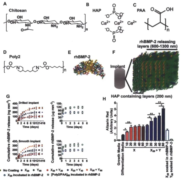
Figure 3.1. Structured coatings for bone regeneration are made up of two composite multilayers. (A to C) The base coating contains chitosan (Chi; 75-85% deacytelated chitin, Mv ~ 100 kDa) and hydroxyapatite [HAP; Calo(PO4)6(OH)2] with poly(acrylic acid) (PAA; Mv -450 kDa) in a bilayer repeat unit. (D and E) The osteogenic factor coating contains a hydrolytically degradable poly(P-amino ester) (Poly2, Mv - 11 kDa) and rhBMP-2 that are alternated with PAA on top of the osteoconductive base coating. (F) Schematic of the two sets of multilayers: osteoconductive and osteoinductive. (G) Cumulative release profile of rhBMP-2 from drilled implants. Data are means ± SEM (n = 9 per coating). (H) rhBMP-2 loading has a dose-dependent effect on calcium
deposition, quantified by alizarin red at 14 days. Data are means ± SEM (n = 6-9). * P < 0.05**P < 0.01, ANOVA with a Tukey post hoc test.
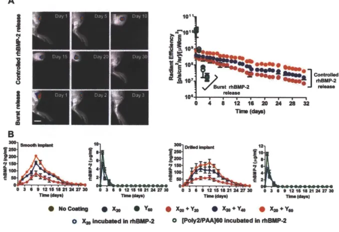
Figure 3.2. In vivo evaluation of rhBMP-2 release. rhBMP-2 was loaded into the
multilayers that coated smooth and drilled PEEK rods and then implanted in the tibias of rats (n = 41-45 per group). (A) Controlled and burst release of fluorescently labeled rhBMP-2 was tracked in vivo over 30 and 3 days respectively. (B) Radiant efficiency at the implant site over time (n = 4-6 per group). (C) Bone marrow flushed out of excised tibiae was assayed for rhBMP-2 using ELISA for smooth and drilled implants. Data are means ± SEM (n = 5-6 per group).
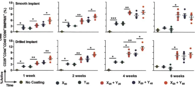
Figure 3.3. Mesenchymal stem cells differentiate into osteoblasts. Five color flow cytometry was used to assess the percentage of osteoblasts in cells isolated from the tibia marrow around smooth and drilled implants. Each point represents individual implants. Means ± SEM (n = 5 per group). *P < 0.05, **P < 0.01, ANOVA with a Tukey post hoc test. FACS plots are provided in Appendix B; fig. B4.
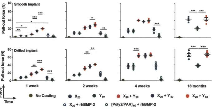
Figure 3.4. Tensile force testing of implants from the rat tibia. Force data from individual implants are presented from smooth and drilled implants. Data are means ± SEM (n = 5 implants per group time point). *p < 0.05; **p < 0.01; ***p < 0.001, ANOVA with a Tukey post hoc test. Interfacial tensile strength data are provided (Appendix B; table B1 and B2).
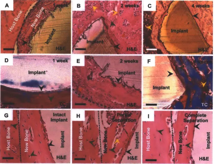
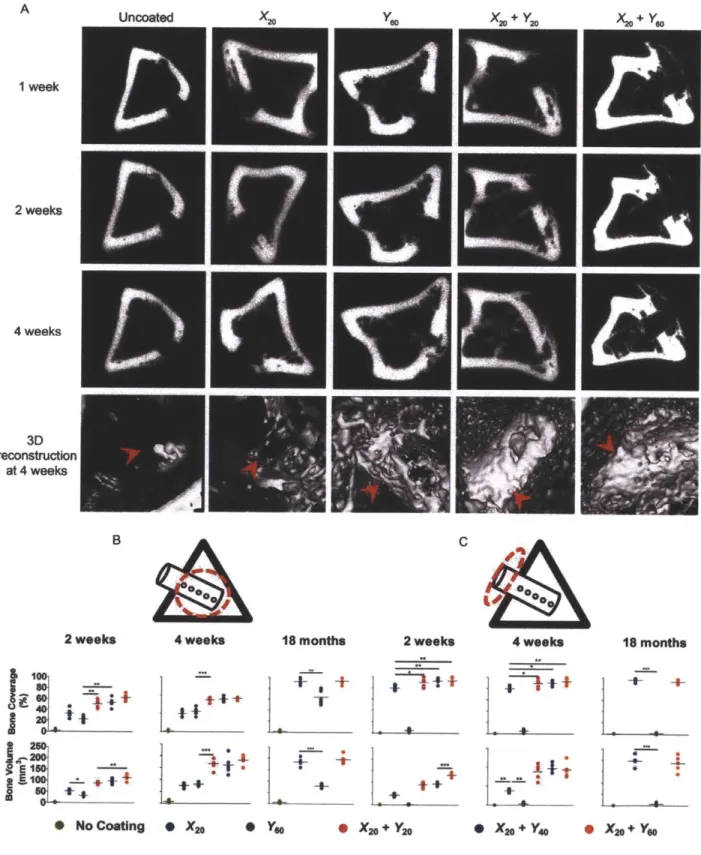
Figure 3.5. Histology of implants with various coating formulations demonstrating bone tissue morphogenesis at the implant interface. (A to F) Implants coated with X2o + Y6o
at 1, 2 and 4 weeks post-implantation demonstrating the process of implant integration with the parent bone tissue. Cement lines (broken black line) are observed on some sections. (G) The plane of fracture in implants with the X2o + Y6o coating is indicated by a broken black line at 4 weeks which depict an intact implant, partial separation from the host bone and complete separation from the host bone. The new bone-implant interface is intact. Sections were viewed under bright field microscopy. Scale bars: (A and C) are 200 pm; (B) and (D to G) are 50 pm. Arrows: black, bone/implant interface; red, active
osteoblasts; dark green, osteocytes; yellow, marrow cells. H&E: hematoxylin & eosin stain, TC: Masson's trichrome stain.
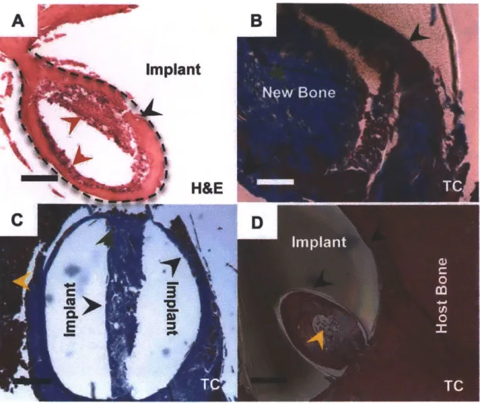
Figure 3.6. Bone deposition in the channels of drilled implants. Representative sections
(n = 5-6 per group) of drilled implants after 4 weeks, which were coated with X20 + Y60. (A) Granulation tissue (broken black line) penetrated the channel and supplied
progenitor cells. (B) Newly deposited bone (blue) matures (red) and (C) gradually filled up the channel at 4 weeks. (D) Bone (blue and red) is present throughout the channel of a drilled implant. Sections were viewed under brightfield microscopy. Scale bars in (A,
B and D) are 100pm and in (C) is 400 pm. Arrows: black, bone/implant interface; red, active osteoblasts; dark green, osteocytes; yellow, marrow cells. H&E: hematoxylin & eosin stain, TC: Masson's trichrome stain.
Figure 3.7. pCT imaging of bone formation on drilled PEEK implants. (A) Radiographs of bone formation around drilled implants with different coatings at 1, 2, and 4 weeks. Red arrows indicate location of the implant. (B and C) The images in (A) were used to quantify bone regeneration at 2 and at 4 weeks within (B) and outside the medullary canal (C) (using regions of interest marked by dotted red circles). Each point represents individual implants. Data are means ± SEM (n = 5-6 per group). *p < 0.05, **p < 0.01, ANOVA with Tukey post hoc test. Data for smooth implants are provided in Appendix B; fig. B11.
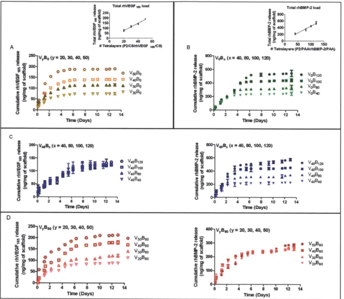
Fig. 4.1. Fabrication of polyelectrolyte multilayer films using the layer-by-layer method in a tetralayer architechture with [Poly2(+)/polyanion(-)/growth factor(+)/polyanion(-)] depicted below. Four different groups were fabricated. For single growth factor films (A) rhBMP-2 PEM films with architecture [Poly2/PAA/rhBMP-2/PAA]x, with x = 40, 80, 100, 120 and (B) rhVEGF165 PEM films with architecture [Poly2/CS/rhVEGF165/CS]y, where y = 20, 30, 40, 50. For dual growth factor films, (C) y = 40 was constant and x = 40, 80, 100, 120. (D) In another formulation, x = 80 was constant and y = 20, 30, 40, 50. The substrate was always a macroporous polycaprolactone/p-tricalcium phosphate (50wt%
PCL/50wt% P-TCP) waffle cylinder scaffold, with diameter = 10mm and height = 2.5mm.
Fig. 4.2. Polyelectrolyte multilayer films were dipped in a Poly2/anion/growth factor/anion tetralayer repeat architecture on a macroporous
polycaprolactone/P-tricalcium phosphate (50wt% PCL/50wt% P-TCP) waffle cylinder scaffold. Growth factor loading was controlled by varying the number of layers dipped on the scaffold. rhBMP-2 released over a period of about 2 weeks, whereas rhVEGF165 eluted completely within 8 days. 4 groups of films were fabricated and incubated in 1x PBS at pH 7.4 over 12 days to study growth factor release from the films. Growth factor release was quantified using ELISA and normalized per milligram of scaffold. Samples were in triplicate and the error bar is the standard deviation. Release from (A) Single growth factor rhVEGF165 release and (inset) total rhVEGF165 loading scales linearly with tetralayer number (R2 =
0.96). (B) Single growth factor rhBMP-2 release and (inset) total rhBMP-2
Fig. 4.3. Pre - osteoblast differentiation assay. Activity of rhBMP-2 released from the polyelectrolyte multilayer films is preserved. MC3T3-E1 cells were exposed to rhBMP-2 released from multilayer films, with the dose normalized per milligram of scaffold weight. Alkaline Phosphatase Assay demonstrates dose dependent early activation of bone
differentiation cascade at Day 5. After 21 days of culture, Alizarin Red Stain confirms the presence of calcium deposits laid down during the differentiation process. Visual inspection of cultures after Alizarin red staining confirms a dose dependent presence of calcium deposits. (A, B and C) Alkaline phosphatase assay at day 3 on cells
differentiated with different release formulations as depicted. (D, E and F) Alizarin red assay at day 14 on cells differentiated with different release formulation as depicted. Representative images of the alizarin red stain are below the bar graph in (D, E, F). A single factor ANOVA test allowed rejection of the null hypothesis for both assays; and a Tukey test between different groups was performed (s.d., n=9, ** p < 0.01; * p < 0.05; ns = not significant; all others p < 0.001).
Fig. 4.4. Proliferation and Migration assays. Activity of rhVEGF165 released from the polyelectrolyte multilayer films is preserved. HUVEC cells exposed to rhVEGF165 dose normalized per milligram of scaffold weight exhibited increased proliferation activity, as measured by BrdU. (A) 40, 80, 100 and 120 tetralayers of rhBMP-2 followed by 40 tetralayers of rhVEGF165. (B) 40 tetralayers of rhBMP-2 followed by 20, 30, 40 and 50 tetralayers of rhVEGF165 and (C) 20, 30, 40 and 50 tetralayers of rhVEGF165. Closure of uniform "scratched" wound gap was monitored in HUVEC cell cultures over a period of 8 hours. Cells were fixed and imaged at 20x magnification (D) 40, 80, 100 and 120 tetralayers of rhBMP-2 followed by 40 tetralayers of rhVEGF165. (E) 40 tetralayers of rhBMP-2 followed by 20, 30, 40 and 50 tetralayers of rhVEGF165 and (F) 20, 30, 40 and 50 tetralayers of rhVEGF165. A single factor ANOVA test allowed rejection of the null
hypothesis for both assays; and a Tukey test between different groups was performed (s.d., n = 9, ** p < 0.01; * p < 0.05; ns = not significant; all others p < 0.001).
Fig. 4.5. Two dimensional microCT scans (2D) and matched three dimensional
reconstructions (3D) of excised PCL-PTCP half disc scaffolds, which were implanted in the intramuscular region of rats. Implants were coated with (i) no growth factor, (ii) 6 pg of single growth factor rhBMP-2 and (iii) 6 pg of single growth factor rhBMP-2 followed
by 4pg of rhVEGF165. The amount of growth factor loaded was determined by
fabricating triplicate companion copies along with the implanted scaffolds, releasing the growth factors in vitro and performing ELISA detection assays. (Top row) Control scaffolds without growth factors produce no detectable bone over the duration of the study. Low levels of backscatter is caused by the polymer. (Middle row) In single growth factor rhBMP-2 films lacking rhVEGF165, bone formation is restricted to the periphery of the scaffold at 4 weeks and 9 weeks. (Bottom row) As a result of increased vascularity,
scaffolds releasing rhVEGF165 demonstrate a smooth, continuous profile in the ectopically formed bone which matures from 4 weeks to 9 weeks to fill the entire
scaffold. In all the images, the bone formed takes the shape of the scaffold and grows inward when VEGF is present. Images are an isosurface rendering at 0.25 surface quality factor at a level threshold of 640, as defined by the proprietary Microview* software from GE Healthcare.
Fig. 4.6. (A) The bone mineral density (BMD) of ectopic bone formed by the rhBMP-2/rhVEGF165 combination scaffolds is 28 ± 4.59% and 32 ± 2.73% higher than bone formed by rhBMP-2 scaffold at 4 and 9 weeks respectively at the periphery of the
higher than the single rhBMP-2 scaffolds indicating that more bone is present in the interior, which matures from 4 to 9 weeks as indicated by the increase in BMD. (C) The trabecular thickness of the bone formed is an important measure of bone maturation, and the trabecular thickness of the bone formed by the combination scaffolds is about 3 times and 4.5 times that of bone formed by rhBMP-2 scaffolds at 4 and 9 weeks. (s.d., n=9, ** p < 0.01; ns = not significant; all others p < 0.001)
Fig. 4.7. Histology sections from rat femur intramuscular implants. (A to D) Bone is absent in control scaffolds releasing no growth factors at 4 and 9 weeks verified by (A, C) H&E staining and (B, D) lack of brilliant blue collagen staining with Masson's
trichrome stain. (A, B) Arrows indicate pockets where the (PCL/PTCP) polymer scaffold is present at 4 weeks which degrades over (C, D) 9 weeks, without any bone growth in the vacant space. (E to H) Bone present in scaffolds releasing single growth factor
rhBMP-2. (E) Presence of trabecular bone with (inset) osteoblasts laying down new bone which is (F) restricted to the periphery of the scaffold at 4 weeks. Of particular interest is (G) the abundant presence of hematopoietic cells surrounding the bone in spaces identified as fatty marrow space (asterisk), where (H) most of the bone
formation is restricted to the periphery at 9 weeks and (inset) as bone matures, it goes from an unorganized collagen matrix structure to a lamellar structure with aligned collagen fibrils that are birefringent under polarized light. (I to L) Bone present in scaffolds releasing rhBMP-2 and rhVEGF165. (I) Spicules of trabecular bone which is present (J) throughout the scaffold as early as 4 weeks. (K) As osteoblasts lay down new bone, spicules mature into (L) bone that fills up the empty spaces throughout the scaffold as it degrades with (inset) a greater number of birefringent aligned collagen fibrils present than fibrils formed when single growth factor rhBMP-2 is released. Bone formed is always within the scaffold. A, C, E, G, I, K are hematoxylin and eosin (H&E) stains at 1Ox objective magnification. B, D, F, H, J, L are Masson's trichrome stains at 2x objective magnification. Images in inset are taken at 20x objective magnification. Figure 5.1. Design of the multilayer polymer "skin". (a) Molecular structures of materials in the system. Hydrophobic PLGA is used to form the membrane. Poly2, PAA, BMP-2 and PDGF-BB are part of the bioactive interface that initiates the bone wound healing cascade. The bisphosphonate molecule alendronate is conjugated to PLGA. (b) Schematic of the phase-inversion membrane formation process. (1) A PLGA-DMF solution is poured on a glass plate. (2) A doctor blade is used to spread the polymer solution uniformly on the glass plate and is (3) immersed into a deionized water bath. (4) The resulting film detaches from the glass substrate. (c) Macroscopic image of the membrane structure that results in a uniform polymer support (scale bar, 8mm). (d) Scanning electron micrographs demonstrating a highly ordered cross section (scale bar,
10pm) (e) PLGA membrane coated with [Poly2/PAA/rhBMP-2/PAA]40 +
[Poly2/PAA/rhPDGF-BB/PAA] 40 (scale bar, 2pm)
Figure 5.2. In vivo and in vitro evaluation of growth factor release. PDGF-BB and BMP-2 were loaded into the multilayers that coated the membrane and then implanted in the critical size defect of a rat calvaria (n = 4-5 per group). (a) In vivo release of PDGF-BB and BMP-2 was tracked for 11 and 20 days respectively (b) In vitro Growth factor
release in single and combination films from PLGA membranes. Data represent the means±s.e.m.
Figure 5.3. pCT imaging of bone repair in live animals. (a) Representative radiographs of bone formation around drilled implants with different coatings at 1, 2, and 4 weeks. Red broken circle indicates the location of the defect in each radiograph and has an 8mm diameter. Defect closure was achieved in all animal groups with different
treatment conditions within 4 weeks. n = 5 per group. (b) The images in (a) were used to quantify bone volume and bone mineral density at 2 and 4 weeks within the regions of interest marked by dotted red circles. Each point represents individual animal. Data are means±s.e.m. (n = 5-6 per group). *p < 0.05, **p < 0.01, ***p < 0.001, ns = not
significant, ANOVA with Tukey post hoc test. All groups are compared with the mechanical properties of the M + Bo.2 +Po.2 group.
Figure 5.4. Histology of new tissue formed with various coating formulations. (a) Each image is a cross section of the calvarial defect after 4 weeks, at which time different levels of bone tissue morphogenesis was observed at the defect site. The broken lines indicate the position of the defect site and are 8mm apart. Collagen is represented by blue and osteocytes (mature bone) is represented by red. Sections were stained with Masson's trichrome stain and viewed under bright field microscopy. (b) Granulation tissue layer at 1, 2 and 4 weeks during bone repair in the M + Bo.2 + PO.2 treatment group. The tissue gradually reduces in thickness from 1 to 4 weeks as bone repair is completed. Pieces of the PLGA membrane were observed in some section (scale bar,
30pm). Arrows: red, PLGA membrane; yellow, granulation tissue layer.
Figure 5.5. Mechanical compression testing of calvaria bone (a) Stiffness and (b)
Failure load from different groups are presented at 4 weeks after implantation. Data are means±s.e.m. (n = 5 implants per group). *p < 0.05; **p < 0.01; ***p < 0.001, ns = not significant, ANOVA with a Tukey post hoc test. All groups are compared with the
mechanical properties of the M + Bo.2 +PO.2 group.
Figure 6.1. Achieving bone tissue level specificity of LbL coated nanoparticles. (A) Aqueous anionic polyelectrolyte, poly(acrylic acid) [PAA, MW 450K], functionalized with the (B) bisphosphonate targeting moiety, alendronate [for high specificity to hydroxyapatite in bone], to yield (C) the aqueous, ligand-functionalized polymer at 40% side-chain functionalization (Figure SI), which is used for complementary, iterative adsorption to the polycationic component (poly-L-lysine, PLL) in the film on the NP substrate, schematically illustrated in (D).
Figure 6.2. in vitro assessment of LbL-targeted QD NPs incubated with 143B osteosarcoma cells. (A) Confocal microscopy of 143B cells incubated with the LbL-targeted QD7o5 core NPs for 2 hours. Blue staining representative of a Hoescht nuclear stain, green representative of a phalloidin stain of the actin filamentous cell structure, and red representative of the nanoparticle fluorescence (QD7o5 fluorescence). Scale bar representative of 20pm. (B) Binding and relative cytotoxicity of 3 bilayer [PLL/PAA-Alendronate] LbL-targeted QD800 core NPs following incubation for 2 hours in 143B cells. Fluorescence emission data corresponds to that of the nanoparticle core (QDaoo).
Figure 6.3. in vivo evaluation of LbL-targeted QD8oo core NPs. (A) Representative live-animal imaging of LbL-targeted QD8oo core NPs following systemic administration to 143B osteosarcoma xenograft-bearing NCR nude mice. Imaging conducted at Aex = 640 nm, Aem = 800 nm for up to 8 days. Hind-flank xenografts identified in the pre-injection image; arrows at 5 minutes and 9 hours indicate binding to native bone tissue. (B) Quantification of fold tumor-specific accumulation normalized to tissue auto-fluorescence pre-injection for the systemically administered LbL-targeted QD800 core NPs, as visualized in (A). n = 3 mice (6 tumors); data presented as mean +/- SEM. (C) Biodistribution data corresponding to endpoint of (A) (quantified as percent recovered fluorescence following harvest of relevant tissue after 8 days post-administration). n = 3 mice (6 tumors); data presented as mean +/- SEM. (D) Co-localization of QD8oo NP core with LbL-film containing labeled PAA-Alendronate700; top row [PAA-Alendronate700 channel] - imaging conducted at Aex = 640 nm, Aem = 700 nm, bottom row [QD800 channel] - imaging conducted at Aex = 640 nm, Aem = 800 nm.
Figure 6.4. in vitro evaluation of LbL-targeted liposomal NPs in 143B cells. (A) Cryo-TEM of LbL-targeted doxorubicin-loaded liposomal NPs. Scale bar representative of 200 nm. (B) Binding (2 hour incubation) and relative cytotoxicity (48 hour incubation) of LbL-targeted empty liposomal NPs over a range of concentrations, based on fluorescence, in 143B cells (Aex = 675 nm, Aem = 710 nm; tracked using PLL7oo as cationic component in LbL film). (C) Confocal microscopy of 143B cells incubated with the LbL-targeted empty liposomal NPs [tracked via PLL700 polycationic component in LbL film] for 2 hours. Blue staining representative of a Hoescht nuclear stain, green representative of a phalloidin stain of the actin filamentous cell structure, and red representative of the nanoparticle fluorescence (PLLcy5.5 polymer shell fluorescence). Scale bar representative of 10 pm. (D) Representative cell-associated NP fluorescence following a 2 hour incubation of 143B cells with the LbL-targeted empty liposomal NPs. (E) in vitro cytotoxicity of LbL-targeted doxorubicin-loaded liposomal NPs and the corresponding (F) uncoated doxorubicin-loaded liposomal control following 48 hour (blue) and 72 hour (red) incubation periods over a range of doxorubicin concentrations. Figure 6.5. Biological evaluation of targeted empty liposomal NPs. LbL-targeted empty liposomal NPs tracked via PLL700 polycationic component in surface coating. Circulation data in (A) normalized to % remaining particles recovered, on the basis of fluorescence [Aex = 640 nm, Aem = 700 nm], immediately following systemic administration to immune proficient BALB/c mice; data presented as mean +/- SEM (n = 3). Two-compartment model fit to determine half-lives displayed. Data includes background subtraction of blood auto-fluorescence. (B) Quantification of fold tumor-specific accumulation normalized to tissue auto-fluorescence pre-injection for the systemically administered LbL-targeted blank liposomal core NPs to 143B xenograft-bearing NCR nude mice; data presented as mean +/- SEM (n = 3). (C) Biodistribution data corresponding to endpoint of (B) (quantified as percent recovered fluorescence [Aex
= 640 nm, Aem = 700 nm] following harvest of relevant tissue after 100 hours post-systemic administration); data presented as mean +/- SEM (n = 3).
Figure 6.6. 143B xenograft tumor remediation following treatment with
xenografts in NCR nude mice for LbL-targeted doxorubicin-loaded liposomal NPs, against untreated and uncoated doxorubicin-liposomal NPs. Untreated control xenografts were sacrificed 19 days post-inoculation of xenograft due to tumor burden exceeding 1 cm. Untargeted and targeted Dox-liposomal formulations were sacked at 30 days post-inoculation following a dose-escalation study with the following treatment regimen: day 10 at 1 mg/kg, day 17 at 2 mg/kg, day 24 at 3 mg/kg. Terminal point shown with final tumor area displayed in (B). Statistics are from an unpaired t-test, two-tailed to compare the untargeted and targeted formulations at each time point. Data presented as mean +/- SEM; n = 4. (C) Caliper measurements for in vivo tumor remediation with repeated dosing of 5 mg/kg for both targeted and untargeted formulations at 22 days, 28 days, and 35 days post-inoculation (displayed as day 0, day 6, and day 13 in (C)). Statistics are from an unpaired t-test, two-tailed to compare the untargeted and targeted formulations; *p < 0.05; **p < 0.01; ***p < 0.001. Data presented as mean +/- SEM; n = 4. Resection of tumors from terminal point of (C), n = 4 for each group, displayed from the final caliper measurement.
Figure 6.7. Representative histology of 143B tumors harvested at 18 days post-treatment corresponding to the terminal point in Figure 6C. (left column) tumors prior to treatment; (right column) tumors at the 18 day terminal point post treatment of 3 repeated injections at 5 mg/kg doxorubicin. (top row) tumors treated with PAA-Alendronate coated doxorubicin-loaded liposomes, (middle row) uncoated dox-loaded liposomes, and (bottom row) untreated animals. Masson's trichrome stain - red = 143B osteosarcoma cells, blue = connective tissue; scale bars representative of 100 pm.
Chapter 1
Introduction
This body of work explores the use of tunable nanoscale polymer coatings that can direct specific cellular processes to drive the repair of tissues and have the potential of revolutionizing the management of disease states. These coatings recapitulate aspects of the natural healing cascade and help integrate prosthetics with the existing body tissues. In addition, new approaches to treat large traumatic and congenital defects are explored, with a particular emphasis on restoring tissue form and function through the use of composite biodegradable implants using an approach which can also be
extended to design targeted therapies for delivery of potent drugs to specific tissues within the body.
1.1. Biomedical implants designed for long-term functionality
Biomedical devices have received significant attention in recent years as the need to repair and replace worn-out or damaged tissues has increased as a result of an increase in the world population, as well as an aging population in developed
economies. These devices, which may be permanent or biodegradable implants, can save and extend lives, replace damaged tissues, restore function, improve mobility, alleviate pain and significantly improve the quality of life of patients suffering from debilitating medical conditions. However, such medical devices also carry the risk of failure that manifests in different forms and can significantly impact the patient's health. There is a significant need to mitigate risks associated with implants which can be
addressed in large part by an understanding of the interactions between the implant and the host environment in which it exists.
Orthopedic implants constitute a large percent of implanted biomedical devices, with over a million joint replacement surgeries occurring each year in North America alone1.
Such implants typically last 15-20 years and allow for pain-free functional movement2.
However, approximately 12% of hip and knee prosthesis fail prematurely, within ten years3. A major clinical issue that limits the success of such implants is failure owing to aseptic loosening and sub-optimal integration with the host tissue, which constitutes
more than half of all joint replacement failures4. Here, the original artificial joint must be removed and replaced with a new implant in a revision arthroplasty procedure. In elderly patients the danger of brittle fracture due to osteoporosis and slower rate of wound healing compounds the complexity of such a procedure. In order to prevent premature implant failure, there has been rising interest in the design and implementation of devices that can 'biologically' integrate with the host tissue for long term functionality.
1.2. Traumatic and congenital defects that result in large tissue loss
The primary treatment and closure of large-area bone defects continues to face major technical challenges. The gold standard for treatment of large bone defects is currently autograft transplantation, which involves transplanting bone from a donor site within the body. It is hampered by the limited supply of donor bone and the potential for
considerable donor site morbidity associated with the tissue harvest5. CMF
reconstruction is particularly challenging due to the complexity of reconstructing the three dimensional facial geometry with fidelity while protecting the underlying delicate
organ systems6. Often multiple surgeries are needed that use complex permanent implant systems that can lead to permanent deformities, functional impairment and an alteration of physical appearance. There is a compelling need for an off-the-shelf device to manage many kinds of bony defects. Current challenges include the engineering of
materials that can recapitulate bone healing and regenerate new bone tissue matrix that restores functional properties and is well-integrated with the native bone.
1.3. Tissue-specific delivery of drug in the body
The delivery of highly potent drugs to specific sites of disease within the body has the potential of reducing side-effects, while increasing the efficacy of the therapy. Bone tissue undergoes cell-mediated remodeling to sustain the structural integrity of the tissue. Disturbances in the physiological processes of osteoblast-mediated bone
deposition and osteoclast-mediated bone resorption are observed in many bone-related disease states, such as osteosarcoma, cancer metastasis to bone, osteoporosis, and
Paget's disease of the bone7, 8. The clinical outcomes for patients with these diseases
made any substantial impact in improving their survival, and new methods for therapy are critical. Engineering a robust delivery platform with bone-tissue level specificity to treat these diseases can improve therapeutic efficacy, lower systemic toxicity, and
improve disease management.
1.4. Controlled Drug Release
Traditional routes of administering medication are topically, orally, intravenously, intramuscularly, subcutaneously, or sublingually. These methods of systemic drug delivery often result in toxic side-effects and impact the efficacy of treatment. Controlled drug delivery, that is also localized, offers significant advantages over these methods. These include requiring lower drug doses, a greater control over drug toxicity and bioavailability, potential for extended release, potential for control over drug release profile directly at the site, and requiring lower number of drug administrations during treatment. There are numerous local drug delivery devices currently approved by the FDA that are routinely utilized10. Some of the prominent areas of use include
drug-eluting stents, catheters, orthopedic devices, and wound dressings. In most of these devices, polymers are utilized to both load and control drug release behavior.
However, engineering challenges exist with respect to designing carriers and delivery systems for controlled drug release. This is particularly true for biological drugs,
including nucleic acids and proteins, where there are additional challenges relating to preserving the activity of these molecules. The use of common biodegradable polymers, including the family of poly(lactic-glycolic acids) (PLGA), or polyanhydrides involve the need to process the polymers under harsh conditions of temperature and/or solvent, and acidic breakdown products of PLGA and similar polymers in confined spaces leads to acidic conditions that are deleterious to the proteins. Direct injection of growth factor to a site leads to the immediate breakdown and clearance before it can begin to exhibit desired results; the growth factor must be sustained at a minimal dose level over prolonged time periods of several weeks to be effective. When multiple growth factors must be delivered, it is desired to introduce appropriate amounts of growth factor at the right time for bone healing. This concept on the importance of timed delivery has also been illustrated with other growth factors examined for bone tissue growth", 12. A lack of
adequate control over drug delivery timescales results in suboptimal response. Most of the current methods do not allow for complex delivery profiles, such as the sequential delivery of multiple therapeutics. In current devices, release is often a combination of drug diffusion and degradation or dissolution of polymer matrix, rather than a stimuli-responsive release.
Very few approaches exist for the highly controlled delivery of multiple drugs from the surfaces of standard medical implants and devices, although there is a true need for such systems, particularly in the form of conformal coatings that can be directly applied to implants such as prostheses without requiring the re-design of the entire implant. Complex, multicomponent or well-controlled release profiles can be obtained using microfabricated devices such as microelectronic chips or microfluidic channels in MEMS 3-16; however such systems require elaborate processing and often utilize an external field for activation, making them unsuitable for simple, structural implant systems such as bone or hip prostheses. A major challenge in the area of passive implantable drug delivery involves the ability to incorporate complex delivery profiles on a range of nonplanar implant geometries. Because the goal of complex or multidrug delivery is more recently a recognized one for implant delivery and tissue engineering1, attempts have been made recently to create tissue engineering scaffolds by
incorporating drug nanoparticles in a polymer matrix as a secondary means of
angiogenic drug release for vascularization'8' 19; however, this approach lacks the ability
to directly tune the release profile during construction of the film and does not provide the ease and flexibility to deliver multiple components in a controlled manner or to produce sequential or complex profiles. Furthermore, traditional polymer delivery systems require the handling of drugs with solvents and/or heat, which can greatly reduce activity of biologic drugs such as nucleic acids and growth factors.
1.5 Layer-by-Layer Assembly (LbL)
One method of drug delivery is via implant coatings, assembled in a very simple approach termed alternating electrostatic layer-by-layer (LBL) or polyelectrolyte
multilayer assembly20 22. The process involves the formation of thin films through the alternating adsorption of positively and negatively charged polymer species at room
conditions. Each adsorbed layer in such films can range from 1 to 100 nm or more in thickness. Total therapeutic film thicknesses range from a few nm to as much as 10 microns. Film growth modulation is achieved by changing deposition solution pH or salt content, and the thickness of each layer can be varied by changing these adsorption conditions"-". The electrostatic layer-by-layer (LbL) assembly technique is versatile, allowing the incorporation of a broad range of functional polymers and other charged species. Adsorption solution dip times can be as low as 1 minute, and new spray-LbL techniques have reduced process times down to seconds per process cycle, making this approach extremely competitive, highly controlled and in many cases superior to film casting using solvents or crosslink gel formation26 27. For this reason, it is possible
to create a multi-drug device with highly tailored release profiles in the form of this simple, conformal thin coating.
The use of polyelectrolyte multilayers as biomaterials for diverse applications has been well-examined by a number of research groups2
-38. In the context of biomedical
applications the LbL assembly method offers several key advantages:
1. Layering active, unmodified drugs: Drug can be directly incorporated into the multilayer thin films, without any modifications, as one of the electrostatically adsorbed layers, and the loading scales with the number of layers in a predictable manner. Biologic drugs such as growth factors, disease regulating peptides, enzymes, and biological signaling molecules can be introduced by interactions with the native charge and/or hydrogen bonding of such molecules with the other film components. Activity assays confirm that these drugs are active after release from the thin films and in some cases, even within the films itself.
2. Combinatorial synergy between complementary materials: Decoupling the
mechanical and physical properties of a substrate from its drug release properties allows provides a broad parameter space to combine and apply materials of complementary functionality. Nano to micrometer size coatings can be applied conformally on a wide variety of complex surfaces with a broad size distribution and variation in geometric complexity. The two may have and continue to retain very different properties (e.x. hydrolytically degradable coatings on non-erodible scaffolds). This can be extended to materials within the coatings themselves that may have very
different physical properties. This makes the approach suitable for biomedical devices, implants, and tissue engineering scaffolds.
3. Local drug release promotes targeted therapy: Targeted and local delivery of therapeutics has emerged as a leading driver in nanomedicine. For releasing potent biologics, local delivery prevents systemic side effects and maintains therapeutically relevant concentrations. Delivering therapeutic protein from implanted medical devices has generally been difficult due to the challenges in processing them together. The ability to locally release therapeutics from several classes of therapeutic proteins, including growth factors from LbL nano to micrometer-scale coatings to mediate the host tissue response is a compelling application.
4. Adaptive, time dependent release: The ability to release different drugs over
different, independent, time periods can be extremely powerful with a broad range of medical applications. Such an approach can serve both as a tool to understand cellular phenomena and one for therapeutic interventions. By spatially ordering these molecules in a LbL film, it is possible to obtain varying levels of control over the release profile of individual therapeutics. Such an approach can be used in the context of tissue
engineering scaffolds and biomedical devices to modulate complex cell behavior for tissue remodeling or regeneration. At a smaller length scale, such an approach can be
used to deliver multiple chemotherapeutic drugs to the same cell to overcome drug resistance and kill tumor cells. Prophylactic and therapeutic vaccination with an antigen and adjuvant may also benefit from this approach. This strategy using simple chemistry to control complex biological processes makes therapeutic materials simpler and more translatable.
LbL has been applied to the area of tissue regeneration, and in particular bone, has in recent years. Applications range from coating implants, scaffolds, capsules, calcium phosphate and polymer scaffolds with materials that facilitate osteogenesis in different settings. Active LbL coated capsules, composed of polypeptides and growth factors were demonstrated to induce bone formation when incubated with embryonic stem cells in vitro and in vivo39. LbL coated TCP/HAP granules were demonstrated to sequester
significant amounts of growth factor, in which the degree of LbL film cross-linking influenced the quantity of growth factor trapped within the films4 0. These granules
induced calcification in vitro and bone formation at an ectopic site in vivo. The manner of growth factor presentation also appears to influence bone formation41. Recent work
demonstrated that when LbL coated implants with growth factor are sterilized using gamma radiation, significant loss in growth factor activity was observed in vitro, but the activity in vivo appeared to be preserved4 2. These studies represent important
advancements in the area of regenerative medicine and implant technology. Approaches to improve upon this technology towards clinical translation would complement this body of work.
1.6. Thesis Overview
This thesis focuses on the development of improved bone implants through the use of ultrathin, multilayered implant coatings that can induce bone tissue formation. Orthopedic implant surfaces are a compelling application because the polyelectrolyte multilayer approach can have direct and immediate benefit in this area due to the need for sequential and controlled delivery of several therapeutic systems. In the proposed work, we will further this approach for the development of coated orthopedic implants for
revision arthroplasty and bone reconstruction, using modeling and new experimental approaches as a guide to gaining desired release, and engineering these systems to enable true bone integration at implant surfaces.
In Chapter 2 an osteophilic modular coating for substrates is described, that can accelerate the regeneration of stable bone tissue. Introduction of hydroxyapatite and rhBMP-2 upregulated osteogenic markers at earlier times in MSCs which resulted in greater calcium deposition in culture, with an upregulation of osteogenic markers43.
In Chapter 3, we demonstrate that a two-part osteophilic coating can promote stable mechanical fixation of an implant in a surrogate rodent model. Osteoconductive
hydroxyapatite (HAP) and osteoinductive bone morphogenetic protein 2 (BMP-2)
contained within the nanostructured coating acted synergistically to induce osteoblastic differentiation of endogenous progenitor cells within the bone marrow, without
indications of a foreign body response4 4.
Towards recapitulating the natural healing cascade, the delivery of multiple growth factors may lead to accelerated tissue development. In Chapter 4, the use of multiple
growth factors from a scaffold surface coating for bone growth is described. PEM films that sequestered BMP-2 and angiogenic VEGF (vascular endothelial growth factor) in different ratios were explored. In vivo, the mineral density of ectopic bone formed de
novo by dual growth factor delivery was approximately 33% higher than when only BMP-2 was introduced, with a higher trabecular thickness. Bone formed throughout the scaffold when both growth factors were released, which suggests more complete remodeling due to an increased local vascular network45.
In Chapter 5, the use of multiple growth factors on a flexible, biodegradable implant surface is described to repair critical size bone defects in a rat calvaria. The polymer "skin" promoted local bone formation that bridged a critical-size defect in the rat calvaria as early as 2 weeks after implantation. Mature, mechanically competent bone
regenerated the native calvaria form, indicating the potential of this approach as an off-the-shelf, cell-free option for bone tissue repair and restoration46.
Lastly, in Chapter 6, a new method to target potent chemotherapeutic drugs to the bone tissue is described. This work investigated an approach for generating functional and targeted drug carriers specifically for treating primary osteosarcoma. This was accomplished via surface modification of drug-loaded nanoparticles with an aqueous polyelectrolyte, poly(acrylic acid) (PAA), side-chain functionalized with alendronate, a potent clinically-used bisphosphonate. Active targeting of 143B xenografts in NCR nude mice with the LbL-targeted doxorubicin liposomes promotes enhanced, prolonged tumor accumulation and significantly improved efficacy47.
Finally, Chapter 7 concludes with a discussion of the primary conclusions from
Chapters 2 through 6. Additionally, Chapter 7 provides suggestions for future work that may advance the current findings of this thesis.
![Fig. 4.1. Fabrication of polyelectrolyte multilayer films using the layer-by-layer method in a tetralayer architechture with [Poly2(+)/polyanion(-)/growth factor(+)/polyanion(-)] depicted below.](https://thumb-eu.123doks.com/thumbv2/123doknet/14753423.581268/103.918.97.771.141.481/fabrication-polyelectrolyte-multilayer-tetralayer-architechture-polyanion-polyanion-depicted.webp)