HAL Id: hal-01619309
https://hal.sorbonne-universite.fr/hal-01619309
Submitted on 19 Oct 2017
HAL is a multi-disciplinary open access
archive for the deposit and dissemination of sci-entific research documents, whether they are pub-lished or not. The documents may come from teaching and research institutions in France or abroad, or from public or private research centers.
L’archive ouverte pluridisciplinaire HAL, est destinée au dépôt et à la diffusion de documents scientifiques de niveau recherche, publiés ou non, émanant des établissements d’enseignement et de recherche français ou étrangers, des laboratoires publics ou privés.
studies
Jean-Yves Brossas, Ernesto Nicolá S Gulin, Margarita Maria, Catalina Bisio,
Manuel Chapelle, Carine Marinach-Patrice, Mallaury Bordessoules, George
Palazon Ruiz, Jérémy Vion, Luc Paris, et al.
To cite this version:
Jean-Yves Brossas, Ernesto Nicolá S Gulin, Margarita Maria, Catalina Bisio, Manuel Chapelle, et al.. Secretome analysis of Trypanosoma cruzi by proteomics studies. PLoS ONE, Public Library of Science, 2017, 12 (10), pp.e0185504. �10.1371/journal.pone.0185504�. �hal-01619309�
Secretome analysis of Trypanosoma cruzi by
proteomics studies
Jean-Yves Brossas1,2,3
*, Julia´n Ernesto Nicola´s Gulin4,5, Margarita Maria
Catalina Bisio4,5, Manuel Chapelle6, Carine Marinach-Patrice1,2, Mallaury Bordessoules1,2, George Palazon Ruiz1, Jeremy Vion1, Luc Paris2,3, Jaime Altcheh4,5, Dominique Mazier1,2,3 1 Centre d’Immunologie et des Maladies Infectieuses, INSERM U1135, Paris, France, 2 Sorbonne
Universite´s, UPMC Univ Paris 06, Paris, France, 3 Service de Parasitologie-Mycologie, Hoˆpital Pitie´-Salpêtrière, AP-HP, Paris, France, 4 Servicio de Parasitologı´a y Enfermedad de Chagas, Hospital de Niños “Dr. Ricardo Gutie´rrez”, Buenos Aires, Argentina, 5 Instituto de Investigaciones en Patologı´as Pedia´tricas, CONICET, Buenos Aires, Argentina, 6 Bruker Daltonics, Champs-sur Marne, France
*jean-yves.brossas@upmc.fr
Abstract
Background
Chagas disease is a debilitating often fatal disease resulting from infection by the protozoan parasite Trypanosoma cruzi. Chagas disease is endemic in 21 countries of the Americas, and it is an emerging disease in other countries as a result of migration. Given the chronic nature of the infection where intracellular parasites persist for years, the diagnosis of T. cruzi by direct detection is difficult, whereas serologic tests though sensitive may yield false-positive results. The development of new rapid test based on the identification of soluble parasitic antigens in serum would be a real innovation in the diagnosis of Chagas disease.
Methods
To identify new soluble biomarkers that may improve diagnostic tests, we investigated the proteins secreted by T. cruzi using mass spectrometric analyses of conditioned culture media devoid of serum collected during the emergence of trypomastigotes from infected Vero cells. In addition, we compared the secretomes of two T. cruzi strains from DTU Tc VI (VD and CL Brener).
Results
Analysis of the secretome collected during the emergence of trypomastigotes from Vero cells led to the identification of 591 T. cruzi proteins. Three hundred sixty three proteins are common to both strains and most belong to different multigenic super families (i.e. TcS, GP63, MASP, and DGF1). Ultimately we have established a list of 94 secreted proteins, common to both DTU Tc VI strains that do not belong to members of multigene families.
Conclusions
This study provides the first comparative analysis of the secretomes from two distinct T. cruzi strains of DTU TcVI. This led us to identify a subset of common secreted proteins that
a1111111111 a1111111111 a1111111111 a1111111111 a1111111111 OPEN ACCESS
Citation: Brossas J-Y, Gulin JEN, Bisio MMC,
Chapelle M, Marinach-Patrice C, Bordessoules M, et al. (2017) Secretome analysis of Trypanosoma cruzi by proteomics studies. PLoS ONE 12(10): e0185504.https://doi.org/10.1371/journal. pone.0185504
Editor: Ana Rodriguez, New York University School
of Medicine, UNITED STATES
Received: February 6, 2017 Accepted: September 13, 2017 Published: October 3, 2017
Copyright:© 2017 Brossas et al. This is an open access article distributed under the terms of the
Creative Commons Attribution License, which permits unrestricted use, distribution, and reproduction in any medium, provided the original author and source are credited.
Data Availability Statement: All relevant data are
within the paper and its Supporting Information files.
Funding: The Bruker Society provided support in
the form of salary for M. Chapelle, but did not have any additional role in the study design, data collection and analysis, decision to publish, or preparation of the manuscript. The specific role of M. Chapelle is articulated in the ‘author contributions’ section. The study was also supported by Ecos-Sud (Grant number: A13S03;
could potentially serve as serum markers for T. cruzi infection. Their potential could now be evaluated, with specific antibodies using sera collected from patients and residents from endemic regions.
Introduction
Trypanosoma cruzi is a protozoan parasite of the order Kinetoplastide and the aetiological
agent of Chagas disease a vector-borne infection with a high prevalence in Central and South America. According to recent estimates, nearly 6 million people are infected with this neglected disease in Latin American countries [1]. Moreover, Chagas disease is now an increasing global health problem because of increased migration of infected persons to non-endemic regions [2].
T. cruzi reproduction is mostly clonal, with occasional events of genetic exchange leading to
the emergence of hybrid genotypes [3]. These features led to a complex population structure, showing remarkable genetic diversity [4]. Biochemical and genetic typing schemes developed throughout the last decades converged in the delineation of six majorT. cruzi evolutionary lin-eages or discrete typing units (DTUs) termed TcI to TcVI [5]. However the remarkable genetic heterogeneity ofT.cruzi could partially account for their wide range of biological features,
eco-epidemiological traits, and the large spectrum of clinical manifestations of Chagas disease [6]. No clear correlation between serodiagnostic test reactivity and molecular diversity of T. cruzi has been observed. Instead, discrepancies between serologic test’s sensitivity may reflect adap-tive immune responses to parasite antigens [7]. In human parasite transmission occurs by vec-torial route when metacyclic trypomastigotes present in blood-sucking triatomine bug faeces penetrate the skin or mucous membranes. However, humans can also become infected via blood transfusion or organ transplantation [8], through the ingestion of tainted food and fluids [9], or via vertical transmission from mother-to-child during pregnancy or delivery [10][11].
The clinical course of theT. cruzi infectionhas three phases: acute, chronic without
symp-toms, and chronic with symptoms [12]. The initial acute phase, lasting for about 2 months, is characterised by high levels of trypomastigotes (the circulating form ofT. cruzi) in the blood.
This is followed by a chronic phase (with or without symptoms) characterized by low trypo-mastigotes levels but with many intra-cellular parasites (atrypo-mastigotes) present in target tissues (cardiac and smooth muscle) that can lead to severe morbidity. During this stage diagnosis is essentially based on the detection for classical (not lytic) anti-T cruzi antibodies in patient
serum. At present, there are no diagnostic criteria for treatment response because anti-T. cruzi
antibodies can persist many years after parasitological cure. Therefore, there is an urgent need to find a means to improve diagnosis in order to evaluate treatment efficacy.
A biomarker is defined as a parameter that can be objectively measured as an indicator of normal or pathogenic biological processes, or as an indicator of pharmacological responses to therapeutic interventions[13] [14]. In infectious diseases, there are two main types of markers: markers from the host and markers from the pathogen[15,16]. Only the latter markers provide evidence for the presence of the pathogen. Such markers can be of different types (carbohy-drate, peptide and proteins or chemical product, lipids, sugar or nucleic acid) and from differ-ent locations (secreted in the serum of infected patidiffer-ents or presdiffer-ent inside infected cells). The detection of circulating parasite excreted-secreted antigens (TESA) in chronically infected per-sons could serve as suitable biomarkers for the infection and treatment follow-up [17,18]. Proteomic approaches present a unique opportunity for identifying new parasite proteins that URL:
www.univ-paris13.fr/cofecub-ecos/ecos-sud). Ecos-Sud had no role in study design, data collection and analysis, decision to publish, or preparation of the manuscript.
Competing interests: The Bruker Society provided
support in the form of salary for M. Chapelle. The cooperation with Bruker society does not alter our adherence to PLOS ONE policies on sharing data and materials.
are potentially detectable in infected patients. We were particularly interested in extracellular proteins (secreted or released by the parasite), as these are often key mediators of host–parasite interactions involved in immune regulation, signalling, or invasion. Many proteomic analysis ofT. cruzi secretome are already published [19,20]. These analyses made it possible to obtain a better understanding of the diverse pathways of secretion used by different parasite stages (epi-mastigote or metacyclic forms) to release many proteins into the extracellular medium and many secreted proteins could be identified. However, these studies have been carried out with parasite stages in the absence of vertebrate host cells, and where secretion was artificially induced. We propose here to complement these studies by analysing the secretome of Vero cells infected with two strains ofT. cruzi DTU TcVI [18,19] CL Brener, the strain used for the first genomic sequence ofT. cruzi [21], with preferential tropism for heart and muscle cells [19] and VD, a strain isolated from a case of congenital Chagas disease, with preferential tro-pism for plancenta [18].
The main objective was to characterize as comprehensively as possible parasitic proteins secreted byT. cruzi (TcVI)-infected cells. The ultimate aim of this first step is to establish a list
of antigens with potential usefulness as antigenic markers in the diagnosis and treatment fol-low-up of Chagas disease.
Materials and methods
Vero cells culture
Vero cells (normal kidney epithelial cells ofCercopithecus aethiops) were obtained from the
Virology Laboratory of the Pitie´ Salpêtrière Hospital (Paris, France). At late exponential growth phase, trypsin-treated Vero cells were subcultured every seven days in RPMI-1640 medium (Life technologies) supplemented with streptomycin/penicillin (Life technologies) and 5% heat-inactivated foetal bovine serum (FBS) (Life technologies). Subcultures were main-tained at 37˚C in a humidified atmosphere of 5% CO2.
Trypanosoma cruzi culture and stocks
CL Brener strain (collection number: MNHN-CEU- 2016–0159) was a gift from Pr. P. Grellier of the Muse´um National d’Histoire Naturelle (Paris, France). VD strain (Tc VI) was isolated from a congenital case of Chagas disease diagnosed at the Parasitology and Chagas Disease Service of the “Dr. Ricardo Gutie´rrez Children’s’ Hospital” (Buenos Aires, Argentina.) Both strains were maintained in CF-1 (Non “Swiss” albino mice from Charles Rivers laboratory) mice prior to their use.
Large-scale trypomastigote production (CL Brener or VD strains) was achieved as follow. At the end of the exponential phase, Vero cells were harvested from subcultures and seeded into 75 cm2flasks with 104cells per cm2, followed by 3 days of incubation at 37˚C in 5% CO2
in air. The cells were then infected withT. cruzi trypomastigotes at a parasite-cell-ratio 1:5.
After 24 hours, the flasks were washed with HBSS buffer in order to remove non-attached cells and free parasites, and were subsequently incubated in RPMI-1640 medium. On day 4 (VD strain) or day 5 (CL Brener strain) post-infection, trypomastigotes were released from the cells. The culture medium was removed and transferred to a centrifuge tube. Attached infected cells were washed with 15 mL of HBSS buffer. The culture medium and wash containing try-pomastigotes were mixed and centrifuged at 200 g for 10 minutes at room temperature to remove host cells and their debris. Subsequently, trypomastigotes were collected by superna-tant centrifugation at 2000 g for 10 minutes, resuspended in 10 mL and counted in a haemo-cytometer using a light microscope. The recovered parasites served for the secretome analysis.
Determining the secretome
The parasite suspension was added to each flask containing Vero cells at 7.104cells per cm2, in a 5:1 parasite-cell ratio. After 24 hours, the cells were washed once with HBSS buffer (without Ca2+and Mg2+) to remove free parasites from the culture media and they were then incubated for 2 days (VD strain) or 3 days (CL Brener strain) in RPMI-1640 medium-5%SBF. After this, the cells were washed five times with HBSS buffer (with Ca2+and Mg2+) to remove all traces of foetal bovine serum (FBS) and then incubated for 24 hours in a medium used for hybridoma culture (PFHM II),.a synthetic culture medium that contains no proteins. After the release of the trypomastigotes into the culture supernatant, the medium was transferred to a 50 mL cen-trifuge tube and cencen-trifuged at 2000 g for 10 minutes at room temperature to remove any con-tamination with host cells,T. cruzi and other debris. The supernatant was collected and
filtered on a 0.22μm membrane to remove any residual cells or parasites, and then dialyzed and concentrated 200 -fold on a Vivaspin1 15R centrifugal concentrator (Vivaspin) at 4˚C. The resultant concentrate was conserved at -80˚C until its use.
One-dimensional electrophoretic analysis
Equal aliquots (40µg) of proteins obtained from infected Vero cells supernatant were submit-ted to a sodium dodecyl sulfate-polyacrylamide gel electrophoresis (12% SDS-PAGE). Proteins were denaturized by incubating samples with Laemlli buffer (0.5 M Tris–HCl, pH 6.8, 50% glycerol, 10% SDS, 5%β-mercaptoethanol, and 0.05% bromophenol blue) at room tempera-ture for 16 hours before electrophoresis. Electrophoresis was carried out running buffer (50 mM Tris, 192 mM glycine and 0.1% SDS). After protein separation, gels were stained with Instant Blue (Expedeon Ltd., Harston, U.K.) for 20 minutes and transferred to distilled water for direct use.
In-gel digestion
Each track was cut into 9 gel bands using a sterile scalpel and these were individually trans-ferred to 1.5 mL Eppendorf sterile tubes. The gel was covered with 200μL of destaining solu-tion (50% acetonitrile, 25 mM ammonium bicarbonate in Milli-Q-H2O) and incubated under
stirring at room temperature for 10 minutes. This step was repeated until the blue colour was removed. The gel pieces were then dehydrated using 200μL of acetonitrile for 5 min at room temperature and air dried for 30 min.
Subsequently, 100μL of buffer (10mM dithiothreitol (DTT), 100 mM ammonium bicar-bonate) were added to reduce proteins for 30 minutes at 56˚C. Next, 100μL of 50 mM iodoa-cetamide buffer was added at room temperature for 15 min. Excess solution was removed and 200μL of acetonitrile were added to dehydrate gel pieces for 5 min at room temperature until white coloration appeared. Excess acetonitrile was removed and the gel was further air dried until complete dryness (30 min, in general).
Trypsin reagent was prepared by adding 1 mL of ice-cold 50mM ammonium bicarbonate to 20μg of trypsin (Promega) (final concentration 20 ng/ μL) and the solution was kept on ice. Then, 30μL of trypsin solution was added to samples to rehydrate gel pieces for 10 min on ice while gently mixing. A total of 30μL of 50 mM ammonium bicarbonate was added to gel pieces and trypsin digestion was carried out overnight at 37˚C. Excess solution was removed and transferred into a new 1.5 mL tube.
30μL of extraction buffer (50% acetonitrile, 1% tri-fluoro-acetic acid (TFA) in Milli-Q-H2O), was sequentially added to gel pieces for incubation for 10 minutes and transferred into the same tube. After a last incubation with extraction buffer, the samples were completely
dried in a vacuum centrifuge and then resuspended in 30μL of buffer A (98% Milli-Q-H2O, 2% acetonitrile, 0.1% TFA).
Mass spectrometry analysis
Nano LC–MS/MS. Protein digests were analyzed by a Q-TOF mass spectrometer (Impact II, Bruker Daltonik GmbH; Bremen, Germany) using a Captive Spray source, interfaced with a nano-HPLC RSLC System (Ultimate 3000 Thermo Scientific). Samples were concentrated on a pre-column (Thermo Scientific, C18 PepMap100, 2 cm× 100 μm ID, 5 μm particle size, 100 A) at a flow rate of 10μL/min using 0.1% tri-fluoro acetic acid. After pre-concentration, pep-tides were separated on a Thermo Scientific C18 PepMap100 UHPLC column (50 cm x 75μm ID, 2μm particles, 100A) at a flow rate of 400 nL/ min using a 90 min linear gradient (buffer A: 2% ACN in 0.1% FA; buffer B 100% ACN in 0.1% FA). Acquisition is data dependent InstantExpertise™ mode (Bruker Daltonik GmbH; Bremen, Germany) for precursor selection based on fixed time (1 MS every 2 sec at 0.25 sec/scan) for MS and as many as possible MSMS in between (1.75 sec) at acquisition time from 0.0625s to 0.25 s/scan based on precursor intensity.
Protein identification with reference to databases. MS/MS raw data were processed using the Data Analysis software (Bruker Daltonik GmbH; Bremen, Germany) to generate the peak lists. The resulting mgf (Mascot Generic File Format) were submitted to Mascot server version 2.5 (MatrixScience–London, UK) through Proteinscape platform version 4.0 (Bruker Daltonics). Files were then searched against a home-built database (38685 entries) made of compiled Chlorocebus sabeus protein database from UniProtKb (19441 entries) andT. cruzi
(strain CL Brener) protein databases from UniProtKb (19244 entries) using trypsin as hydroly-sis enzyme. Mass tolerances were set at 10 ppm and 0.01 Da for precursor and product ions, respectively. Two missed cleavages per peptide were allowed considering both tryptic and semi-tryptic cleavages, while the search included fixed carbamidomethylation of Cysteine, and the following variable modifications: N-terminal acetylation, deamidation of asparagines and glutamines and, methionine oxidation. Searches were filtered using both a minimum peptide ion score of 20 and a false discovery rate (FDR) <1% calculated using Percolator.
Results
We opted to analyse all the proteins secreted from Vero cells infected with trypomastigotes of either one of two strains, CL Brener and VD, as a means to derive a list of proteins that could be potential biomarkers for aT. cruzi infection.
In ourin vitro infection model, following the multiplication of the intracellular amastigotes
and their differentiation into trypomastigotes, the parasites are released from the cells in the culture medium. The culture supernatant is defined in this study as the "secretome", and it includes: 1) soluble proteins from Vero cells, 2) parasite proteins secreted into Vero cells cyto-plasm that are then released into the culture media, and 3) all the trypomastigote proteins that have been directly secreted into the culture supernatant.
Comparing the secretome of two T. cruzi strains
To increase the validity of the results we selected either proteins whose LC-MSMS score was over 35 and with at least two peptides, or proteins with a score over 50 but with only one pep-tide identified. In this way, we identified 441 parasites proteins (Additional File 1) from the secretome derived from Vero cells infected with the CL Brener strain, and, a total of 515 pro-teins (Additional File 2) from Vero cells infected with the VD strain. Since Vero lineage was isolated from kidney epithelial cells extracted from an African green monkey and this study
focuses on parasite proteins, those derived from the Vero cells were excluded from further analyses.
A total of the 591T. cruzi proteins were found in the secretomes of the 2 strains, 363 were
common to both strains (additional file 3), while 78 were exclusive to CL Brener strain and 151 were detected only from the VD strain (seeFig 1).
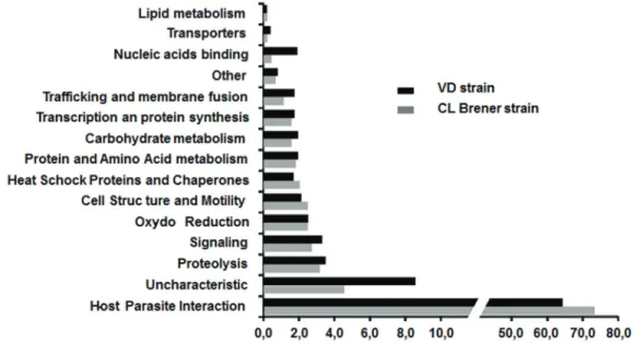
In order to ascertain the possible role of these secreted proteins, the 363 specific proteins were assigned to 15 functional groups based on information from the literature (Mainly the work of E. Bayer santoset al) and UniprotKB annotation (seeFig 2).
For both strains, approximately 70% of the proteins belong to the host-parasite interaction category that plays a very important role in virulence. A significant difference is remarkable; the number of identified proteins in the secretome with unknown function for the VD strain (44 proteins) is slightly more twice that for the CL Brener strain (20 proteins). For the VD strain, this subset represents more than 8% of the total proteins identified. A similar difference was noticed for the proteins involved in nucleic acid binding, but with a much lower number
Fig 1. Overlap between secretomes of two different T. cruzi strains DTU Tc VI. A total of 591 proteins of
T. cruzi were identified. We note that 78 proteins are specific to the CL Brener strain whereas 151 proteins are specific to VD strain. However, 363 proteins are common to both strains.
https://doi.org/10.1371/journal.pone.0185504.g001
Fig 2. Functional categories classification of T. cruzi proteins from CL Brener and VD strains. Proteins were
classified into 15 functional categories using literature and UniprotKB annotation. Y-axis, categories are indicated. X-axis shows the percentage of each category for each strain.
of proteins (10 proteins versus 2 proteins, respectively). For the other categories, there was no significant difference between the two strains. The set ofT. cruzi proteins identified for each of
the strains is provided in Additional Files 1 and 2.
Multigenic superfamilies identified in the secretomes
Among the 591 proteins identified in bothT. cruzi secretomes, 379 (64%) correspond to those
encoded by different multigenic superfamilies. Trans-sialidase proteins (TcS) and trans-siali-dase like proteins are the major families founded in the secretomes of the two strains (315 pro-teins for TcS, 18 propro-teins for DGF1, 15 propro-teins for Masps and 31 propro-teins for GP63 surface proteins). A total of 315 TcS proteins from theT. cruzi secretomes have been identified for the
2 strains. Among these proteins, 46 are fragments of TcS. We can remark that 184 complete TcS proteins were common to both strains. This implies that 42 proteins are exclusively found in CL Brener strain and 43 for VD strain.
Since, previous studies have shown the presence of 508 complete TcS genes that can be clas-sified into 8 Groups [22], we analysed the expression profile of these TcS in the two secretomes and classified these TcS proteins into groups I to VIII, according to previously described clas-ses. For a better description of the expression of different TcS groups, we derived the ratio between the number of TcS identified in our analysis, and that in each group (seeFig 3).
Discussion
A reliable biomarker for theT. cruzi infection could be of different nature (protein, lipid, sugar
or nucleid acid) but it must be specific toT. cruzi and ubiquitous in the different T. cruzi
geno-types. The objective of this work was to obtain and compare the secretomes of twoT. cruzi
strains infecting Vero celIs, as a way to focus on protein biomarkers for Chagas disease. For this study we chose to work with twoT. cruzi strains of the same DTU (TcVI). The
genetic intra-DTU variability in the parasite was poorly studied. TheT. cruzi CL Brener
genome (TcVI) was published in 2005 [21]. Other genomes, such as that of the Sylvio X10/1 strain and DM28c (TcI), have also been published [23,24]. The VD strain was isolated from a case of congenital Chagas disease and its genome is not sequenced. The comparison of two strains of TcVI genome but with different tropism increases the degree of selection of the most important secreted proteins and eliminated the strain-dependent proteins.
Fig 3. Analyse of trans-sialidase (TcS) proteins found in secretome of 2 strains. Classification of TcS
proteins for each strain into 8 group previously described [22]. Groups IV, V and VI are less than I, II, III groups VII and VIII for two strains. On the x-axis, the number of group is indicated. The Y-axis shows the percentage of each TcS identified in our analyses.
Secretome comparison
We have identified 515 proteins for VD strain compared to 441 proteins for the CL Brener strain. The result of the comparison shows a high proportion (more than 60%) of protein secreted common to bothT. cruzi strains. It would seem that gene expression in both T. cruzi
strain of the same DTU is relatively homogeneous. This high degree of homology makes possi-ble to identify proteins secreted by all strains ofT. cruzi and thus potential markers. This result
is very different from that obtained by Geigeret al. who analyzed the secretome of T. brucei
and found a small number of proteins common to the two strains of the same group (36 pro-teins common to the Feo strain and the strain OK out of a total of 270 identified propro-teins [25].
After functional classification of the parasite proteins, we noted that the proportion of iden-tified proteins involved in proteolysis, signalling, oxydo-reduction, cell structure and motilities was quite different for the two strains. This result suggests that proteins involved in metabolic pathways of stress response, protein folding, proteolysis, and oxidoreductase activity, along with proteins that can interact with different signalling pathways, are important forT. cruzi’s
capacity to infect its host cell, and that these are probably more conserved in allT. cruzi strains.
Furthermore, proteins involved in DNA binding are also more numerous in the secretome of the VD strain as compared to that of the CL Brener strain. This class of proteins plays an essen-tial role in the control of gene expression inT. cruzi, modulating RNA processing stability,
turnover, and translation [26].
The VD strain secretome contained more proteins with unknown functions than the CL Brener strain. The VD strain was isolated from a pediatric patient with congenital infection. As previously noted fromin vivo investigations [27,28], in our study the VD strain seemed more virulent than the CL Brener strain due the faster emergence from infected Vero cells. The hypothetical proteins of unknown function could be involved in virulence, and also be a source of biomarkers candidates for strain discrimination and for further studies to under-stand the underlying mechanisms of pathogenicity.
However, more than 70% of the proteins that have been identified in the secretome of the two strains belong to the host-parasite interaction category, and these play a very important role as virulence factors. In this class there are essentially four major multigenic proteins fami-lies. A remarkable feature of theT. cruzi genome is the massive expansion of these multigenes
family that encodes polymorphic surface proteins such as trans-sialidase (TcS), mucin and mucin-associated surface proteins (MASPs), dispersed gene family 1 (DGF-1), and gp63 pepti-dases. The gene families coding for these proteins represent about 10% to 30% of theT. cruzi
genome [29]. The high sequence variability of these different gene families could confer upon the parasite the ability to invade a significant number of cell types, as well as to have pleiotropic tissue tropisms, as described previously inin vitro, and in in vivo and clinical studies [30] where a wide variety of syndromes associated withT. cruzi infection were described.
The trans-sialidase and tran-sialidase like gene (TcS) super family represents the largestT. cruzi gene family, with more than 1,400 genes. However, only half of these genes are
appar-ently functional [21]. The TcS superfamily is composed of glycosylphosphatidylinositol pro-teins anchored onT. cruzi surface, but they are also released to the extracellular space. The TcS
gene family is highly polymorphic and only a few members have critical residues necessary for catalytic activity [31]. The very large number of different TcS proteins found in both secre-tomes is not unexpected. It has been shown that TcS expression is highly induced when cells become full of parasites and when amastigotes transform to trypomastigotes [32]. Thus, it would be possible to correlate the presence of large amounts of TcS into culture medium with cell host rupture. Finally, Affranchino JLet al. reported that Vero cells infected with
[33]. However, our study provides a more detailed description (number and identification) of the TcS members expressed during the life cycle ofT. cruzi (DTU TcVI). Recently, a
compara-tive analysis of TcS sequences (508 complete genes) in theT. cruzi CL Brener strain genome
led to the differentiation of eight different groups [22]. These authors further showed that the majority of TcS transcripts of all groups are present both in trypomastigotes and amastigotes forms. However, some transcripts are highly expressed in trypomastigote stage whereas other transcripts are expressed in amastigotes. It is interesting to note that Freitas LM and their col-leagues found a very low level of expression for two genes from TcS group V in all the develop-mental stages [22]. The protein structure of the TcS groups V and VI are very similar. Trans-sialidases of these two groups have one signal peptide, a single asp box, a canonical VTV motif and a GPI anchor [22]. Our study appears to confirm these observations since we observed that more than half of the TcS proteins belonging to groups I, II and III, are expressed (19, 117 and 15 proteins, respectively). This percentage increases up to 80% for the VII and VIII groups (17 and 46 sequences, respectively). While for group IV the expression of TcS is less than 40%, and for groups V and VI less than 25% (227 and 39, respectively). This observation is notewor-thy, because groups V and VI represent more than 60% of the trans-sialidases (TcS).
Finally, antibodies against two repeated epitopes, SAPA epitope (PVDSSAHG/STPST) and TcD epitope (PKPAE) of trans-sialidases were found in the sera of patients with Chagas’ dis-ease in acute and chronic phase [34]. Our sequence analysis shows trans-sialidases that possess the SAPA epitope (Group I) and the TcD epitope (Group IV) in the secretome of both ana-lysed strains. There is a high homology in the TcS group expressed in both strains from same DTU (TcVI), we could suppose that some variations in their expression might be related to the genetic difference between strains. Numerous proteomic studies have revealed that theT. cruzi
secretomes contain many proteins. The first secretome analysis has been made from ‘reservo-some’ fraction belonging to Dm28c strain (TcI) epimastigotes [19], suggesting that there are several possible pathways for the secretion of proteins fromT. cruzi. In another study [25] the authors identifiedT. brucei proteins from purified microvesicles extracted from infected rats
sera.
In the latest and most comprehensive study [20], the authors identified two types of micro-vesicles released by epimastigotes and metacyclic forms of Dm28cT. cruzi strain. All these
studies pointed to the use of microvesicles to deliver cargo of many proteins into host cells. The role of the proteins released from microvesicles is fundamental in Chagas disease. For example, Cestariet al. observed microvesicles release from infected cells into bloodstream that
could allow immune evasion by inhibiting C3 convertase [35]. The protein content of the microvesicles was found to be ubiquitous, contributing to virulence, infectivity and immunity in many pathogens such asTrypanosoma spp. and other related parasites such as Leishmania spp. [36] andPlasmodium spp. [37], but also from bacteria [38] or virus [39].
Nevertheless, all these studies were carried out without the host cell context. In the present study, we analyzed the secretome ofT. cruzi in the presence of host cells and particularly at the
time when trypomastigotes are released from the cell.
In our model we have identified a set of parasite proteins that have been released from the host cells by secretion, by microvesicles, or directly from the cell cytoplasm to the culture medium. These proteins are linked to biochemical processes that enable the transformation of amastigotes in trypomastigotes, the multiplication of amastigotes, the release of trypomasti-gotes from host cells and finally invasion of new cells. In the culture medium, we had access to all the proteins secreted or excreted by the parasite.
Our data show that the majority of the proteins identified for both strains were found in the microvesicles of the epimastigote and the metacyclic forms ofT.cruzi. However, we have also
such as the 10 KDa heat shock protein [20]. This result suggests that there are slight differences in the secretome of trypomastigote and amastigote forms ofT. cruzi. Although these parasitic
proteins are not required for cell invasion (metacyclic form) they may have a role in survival within host cell cytoplasm.
Short list of parasite proteins as a potential marker of infection with T.
cruzi
Although the protein superfamilies (TcS, GP63, MASP, and DGF1) are highly represented in the secretome of both strains, they cannot be used as biomarkers forT. cruzi infection. The
sequence identity of the TcS is low and is often limited to common motifs (Asp-box, GPI anchor, VTV and FRIP box). These motifs include a maximum of ten amino acids. In addition it should be noted that these proteins have sequences that are fairly variable between the vari-ousT. cruzi strains. Pattern searches by the protein BLAST algorithm from National Center
for Biotechnology Information (NCBI)show that the TcD epitope is repeated 430 times in the genome ofT. cruzi strain CL Brener while it is identified only 5 times in strain DM28C.
Simi-larly we did not find the SAPA epitope In the DM28C strain whereas in the strain CL Brener this pattern is repeated 80 times. So we propose a selected list of proteins (seeTable 1) that could potentially serve as markers of an infection withT. cruzi. Indeed, these proteins are
secreted, clearly produced abundantly, and are common to at least two strains of TcVI. The list includes 94 proteins of which 8 are represented by two isoforms and 18 have no known function. These various proteins might also be future therapeutic targets and/or mark-ers of Chagas disease. The protein Q4CVJ1 was not identified in the various secretomes pub-lished forT.cruzi, but it has been found in high-throughput screening and is highly expressed
and immunogenic; moreover, 68% of sera from patients with Chagas disease recognize this antigen [40]. Finally, the proteins with unknown function, Q4CTF0 and Q4CUB2, have been described as part of the last multigenic family (about 40 members in Cl Brener) subdivided into 3 subfamilies TcTASV-A, B and C. Q4CTF0 is part of the subgroup family TcTASV-A while Q4CUB2 is part of the TcTASV-C family. This subfamily (8 proteins) was identified on the surface of trypomastigotes ofT. cruzi and in (strain CL Brener, Sylvio and RA). About 30%
of human sera infected byT. cruzi reacted with TcTASV-C [41].
We note that for some proteins in this list, Microtubule-associated protein, Heat shock pro-teins (HSP70), Complement regulatory protein and Flagellar Calcium-binding protein, spe-cific antibodies can be found in the acute and chronic phase sera of patients with Chagas’ disease [38]–[42].
The EMBRARIO laboratory (Brazil) offers a confirmatory test (HBK 740 Imunoblot Linhas) based on a multi-epitope recombinant peptide, including the TC-24 epitope (Flagellar calcium-binding protein) and the MAP repeated epitope (Microtubule-associated protein) [43,44].
Some proteins have already been identified as potential markers in other infections. For example, the fructose biphosphate aldolase enzyme, a key enzyme in the glycolytic pathway, was found in plasma of patients infected withP. falciparum [45]).
The abundance of the parasite proteins and their relative stability in serum are two criteria to help in the identification of suitable markers forT. cruzi infection. In our study the strategy
adopted to obtain theT. cruzi secretome may eliminate the most labile proteins since the
pres-ence of proteases in the culture supernatant is likely to degrade many of the secreted proteins.
Conclusion
The development and availability of a simple, inexpensive, easy to handle, sensitive and spe-cific method to effectively demonstrate the presence ofT. cruzi from a blood sample is a major
Table 1. List of secreted proteins that are common in both T.cruzi strains without of multigene proteins.
Proteins UNIPROT Accession number MW (Kda)
10 kDa heat shock protein, putative Q4DFA8 10.7
40S ribosomal protein S15a, putative Q4E0N6 14.7
40S ribosomal protein S18, putative Q4E093 17.5
Actin, putative Q4D7A6 38.1
Adenosylhomocysteinase Q4D455 48.4
ADP-ribosylation factor 1, putative Q4D7Y8 20.7
Alpha tubulin, putative Q4CLA1 49.8
Arginine kinase, putative Q4CWA5 40.2
ATP synthase subunit beta Q4DTX7 55.7
Beta tubulin, putative Q4DQP2 49.7
Calcium-binding protein, putative Q4D1Q2 19.6
Calmodulin, putative (Fragment) Q4D2S5 9.5
Calpain-like cysteine peptidase, putative Q4D066 12.8
Calreticulin, putative Q4DDX3 46.2
Chaperonin HSP60, mitochondrial Q4DYP5 59.1
Complement regulatory protein, putative Q4DQ07 113.7
Cysteine peptidase C Q4DQB0 36.7
Dynein light chain, putative Q4E4N7 10.4
Elongation factor 1-alpha (Fragment) Q4CXI2 42.8
Elongation factor 2, putative Q4D3T1 94.1
Enolase, putative Q4DZ98 46.4
Glucose-regulated protein 78, putative Q4D620 71.3
Glutamate dehydrogenase Q4D5C2 45
Glutathione peroxidase Q4DEJ5 19.7
Glyceraldehyde 3-phosphate dehydrogenase, putative Q4D3Y9 14.7
Glyceraldehyde 3-phosphate dehydrogenase, putative Q4DHF0 39
Heat shock 70 kDa protein, mitochondrial, putative Q4CVR9 70.9
Heat shock 70 kDa protein, putative (Fragment) Q4CU95 40.8
Heat shock 70 kDa protein, putative (Fragment) Q4DAZ6 30.1
Heat shock protein 70 (HSP70), putative Q4DTM9 70.9
Heat shock protein 85, putative Q4CQS6 80.7
IgE-dependent histamine-releasing factor, putative Q4CW52 19.6
Isocitrate dehydrogenase [NADP] Q4E4L7 46.8
Lysosomal alpha-mannosidase, putative Q4DXL4 111.2
Malate dehydrogenase (Fragment) Q4D4A0 31.5
Microtubule-associated protein, putative (Fragment) Q4CMT2 85.2
NAD/FAD dependent dehydrogenase, putative Q4CVH0 43
Neutral sphingomyelinase activation associated protein Q4DSD4 90.5
Nucleoside diphosphate kinase Q4E256 16.9
Peptidyl-prolyl cis-trans isomerase Q4E4L9 18.8
Peptidyl-prolyl cis-trans isomerase Q4DPB9 21.9
Phosphoglycerate kinase Q4D193 44.4
Proteasome regulatory ATPase subunit 1, putative Q4D9J1 48.5
Proteasome regulatory ATPase subunit 2, putative Q4D0B9 49
Rab7 GTP binding protein, putative Q4E4T4 23.9
Ras-related protein rab-2a, putative (Fragment) Q4DM40 10.5
Ras-related protein rab-5, putative (Fragment) Q4D504 20.4
challenge for the diagnosis of Chagas disease. Identification of one or more proteins (biomark-ers) in the patient’s serum will be clear evidence for the presence of the parasite.
This study provides the first comparative overview of the secretomes of cells infected by the
T. cruzi CL Brenner and the VD strains. Not surprisingly, the majority of excreted proteins
belong to the four multigenic gene families (TcS, MASPs, DGF1, and gp63 surface protein), that unfortunately cannot be used as infection marker because of their very high, inter-species, degree of variability.
Some of the proteins identified had not been studied previously, particularly from the VD strain, and some may be new potential therapeutic targets. We have thus established a list of 94 secreted proteins, common to both strains and that do not belong to members of multigene
Table 1. (Continued)
Proteins UNIPROT Accession number MW (Kda)
S-adenosylmethionine synthase Q4CSC4 43.5
Serine carboxypeptidase S28, putative Q4DM56 72.1
Serine/threonine-protein phosphatase Q4D9Y4 35
Seryl-tRNA synthetase, putative (Fragment) Q4CW46 25.7
Small GTP-binding protein Rab1, putative Q4CZR0 22.8
Superoxide dismutase Q4D5A6 21.9
Transitional endoplasmic reticulum ATPase, putative Q4DWB5 86.1
tRNA synthetase, putative (Fragment) Q4E397 78.8
Tryparedoxin peroxidase, putative Q4CM56 22.4
Tryparedoxin peroxidase, putative Q4CX87 25.5
Ubiquitin-activating enzyme E1, putative Q4DYM1 114.3
Ubiquitin-conjugating enzyme E2, putative Q4CTN0 17.5
Cofilin/actin depolymerizing factor, putative Q4D8D3 Q4CVE9 15.7 15.7
14-3-3 protein, putative Q4DJB6 Q4DRH6 29.9 29.9
Cysteine peptidase inhibitor Q4DH32 Q4DY71 12 12.1
Cysteine peptidase, putative Q4DW02 Q4E0J7 49,9 49.8
Cytochrome c, putative Q4D480 Q4CV48 12.2 12.2
Fructose-bisphosphate aldolase Q4D0Q0 Q4D4R9 40.8 40.8
Serine carboxypeptidase (CBP1), putative Q4CMQ4 Q4DTP7 59,5 59.5
Uncharacterized protein Q4D3H5 Q4DNJ6 16.7 16.8
Uncharacterized protein Q4CNH1 66
Uncharacterized protein Q4CPM9 112.1
Uncharacterized protein Q4CPX4 23.1
Uncharacterized protein Q4CUB2 36.5
Uncharacterized protein Q4CUQ4 23
Uncharacterized protein Q4CVJ1 16.4 Uncharacterized protein Q4CW22 13 Uncharacterized protein Q4CW23 13.5 Uncharacterized protein Q4D1D9 323 Uncharacterized protein Q4D6D8 21.1 Uncharacterized protein Q4DPV6 24.4 Uncharacterized protein Q4DT54 23.6
Uncharacterized protein Q4DUX0 22.3
Uncharacterized protein (Fragment) Q4CQX1 41.3
Uncharacterized protein (Fragment) Q4CTF0 21.3
Uncharacterized protein (Fragment) Q4DHN4 38.1
families. These proteins are secreted in large quantities and are relatively stable in presence of many proteases. These conditions are necessary to identify good serum markers. This descrip-tive study provides the first step toward the identification of potential diagnostic markers in the sera ofT. cruzi-infected persons.
Supporting information
S1 Table. Secreted parasites proteins identified in T. cruzi CL Brener strains separated on 1D gel and their classification according to functional categories (informations from liter-ature and UniprotKB annotation). The proteins secreted have selected with LCMSMS score was over 35 and with at least two peptides identified, or with a score over 50 but with only one peptide identified. For each protein, the number of matched proteins and peptides and the highest score are described.
(PDF)
S2 Table. Secreted parasites proteins identified inT. cruzi VD strains separated on 1D gel and their classification according to functional categories (informations from literature and UniprotKB annotation). The proteins secreted have selected with LCMSMS score was over 35 and with at least two peptides identified, or with a score over 50 but with only one pep-tide identified. For each protein, the number of matched proteins and peppep-tides and the highest score are described.
(PDF)
S3 Table. Secreted parasites proteins identified inT. cruzi VD and CL Brener strains (com-mon proteins) separated on 1D gel. For each protein, the number of matched proteins and peptides and the highest score for CL Brener (white box) and VD (gray box) strain are described.
(PDF)
Acknowledgments
We would to like to thanks Georges Snounou and Angelita Rebollo for fruitful scientific discussion.
We thank Dr. Marı´a Elisa Solana (Facultad de Medicina, Instituto de Investigaciones en Microbiologı´a y Parasitologı´a Me´dica (IMPAM),UBA-CONICET, Universidad de Buenos Aires) to maintain of the VD strain of T. cruzi".
Author Contributions
Conceptualization: Jean-Yves Brossas, Julia´n Ernesto Nicola´s Gulin, Margarita Maria Cata-lina Bisio, Carine Marinach-Patrice, George Palazon Ruiz.
Data curation: Jean-Yves Brossas. Formal analysis: Jean-Yves Brossas.
Funding acquisition: Jaime Altcheh, Dominique Mazier.
Investigation: Jean-Yves Brossas, Manuel Chapelle, Mallaury Bordessoules, George Palazon Ruiz, Jeremy Vion.
Methodology: Jean-Yves Brossas.
Project administration: Jaime Altcheh, Dominique Mazier. Resources: Jean-Yves Brossas.
Supervision: Luc Paris, Jaime Altcheh, Dominique Mazier. Validation: Jean-Yves Brossas.
Writing – original draft: Jean-Yves Brossas, Julia´n Ernesto Nicola´s Gulin, Margarita Maria Catalina Bisio.
Writing – review & editing: Jean-Yves Brossas, Julia´n Ernesto Nicola´s Gulin, Margarita Maria Catalina Bisio, Jaime Altcheh, Dominique Mazier.
References
1. WHO | 6 February 2015, vol. 90, 6 (pp. 33–44) [Internet]. WHO. [cited 2017 Jan 4]. Available from:
http://www.who.int/wer/2015/wer9006/en/
2. Steverding D. The history of Chagas disease. Parasit Vectors. 2014 Jul 10; 7:317.https://doi.org/10. 1186/1756-3305-7-317PMID:25011546
3. Messenger LA, Miles MA. Evidence and importance of genetic exchange among field populations of Trypanosoma cruzi. Acta Trop. 2015 Nov; 151:150–5.https://doi.org/10.1016/j.actatropica.2015.05. 007PMID:26188331
4. Tibayrenc M, Ayala FJ. The population genetics of Trypanosoma cruzi revisited in the light of the pre-dominant clonal evolution model. Acta Trop. 2015 Nov; 151:156–65.https://doi.org/10.1016/j. actatropica.2015.05.006PMID:26188332
5. Zingales B, Andrade SG, Briones MRS, Campbell DA, Chiari E, Fernandes O, et al. A new consensus for Trypanosoma cruzi intraspecific nomenclature: second revision meeting recommends TcI to TcVI. Mem Inst Oswaldo Cruz. 2009 Nov; 104(7):1051–4. PMID:20027478
6. Zingales B, Miles MA, Campbell DA, Tibayrenc M, Macedo AM, Teixeira MMG, et al. The revised Trypa-nosoma cruzi subspecific nomenclature: rationale, epidemiological relevance and research applica-tions. Infect Genet Evol J Mol Epidemiol Evol Genet Infect Dis. 2012 Mar; 12(2):240–53.
7. Martin DL, Marks M, Galdos-Cardenas G, Gilman RH, Goodhew B, Ferrufino L, et al. Regional variation in the correlation of antibody and T-cell responses to Trypanosoma cruzi. Am J Trop Med Hyg. 2014 Jun; 90(6):1074–81.https://doi.org/10.4269/ajtmh.13-0391PMID:24710614
8. Gascon J, Bern C, Pinazo M-J. Chagas disease in Spain, the United States and other non-endemic countries. Acta Trop. 2010 Aug; 115(1–2):22–7.https://doi.org/10.1016/j.actatropica.2009.07.019
PMID:19646412
9. Segovia M, Carrasco HJ, Martı´nez CE, Messenger LA, Nessi A, Londoño JC, et al. Molecular epidemio-logic source tracking of orally transmitted Chagas disease, Venezuela. Emerg Infect Dis. 2013 Jul; 19 (7):1098–101.https://doi.org/10.3201/eid1907.121576PMID:23768982
10. Carlier Y, Torrico F, Sosa-Estani S, Russomando G, Luquetti A, Freilij H, et al. Congenital Chagas dis-ease: recommendations for diagnosis, treatment and control of newborns, siblings and pregnant women. PLoS Negl Trop Dis. 2011 Oct; 5(10):e1250.https://doi.org/10.1371/journal.pntd.0001250
PMID:22039554
11. Freilij H, Altcheh J. Congenital Chagas’ disease: diagnostic and clinical aspects. Clin Infect Dis Off Publ Infect Dis Soc Am. 1995 Sep; 21(3):551–5.
12. Chagas’ Disease—NEJM [Internet]. [cited 2017 Apr 28]. Available from:http://www.nejm.org/doi/full/ 10.1056/NEJMra1410150
13. Biomarkers Definitions Working Group. Biomarkers and surrogate endpoints: preferred definitions and conceptual framework. Clin Pharmacol Ther. 2001 Mar; 69(3):89–95.https://doi.org/10.1067/mcp. 2001.113989PMID:11240971
14. Hulka BS. ASPO Distinguished Achievement Award Lecture. Epidemiological studies using biological markers: issues for epidemiologists. Cancer Epidemiol Biomark Prev Publ Am Assoc Cancer Res Cosponsored Am Soc Prev Oncol. 1991 Dec; 1(1):13–9.
15. McNerney R, Maeurer M, Abubakar I, Marais B, McHugh TD, Ford N, et al. Tuberculosis diagnostics and biomarkers: needs, challenges, recent advances, and opportunities. J Infect Dis. 2012 May 15; 205 Suppl 2:S147–158.
16. Jain P, Chakma B, Patra S, Goswami P. Potential Biomarkers and Their Applications for Rapid and Reliable Detection of Malaria. BioMed Res Int [Internet]. 2014 Apr 2 [cited 2017 Apr 26];2014. Available from:https://www.hindawi.com/journals/bmri/2014/852645/abs/
17. de Araujo FF, Nagarkatti R, Gupta C, Marino AP, Debrabant A. Aptamer-based detection of disease biomarkers in mouse models for chagas drug discovery. PLoS Negl Trop Dis. 2015 Jan; 9(1):e3451.
https://doi.org/10.1371/journal.pntd.0003451PMID:25569299
18. Umezawa ES, Nascimento MS, Stolf AM. Enzyme-linked immunosorbent assay with Trypanosoma cruzi excreted-secreted antigens (TESA-ELISA) for serodiagnosis of acute and chronic Chagas’ dis-ease. Diagn Microbiol Infect Dis. 2001 Mar; 39(3):169–76. PMID:11337184
19. Sant’Anna C, Nakayasu ES, Pereira MG, Lourenc¸o D, de Souza W, Almeida IC, et al. Subcellular prote-omics of Trypanosoma cruzi reservosomes. Proteprote-omics. 2009 Apr; 9(7):1782–94.https://doi.org/10. 1002/pmic.200800730PMID:19288526
20. Bayer-Santos E, Aguilar-Bonavides C, Rodrigues SP, Cordero EM, Marques AF, Varela-Ramirez A, et al. Proteomic analysis of Trypanosoma cruzi secretome: characterization of two populations of extra-cellular vesicles and soluble proteins. J Proteome Res. 2013 Feb 1; 12(2):883–97.https://doi.org/10. 1021/pr300947gPMID:23214914
21. El-Sayed NM, Myler PJ, Bartholomeu DC, Nilsson D, Aggarwal G, Tran A-N, et al. The genome sequence of Trypanosoma cruzi, etiologic agent of Chagas disease. Science. 2005 Jul 15; 309 (5733):409–15.https://doi.org/10.1126/science.1112631PMID:16020725
22. Freitas LM, Santos SL dos, Rodrigues-Luiz GF, Mendes TAO, Rodrigues TS, Gazzinelli RT, et al. Genomic Analyses, Gene Expression and Antigenic Profile of the Trans-Sialidase Superfamily of Trypa-nosoma cruzi Reveal an Undetected Level of Complexity. PLOS ONE. 2011 Oct 19; 6(10):e25914.
https://doi.org/10.1371/journal.pone.0025914PMID:22039427
23. Franze´n O, Ochaya S, Sherwood E, Lewis MD, Llewellyn MS, Miles MA, et al. Shotgun sequencing analysis of Trypanosoma cruzi I Sylvio X10/1 and comparison with T. cruzi VI CL Brener. PLoS Negl Trop Dis. 2011 Mar 8; 5(3):e984.https://doi.org/10.1371/journal.pntd.0000984PMID:21408126
24. Grisard EC, Teixeira SMR, de Almeida LGP, Stoco PH, Gerber AL, Talavera-Lo´pez C, et al. Trypano-soma cruzi Clone Dm28c Draft Genome Sequence. Genome Announc. 2014 Jan 30; 2(1).
25. Geiger A, Hirtz C, Be´cue T, Bellard E, Centeno D, Gargani D, et al. Exocytosis and protein secretion in Trypanosoma. BMC Microbiol. 2010 Jan 26; 10:20.https://doi.org/10.1186/1471-2180-10-20PMID:
20102621
26. De Gaudenzi JG, Noe´ G, Campo VA, Frasch AC, Cassola A. Gene expression regulation in trypanoso-matids. Essays Biochem. 2011; 51:31–46.https://doi.org/10.1042/bse0510031PMID:22023440
27. Gulin JEN, Eagleson MA, Postan M, Cutrullis RA, Freilij H, Bournissen FG, et al. Efficacy of voricona-zole in a murine model of acute Trypanosoma cruzi infection. J Antimicrob Chemother. 2013 Apr; 68 (4):888–94.https://doi.org/10.1093/jac/dks478PMID:23212113
28. Risso MG, Garbarino GB, Mocetti E, Campetella O, Gonzalez Cappa SM, Buscaglia CA, et al. Differen-tial expression of a virulence factor, the trans-sialidase, by the main Trypanosoma cruzi phylogenetic lineages. J Infect Dis. 2004 Jun 15; 189(12):2250–9.https://doi.org/10.1086/420831PMID:15181573
29. De Pablos LM, Osuna A. Multigene families in Trypanosoma cruzi and their role in infectivity. Infect Immun. 2012 Jul; 80(7):2258–64.https://doi.org/10.1128/IAI.06225-11PMID:22431647
30. Tonelli RR, Giordano RJ, Barbu EM, Torrecilhas AC, Kobayashi GS, Langley RR, et al. Role of the gp85/trans-sialidases in Trypanosoma cruzi tissue tropism: preferential binding of a conserved peptide motif to the vasculature in vivo. PLoS Negl Trop Dis. 2010 Nov 2; 4(11):e864.https://doi.org/10.1371/ journal.pntd.0000864PMID:21072227
31. Schenkman S, Eichinger D, Pereira ME, Nussenzweig V. Structural and functional properties of Trypa-nosoma trans-sialidase. Annu Rev Microbiol. 1994; 48:499–523.https://doi.org/10.1146/annurev.mi. 48.100194.002435PMID:7826016
32. Frevert U, Schenkman S, Nussenzweig V. Stage-specific expression and intracellular shedding of the cell surface trans-sialidase of Trypanosoma cruzi. Infect Immun. 1992 Jun; 60(6):2349–60. PMID:
1375197
33. Affranchino JL, Ibañez CF, Luquetti AO, Rassi A, Reyes MB, Macina RA, et al. Identification of a Trypa-nosoma cruzi antigen that is shed during the acute phase of Chagas’ disease. Mol Biochem Parasitol. 1989 May 15; 34(3):221–8. PMID:2499788
34. Frasch AC, Cazzulo JJ, Aslund L, Pettersson U. Comparison of genes encoding Trypanosoma cruzi antigens. Parasitol Today Pers Ed. 1991 Jun; 7(6):148–51.
35. Cestari I, Ansa-Addo E, Deolindo P, Inal JM, Ramirez MI. Trypanosoma cruzi immune evasion medi-ated by host cell-derived microvesicles. J Immunol Baltim Md 1950. 2012 Feb 15; 188(4):1942–52.
36. Silverman JM, Chan SK, Robinson DP, Dwyer DM, Nandan D, Foster LJ, et al. Proteomic analysis of the secretome of Leishmania donovani. Genome Biol. 2008; 9(2):R35. https://doi.org/10.1186/gb-2008-9-2-r35PMID:18282296
37. Mantel P-Y, Hoang AN, Goldowitz I, Potashnikova D, Hamza B, Vorobjev I, et al. Malaria-infected eryth-rocyte-derived microvesicles mediate cellular communication within the parasite population and with the host immune system. Cell Host Microbe. 2013 May 15; 13(5):521–34.https://doi.org/10.1016/j. chom.2013.04.009PMID:23684304
38. Ståhl A, Arvidsson I, Johansson KE, Chromek M, Rebetz J, Loos S, et al. A novel mechanism of bacte-rial toxin transfer within host blood cell-derived microvesicles. PLoS Pathog. 2015 Feb; 11(2): e1004619.https://doi.org/10.1371/journal.ppat.1004619PMID:25719452
39. Mercier SK, Donaghy H, Botting RA, Turville SG, Harman AN, Nasr N, et al. The microvesicle compo-nent of HIV-1 inocula modulates dendritic cell infection and maturation and enhances adhesion to and activation of T lymphocytes. PLoS Pathog. 2013 Oct; 9(10):e1003700.https://doi.org/10.1371/journal. ppat.1003700PMID:24204260
40. Cooley G, Etheridge RD, Boehlke C, Bundy B, Weatherly DB, Minning T, et al. High throughput selec-tion of effective serodiagnostics for Trypanosoma cruzi infecselec-tion. PLoS Negl Trop Dis. 2008 Oct 8; 2 (10):e316.https://doi.org/10.1371/journal.pntd.0000316PMID:18841200
41. Bernabo´ G, Levy G, Ziliani M, Caeiro LD, Sa´nchez DO, Tekiel V. TcTASV-C, a Protein Family in Trypa-nosoma cruzi that Is Predominantly Trypomastigote-Stage Specific and Secreted to the Medium. PLOS ONE. 2013 juil; 8(7):e71192.https://doi.org/10.1371/journal.pone.0071192PMID:23923058
42. Meira WSF, Galvão LMC, Gontijo ED, Machado-Coelho GLL, Norris KA, Chiari E. Use of the Trypano-soma cruzi recombinant complement regulatory protein to evaluate therapeutic efficacy following treat-ment of chronic chagasic patients. J Clin Microbiol. 2004 Feb; 42(2):707–12.https://doi.org/10.1128/ JCM.42.2.707-712.2004PMID:14766840
43. Krautz GM, Galvão LM, Canc¸ado JR, Guevara-Espinoza A, Ouaissi A, Krettli AU. Use of a 24-kilodalton Trypanosoma cruzi recombinant protein to monitor cure of human Chagas’ disease. J Clin Microbiol. 1995 Aug; 33(8):2086–90. PMID:7559953
44. Ferna´ndez-Villegas A, Pinazo MJ, Maraño´n C, Thomas MC, Posada E, Carrilero B, et al. Short-term fol-low-up of chagasic patients after benzonidazole treatment using multiple serological markers. BMC Infect Dis. 2011 Jul 31; 11:206.https://doi.org/10.1186/1471-2334-11-206PMID:21801456
45. The´ze´nas ML, Huang H, Njie M, Ramaprasad A, Nwakanma DC, Fischer R, et al. PfHPRT: a new bio-marker candidate of acute Plasmodium falciparum infection. J Proteome Res. 2013 Mar 1; 12(3):1211– 22.https://doi.org/10.1021/pr300858gPMID:23339668
![Fig 3. Analyse of trans-sialidase (TcS) proteins found in secretome of 2 strains. Classification of TcS proteins for each strain into 8 group previously described [22]](https://thumb-eu.123doks.com/thumbv2/123doknet/14634373.548612/8.918.300.697.114.335/analyse-sialidase-proteins-secretome-classification-proteins-previously-described.webp)

