. . . .
. . . .
T-cadherin is present on endothelial
microparticles and is elevated in plasma
in early atherosclerosis
Maria Philippova
1, Yves Suter
2, Stefan Toggweiler
2, Andreas W. Schoenenberger
3,
Manjunath B. Joshi
1, Emmanouil Kyriakakis
1, Paul Erne
2, and The´re`se J. Resink
1*
1
Department of Biomedicine, Laboratory for Signal Transduction, Basel University Hospital, ZLF 316 Hebelstrasse 20, CH 4031, Basel, Switzerland;2
Division of Cardiology,
Kantonsspital Luzern, Luzern, Switzerland; and3
Department of Geriatrics and General Internal Medicine, University of Bern Hospital, Inselspital Bern, Switzerland Received 16 February 2010; revised 15 April 2010; accepted 10 May 2010; online publish-ahead-of-print 28 June 2010
Aims The presence of endothelial cell (EC)-derived surface molecules in the circulation is among hallmarks of endothelial
activation and damage in vivo. Previous investigations suggest that upregulation of T-cadherin (T-cad) on the surface of ECs may be a characteristic marker of EC activation and stress. We investigated whether T-cad might also be shed from ECs and in amounts reflecting the extent of activation or damage.
Methods and results
Immunoblotting showed the presence of T-cad protein in the culture medium from normal proliferating ECs and higher levels in the medium from stressed/apoptotic ECs. Release of T-cad into the circulation occurs in vivo and in association with endothelial dysfunction. Sandwich ELISA revealed negligible T-cad protein in the plasma of healthy volunteers (0.90 + 0.90 ng/mL, n ¼ 30), and increased levels in the plasma from patients with non-significant atherosclerosis (9.23 + 2.61 ng/mL, n ¼ 63) and patients with chronic coronary artery disease (6.93 + 1.31 ng/mL, n ¼ 162). In both patient groups there was a significant (P ¼ 0.043) dependency of T-cad and degree of endothelial dysfunction as measured by reactive hyperaemia peripheral tonometry. Flow cytometry analysis showed that the major fraction of T-cad was released into the EC culture medium and the plasma as a surface component of EC-derived annexin V- and CD144/CD31-positive microparticles (MPs). Gain-of-function and loss-of-function studies demonstrate that MP-bound T-cad induced Akt phosphorylation and activated angiogenic behaviour in target ECs via homophilic-based interactions.
Conclusion Our findings reveal a novel mechanism of T-cad-dependent signalling in the vascular endothelium. We identify T-cad as an endothelial MP antigen in vivo and demonstrate that its level in plasma is increased in early atherosclerosis and correlates with endothelial dysfunction.
-Keywords Atherosclerosis † Endothelial dysfunction † Plasma biomarkers † Endothelial microparticles † T-cadherin
Introduction
Atherosclerosis is a chronic inflammatory disease of large arteries that is initiated by activation and injury of the vascular endothelium upon exposure to multiple cardiovascular risk factors. There is mounting interest in detecting and influencing both the develop-ment of atherosclerosis at its non-significant stages and the shift from ‘indolent disease’ to acute disease. Studies have demon-strated that endothelial activation and dysfunction detected by
measuring changes in endothelium-dependent vasodilatation or by analysing plasma levels of soluble inflammatory markers (e.g. high-sensitivity C-reactive protein, CD40L) can be a prognostic
tool predicting future coronary events.1–4
Among the hallmarks of endothelial activation and damage is the presence of endothelial cell (EC)-derived surface molecules in the circulation. Plasma levels of soluble forms of endothelial membrane proteins (e.g. VCAM-1, E-selectin and P-selectin, LOX-1 receptor) have variously been shown to reflect the severity and the stage of
*Corresponding author. Tel:+41 61 265 2422, Fax: +41 61 265 2350, Email:[email protected]
the disease.5 EC membrane proteins (e.g. E-selectin, ICAM-1, VE-cadherin) are also present in the circulation on microparticles (MPs), defined as vesicles ,1 mm in size shed from the plasma membrane by exocytotic budding in response to cell activation,
injury, or apoptosis.6–8Cellular origin (ECs, leucocytes,
lympho-cytes, erythrolympho-cytes, and platelets), amount, and protein compo-sition of MPs in the circulation have been suggested to be useful clinical markers for a great variety of diseases with vascular
invol-vement and hypercoagulability.6–8 In addition to their biological
actions in inflammation, immune responses and coagulation MPs are capable of directly stimulating intracellular signalling and elicit-ing cellular responses, such as proliferation, survival, adhesion,
che-motaxis, and intercellular communication.7,8
Accumulating evidence supports a role for cell surface T-cadherin (T-cad) survival responses of vascular cells to activation
and injury of the vasculature.9,10 T-cad expression is increased
in vivo in atherosclerotic lesions from human aorta,11in
experimen-tal restenotic lesions of the carotid artery,12 and in proliferating
ECs and smooth muscle cells in vitro.13Upregulation of T-cad in
ECs also occurs during oxidative and endoplasmic reticulum
stress and functions to diminish stress-induced apoptosis.14,15
Overexpression and ligation of T-cad on the EC surface activate signalling pathways (PI3-kinase, Akt, GSK3b) important for cell
survival, proliferation and angiogenesis.9,14,16
The foregoing studies utilized artificial ligands (i.e. antibodies or recombinant protein) to induce T-cad ligation. The present study examines (i) whether T-cad might also be shed from ECs under conditions of stress and in what form, (ii) whether the shed T-cad might be a functionally relevant and naturally occurring ligand for cell surface-expressed T-cad, and (iii) whether T-cad might be present in the circulation in association with endothelial activation/damage.
Methods
In vitro studies
Established protocols were used for culture of EC, viral transduction, Western blotting, MP isolation, flow cytometry, electron microscopy, sandwich ELISA, and angiogenesis in vitro (see Supplementary material online).
Clinical study
The study complies with the Declaration of Helsinki and was approved by the regional ethical committee. Informed consent was obtained from all subjects. They include 225 consecutive patients who underwent coronary angiography at the Luzerner Kantonsspital and a group of 30 volunteers younger than 35 years and without any cardiovascular risk factors. Intravascular ultrasound (IVUS) and quantification of IVUS in the patients was
carried out using Volcano equipment.17Based on important
differ-ences in clinical and angiographic presentation, patients were divided into two groups of those with chronic coronary artery disease (CAD) (n ¼ 163) or with non-significant atherosclerosis (n ¼ 63). Definition of chronic CAD was angiographically and IVUS-proven relevant coronary stenosis (lesions greater than
50%) of at least one vessel and with either angina and/or ST depression during exercise. Definition of non-significant athero-sclerosis was diffuse luminal irregularities without relevant lesions (lesions less than 50%) as determined angiographically and by IVUS. The indication for exam in these patients was symptoms of angina or dyspnoea or in the course of investigation of arrhyth-mias or palpitations. In the group of patients with chronic CAD, mean grade of stenosis was 66.7 + 26.1%, mean plaque burden was 48.6 + 10.5%, and mean necrotic core volume (% of plaque volume) was 17.2 + 8.7%. In the group of patients with non-significant atherosclerosis, mean grade of stenosis was 6.1 + 15.2%, mean plaque burden was 31.6 + 12.4%, and mean necrotic core volume (% of plaque volume) was 9.5 + 8.1%; all P , 0.001 in comparison with the group of patients with chronic CAD.
Measurements of reactive hyperaemia-peripheral arterial tono-metry (RH-PAT) and of T-cad in plasma were performed following protocols detailed in Supplementary material online.
Statistical analysis
For in vitro studies, differences were variously determined with one-way or two-way repeated-measures analysis of variance (ANOVA) with Tukey’s or Bonferroni’s multiple comparison test, respectively, using Prism 5.0 software (GraphPad Software, San Diego, CA, USA), or three-way repeated-measures ANOVA with Bonferroni’s multiple comparison test using STATA Version 9.2 (STATA Corporation, College Station, TX, USA). Statistical analysis of data from patients and healthy volunteers was performed using STATA Version 9.2. Continuous variables were compared using Student’s t-test assuming normal distributions or the Wilcoxon rank sum test for variables with non-normal distributions.
Dichot-omous variables were compared using the x2-test or Fisher’s exact
test when cell counts were ,5. In patients, Spearman’s correlation using continuous variables and logistic regression models using dichotomous variables were used to detect associations between T-cad plasma levels and the degree of ED as assessed by RH-PAT. For the purpose of logistic regression analysis, RH-PAT index was dichotomized at a value of 2.0 and T-cad values were dichotomized at a detection limit of 1 ng/mL. A significance level of 0.05 was assumed for all tests.
Results
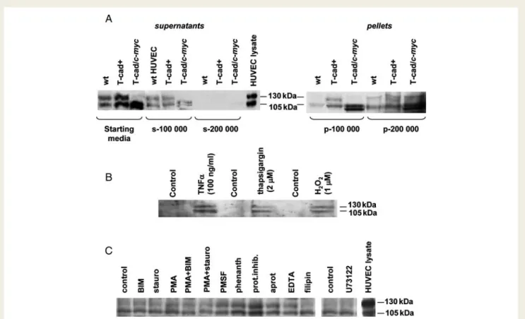
T-cadherin can be detected in culture
supernatants from endothelial cells
To determine whether T-cad protein is released from EC, con-ditioned media were harvested from monolayer cultures of par-ental human umbilical vein endothelial cells (HUVECs) and
HUVEC transduced with native T-cad protein or
T-cad-c-myc-tagged protein following a 24 h culture period. Immu-noblotting demonstrated the presence of T-cad protein in media
(Figure1A). Different agents inducing endothelial stress/apoptosis
(TNF-a, hydrogen peroxide, and thapsigargin) increased the
content of T-cad in the medium (Figure 1B). Release of T-cad
into the medium was not prevented by inclusion of various com-pounds that can inhibit the catalytic cleavage of membrane
proteins from the cell surface, including protein kinase inhibitors—
bisindoylmaleimide, staurosporine, and phorbol myristate
acetate; protease inhibitors—phenylmethylsulphonyl fluoride,
Sigma proteolytic cocktail, and aprotinin; divalent cation scavenger ethylenediammine tetraacetic acid; lipid-raft disrupting agent
filipin; phospholipase D metalloproteinase inhibitor
1,10-phenanthroline; phospholipase C inhibitor U73122 (Figure 1C).
However, ultracentrifugation of conditioned media resulted in the loss of T-cad from supernatants with simultaneous
accumu-lation of the protein in the pellets (Figure1A), suggesting that the
major fraction of T-cad was released in association with MPs rather than as soluble or cleaved protein.
T-cadherin is present on the surface
of microparticles released from
endothelial cells
To determine whether endothelial MPs harbour T-cad, MPs were isolated from TNF-a-treated HUVEC culture supernatants and
characterized by electron microscopy (Figure2A), immunoblotting
(Figure 2B), and flow cytometry (Figure 2C – F). Isolated MPs had
diameters of less than 1 mm (Figure2A). The number of collected
MPs was 190 + 35/103 cells (mean + SD of five isolations)
(Figure 2C). T-cad was present on MPs collected from culture
supernatants of parental and T-cad-overexpressing HUVEC
(Figure 2B). Flow cytometry analysis showed that MPs from
par-ental HUVEC stained positively for annexin V, a characteristic feature of these apoptotic vesicles exposing phosphatidylserine
on their outer membrane surface,6–8 and annexin binding was
decreased in the absence of Ca2+, confirming the specificity of
the staining (Figure2D). MPs isolated from HUVEC culture
super-natants were, as expected, double-positive for CD31 and annexin
V (53% of all MPs) (Figure2E). With immunolabelling for T-cad and
annexin V, 67% of all the MP population were double-positive, con-trasting with minimal double-positivity in control samples labelled
with either annexin V in the absence of Ca2+ (8%) or isotype
control IgG (14%) (Figure2F).
Figure 1 T-cadherin (T-cad) is present in culture supernatants from endothelial cells (ECs) in vitro. (A) Immunoblotting for T-cad in 24 h culture media collected from parental human umbilical vein endothelial cells (HUVEC) (wt), and HUVEC-overexpressing human T-cad (T-cad+) or c-myc-tagged T-cad protein (T-cad/c-myc). Ultracentrifugation of starting media at 100 000 g and 200 000 g resulted in the loss of T-cad from supernatants (s-100 000 and s-200 000) with simultaneous accumulation of protein in the pellets (p-100 000 and p-200 000). (B) Immunoblotting for T-cad in media from parental HUVEC after 5 h of treatment with indicated stress-inducing agents or vehicle controls. (C ) Immunoblotting for T-cad in media from parental HUVEC after 8 h of treatment with 100 ng/mL TNF-a in the absence (control) or pres-ence of various inhibitors of enzymes inducing protein cleavage: bisindoylmaleimide (BIM, 1026mol/L), staurosporine (stauro, 1029mol/L), phorbol myristate acetate (PMA, 1026mol/L), phenylmethylsulphonyl fluoride (PMSF, 1026mol/L), Sigma proteolytic cocktail (prot.inhib., 1:100), aprotinin (aprot, 1026mol/L), EDTA (1 mmol/L), filipin (8× 1026mol/L), 1,10-phenanthroline (phenanth., 1023mol/L), and U73122 (1025mol/L).
Endothelial microparticles expressing
T-cadherin on their surface can induce
T-cadherin-dependent signalling and
angiogenic behaviour in endothelial cells
In vitro homophilic ligation of cell surface T-cad by recombinant T-cad protein or agonistic antibody induces intracellular signalling
and the angiogenic phenotype in ECs.16,18 How such ligation
might occur in vivo is unclear, since in intact EC monolayers T-cad, like other GPI-anchored molecules, is present on the
apical surface of cells and not at sites of intercellular contacts.19
MPs are recognized as diffusible vectors for the transfer of biologi-cal information from one cell to another (homotypic or
heteroty-pic) within proximal or remote tissues.7,8To test the postulate that
T-cad-carrying MPs represent ligands inducing T-cad-dependent
signalling and behaviour in target ECs, we performed
gain-of-function experiments. MPs collected from
T-cad-overexpressing ECs (MP-T) and control vector-transduced ECs (MP-E) were analysed for their ability to induce Akt phosphoryl-ation in HUVEC, human aortic endothelial cells (HAECs), and human microvascular endothelial cell line (HMEC-1). Comparable results were obtained for all EC types. Both MP-E and MP-T induced a quick Akt phosphorylation response in parental EC, which was observed within 5 min after addition of MPs to cell
cul-tures, reached its peak within 10 – 15 min (Figure3A, images herein
show data for HUVEC and HAEC only), and was normalized within 45 min (not shown). The response to MP-T during the first 10 min was significantly more pronounced than that induced by MP-E
(Figure3A). To exclude that signalling might be induced by traces
of foetal calf serum or cytokines in MP preparations, we treated HUVEC with equal volumes of the final phosphate-buffered saline (PBS) supernatants collected during the preparation of
Figure 2 T-cad is present on microparticles (MPs) released from apoptotic ECs. (A) Electron microscopic characterization of MPs isolated from TNF-a-treated (4 mmol/L, 24 h) HUVEC. (B) Immunoblotting for T-cad in MPs isolated from TNF-a-treated parental (wt) and T-cad-overexpressing HUVEC (T-cad+). (C–F) Flow cytometry of MPs from TNF-a-treated HUVEC. (C) MPs were defined on a dot-plot histo-gram (region R2) and counted using an internal standard of calibrating TruCOUNT beads (R1). (D) Staining with annexin V – FITC conjugate with the confirmation of specific annexin V binding by staining in the absence of Ca2+. (E) MPs are double-positive for annexin V and CD31. Isotype control IgG-PE conjugate was used as negative control. (F ) MPs are double-positive for annexin V and T-cad. Negative controls: labelling with isotype control IgG and annexin V in the absence of Ca2+. Experiments were performed on at least three separate occasions.
MP-E and MP-T (sup-E and sup-T, respectively). No Akt phos-phorylation was observed in response to either sup-E or sup-T
(Figure3B).
To validate T-cad-specific effects of MP-T on Akt phosphoryl-ation, we performed gain-of-function and loss-of-function experi-ments using T-cad-overexpressing and T-cad-silenced HMEC-1. Firstly, we examined the responses of T-cad-silenced (shT) and control shRNA-transduced (shC) HMEC-1 to MP-T and MP-E
derived from T-cad-overexpressing and control-transduced
HMEC-1, respectively. The Akt phosphorylation response of
shC-HMEC-1 to MP-T was greater than that to MP-E (Figure3C).
In contrast, for shT-HMEC-1, the Akt phosphorylation responses
to MP-E and MP-T were comparable (Figure 3C). Secondly, we
examined the response of parental HMEC-1 to MPs collected from shT-HMEC-1 or shC-HMEC-1. MPs from T-cad-silenced HMEC-1 induced a markedly weaker and delayed response than
Figure 3 Endothelial MPs carrying T-cad induce T-cad-dependent Akt phosphorylation in ECs. (A) MPs from control-transduced (MP-E) and T-cad-overexpressing (MP-T) cultures of HUVEC, human aortic endothelial cells (HAEC), or human microvascular endothelial cell line (HMEC-1) were added (10 mg/mL) or not (control) to the corresponding parental cultures (data for HMEC-1 not shown). (B) As negative controls, parental HUVECs were treated with equal volumes of the final PBS supernatants collected during the preparation of MP-E and MP-T (sup-E and sup-T, respectively). (C ) T-cad-silenced (shT) or control vector-transduced (shC) HMEC-1 treated with HMEC-1-derived MP-E or MP-T. (D) Parental HMEC-1 treated with MP collected from shT or shC HMEC-1. (A – D) Whole cell lysates prepared at the indicated times (minutes) were immunoblotted for phospho-Akt or GAPDH as internal loading control. Total Akt levels in ECs were not altered by MP-E, MP-T, sup-E, or sup-T (data not shown). Representative blots are shown and data in histograms are given as mean + SEM (n ¼ at least 3). Two-way (A, B, D) and three-way (C ) ANOVA; *P , 0.05; **P , 0.01; ***P , 0.001;§P , 0.001.
MP from shC-HMEC-1 (Figure3D). These data support that acti-vation of intracellular signalling in target EC by MP-T depended upon T-cad homophilic interactions.
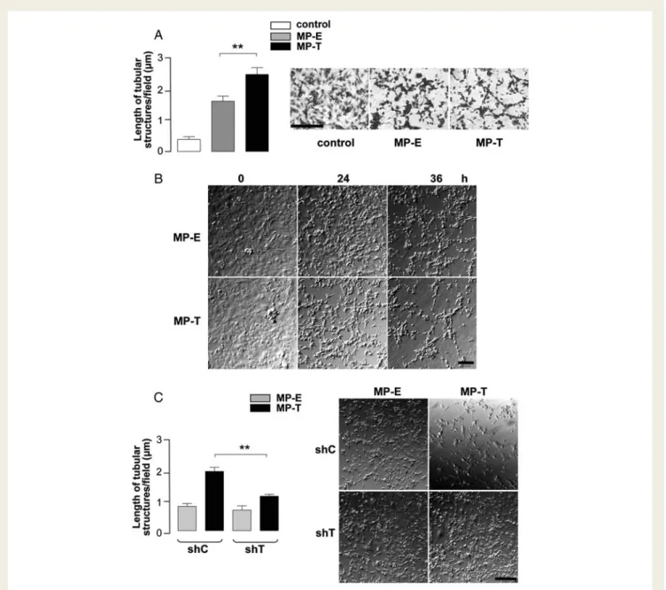
Next, we investigated whether effects of T-cad-carrying MP on intracellular signalling might translate into relevant biological effects. Angiogenic behaviour of EC was selected as the functional read-out since in vitro and in vivo studies have identified T-cad as a
proangiogenic molecule.16,18Moreover, an important action of MP
in the vascular system is their ability to induce angiogenesis.7,8,20
MP-E and MP-T were added to HMEC-1 monolayers and their effects on angiogenic behaviour were monitored by time-lapse
videomicroscopy. In parental HMEC-1, both types of MP induced a proangiogenic phenotype and formation of net structures, but for MP-T the total length of tubular structures was greater
(Figure 4A), and onset of network formation occurred earlier
(Figure 4B). The angiogenic response of T-cad-silenced HMEC-1
(shT) to MP-T was significantly lower than that of control
HMEC-1 (shC) (Figure 4C). These data demonstrate that
MP-harboured T-cad can elicit a biologically relevant and T-cad-dependent (i.e. angiogenic) response, and further confirm homophilic interaction of MP-T with T-cad molecules expressed on the surface of target cells.
Figure 4 Endothelial MPs expressing T-cad on their surface facilitate angiogenesis in vitro. MP-E or MP-T were added (10 mg/mL) to parental HMEC-1 monolayers (A, B), T-cad-silenced (shT), and control-transduced (shC) HMEC-1 (C ). Formation of tubular structures was monitored by time-lapse videomicroscopy over 36 h. Morphometric analysis of the length of tubular structures (A, C) was performed on either videomicro-scopy still-frames or after fixation/staining of cells. Images depict fixed/stained cells (A, 36 h) and still-frames after 0, 24, and 36 h (B) or 24 h (C ). Scale bars ¼ 0.1 mm (A, B) or 0.2 mm (C ). Data in histograms (mean + SEM, n ¼ 3) present morphometric analysis at 24 h. One-way (A) and two-way (C ) ANOVA; **P , 0.01.
T-cadherin is present in human plasma at
increased levels during early stages of
atherosclerosis and correlates with
endothelial dysfunction
We next addressed whether T-cad might be present in the plasma of human donors and whether plasma levels of T-cad reflect in vivo ED and/or injury. The clinical characteristics of the study
popu-lations are shown in Table 1; there was a population of young
healthy volunteers, a population of middle-aged patients with inter-mediate atherosclerotic risk and non-significant atherosclerosis, and a slightly older population with highly prevalent cardiovascular risk factors and documented chronic CAD.
ELISA of plasma samples demonstrated the presence of T-cad in 13.3% of healthy volunteers, in 42.9% of patients with non-significant atherosclerosis, and in 34.6% of patients with chronic
CAD (Figure5A). Levels of T-cad in both patient groups were
sig-nificantly higher than healthy volunteers (Figure 5A and B), even
with exclusion from the analysis of patients with outlying high
T-cad plasma levels (Figure 5B). Levels of T-cad between the
patient groups did not differ significantly (Figure5B).
In patients with chronic CAD or non-significant atherosclerosis, Spearman’s correlation using continuous variables revealed a sig-nificant (P ¼ 0.043) dependency of T-cad and degree of ED as
measured by RH-PAT (Figure 5C). Patients with elevated T-cad
had a lower RH-PAT index (rho ¼ 20.187), further suggesting a link between ED and T-cad release into the circulation. To further evaluate the relationship between T-cad and RH-PAT, we
divided the RH-PAT scores into three categories1–4and the
sub-jects into T-cad producers or non-producers, and performed a
x2
-test of independence on the counts. The Fisher’s exact test showed a P-value of 0.046.
In multivariate logistic regression adjusted for age, gender, cardi-ovascular risk factors (smoking, diabetes, hypertension, hyperlipi-daemia, and family history), and statin use, only a lower RH-PAT index was significantly associated with elevated T-cad (OR 2.2
for elevated T-cad in patients with lower vs. higher RH-PAT;
Table2).
Microparticles isolated from plasma
express T-cadherin on their surface
To analyse whether T-cad is released into the circulation on the surface of endothelial MP, we isolated and characterized MP from the plasma of human donors. Analysis was performed on several plasma samples (n ¼ 7) identified as positive for T-cad
protein in ELISA. Figure 6 shows representative stainings of
samples from patients with non-significant atherosclerosis. Triple staining for endothelial protein CD31/PECAM-1, T-cad, and annexin V demonstrated that 40% of isolated MP (953 +
112 events/ml, average from three measurements) were CD31+
and annexin V+, thus representing endothelial apoptotic MP. Of
these, 18% were also T-cad+ (Figure 6A, upper panels; lower
panels show negative control stainings). Since CD31 is also present on platelets and leucocytes, we validated the EC origin
of T-cad+-MP using EC-specific marker, CD144/VE-cadherin.
Twenty-five percent of CD144+-MP (total number 242 +
70 events/ml) were T-cad+ (Figure 6B, right upper panel; lower
panel shows negative control staining). To illustrate that levels of T-cad determined by ELISA reflect the level of EC-derived MP-bound T-cad in plasma, we compared the analysis presented
in Figure6B with a parallel analysis of plasma samples from patients
and healthy controls containing negligible T-cad on ELISA; these
samples exhibited very low levels of CD144+/T-cad+-MP
(Sup-plementary material online, Figure S1). Staining for other cell-type
markers (Figure6C) showed the presence of MP originating from
platelets (602 + 142 events/ml positive for CD41/GpIIb), erythro-cytes (141 + 33 events/ml positive for CD235a/glycophorin A), and leucocytes (94 + 29 events/ml positive for CD45). Negligible levels of double-positivity were detected in samples co-stained
for T-cad and any of these markers (Figure 6C, upper panels;
. . . .
. . . .
. . . . Table 1 Clinical characteristics of healthy volunteers and participating patients
Healthy volunteers (n 5 30) Non-significant atherosclerosis (n 5 63) Chronic CAD (n 5 162) P-values (between-patient groups) Age (years) 27.3 + 3.1 55.9 + 9.9 62.3 + 9.3 ,0.001 Gender 20 females/10 males 29 females/34 males 40 females/122 males 0.002 Risk factors Smoking 0/30 (0.0%) 24/63 (38.1%) 94/162 (58.0%) 0.007 Diabetes 0/30 (0.0%) 6/63 (9.5%) 35/162 (21.6%) 0.035 Hypertension 0/30 (0.0%) 30/63 (47.6%) 103/162 (63.6%) 0.029 Hyperlipidaemia 0/30 (0.0%) 25/63 (39.7%) 116/162 (71.6%) ,0.001 Family history 0/30 (0.0%) 23/63 (36.5%) 58/162 (35.8%) 0.921 Medications Beta-blockers 0/30 (0.0%) 28/63 (44.4%) 104/162 (64.2%) 0.007 ACE-inhibitors 0/30 (0.0%) 17/63 (27.0%) 81/162 (50.0%) 0.002 Ca2+antagonists 0/30 (0.0%) 5/63 (7.9%) 36/162 (22.2%) 0.013 Statins 0/30 (0.0%) 25/63 (39.7%) 143/162 (88.3%) ,0.001
negative control labelling is shown in lower panels), supporting the EC origin of T-cad harbouring MPs in plasma.
Discussion
We have previously demonstrated that T-cad protein expression on EC is upregulated under conditions leading to EC activation
and damage.13,14 Here, we provide evidence that T-cad can be
shed from EC and in amounts reflecting the extent of activation or damage. Firstly, in vitro studies demonstrated that T-cad protein is present on MP released from stressed/apoptotic EC, and that these MP can induce T-cad-dependent signalling and a proangiogenic functional response in target cells. Secondly, release of T-cad from EC occurs in vivo in association with ED;
Figure 5 T-cad in the plasma of human donors is increased at early stages of atherosclerosis: T-cad levels in the plasma of healthy subjects (n ¼ 30), patients with non-significant atherosclerosis (n ¼ 63), patients with chronic CAD (n ¼ 162) was measured by sandwich ELISA. (A) Error bars in the scatter plot are mean + SEM. (B) T-cad levels are given as mean + SEM. P-values indicate significance of difference between subject groups as determined with two-sample Wilcoxon rank-sum (Mann – Whitney) test; comparison with healthy volunteersa,
com-parison between the patient groups either withoutbor withcexclusion of patients with outlying high plasma T-cad levels. (C ) The correlation between plasma T-cad levels and the degree of ED in the patients (n ¼ 225) as measured by RH-PAT was estimated with the Spearman’s method using continuous variables (r ¼ 20.19, P , 0.05) and is depicted using linear regression with 95% confidence bands (interrupted lines).
negligible T-cad protein was detected in the plasma of healthy human donors, but its level was increased in the plasma from patients with non-significant atherosclerosis and patients with chronic CAD.
Cell surface molecules can be released into the circulation by several mechanisms. Proteolytic cleavage of the extracellular domains of transmembrane polypeptides by matrix metallopro-teases, serine prometallopro-teases, and disintegrins, known as ectodomain shedding, has been demonstrated for growth factor receptors,
proteoglycans, and adhesion molecules.21 Lipid-anchored
mol-ecules, such as GPI-proteins or gangliosides can be cleaved from
the plasma membrane by phospholipases.22 In addition to being
a mechanism of regulating surface protein expression, enzymatic cleavage is involved in the control of various cell functions since soluble protein fragments can retain their biological activity and
act as ligands inducing signalling events in neighbouring cells.6–8
Shedding of endothelial adhesion molecules modulates adhesive properties of luminal ECs and recruitment of inflammatory cells
to the vessel wall, thus modulating inflammatory responses.23
Clea-vage of extracellular domains of N- and E-cadherin modulates cell
adhesive and migratory behaviour.24 Our in vitro data obtained
using inhibitors of shedding-inducing enzymes and ultracentrifuga-tion argues for T-cad release from EC not as soluble protein, but rather in association with some kind of cellular particles. Flow cytometry confirmed the presence of T-cad on the surface of endothelial apoptotic MP (or ectosomes), both in conditioned culture medium and in the plasma of human donors in vivo. Not-withstanding, proteolysis or lipolysis as alternative mechanisms for shedding surface T-cad cannot be completely excluded, since trace amounts of T-cad were occasionally detected in medium clarified by ultracentrifugation.
In addition to their characteristic loss of plasma membrane asymmetry and exposure of phosphatidylserine (annexin V positiv-ity) on the outer leaflet, MPs harbour membrane and cytoplasmic
components of the cell of origin.6–8Our flow cytometry analysis
of MPs from human plasma showed that MPs of platelet,
erythrocyte, or leucocyte origin were negative for T-cad. EC origin of T-cad-containing MPs was demonstrated by their positiv-ity for EC-specific marker CD144/VE-cadherin. Exosomes are another type of cell-derived microvesicles found in extracellular medium in vitro and in the circulation. They are smaller than MPs and more homogenous in size (approximately 50 – 90 nm), orig-inate from intracellular multivesicular bodies, are released from the cell via exocytic fusion with the plasma membrane, and are
dis-tinct from MPs in membrane composition and function.25 Since
exosomes will pellet together with MPs during centifugation at 100 000 g, it should be considered that microvesicular-associated T-cad is not restricted to MPs and might also be present on exo-somes. We did not specifically address this issue. Nevertheless, exosomes are too small for easy detection by flow cytometry
and usually require immunocoupling to carrier beads.25Our flow
cytometry analysis shows that microvesicles corresponding in size to MPs and carrying endothelial plasma membrane markers are positive for T-cad, permitting us to conclude that a significant pool of microvesicles carrying T-cad represent MPs.
The precise mechanisms leading to increased elevation of MP-bound T-cad in the plasma of patients with non-significant ather-osclerosis and patients with chronic CAD are unclear. T-cad is
upre-gulated on ECs early upon activation and stress in vitro,13–15 is
elevated on ECs during atherosclerosis,11and can undergo
redistri-bution within the plasma membrane of activated vascular cells.19
Therefore, local conditions of EC activation owing to endothelial shear stress together with the presence of proatherogenic
inflam-matory, thrombotic, apoptotic, or oxidative substances,26,27 and a
possible concomitant upregulation of T-cad gene expression or even redistribution of protein within the plasma membrane are factors that might underlie increased membrane vesiculation and/or increased levels of T-cad in blebbing plasma membrane domains.
There is considerable interest in plasma biomarkers that provide information about the functional status of the cardiovascular system at early stages of atherogenesis before acute clinical mani-festations take place. Since recent IVUS studies demonstrate high incidence of coronary atherosclerotic lesions even in
asympto-matic teenagers and young adults,28of special importance would
be markers that help to detect not only intimal thickening but the shift from silent to acute disease that manifests itself in func-tional abnormalities. Promising candidates include markers of acute or chronic inflammatory processes occurring in the coronary vessel wall during atherogenesis, such as soluble forms of endo-thelial adhesion molecules, or C-reactive protein for which even a small elevation was shown to be a powerful independent predic-tor of future vascular events in apparently healthy asymptomatic
individuals.4,29 Accumulating evidence supports a prognostic role
of ED in CAD;1,3 however, only von Willebrand factor and
E-selectin are viewed as biochemical markers specifically reflecting ED. Since our patient subgrouping was based on angiographic and IVUS parameters, an elevation in plasma T-cad cannot be unequi-vocally interpreted as a criterion for distinguishing between
indo-lent and functionally relevant disease. Nevertheless, ED
represents one the earliest functional disturbances that precede acute coronary events. We found both the presence of T-cad plasma in patients with non-significant atherosclerosis and an
. . . . Table 2 Multivariate logistic regression
OR (95%CI) P-value Age (OR per year increase) 1.00 (0.97 – 1.03) 0.782 Gender (OR for female vs. male) 1.61 (0.84 – 3.10) 0.155 Smoker (OR for smoker vs.
non-smoker)
1.33 (0.73 – 2.44) 0.356 Diabetes (OR for diabetes vs. no
diabetes)
0.92 (0.43 – 1.99) 0.840 Hypertension (OR for hypertension vs.
no hypertension)
0.95 (0.53 – 1.72) 0.873 Hyperlipidaemia (OR for
hyperlipidaemia vs. no hyperlipidaemia)
0.70 (0.37 – 1.33) 0.282
Family history (OR for family history vs. no family history)
1.04 (0.57 – 1.88) 0.907 Statin use (OR for statin vs. no statin) 1.00 (0.49 – 2.05) 0.998 RH-PAT index (OR for low vs. high) 2.19 (1.16 – 4.16) 0.016
association between T-cad levels and degree of ED. Therefore, it is plausible to consider T-cad as a marker of early-stage athero-sclerosis, which is clinically silent but characterized by disturbances in endothelial function.
Interestingly, although ELISA clearly demonstrated elevation of T-cad in plasma in patients with atherosclerosis when compared with healthy subjects, T-cad levels between patients with chronic CAD and those with non-significant atherosclerosis (albeit slightly higher) were not significantly different. This might be explained by the assumption that T-cad levels reflect not so much the actual
vessel occlusion but rather the state of endothelial activation and
dysfunction. Our previous in vitro studies on oxidative stress14
and endoplasmic reticulum stress30 show that T-cad expression
on EC is upregulated within 2 – 3 h of treatment with pro-inflammatory and pro-apoptotic agents and thereafter declines when manifestations of acute cell damage and death become obvious. Here, T-cad upregulation represents a protective mech-anism that can shift the balance in cellular stress response to the prosurvival signalling branches, which is only later followed by alarm and apoptotic phases. In vivo, this phenomenon might
Figure 6 Flow cytometry characterization of T-cad-harbouring MPs in human plasma. (A) Representative dot-plot graphs show triple staining of MP preparations for CD31, T-cad, and annexin V (upper panels) or triple staining with matching isotype control IgG conjugates and annexin V labelling in the absence of Ca2+performed for negative controls (lower panels). (B) Double stainings of MP for T-cad and VE-cadherin (upper panel) or respective isotype controls (low panels). (C ) Double stainings of MP for T-cad, GpIIb, GFA, or CD-45 (upper panels, left to right) and double staining with matching isotype control IgG conjugates (lower panels).
translate into an attempt of vascular cells to limit tissue dysfunction and damage at the onset of atherosclerosis.
Many studies have demonstrated that MP shedding is a hallmark of endothelial activation and injury. Circulating MP levels correlate with the degree of ED, impaired vasodilatation in patients with chronic and end-stage renal failure, and acute coronary syndromes, and are increased in patients with hypercholesterolaemia,
hyper-tension, and diabetes.5–7 Two recent papers present evidence
for potential usefulness of endothelial MP levels in the identifi-cation of patients at immediate risk of acute cardiovascular events. Assessment of endothelial dysfunction by measurement of endothelial MP independently predicted cardiovascular events in patients with coronary heart disease suggesting this readout as an important component of a multiple biomarker strategy of
improving risk stratification.31Bernard et al. showed an association
between CD144+-MP and the presence of coronary non-calcified
vulnerable plaques indicating that elevation of endothelial MP may
predict acute thrombotic events in type 2 diabetic patients.32
Identification of T-cad as an endothelial MP antigen in vivo has potential value with respect to the detection of endothelial acti-vation/injury. However, it must be recognized that considerable technical difficulties are associated with standardized isolation, measurement, and characterization of MP, and utilization of MP as a reproducibly quantifiable parameter is far from the main-stream clinical analysis. Although cell-free T-cad is mostly MP-bound, we successfully applied ELISA-based methodology to measure T-cad in human plasma. This methodology offers a more reliable and simple technical opportunity to detect ED characterized by the elevation of ‘biomarker’ endothelial MP in plasma.
A limitation of the study is that healthy subjects in the control group were significantly younger than patients in both groups with atherosclerosis. However, it seems unlikely that age is a potential confounder that results in elevated plasma T-cad in our patient groups. In our multivariate logistic regression analysis, age does not show up as a factor (OR 1.0). This is important since the other variables are age-prone factors. On the other hand, a lower RH-PAT index (ED) shows up with an OR 2.2 (P , 0.016), which underscores that the level of T-cad in plasma is a factor that relates to endothelial activation/dysfunction, and, irrespective of age, may reflect a prevailing state of vascular activation and injury.
Relevance for an association between T-cad in plasma and ED is underscored by the recent genome-wide association studies aimed at identification of genetic variants contributing to cardiovascular
disease. The Framingham Heart Study33and Org et al.34
demon-strated correlations between single nucleotide polymorphisms within intron 11 and promoter regions of CDH13 gene and long-term average diastolic and systolic blood pressure phenotypes and arterial stiffness, indicating a role for T-cad in progression of hyper-tension. The impact of T-cad within the complicated network of processes contributing to cardiovascular disorders is seemingly broad. T-cad has been identified as a receptor for adipose
tissue-derived adiponectin,35 a hormone that profoundly affects
pro-gression of atherosclerosis and diabetes by modulating insulin sen-sitivity, lipoprotein metabolism, and EC function. T-cad also acts as
a receptor for low-density lipoproteins,36suggesting a role in the
regulation of EC function in hypercholesterolaemia.
Our study has also revealed a novel mechanism of
T-cad-dependent signalling in the vasculature. Like other members of the cadherin superfamily, T-cad is capable of homophilic – ligation interactions mediating intercellular adhesion and inducing intracellular signalling events. Downstream signalling pathways seem to be activated by both T-cad ligation and
upregula-tion.13,14,16,18Ligation of T-cad on the cell surface is equivalent in
vivo to trans-interactions between T-cad molecules on neighbour-ing cells and can be mimicked in vitro by the addition of recombi-nant T-cad protein or agonistic antibody to T-cad-expressing cells. In the case of T-cad overexpression, downstream signalling might be induced either by cis-clustering of T-cad molecules within the plasma membrane or by increased engagement of T-cad-signalling adaptor molecules. In the intact endothelial mono-layer, T-cad is expressed only on the apical surface and not in the
intercellular contacts.19Such a cellular localization pattern in vivo
(i.e. facing the vessel lumen) restricts opportunities for T-cad ligation-induced signalling. By demonstrating that T-cad-harboured MP can induce Akt phosphorylation and the proangiogenic pheno-type in target EC, we have identified MP as a novel physiological ligand capable of initiating T-cad-dependent signal transduction in the endothelium. These T-cad-mediated homophilic-based inter-actions may occur both in a paracrine or in an autocrine manner, i.e. induce signalling in distant ECs or ECs from which MPs were released.
Depending on the origin and protein composition, the outcome of MP binding to endothelial and blood cells might be adverse (e.g. promotion of thrombotic events) or beneficial (e.g. tissue
regener-ation).7,8 In vivo, the interaction of plasma-delivered
T-cad-harbouring MP to the luminal endothelium, accompanied by an upregulation of surface T-cad expression on activated/injured EC, might function to regulate patho(physiological) processes involving endothelial activation and stress, and therefore influence vessel remodelling.
Supplementary material
Supplementary material is available at European Heart Journal online.
Acknowledgements
Dr Salima Sadallah (Laboratory for Immunonephrology, Depart-ment of Biomedicine, Basel University Hospital) and Prof. Gennaro de Libero (Laboratory for Experimental Immunology, Department of Biomedicine, Basel University Hospital) are thanked for helpful advice and discussions regarding FACS analysis of microparticles.
Funding
This work was supported by the Swiss National Science Foundation (grant no. 310000 – 118468/1), the Kamillo Eisner Stiftung, the Schweizerische HerzStiftung, the Herzkreislauf Stiftung, Astra Zeneca (Grant-in-Aid 2008), and the Novartis Foundation for Medical Biological Research.
References
1. Bonetti PO, Barsness GW, Keelan PC, Schnell TI, Pumper GM, Kuvin JT, Schnall RP, Holmes DR, Higano ST, Lerman A. Enhanced external counterpulsa-tion improves endothelial funccounterpulsa-tion in patients with symptomatic coronary artery disease. J Am Coll Cardiol 2003;41:1761 – 1768.
2. Bonetti PO, Pumper GM, Higano ST, Holmes DR Jr, Kuvin JT, Lerman A. Nonin-vasive identification of patients with early coronary atherosclerosis by assessment of digital reactive hyperemia. J Am Coll Cardiol 2004;44:2137 – 2141.
3. Deanfield JE, Halcox JP, Rabelink TJ. Endothelial function and dysfunction: testing and clinical relevance. Circulation 2007;115:1285 – 1295.
4. Nakou ES, Liberopoulos EN, Milionis HJ, Elisaf MS. The role of C-reactive protein in atherosclerotic cardiovascular disease: an overview. Curr Vasc Pharmacol 2008;6: 258 – 270.
5. Constans J, Conri C. Circulating markers of endothelial function in cardiovascular disease. Clin Chim Acta 2006;368:33 – 47.
6. Boulanger CM, Amabile N, Tedgui A. Circulating microparticles: a potential prog-nostic marker for atherosclerotic vascular disease. Hypertension 2006;48:180–186. 7. Chironi GN, Boulanger CM, Simon A, Dignat-George F, Freyssinet JM, Tedgui A.
Endothelial microparticles in diseases. Cell Tissue Res 2009;335:143 – 151. 8. Hugel B, Martinez MC, Kunzelmann C, Freyssinet JM. Membrane microparticles:
two sides of the coin. Physiology (Bethesda) 2005;20:22 – 27.
9. Philippova M, Joshi MB, Kyriakakis E, Pfaff D, Erne P, Resink TJ. A guide and guard: the many faces of T-cadherin. Cell Signal 2009;21:1035 – 1044.
10. Resink TJ, Philippova M, Joshi MB, Kyriakakis E, Erne P. Cadherins and cardiovas-cular disease. Swiss Med Wkly 2009;139:122 – 134.
11. Ivanov D, Philippova M, Antropova J, Gubaeva F, Iljinskaya O, Tararak E, Bochkov V, Erne P, Resink T, Tkachuk V. Expression of cell adhesion molecule T-cadherin in the human vasculature. Histochem Cell Biol 2001;115:231 – 242. 12. Kudrjashova E, Bashtrikov P, Bochkov V, Parfyonova Y, Tkachuk V, Antropova J,
Iljinskaya O, Tararak E, Erne P, Ivanov D, Philippova M, Resink TJ. Expression of adhesion molecule T-cadherin is increased during neointima formation in exper-imental restenosis. Histochem Cell Biol 2002;118:281 – 290.
13. Ivanov D, Philippova M, Allenspach R, Erne P, Resink T. T-cadherin upregulation correlates with cell-cycle progression and promotes proliferation of vascular cells. Cardiovasc Res 2004;64:132 – 143.
14. Joshi MB, Philippova M, Ivanov D, Allenspach R, Erne P, Resink TJ. T-cadherin pro-tects endothelial cells from oxidative stress-induced apoptosis. FASEB J 2005;19: 1737 – 1739.
15. Kyriakakis E, Joshi MB, Philipova M, Erne P, Resink TJ. Endoplasmic reticulum stress and the role of T-cadherin. FEBS J 2008;275(Suppl. 1):411.
16. Philippova M, Banfi A, Ivanov D, Gianni-Barrera R, Allenspach R, Erne P, Resink T. Atypical GPI-anchored T-cadherin stimulates angiogenesis in vitro and in vivo. Arter-ioscler Thromb Vasc Biol 2006;26:2222 – 2230.
17. Suter Y, Schoenenberger AW, Toggweiler S, Jamshidi P, Resink T, Erne P. Intravas-cular ultrasound-based left main coronary artery assessment: comparison between pullback from left anterior descending and circumflex arteries. J Inv Cardiol 2009;21:457 – 460.
18. Ivanov D, Philippova M, Tkachuk V, Erne P, Resink T. Cell adhesion molecule T-cadherin regulates vascular cell adhesion, phenotype and motility. Exp Cell Res 2004;293:207 – 218.
19. Philippova M, Ivanov D, Tkachuk V, Erne P, Resink TJ. Polarisation of T-cadherin to the leading edge of migrating vascular cells in vitro: a function in vascular cell motility? Histochem Cell Biol 2003;120:353 – 360.
20. Martinez MC, Tesse A, Zobairi F, Andriantsitohaina R. Shed membrane micropar-ticles from circulating and vascular cells in regulating vascular function. Am J Physiol Heart Circ Physiol 2005;288:H1004 – H1009.
21. Arribas J, Borroto A. Protein ectodomain shedding. Chem Rev 2002;102: 4627 – 4638.
22. Lauc G, Heffer-Lauc M. Shedding and uptake of gangliosides and
glycosylphosphatidylinositol-anchored proteins. Biochim Biophys Acta 2006;1760: 584 – 602.
23. Blankenberg S, Barbaux S, Tiret L. Adhesion molecules and atherosclerosis. Ather-osclerosis 2003;170:191 – 203.
24. Horstman LL, Jy W, Jimenez JJ, Ahn YS. Endothelial microparticles as markers of endothelial dysfunction. Front Biosci 2004;9:1118 – 1135.
25. Thery C, Zitvogel L, Amigorena S. Exosomes: composition, biogenesis and func-tion. Nat Rev Immunol 2002;2:569 – 579.
26. Chatzizisis YS, Coskun AU, Jonas M, Edelman ER, Feldman CL, Stone PH. Role of endothelial shear stress in the natural history of coronary atherosclerosis and vas-cular remodeling: molevas-cular, cellular, and vasvas-cular behavior. J Am Coll Cardiol 2007; 49:2379 – 2393.
27. Lynch SF, Ludlam CA. Plasma microparticles and vascular disorders. Br J Haematol 2007;137:36 – 48.
28. Tuzcu EM, Kapadia SR, Tutar E, Ziada KM, Hobbs RE, McCarthy PM, Young JB, Nissen SE. High prevalence of coronary atherosclerosis in asymptomatic teen-agers and young adults: evidence from intravascular ultrasound. Circulation 2001; 103:2705 – 2710.
29. Packard RR, Libby P. Inflammation in atherosclerosis: from vascular biology to biomarker discovery and risk prediction. Clin Chem 2008;54:24 – 38.
30. Kyriakakis E, Philippova M, Joshi MB, Pfaff D, Bochkov V, Afonyushkin T, Erne P, Resink TJ. T-cadherin attenuates the PERK branch of the unfolded protein response and protects vascular endothelial cells from endoplasmic reticulum stress-induced apoptosis. Cell Signall 2010; doi:10.1016/j.cellsig.2010.04.008. 31. Nozaki T, Sugiyama S, Koga H, Sugamura K, Ohba K, Matsuzawa Y, Sumida H,
Matsui K, Jinnouchi H, Ogawa H. Significance of a multiple biomarkers strategy including endothelial dysfunction to improve risk stratification for cardiovascular events in patients at high risk for coronary heart disease. J Am Coll Cardiol 2009;54: 601 – 608.
32. Bernard S, Loffroy R, Serusclat A, Boussel L, Bonnefoy E, Thevenon C, Rabilloud M, Revel D, Moulin P, Douek P. Increased levels of endothelial micro-particles CD144 (VE-Cadherin) positives in type 2 diabetic patients with coronary noncalcified plaques evaluated by multidetector computed tomography (MDCT). Atherosclerosis 2009;203:429 – 435.
33. Levy D, Larson MG, Benjamin EJ, Newton-Cheh C, Wang TJ, Hwang SJ, Vasan RS, Mitchell GF. Framingham Heart Study 100K Project: genome-wide associations for blood pressure and arterial stiffness. BMC Med Genet 2007; 8(Suppl. 1):S3.
34. Org E, Eyheramendy S, Juhanson P, Gieger C, Lichtner P, Klopp N, Veldre G, Doring A, Viigimaa M, Sober S, Tomberg K, Eckstein G, Kelgo P, Rebane T, Shaw-Hawkins S, Howard P, Onipinla A, Dobson RJ, Newhouse SJ, Brown M, Dominiczak A, Connell J, Samani N, Farrall M, Caulfield MJ, Munroe PB, Illig T, Wichmann HE, Meitinger T, Laan M. Genome-wide scan identifies CDH13 as a novel susceptibility locus contributing to blood pressure determination in two European populations. Hum Mol Genet 2009;18:2288 – 2296.
35. Hug C, Wang J, Ahmad NS, Bogan JS, Tsao TS, Lodish HF. T-cadherin is a recep-tor for hexameric and high-molecular-weight forms of Acrp30/adiponectin. Proc Natl Acad Sci USA 2004;101:10308 – 10313.
36. Kipmen-Korgun D, Osibow K, Zoratti C, Schraml E, Greilberger J, Kostner GM, Jurgens G, Graier WF. T-cadherin mediates low-density lipoprotein-initiated cell proliferation via the Ca(2+)-tyrosine kinase-Erk1/2 pathway. J Cardiovasc Phar-macol 2005;45:418 – 430.