Introduction
Since the introduction of the bilateral sagittal split osteotomy (BSSO) ( Trauner and Obwegeser, 1955 ; Obwegeser, 1957 ), the procedure has gained popularity, especially when combined with rigid internal fi xation (RIF). Several modifi cations of the BSSO technique have been proposed ( Dal Pont, 1961 ; Hunsuck, 1968 ; Gallo et al. , 1976 ; Epker, 1977 ).
Nowadays, the trend for skeletal Class III treatment is a combination of a Le Fort I advancement osteotomy of the maxilla and a BSSO of the mandible ( Bell et al. , 1986 ; Sinclair and Proffi t, 1990 ; Bailey et al. , 1995 ). Bailey et al. (1995) , in a review of a surgical-orthodontic database, reported that 50 per cent of all cases before 1985 were treated by isolated mandibular setback, while only 15 per cent underwent maxillary advancement and one-third had bimaxillary surgery. From 1990 to 1992, isolated mandibular setback was performed in only 9 per cent, while 40 per cent had maxillary advancement and 50 per cent two-jaw surgery.
Compared with mandibular advancement after BSSO with RIF, the same procedure with mandibular setback has not been widely reported. Mobarak et al. (2000) examined
Stability of hard tissue profi le after mandibular setback in
sagittal split osteotomies: a longitudinal and long-term follow-up
study
Christof Urs Joss * and Urs Walter Thüer **
Departments of Orthodontics, * University of Geneva and ** University of Bern, Switzerland
SUMMARY The aim of the study was to conduct a long-term follow-up on the stability of the hard tissues after bilateral sagittal split osteotomy (BSSO) with rigid internal fi xation (RIF)to set back the mandible and to compare it with that of mandibular advancement performed by the same team of surgeons and with the same examination protocol.
Seventeen consecutive patients (6 females and 11 males) could be re-examined 12.7 years (T5) after surgery. The previous examinations were before surgery (T1), 5 days (T2), and 6.6 (T3) and 14.4 (T4) months after surgery. Lateral cephalograms were traced by hand, digitized, and evaluated with the Dentofacial Planner® software program. The x -axis for the system of co-ordinates ran through sella (point zero) and the line nasion-sella-line minus 7 degrees. The program determined the x - and y -values of each variable and the usual angles and distances. The effects of treatment were determined with Wilcoxon matched pairs, signed ranks test, with Bonferroni adjustment, and the relationship between variables with Spearman rank correlation coeffi cient.
Relapse at point B was 0.94 mm or 15 per cent and at pogonion 1.46 mm or 21 per cent of the initial setback at T5. Relapse was mainly short-term (T4 – T2), 13 per cent for point B and 17 per cent for pogonion. Gender correlated signifi cantly with relapse (T5 – T2) at point B ( P = 0.002) and pogonion ( P = 0.021), i.e. females in contrast to males showed further distalization of the mandible instead of relapse. No correlations were seen for age or the amount of surgical setback.
The long-term results in mandibular setback patients were more stable when compared with the mandibular advancement patients examined previously. The initial soft tissue profi le, the initial growth direction, and the remodelling processes of the hard tissues must be considered as reasons for long-term relapse. Growth direction positively infl uenced the long-term results in females: further distalization of the mandible occurred.
80 patients after BSSO and RIF 3 years post-operatively. They found that relapse at point B was 19 per cent and at pogonion 26 per cent of the initial setback. Most of the relapse (72 per cent) took place during the fi rst 6 months after surgery through a tendency of the proximal segment to return to its original inclination.
Proffi t et al. (1991) examined patients 1-year post-operatively after BSSO with RIF and 29 after BSSO with wire fi xation (WF). They found a relapse in pogonion of 91 (RIF) and 51 (WF) per cent. The relapse at point B was 62 and 47 per cent, respectively.
A review of studies on mandibular setback with RIF or WF show, for pogonion, a relapse between 2 and 91 per cent and for point B between 7 and 62 per cent. The follow-ups were conducted between 4 weeks and more than 5 years post-operatively ( Pepersack and Chausse, 1978 ; Paulus and Steinhäuser, 1982 ; Hadjianghelou et al. , 1985 ; Kobayashi et al. , 1986 ; Phillips et al. , 1986 ; Komori et al. , 1987 ; Sorokolit and Nanda, 1990 ; Proffi t et al. , 1991 ; Ingervall et al. , 1995 ; Schatz and Tsimas, 1995 ; Mobarak et al. , 2000 ). The aim of the present study was to compare the stability of mandibular setback with BSSO and RIF with
that of mandibular advancement performed by the same team of surgeons and with the same examination protocol.
Subjects and methods
As a continuation of a previous study ( Ingervall et al. , 1995 ), 17 consecutive patients (6 females and 11 males), aged 18.9 to 40.5 years (mean age 27.1 years), who underwent a solitary mandibular setback procedure at the Department of Craniomaxillofacial Surgery, University of Bern, in the years 1986 – 1989 were studied prospectively. All patients were Caucasian.
The patients had a moderate or marked mesial occlusion that was corrected with a BSSO of the mandible and RIF. The sagittal splits were fi xed with three titanium lag screws (diameter 3.5 mm) on each side. None of the patients, who were referred by various orthodontists, underwent a genioplasty. The surgery was performed by one of four senior surgeons at the department. The surgical technique ( Raveh et al. , 1988 ) was the same for all patients, and each of the surgeons was experienced in this procedure. No splint was used for stabilization of the mandible during surgery, but maxillomandibular fi xation was used for 4 – 8 days post-surgery.
The skeletal changes resulting from the surgical procedure and their stability were evaluated on profi le cephalograms. The cephalograms were obtained with the teeth in the intercuspal position and included a linear enlargement of 3.3 per cent. The cephalograms were taken with the subject standing upright and trying to assume a natural position of the head, with the lips relaxed.
The cephalogram at T1 was taken 0 – 5 days (mean 1 day) before surgery, the second (T2) between 3 and 9 days (mean 5 days), at T3 between 4.2 and 9.7 months (mean 6.6 months), at T4 between 11.5 and 18.7 months (mean 14.4 months), and at T5 between 11.1 and 14.0 years (mean 12.7 years) after surgery.
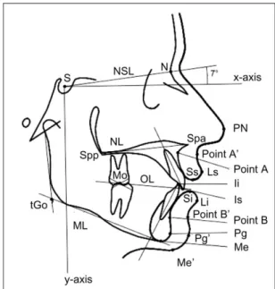
The cephalometric analysis was carried out by one examiner (CUJ) and included the reference points and lines shown in Figure 1 . The cephalogram was then traced and the reference points were digitized with the Dentofacial Planner® (Dentofacial Software Inc., Toronto, Ontario, Canada). The author was blind to the degree of mandibular setback and the date of the post-operative radiograph. Conventional cephalometric variables as well as the co-ordinates of the reference points were calculated by the computer. The co-ordinate system had its origin at sella, and its x -axis formed an angle of 7 degrees to the reference line nasion-sella-line (NSL) ( Figure 1 ). Overjet and overbite were calculated from the co-ordinates of the points incision superior and incision inferior.
The systematic and accidental errors of the cephalometric analysis were evaluated by duplicate determination of 11 cephalograms selected at random. These cephalograms were retraced and remeasured for a second time by the same examiner 2 weeks after the fi rst assessment. No systematic
errors were found when the values were evaluated with a paired t -test. The accidental errors stomion inferior (si) were calculated with the formula
= d
n
2
2
∑ ,
where d is the difference between the repeated measurements and n is the number of duplicate determinations ( Dahlberg, 1940 ). Most of the angular variables and co-ordinates of the skeletal reference points had accidental errors smaller than 1.0 degree or 1.0 mm, respectively ( Table 1 ).
Statistical analysis
The effects of treatment, i.e. the differences between the variables and co-ordinates at T1 and T2, T4 and T5, T2 and T5, as well as T1 and T5, were tested with the Wilcoxon ’ s matched-pairs, signed-ranks test. To increase the level of signifi cance, Bonferroni adjustments were carried out. The relationships between variables were analyzed with the Spearman rank correlation coeffi cient.
Results
Table 2 shows the selected variables before (T1) and 12.7 years after surgery (T5). The mean changes, standard deviations, and ranges before surgery and during subsequent observation periods are given in Tables 3 and 4 and Figures 2 and 3 . Negative values imply a backward and positive
Figure 1 Reference points and lines used in the cephalometric analysis. S, sella; NSL, nasion-sella-line; N, nasion; x , horizontal reference plane; NL nasal line; Ils, upper incisal line; Spp, posterior nasal spine; Spa, spine anterior nasal; PN, pronasion (point A ′ ); Mo, molar; OL, occlusal line; Ss, stomion superior; Ls, labrale superior (point A); Ii, incision inferior; Si, stomion inferior; Li, labrale inferior; Is, incision superior; tGo, tangent gonion; ML, mandibular line (point B; point B ′ ); Pg, pogonion; Pg ′ , soft tissue pogonion; Me, menton; Me ′ , soft tissue menton; y , vertical reference plane.
values a forward movement of the point in the horizontal plane. Negative values imply an upward and positive values a downward movement of the point in the vertical plane. Skeletal changes
Horizontal skeletal changes. The mean setback of the mandible immediately post-surgery (T2 – T1) was 6.29 mm at point B, 6.97 mm at pogonion, and 6.98 mm at incision inferior. Between T5 and T2, the mean relapse at point B was 0.94 mm [ P = 0.124, not signifi cant (ns)] and represented 15 per cent of the surgical setback. The mean relapse at pogonion (T5 – T2) was 1.46 mm ( P = 0.07, ns), i.e. 21 per cent of the surgical setback. The mean relapse (T5 – T2) of 1.67 mm at incision inferior was not signifi cant and represented 24 per cent of the initial setback (T2 – T1).
The mandibular setback relapsed to some degree in nine subjects; in three subjects the relapse was complete, but in six it was less than 50 per cent of the initial setback. Further,
posterior movement of the mandible (point B) was seen in eight patients at T5.
Vertical skeletal changes. The fi nal result (T5 – T1) showed an upward movement of menton of 0.96 mm ( P = 0.093). Tangent gonion also moved upward by 1.53 mm ( P = 0.039) in the post-operative relapse period (T5 – T2). There was a non-signifi cant upward movement of 2.11 mm ( P = 0.113) (T5 – T1).
The post-surgical relapse (T5 – T2) of menton showed no signifi cant ( P = 0.368) change, 0.35 mm. Pogonion had a signifi cant ( P = 0.008) downward movement of 1.52 mm at T5 – T2, but the fi nal result (T5 – T1) showed a non-signifi cant ( P = 0.356) upward movement of 0.94 mm.
Angular changes and distances. There was a mean decrease of SNB (3.76 degrees) immediately after surgery (T2 – T1) and an increase of 3.55 degrees in ANB. The mean relapse after 12.7 years (T5 – T2) for SNB was 0.31 degrees ( P = 0.344, ns). The fi nal result (T5 – T1) was still very signifi cant ( P = 0.001) with a decrease of 3.23 degrees. The angles NSL/mandibular line (ML) and nasal line/ML showed no signifi cant changes (T5 – T2, T5 – T1).
There was a signifi cant ( P = 0.009) decrease of 0.88 mm in overjet demonstrating post-surgical relapse (T5 – T2). The fi nal result (T5 – T1) showed a highly signifi cant increase ( P = 0.000) of 5.87 mm. The net effect on overjet (mean of 5.87) was 87.6 per cent of the improvement achieved during surgery.
Overbite showed a decrease ( P = 0.301, ns) in the post-surgical relapse period (T5 – T2) and an increase ( P = 0.266, ns) in the fi nal result (T5 – T1). In spite of the surgical setback, the short-term surgical result (T2 – T1) for overbite showed only a minimal change of 1.16 mm. The range of variation for the changes in overbite was, however, considerable (8.2 mm).
Table 1 Accidental errors si of the cephalometric analysis.
Variable Si (°) Reference point
(skeletal) Si (mm) x y SNA 0.57 N 0.56 0.08 SNB 0.34 Point A 0.83 0.52 ANB 0.57 Point B 0.66 0.96 NL/NSL 0.82 Incision superior 0.57 0.52 ML/NSL 0.45 Incision inferior 0.52 0.30 ML/NL 0.89 Pogonion 0.84 0.82 N – Spa 0.58 Menton 0.72 0.42 Spa – Me 0.54 tGonion 1.09 0.32 N – Me 0.37 Spa 0.69 0.59 S – tGo 0.30 Spp 0.63 0.48
Table 2 Values for selected cephalometric variables before (T1) and 12.7 years after (T5) surgery.
T1 T2
Mean SD Range Mean SD Range
SNA (°) 78.30 5.09 69.1 to 90.0 78.78 5.05 68.2 to 88.1 SNB (°) 83.18 5.95 93.7 to 71.0 79.74 5.58 69.0 to 89.8 ANB (°) − 4.88 4.02 − 11.7 to 2.2 − 0.95 2.59 − 5.0 to 3.1 NSL/NL (°) 7.99 5.05 − 2.7 to 18.6 9.13 4.88 1.8 to 20.2 NSL/ML (°) 34.05 8.06 21.6 to 48.8 34.49 8.49 50.3 to 21.9 NL/ML (°) 26.08 7.73 15.5 to 37.2 25.36 8.16 15.1 to 40.1 Gonion angle (°) 131.32 6.22 120.0 to 141.3 126.45 7.70 114.3 to 139.6
Anterior face height (N – Me) (mm)
124.61 11.39 97.3 to 147.4 124.14 10.20 99.2 to 144.1
Upper face height (N – Spa) (mm)
52.18 4.15 44.6 to 61.2 52.59 4.05 45.3 to 62.1
Lower face height (Spa – Me) (mm)
73.32 9.61 52.7 to 93.9 72.61 8.12 54.0 to 89.1
Posterior face height (S – tGo) (mm)
82.49 10.85 65.3 to 100.9 81.18 10.93 59.3 to 101.8
Overjet (mm) − 3.15 3.99 − 11.3 to 2.0 2.20 1.70 − 0.5 to 5.8
Correlations. No signifi cant correlations were found for the age of the patients and the amount of surgical setback at any time point. Gender correlated very signifi cantly ( P = 0.002, R = − 0.691) with relapse (T5 – T2) at point B ( x -value), i.e. females in contrast to males showed further distalization of the mandible instead of relapse. The x -value for pogonion ( P = 0.021 and R = − 0.553) and incision inferior ( P = 0.010 and R = − 0.603) showed similar correlations. Thus, relapse of SNB (T5 – T2 and T5 – T4) was also signifi cantly correlated with gender (both P = 0.034, R = − 0.516; R = − 0.517, respectively).
In the period from T5 to T4, the same correlations were seen between gender and the x -value of point B ( P = 0.003, R = − 0.680), pogonion ( P = 0.007, R = − 0.629), and incision inferior ( P = 0.012, R = − 0.592).
Discussion
In contrast to previous studies on mandibular advancement ( Ingervall et al. , 1995 ; Joss and Thüer, 2007 ), the setback group contained only 6 females and 11 males. The surgical sagittal setback of the mandible was 6.29 mm at point B and 6.97 mm at pogonion. Several authors found similar mean values after setback surgery ( Hadjianghelou et al. , 1985 ; Komori et al. , 1987 ; Schatz and Tsimas, 1995 ; Mobarak et al. , 2000 ).
The mean post-surgical relapse (T5 – T2) at point B was 0.94 mm and at pogonion 1.46 mm, which represented 15 and 21 per cent, respectively, of the initial setback. Unfortunately, a lack of reported long-term follow-up studies is evident, with only two studies in the literature with which to compare the present data ( Pepersack and Chausse, 1978 ; Mobarak et al. , 2000 ).
Pepersack and Chausse (1978) studied 43 patients with BSSO without RIF more than 5 years post-operatively. They found a relapse of 12 per cent for point B and 14 percent for pogonion of the initial setback of the mandible. Mobarak et al. (2000) showed a relapse of 19 per cent at point B 3 years post-operatively. At pogonion the relapse was 26 per cent of the initial setback.
Analysis of the data showed that there was a mean sagittal decrease of 0.4 mm at point B from T2 to T3 and T3 to T4. However, the decrease from T4 to T5 was only 0.1 mm. The major part of the relapse (13 per cent) took place shortly after surgery. Between 14.4 months and 12.7 years after surgery, no signifi cant antero – posterior change was seen ( Figure 3 ).
Relapse in the setback group in this investigation was smaller compared with the fi ndings in mandibular advancement patients ( Ingervall et al. , 1995 ; Joss and Thüer, 2007 ), where, 12.7 years post-surgery, the relapse at point B and pogonion was 50 and 60 per cent, respectively. The
Table 3 Changes (mm or degree) in the variables and co-ordinates as a result of surgery at T2 – T1, 5 days after surgery to before surgery; T5 – T4, 12.7 years to 14.4 months after surgery.
Variable or co-ordinate T2 – T1 T5 – T4
Mean SD Range Mean SD Range
Horizontal [ x -value (mm)] Point A − 0.06 ns 0.47 − 1.1 to 1.0 1.19 ns 1.86 − 2.4 to 6.1
Point B − 6.29 *** 4.32 − 13.7 to − 1.0 0.14 ns 2.24 − 4.0 to 3.8
Pogonion − 6.97 *** 6.40 − 21.1 to 0.2 0.24 ns 2.45 − 4.4 to 4.5
tGonion − 7.65 *** 4.35 − 16.0 to − 1.0 0.98 ns 2.68 − 3.0 to 6.9
Incision superior − 0.28 ns 0.97 − 1.9 to 1.2 0.64 ns 2.76 − 3.4 to 8.4
Incision inferior − 6.98 *** 3.32 − 12.5 to − 3.1 0.68 ns 2.87 − 3.8 to 9.1
Vertical ( y -value [mm]) Point B − 0.82 ns 2.14 − 3.1 to 3.8 − 1.13 ns 2.07 − 5.3 to 1.4
Pogonion − 0.88 ns 2.12 − 3.6 to 4.9 1.58 * 2.02 − 2.7 to 4.2
Menton − 0.70 ns 1.85 − 2.6 to 3.6 0.22 ns 1.66 − 2.7 to 2.3
tGonion − 0.11 ns 2.73 − 5.2 to 5.3 0.47 ns 2.14 − 3.1 to 4.4
Incision superior 0.27 ns 0.68 − 1.6 to 1.2 0.29 ns 1.53 − 2.6 to 2.9
Incision inferior − 0.89 ns 2.26 − 3.8 to 2.8 0.52 ns 1.52 − 2.7 to 3.0
Angle (degree) and linear measurements (mm) SNA − 0.19 ns 0.47 − 0.9 to 0.9 0.91 ns 1.55 − 1.4 to 4.1 SNB − 3.76 *** 2.47 − 8.5 to − 0.9 − 0.22 ns 1.10 − 2.5 to 1.3 ANB 3.55 *** 2.30 0.4 to 7.6 1.12 * 1.39 − 1.4 to 4.5 NSL/NL 0.23 ns 0.61 − 0.7 to 1.3 0.91 ns 2.03 − 2.4 to 5.3 NSL/ML − 0.68 ns 3.24 − 5.7 to 5.6 − 0.09 ns 1.90 − 3.3 to 3.4 NL/ML − 0.94 ns 3.21 − 5.4 to 5.7 − 0.98 ns 2.41 − 4.5 to 4.6 Gonion angle − 8.33 *** 4.39 − 17.3 to − 3.0 0.49 ns 2.59 − 4.4 to 5.8 Overjet (mm) 6.70 *** 3.70 1.6 to 13.1 − 0.05 ns 0.97 − 1.6 to 2.6 Overbite 1.16 ns 2.57 − 3.6 to 4.6 − 0.23 ns 1.17 − 2.5 to 2.5 Spa-Me 0.14 ns 2.22 − 3.2 to 5.4 − 0.29 ns 2.01 − 5.5 to 2.5 N-Me − 0.14 ns 1.87 − 2.6 to 3.7 0.28 ns 1.71 − 2.5 to 2.6 S-tGo 0.40 ns 2.50 − 5.5 to 5.3 0.45 ns 1.98 − 2.3 to 4.3
majority of relapse (33 per cent) occurred between 5 days and 13.9 months after surgery. It was considered that soft tissue stretching in the mandibular advancement patients resulted in a signifi cant relapse.
Gender correlated very signifi cantly ( P = 0.002) with post-surgical relapse (T5 – T2) at point B ( x -value), i.e. females showed, in contrast to males, further distalization of the mandible. The same correlation was seen for the x -values in point B ( P = 0.021) and incision inferior ( P = 0.010). For this reason, relapse of SNB (T5 – T2 and T5 – T4) was signifi cantly correlated with gender ( P = 0.034). There were no correlations with the patient ’ s age or the amount of initial setback.
In the period from T2 to T5, eight patients (fi ve females and three males) had an additional setback of the mandible. For three males, point B and pogonion were found to be more anterior than prior to surgery. Each of these patients had only a small sagittal setback of the mandible, e.g. at point B between 1.9 and 2.1 mm and at pogonion between 0.6 and 1.4 mm.
Behrents (1985a , b) stated that in view of their initial growth tendency, orthodontically treated male Class III patients are more prone to relapse than female Class III patients. On the other hand, orthodontically treated female Class II subjects will probably, due to normal growth or the remodelling processes, experience more relapse after BSSO
advancement than males. The chin, together with the surrounding soft tissue, grows forward and downward in males while in females it grows mostly downward but neither forward nor backward. In males, the mandible rotates anteriorly but in females rather posteriorly. The growth and remodelling processes in the present females at T5 demonstrated an improvement of the initial result after surgery but in males a deterioration. Among factors which could contribute to this relapse are further growth as well as mandibular remodelling. No surgical splint was used for identifying the correct occlusion during surgery, which could have also negatively infl uenced the short-term relapse of the mandible.
The tongue and its adaptation to the new environment of the shortened mandible could also play an important role in long-term relapse. The position and the size of the tongue, without any tongue resection, will be the same after surgery and could lead to increased mandibular pressure in a forward direction.
A stable result at T5 with mandibular setback surgery was easier to achieve than with mandibular advancement surgery ( Joss and Thüer, 2007 ). A possible explanation could be that it is easier to place the condyles in their fossa in mandibular setback than in advancement patients, where the posterior tension of the musculature and soft tissue plays an important role.
Table 4 Changes (mm or degree) in the variables and co-ordinates of a result of surgery at T5 – T2: 12.7 years to 5 days after surgery; T5 – T1: 12.7 years after to before surgery.
Variable or
co-ordinate
T5 – T2 T5 – T1
Mean SD Range Mean SD Range
Horizontal [ x -value (mm)] Point A 0.94 ns 1.94 − 2.1 to 6.4 − 0.32 ns 0.69 − 2.2 to 0.6
Point B 0.94 ns 2.34 − 2.2 to 5.4 − 5.49 ** 4.00 − 13.0 to 0.9
Pogonion 1.46 ns 2.86 − 2.3 to 6.0 − 5.75 * 5.62 − 17.5 to 2.4
tGonion 2.28 ns 3.88 − 4.3 to 12.6 − 6.35 ** 2.54 − 12.1 to − 2.2
Incision superior 0.79 ns 2.36 − 2.0 to 6.8 − 0.12 ns 1.33 − 2.5 to 2.0
Incision inferior 1.67 ns 2.70 − 2.6 to 9.2 − 5.99 ** 3.52 − 13.1 to 0.2
Vertical [ y -value (mm)] Point B − 1.12 ns 2.5 − 4.9 to 3.2 − 0.81 * 1.78 − 3.1 to 2.3
Pogonion 1.52 * 1.93 − 2.0 to 5.6 − 0.94 ns 1.94 − 3.0 to 2.6
Menton − 0.35 ns 1.76 − 3.3 to 2.8 − 0.96 ns 1.16 − 2.7 to 1.2
tGonion − 1.53 ns 2.66 − 5.0 to 2.3 − 2.11 ns 1.88 − 6.0 to 1.0
Incision superior − 0.01 ns 1.57 − 3.2 to 2.4 − 0.03 ns 0.68 − 1.4 to 1.0
Incision inferior 0.38 ns 1.98 − 3.4 to 4.1 − 1.04 ns 1.86 − 3.9 to 1.7
Angle (degree) and linear
measurements (mm) SNA (°) 0.68 ns 1.71 − 2.1 to 4.8 − 0.43 ns 1.01 − 3.4 to 1.2 SNB 0.31 ns 1.17 − 1.3 to 2.7 − 3.23 ** 2.33 − 7.6 to 0.6 ANB 0.38 ns 1.51 − 2.2 to 3.8 2.81 *** 2.42 − 1.2 to 7.4 NSL/NL 0.91 ns 1.95 − 3.7 to 4.7 1.14 ns 1.82 − 3.3 to 4.5 NSL/ML 1.12 ns 2.34 − 3.2 to 5.7 0.44 ns 3.02 − 4.6 to 5.8 NL/ML 0.22 ns 3.18 − 6.6 to 5.8 − 0.71 ns 3.08 − 6.1 to 4.1 Gonion angle 3.45 ns 4.85 − 3.4 to 12.1 − 5.36 ** 2.47 − 8.7 to − 0.2 Overjet (mm) − 0.88 * 1.07 − 2.4 to 1.9 5.87 *** 4.23 0.5 to 15.1 Overbite − 0.39 ns 1.80 − 3.2 to 3.4 1.01 ns 2.22 − 2.1 to 3.9 Spa – Me − 0.85 ns 2.49 − 5.0 to 4.7 − 0.71 ns 2.05 − 4.8 to 3.1 N – Me − 0.34 ns 1.82 − 3.7 to 3.5 − 0.47 ns 1.96 3.5 to 2.7 S – tGo − 1.72 ns 2.60 − 5.5 to 2.9 − 1.32 ns 3.12 − 8.5 to 1.9
The tension in mandibular setback is due to compression and a lack of stretched tissue, in agreement with the fi ndings of Kundert and Hadjianghelou (1980) and Ingervall et al. (1995) .
SNA remained the same at the fi nal examination (T5 – T1). SNB did not change in the post-surgical relapse period (T2 – T5) and remained at, 3.23 degrees, very signifi cantly decreased. Compared with the normal SNB of 79.7 degrees for males and 78.3 degrees for females from age 31 to 50 ( Behrents, 1985a , b) , the mean angle was 79.7 degrees in both genders.
Conclusions
Skeletal relapse 12.7 years after BSSO setback surgery was 15 per cent for point B and 21 per cent for pogonion, with 13
and 17 per cent of the total relapse occurring between 5 days and 14.4 months for point B and pogonion, respectively.
Gender correlated signifi cantly with relapse at point B and pogonion, i.e. females, in contrast to males, showed further distalization of the mandible instead of relapse.
Compared with BSSO and RIF for mandibular advancement surgery, the long-term results in setback surgery are more stable.
Among the reasons for the relapse, the surgical technique, the initial growth direction, and remodelling processes must be mentioned. Growth direction positively infl uenced the long-term outcome of setback surgery in female patients as they showed even further distalization of the mandible.
Address for correspondence
Dr Christof Joss Faculté de médecine
Section de médecine dentaire Rue Barthélémy-Menn 19 CH-1205 Genève
Switzerland
E-mail: [email protected]
Acknowledgements
The authors would like to thank Michael Vock, Department of Statistics, University of Bern, for his kind help with the statistical analysis.
References
Bailey L T J , Proffi t W R , White R P 1995 Trends in surgical treatment of Class III skeletal relationships . International Journal of Adult Orthodontics and Orthognathic Surgery 10 : 108 – 118
Behrents R G (ed.) 1985a Growth in the aging craniofacial skeleton. Monograph No 17, Craniofacial Growth Series . Center for Human Growth and Development, The University of Michigan , Ann Arbor
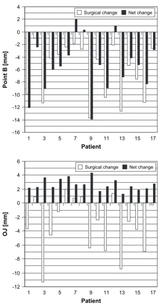
-16 -14 -12 -10 -8 -6 -4 -2 0 2 4 1 3 5 7 9 11 13 15 17 Patient Point B [mm]
Surgical change Net change
1 3 5 7 9 11 13 15 17 Patient -12 -10 -8 -6 -4 -2 0 2 4 6 OJ [mm]
Surgical change Net change
Figure 2 Surgical and net effects 12.7 years post surgery of the setback of point B (a) and the change in overjet (b) in individual patients. Numbers 1 – 6 are female patients.
40.00 50.00 60.00 70.00 80.00 90.00 T1 T2 T3 T4 T5 Follow-up X-value (mm) B-point Pogonion
T1: before surgery; T2: 5 days after surgery;
T3:6.6 months after surgery T4: 14.4 months after surgery; T5: 12.7 years after surgery
Figure 3 Surgical and net effects at different follow-ups of the setback at point B and pogonion in individual patients.
Behrents R G 1985b An atlas of growth in the aging craniofacial skeleton. Monograph No 18, Craniofacial Growth Series. Center for Human Growth and Development, The University of Michigan , Ann Arbor Bell W H , Jacobs J D , Quejada J G 1986 Simultaneous repositioning of the
maxilla, mandible, and chin . American Journal of Orthodontics 89 : 28 – 50 Dahlberg G 1940 Statistical methods for medical and biological students .
Interscience Publications , New York
Dal Pont G 1961 Retromolar osteotomy for the correction of prognathism . Journal of Oral Surgery 19 : 42 – 47
Epker B 1977 Modifi cations in the sagittal osteotomy of the mandible . Journal of Oral Surgery 35 : 157 – 159
Gallo W , Moss M , Gaul J 1976 Modifi cation of the sagittal split ramus osteotomy for retrognathia . Journal of Oral Surgery 34 : 178 – 179 Hadjianghelou O , Luder H-U , von Weydlich G 1985 Rezidivursachen
nach Korrektur der Progenie mit sagittaler Spaltung . Informationen aus der Kieferorthopädie 17 : 185 – 204
Hunsuck E E 1968 A modifi ed intraoral sagittal splitting technique for correction of mandibular prognathism . Journal of Oral Surgery 26 : 250 – 253
Ingervall B , Thüer U , Vuillemin T 1995 Stability and effect on the soft tissue profi le of mandibular setback with sagittal split osteotomy and rigid internal fi xation . International Journal of Adult Orthodontics and Orthognathic Surgery 10 : 15 – 25
Joss C U , Thüer U W 2008 Stability of hard and soft tissue profi le after mandibular advancement in sagittal split osteotomies: a longitudinal and long-term follow-up study . European Journal of Orthodontics 30 : 16 – 23
Kobayashi T , Watanabe I , Ueda K , Nakajima T 1986 Stability of the mandible after sagittal ramus osteotomy for correction of prognathism . Journal of Oral and Maxillofacial Surgery 44 : 693 – 697
Komori E , Aigase K , Sugisaki M , Tanabe H 1987 Skeletal fi xation versus skeletal relapse . American Journal of Orthodontics and Dentofacial Orthopedics 92 : 412 – 421
Kundert M , Hadjianghelou O 1980 Condylar displacement after sagittal splitting of the mandibular rami . Journal of Maxillo-Facial Surgery 8 : 278 – 287
Mobarak K A , Krogstad O , Espeland L , Lyberg T 2000 Long-term stability of mandibular setback surgery: a follow-up of 80 bilateral sagittal split osteotomy patients . International Journal of Adult Orthodontics and Orthognathic Surgery 15 : 83 – 95
Obwegeser H 1957 The surgical correction of mandibular prognathism and retrognathia with consideration of genioplasty . Journal of Oral Surgery 10 : 677 – 689
Paulus G W , Steinhäuser E W 1982 A comperative study of wire osteosynthesis versus bone screws in the treatment of mandibular prognathism . Oral Surgery, Oral Medicine, and Oral Pathology 54 : 2 – 6
Pepersack W J , Chausse J M 1978 Long term follow-up of the sagittal splitting technique for correction of mandibular prognathism . Journal of Maxillo-Facial Surgery 6 : 117 – 140
Phillips C , Zaytoun H S , Thomas P M , Terry B C 1986 Skeletal alterations following TOVRO or BSSO procedures . International Journal of Adult Orthodontics and Orthognathic Surgery 3 : 203 – 213
Proffi t W R , Phillips C , Dann C , Turvey T A 1991 Stability after surgical-orthodontic correction of skeletal Class III malocclusion. I. Mandibular setback . International Journal of Adult Orthodontics and Orthognathic Surgery 6 : 7 – 18
Raveh J , Vuillemin T , Lädrach K , Sutter F 1988 New techniques for reproduction of the condyle relation and reduction of complications after sagittal ramus split osteotomy of the mandible . Journal of Oral and Maxillofacial Surgery 46 : 751 – 757
Schatz J-P , Tsimas P 1995 Cephalometric evaluation of surgical-orthodontic treatment of skeletal Class III malocclusion . International Journal of Adult Orthodontics and Orthognathic Surgery 10 : 173 – 180
Sinclair P M , Proffi t W R 1990 Class III problems: mandibular excess/ maxillary defi ciency. Mosby , St Louis
Sorokolit C A , Nanda R S 1990 Assessment of the stability of mandibular setback procedures with rigid fi xation . Journal of Oral Maxillofacial Surgery 48 : 817 – 822
Trauner R , Obwegeser H 1955 Zur Operationstechnik bei der Progenie und anderen Unterkieferanomalien . Deutsche Zahn-, Mund- und Kieferheilkunde 23 : 1 – 26