An armadillo-domain protein participates in a telomerase interaction network
Ladislav Dokládal
1,2,6· Eva Benková
3· David Honys
4· Nikoleta Dupľáková
4· Lan-Ying Lee
5· Stanton B. Gelvin
5· Eva Sýkorová
1Key message Arabidopsis and human ARM protein interact with telomerase. Deregulated mRNA levels of DNA repair and ribosomal protein genes in an Arabidopsis arm mutant suggest non-telomeric ARM function. The human homolog ARMC6 interacts with hTRF2.
Abstract Telomerase maintains telomeres and has proposed non-telomeric functions. We previously identified interaction of the C-terminal domain of Arabidopsis telomerase reverse transcriptase (AtTERT) with an armadillo/β-catenin-like repeat (ARM) containing protein. Here we explore protein–protein interactions of the ARM protein, AtTERT domains, POT1a, TRF-like family and SMH family proteins, and the chromatin remodeling protein CHR19 using bimolecular fluorescence complementation (BiFC), yeast two-hybrid (Y2H) analysis, and co-immunoprecipitation. The ARM protein interacts with both the N- and C-terminal domains of AtTERT in different cellular compartments. ARM interacts with CHR19 and TRF- like I family proteins that also bind AtTERT directly or through interaction with POT1a. The putative human ARM homolog co-precipitates telomerase activity and interacts with hTRF2 protein in vitro. Analysis of Arabidopsis arm mutants shows no obvious changes in telomere length or telomerase activity, suggesting that ARM is not essential for telomere maintenance.
The observed interactions with telomerase and Myb-like domain proteins (TRF-like family I) may therefore reflect possible non-telomeric functions. Transcript levels of several DNA repair and ribosomal genes are affected in arm mutants, and ARM, likely in association with other proteins, suppressed expression of XRCC3 and RPSAA promoter constructs in luciferase reporter assays. In conclusion, ARM can participate in non-telomeric functions of telomerase, and can also perform its own telomerase-independent functions.
Keywords Armadillo/β-catenin-like repeat · ARMC6 · AtTERT · Homologous recombination · Protein–protein interaction · Telomerase activity
Introduction
Telomerase is a conserved ribonucleoprotein complex that is responsible for telomere synthesis (Greider and Blackburn 1985, 1987), compensating telomere shortening in cells with
Electronic supplementary material.
Eva Sýkorová [email protected]
1 Institute of Biophysics, The Czech Academy of Sciences, Královopolská 135, 61265 Brno, Czech Republic
2 Laboratory of Functional Genomics and Proteomics, NCBR, Faculty of Science, Masaryk University, Brno, Czech Republic
3 Institute of Science and Technology Austria, 3400 Klosterneuburg, Austria
4 Institute of Experimental Botany, The Czech Academy of Sciences, Rozvojova 263, 16502 Prague, Czech Republic
5 Department of Biological Sciences, Purdue University, West Lafayette, IN 47907-1392, USA
6 Present Address: Department of Biology, Faculty of Science and Medicine, University of Fribourg, Fribourg, Switzerland
http://doc.rero.ch
1VCMJTIFEJO1MBOU.PMFDVMBS#JPMPHZ o
XIJDITIPVMECFDJUFEUPSFGFSUPUIJTXPSL
high proliferative needs such as in animal embryonic, stem, and cancer cells (reviewed in Blasco 2005), plant meris- tematic cells, and tissue culture cells (Fajkus et al. 1996;
Fitzgerald et al. 1996). Telomerase activity requires two core subunits, telomerase reverse transcriptase (TERT) and telomerase RNA, which are associated with additional pro- teins to form the telomerase complex in vivo (Collins 2006).
In most organisms, the TERT subunit has an evolutionarily conserved primary structure comprising a telomerase essen- tial N-terminal (TEN) domain, a telomerase RNA binding domain, a central reverse transcriptase (RT) domain, and a C-terminal extension (CTE). Whereas the RT domain is responsible for telomerase enzymatic activity, the N- and C-terminal parts of TERT also represent potential targets for telomerase accessory and regulatory proteins. In plants, tandem affinity purification of Arabidopsis thaliana TERT showed that AtPOT1a protein is a component of the Arabi- dopsis telomerase holoenzyme and interacts with AtTERT in vivo (Majerska et al. 2017). This observation is consist- ent with previous findings that the AtTERT-V(I8) splicing isoform physically interacts with POT1a (Rossignol et al.
2007), pot1a mutants display progressive telomere short- ening, and telomerase activity can be immunoprecipitated using POT1a antibodies (Surovtseva et al. 2007). Protein components associated with Arabidopsis telomeres (see Prochazkova Schrumpfova et al. 2016 for review) differ sig- nificantly from human shelterin formed by proteins TRF1, TRF2, TIN2, TPP1, RAP1 and POT1 (de Lange 2005).
Human POT1 binds the telomeric 3ƍ DNA overhang and is not a component of the human telomerase holoenzyme complex. However, the shelterin subcomplex POT1-TPP1 binds telomeric DNA and its interaction (via TPP1) with the TEN domain of hTERT increases telomerase proces- sivity (Zaug et al. 2010). The key components of human shelterin are the Myb domain-containing proteins TRF1 and TRF2 that bind double-strand telomeric DNA (Broc- coli et al. 1997; Chong et al. 1995). Based on sequence similarities with hTRF Myb domains, the TRF-like (TRFL) proteins (family I and II) were identified in Arabidopsis.
However any function of these proteins in telomere main- tenance has not previously been demonstrated (Fulcher and Riha 2015). Telomeric functions in vivo were recently dem- onstrated for the plant-specific Single Myb Histone (SMH) proteins (Dvorackova et al. 2015; Schrumpfova et al. 2014).
Moreover, the SMH proteins may function as chromatin modulators: they bind promoter regions of genes involved in ribosome biogenesis (Schrumpfova et al. 2016; Zhou et al.
2016) and of other genes containing a telobox, a regulatory motif with a sequence identical to that of the plant telom- eric repeat (AAA CCC T)n (Regad et al. 1994). Similar to SMH proteins, the TRF-like family I proteins can bind telo- meric dsDNA in vitro (Karamysheva et al. 2004) and their putative homologs from parsley and maize are implicated
in regulating gene expression (da Costa e Silva et al. 1993;
Lugert and Werr 1994; Nagaoka and Takano 2003).
Non-canonical activities of plant telomerase have not yet been reported. Several studies have described non-telomeric (non-canonical) functions of mammalian telomerase. These include involvement in regulating cellular processes such as apoptosis, cellular proliferation, and cell cycle progres- sion. Moreover, hTERT expression and telomerase activity are linked to tumorigenesis by telomere length-independent mechanisms (reviewed in Majerska et al. 2011). The most discussed function is the participation of mammalian tel- omerase and armadillo/β-catenin proteins in the Wnt path- way that was first identified for its role in carcinogenesis.
However, similar effector proteins in plants are unknown (see Sharma et al. 2014 for review). Our previous search for AtTERT partners identified two proteins from an Arabi- dopsis cDNA library that interacted with the CTE domain (Lee et al. 2012). One of these was an RNA-recognition- motif (RRM)-containing protein that also interacted with proteins responsible for transcriptional and post-transcrip- tional regulation (Dokladal et al. 2015). The second protein of unknown function was an armadillo/β-catenin-like repeat (ARM, encoded by AT4G33945) protein that interacted with the CTE domain of AtTERT, predominantly in the cyto- plasm of BY-2 protoplasts (Lee et al. 2012). The ARM pro- tein contains four armadillo/β-catenin-like repeats. Except for the presence of these repeats, its primary structure does not resemble mammalian or plant armadillo proteins with clear biological functions, such as importins, kinesins, ubiq- uitin ligases, or kinases (see Coates 2003; Tewari et al. 2010, and references herein). However, in searching for putative telomere links we found a putative human homologue of the ARM protein ARMC6 among proteins that co-purified with hTRF2 (Giannone et al. 2010), a core component of the human shelterin complex. The function of ARMC6 has not yet been determined.
Here, we report the localization of the Arabidopsis ARM protein and mutual protein–protein interactions of ARM, Arabidopsis telomerase holoenzyme (AtTERT and POT1a), Myb domain-containing proteins (TRFL and SMH fami- lies), and the chromatin remodeling protein CHR19. Tel- omere length and telomerase activity were not influenced by disruption of the ARM gene, whereas mRNA levels of several ribosomal genes and genes involved in the homolo- gous recombination pathway were altered in arm mutants.
A putative human homolog of ARM co-precipitated human telomerase activity in vitro, suggesting that the interaction between ARM proteins and telomerases may be evolutionar- ily conserved.
http://doc.rero.ch
Materials and methods Plant material
Arabidopsis T-DNA insertion lines SALK_063839C (arm- 1) and SALK_150486C (arm-2) (Alonso et al. 2003) were obtained from the Nottingham Arabidopsis Stock Centre.
The tert line (SAIL_284_B07) was described by Fojtova et al. (2011). Seeds were surface sterilized and germinated on 0.8% (w/v) agar plates supplemented with 1/2 Murashige and Skoog media (MS; cat. n. M0255.0050; Duchefa, http://
www.duche fa-bioch emie.com) and 1% (w/v) sucrose. Seed- lings were potted after 7 days and further grown under con- ditions of 16 h light, 21 °C and 8 h dark, 19 °C, illumina- tion 150 μmol m
−2s
−1. Individual plants from each T-DNA insertion line were genotyped (see Supplemental Table S1 for primer sequences) and homozygous mutant plants were grown. Nicotiana benthamiana plants were grown under conditions of 8 h light, 21 °C and 16 h dark, 19 °C, illumi- nation 100 μmol m
−2s
−1.
Telomere length and telomerase activity analyses
Terminal restriction fragment (TRF) analysis, the conven- tional telomere repeat amplification protocol (TRAP), and the quantitative TRAP assays were performed as described (Fojtova et al. 2011). Mean telomere length values were cal- culated using TeloTool software (Gohring et al. 2014). The TRAP assay with co-immunoprecipitated human telomerase is described in Supplemental materials and methods.
Entry clone generation
Sequences encoding the coding regions of ARM ( AT 4 G 3 3 9 4 5 ) , T R F L 3 ( AT 1 G 1 7 4 6 0 ) , T R F L 6 ( AT 1 G 7 2 6 5 0 ) , T R F L 9 ( AT 3 G 1 2 5 6 0 ) , T R F L 1 1 (AT5G58340) and POT1a proteins (AT2G05210) were amplified from 7-day-old seedling cDNA, and TRFL2 (AT1G07540) from pollen cDNA using Phusion HF DNA polymerase (Finnzymes, http://www.therm oscie ntifi cbio.
com/finnz ymes) according to the manufacturer’s instruc- tions. Primers used for cloning are listed in Supplemental Table S1. PCR products were cloned into pDONR/Zeo (Invitrogen, http://www.lifet echno logie s.com) using Gate- way technology. An entry clone encoding CHR19 (stock no.
U16514, AT2G02090) was obtained from the ABRC (http://
www.arabi dopsi s.org/). Entry clones encoding AtTERT (AT5G16850) fragments TEN(1–233), RID1(1–271), Fw3N-NLS(229–582), RT(597–987), CTE2(958–1123) were prepared previously (Zachova et al. 2013).
Yeast two hybrid (Y2H) and co-immunoprecipitation (co-IP) analyses
Y2H experiments were performed using the Matchmaker™
GAL4-based two-hybrid system (Clontech, http://www.
clont ech.com). cDNA sequences encoding ARM, TRFL2, TRFL3, TRFL6, TRFL9, TRFL11, POT1a, CHR19 and TERT fragments were subcloned from their entry clones into the destination vectors pGADT7-DEST and pGBKT7- DEST. TRB1 (AT1G49950), TRB2 (AT5G67580), and TRP1 (AT5G59430) constructs in pGADT7 and pGBKT7 vectors were prepared previously (Kuchar and Fajkus 2004).
Each bait/prey combination was co-transformed into Sac- charomyces cerevisiae PJ69-4a and Y2H analysis was per- formed as described in (Schrumpfova et al. 2014) using TRB1–RID interaction as a positive control. Protein expres- sion was verified by immunoblotting using mouse anti-HA (kindly provided by Bořivoj Vojtěšek, Masaryk university Brno, Czech Republic) or mouse anti-myc primary antibod- ies and a HRP-conjugated anti-mouse secondary antibody (both Sigma-Aldrich, http://www.sigma -aldri ch.com) (Sup- plemental Fig. S1a). In vitro translation of bait/prey proteins was performed using the same constructs as in the Y2H sys- tem (vectors pGADT7-DEST and pGBKT7-DEST with min- imal T7 promoter) and a TNT quick coupled transcription/
translation system (Promega, https ://www.prome ga.com) in 25/50 μl reaction volumes according to the manufacturer’s instructions. Prey proteins were radioactively labeled using
35
S-Met (Hartmann Analytic, Germany). The co-IP pro- cedure was performed as described by Schrumpfova et al.
(2014) using mouse anti-myc antibody (9E10, Sigma) and magnetic beads Dynabeads® Protein G (Life Technologies, https ://www.therm ofish er.com). Proteins in input, unbound, and bound fractions were separated by 12.5% SDS-PAGE, blotted onto Amersham Hybond-ECL membranes (GE Healthcare, http://www.gelif escie nces.com/), and analyzed using an FLA7000 imager (Fujifilm).
Bimolecular fluorescence complementation (BiFC)
pSAT1-nEYFP-C1::RID1, pSAT1-nEYFP-C1::TRP1, pSAT1-cEYFP-C1-B::RID1, and pSAT5-DEST-cEYFP- C1(B)::ARM constructs used for BiFC in tobacco BY-2 protoplasts were prepared previously (Lee et al. 2012;
Schrumpfova et al. 2014). Tobacco BY-2 protoplasts were isolated and transfected as described (Tenea et al. 2009; Lee et al. 2012). To label cell nuclei, we co-transfected a plasmid expressing mRFP fused to the nuclear localization signal of the VirD2 protein from Agrobacterium tumefaciens (mRFP- VirD2(NLS); Citovsky et al. 2006). For BiFC in N. bentha- miana leaves, cDNA sequences encoding TRFL2/3/6/9, CHR19, ARM, and TERT fragments (RID1 and RT) were subcloned from their entry clones into the binary destination
http://doc.rero.ch
vectors pE-SPYNE-GW (for N-terminal nYFP tag) and/or pE-SPYCE-GW (for N-terminal cYFP tag) that were kindly provided by Caroline Mayer and Wolfgang Dröge-Laser (University of Göttingen; Mayer and Dröge-Laser, unpub- lished results). The constructs were electroporated into Agro- bacterium tumefaciens GV3101 and the transformants were selected on YEP medium containing 40 μg/ml carbenicillin.
Agrobacterium cultures with nYFP-, cYFP-, p19 [to prevent gene silencing (Voinnet et al. 2000)], and pK7RWG2::AT- HOOK (AT1G48610; to label cell nuclei) constructs were grown to A
600= 0.8 and used for transient expression in N. benthamiana leaves via syringe infiltration in a ratio of 1:1:1:1. After 3 days incubation, fluorescence was observed using a Zeiss Observer.Z1 equipped with an LSM780 confo- cal unit. Protein expression was tested by immunoblotting using an anti-GFP primary antibody (Roche, http://www.
roche .com) and a HRP-conjugated anti-mouse secondary antibody (Sigma-Aldrich, http://www.sigma -aldri ch.com).
Protein extracts were prepared according to Heinekamp et al.
(2002). As negative controls, we used nYFP- and cYFP- GAUT10 (AT2G20810) constructs. The pMDC43::ARM construct was used to visualize GFP-ARM subcellular localization in N. benthamiana leaves, and the PM-RB con- struct described by Nelson et al. (2007) was used as a plasma membrane marker.
Analysis of human proteins
We prepared constructs of armadillo repeat-containing pro- tein 6 (ARMC6) coding for isoform 2 (Genebank accession NP_219483.1) in pBluescript-SK or pDONRZeo vectors using sequence-specific PCR primers and cDNA from the human cell line MCF7 using Superscript III reverse tran- scriptase (Invitrogen, http://www.lifet echno logie s.com).
The constructs TRF2 and TPP1 in pDONR/Zeo, and Rap1 in pHGWA (Janouskova et al. 2015) were kindly provided by Ctirad Hofr (CEITEC MU Brno, Czech Republic). The ARMC6, TRF2, and TPP1 inserts were introduced into pGBKT7-DEST and pDEST17 vectors using Gateway tech- nology (Invitrogen, http://www.lifet echno logie s.com) or into the pTriEx4 vector by ligation. The construct hTERT-hTR in pBluescript-SK (Bachand et al. 2000) was kindly provided by Chantal Autexier (McGill University, Montreal, Quebec, Canada). Preparation of constructs of human proteins cre- ated for this work, in vitro expression in RRL, co-immuno- precipitation, and TRAP assays are described in detail in Supplemental Materials.
RNA isolation and RT-qPCR analysis
Various Arabidopsis pollen developmental stages were col- lected according to (Dupl’akova et al. 2016) and RNA was
isolated using a Plant RNeasy Kit (Qiagen, http://www.
qiage n.com) according to the manufacturer’s instructions, and further purified by DNaseI treatment (see Supplemental Material for details and specific RT-qPCR conditions). RNA isolation from other plant tissues and reverse transcription were performed as described (Fojtova et al. 2011; Ogrocka et al. 2012). Calli were derived from 7-day-old seedlings, propagated on cultivation medium with 1 μg ml
−11-naph- thaleneacetic acid and 1 μg ml
−12,4-dichlorophenoxyacetic acid, and subcultured monthly onto fresh medium.
Transcript levels relative to a ubiquitin reference gene (ubi-10) were analyzed in plant tissues using FastStart SYBR Green Master (Roche). A 1 μl of cDNA was added to the 20 μl reaction mix; the final concentration of each forward and reverse primer (Supplemental Table S1) was 0.5 μM. Reactions were performed in triplicate. PCR cycle conditions consisted of 10 min of initial denaturation (95 °C) followed by 40 cycles of 20 s at 95 °C, 30 s at 55 °C, and 1 min at 72 °C. SYBR Green I fluorescence was monitored after each extension step. The amount of the respective tran- script was determined for at least two biological replicates (for details see Supplemental Table S3) using the ΔΔCt method (Pfaffl 2004).
Luciferase assay
Sequences 500 or 1000 bp upstream from the translation start of genes encoding RPSAA (AT1G72370) and XRCC3 (AT5G57450) were fused with the firefly luciferase cod- ing sequence and cloned into pM42GW7 (“reporter”). The ARM coding sequence was cloned into the expression vector p2GW7 under control of the CaMV 35S promoter (“effec- tor”). A p2GW7::GUS construct was used as a reference.
Arabidopsis protoplasts were freshly prepared from a root cell suspension culture (Simaskova et al. 2015) and trans- fected with 3 μg each of reporter- and effector/reference- encoding plasmids and 1 μg of plasmid encoding control Renilla luciferase. Both firefly and Renilla luciferase activi- ties were measured 16 h after transfection using the Dual- Luciferase Reporter Assay System (Promega) on a Synergy H1 microplate reader (Bio-Tek) at 12 one-second intervals.
To eliminate the effect of different transfection efficiencies, total firefly luciferase activity was divided by total Renilla luciferase activity (fluc/rluc). Relative promoter activity was calculated as (fluc/rluc)
effector:(fluc/rluc)
reference. The experi- ment was performed using three biological replicas.
http://doc.rero.ch
Results
The ARM protein interacts with the N- and C-terminal domains of AtTERT
By BiFC screening of a cYFP-tagged cDNA library in tobacco BY-2 protoplasts (Lee et al. 2012), we previ- ously identified the ARM protein as an interacting part- ner with the C-terminal domain (CTE2) of AtTERT. To verify this interaction, we employed a yeast-two-hybrid (Y2H) system. The results confirmed ARM–CTE2 inter- action using histidine growth selection (Fig. 1a). We fur- ther investigated ARM interactions with other AtTERT protein fragments covering all conserved TERT regions [TEN, RID1, Fw3N-NLS, RT; naming convention is from Zachova et al. (2013)], and with another component of the Arabidopsis telomerase holoenzyme complex, POT1a (summarized in Supplemental Table S2). Among these, ARM interaction with the N-terminal TERT fragment RID1 was observed on both histidine and stringent adenine selection plates (Fig. 1a). Using BiFC, we confirmed that ARM-RID1 interaction occurred exclusively within the nucleus, including the nucleolus, of tobacco BY-2 pro- toplasts (Fig. 1b). The previously described interaction, ARM–CTE2, occurred predominantly in the cytoplasm (Lee et al. 2012). To determine the natural localization of the ARM protein, we transiently expressed a GFP–ARM fusion protein in N. benthamiana leaves. In addition to nuclear and cytoplasmic localization, the GFP–ARM pro- tein co-localized with a plasma membrane marker (Fig. 1c and Supplemental Fig. S2). Variable localization of ARM protein and its interactions suggests ARM may perform its biological functions outside of the nucleus in addition to possible ARM-mediated functions of telomerase.
The ARM protein interacts with TRF-like family I proteins
We identified a putative human homologue (ARMC6) of the AtARM protein in the dataset of (Giannone et al. 2010) among proteins that co-purify with the telomere binding protein hTRF2. We therefore investigated interactions of the AtARM protein with representative members of the TRF-like family I (TRP1, TRFL2 and TRFL9) and the TRF-like family II (TRFL3, TRFL6, TRFL11). Despite their sequence similarity to human TRFs, these proteins do not function in telomere maintenance (Fulcher and Riha 2015). Thus, we also tested telomeric proteins from the plant-specific SMH family (TRB1, TRB2). TRP1 and TRFL9 proteins are distant relatives within the TRF-like I family and they can form heterodimers in vitro (Kar- amysheva et al. 2004). TRFL2 shows specific expression
in the gametophyte (Fig. 1d). TRFL3 and TRFL6 are close relatives among members of the TRF-like family II (Karamysheva et al. 2004; Yanhui et al. 2006) but they show strikingly different transcriptional profiles with highly induced TRFL3, but not TRFL6, gene expression in response to DNA damage (Culligan et al. 2006) and in tert−/− mutants with eroded telomeres (Amiard et al.
2014). TRFL11 was identified among proteins that co- purified with AtTERT (Majerska et al. 2017). Investi- gation of protein–protein interactions by Y2H revealed positive interactions of two proteins from the TRF-like family I (TRP1 and TRFL2) with the ARM protein on his- tidine growth selection (Supplemental Table S2, Fig. 1a).
Using BiFC, the TRP1–ARM and TRFL2–ARM interac- tions were detected in nuclei of tobacco BY-2 protoplasts and N. benthamiana leaves, respectively (Fig. 1b). For an unknown reason, TRFL9 interactions in Y2H were not reproducible; we therefore tested co-IP of proteins expressed in a rabbit reticulocyte lysate (RRL) in vitro.
Using this assay, TRFL9–ARM interaction was negative (Supplemental Table S2, Fig. 1e), whereas we observed a positive interaction of TRFL9 with ARM using BiFC (Fig. 1b). It is possible that this BiFC interaction is not direct but rather mediated by another protein in vivo, or it may depend on a specific posttranslational modification that is available in plant cells but not in yeast or in an in vitro system. The ARM protein did not interact with either the TRF-like family II protein TRFL11, or with the SMH proteins TRB1 and TRB2 using a Y2H analysis (Supplemental Table S2, Fig. 1a). Interactions of the TRF- like family II proteins TRFL3 and TRFL6 could not be investigated in a Y2H because the BD-TRFL3/6 constructs alone showed growth on plates with high 3-aminotriazole concentrations, and AD-TRFL3/6 constructs were not expressed in yeast (Supplemental Fig. S1a). Importantly, no interaction was observed between the TRFL3/6 proteins and the ARM protein using BiFC in N. benthamiana leaves (Fig. 1b), despite proven protein expression (Supplemental Fig. S1a). In conclusion, our results demonstrate interac- tions of the ARM protein exclusively with TRF-like family I proteins (TRP1, TRFL2, TRFL9) using the techniques of BiFC, Y2H, and/or co-IP (summarized in Supplemental Table S2 and Fig. 1f).
Putative protein–protein interaction network of AtTERT, ARM, and Myb domain-containing proteins
Positive interactions of the ARM protein with two AtTERT domains and with TRFL family I proteins encouraged us to test for interactions of TRFL proteins with telomerase holoenzyme components represented by AtTERT fragments and POT1a. Investigation of a putative protein–protein
http://doc.rero.ch
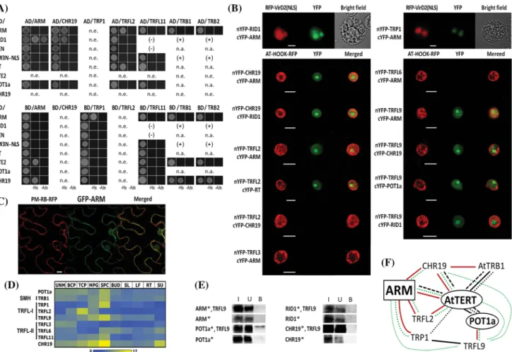
Fig. 1 Protein–protein interactions and subcellular localization of the ARM protein. a Yeast two-hybrid assay (Y2H), b bimolecular fluorescence complementation (BiFC) and e co-immunoprecipi- tation were used to study interactions among ARM, TERT frag- ments (RID1, TEN, FW3N-NLS, RT, and CTE2), POT1a, CHR19, and TRF-like proteins. The results are summarized in Supplemental Table S2. a Y2H experiments were performed using protein con- structs expressed in the vectors pGADT7-DEST (AD) and pGBKT7- DEST (BD). Each bait/prey combination was co-transformed into Saccharomyces cerevisiae PJ69-4a and positive interactions were detected using histidine (−His) or histidine and adenine (−His,
−Ade) growth selection. Interactions of TRFL11 and TRB pro- teins indicated (+/−) were reported in Majerska et al. (2017) and Schrumpfova et al. (2014), respectively; n.a. not analyzed; n.e. not expressed. b In BiFC assays, interactions between n/cYFP constructs of ARM and RID1 or TRP1 were detected in the nucleus, includ- ing the nucleolus, of tobacco BY-2 protoplasts (upper panels) and between ARM and TRFL2, TRFL9 or CHR19 in N. benthamiana leaf cells (bottom panels; only the nucleus is visible). Similar posi- tive results were found for CHR19–RID1, CHR19–TRFL9, TRFL2–
RT, TRFL9–RID1, and TRFL9–POT1a interactions. Negative BiFC results indicated lack of interaction between TRFL2 and CHR19, and between ARM and TRF-like family II proteins (TRFL3, TRFL6, bottom panels), and with RT fragment (Supplemental Fig. S1b). n/
cYFP constructs and a mRFP-VirD2-NLS nuclear marker or p19 and AT-HOOK-RFP nuclear marker were co-transfected into protoplasts (upper panel) or Agrobacterum infiltrated into leaf cells (bottom
panels), respectively. n/cYFP-GAUT10 constructs served as nega- tive controls (Supplemental Fig. S1c). c GFP–ARM localized in the nucleus and in the plasma membrane of N. benthamiana leaf cells (lower panel, see also Supplemental Fig. S2), using a PM-RB-RFP construct to label the plasma membrane. YFP fluorescence (green), mRFP fluorescence (red). Scale bars (b, c) indicate 20 μm. d Heat map showing expression profiles of selected genes in various Arabi- dopsis tissues selected from publicly available transcriptomic data- sets (Duplakova et al. 2007, see Supplemental Material for details).
Expression signals are presented as log2 values. UNM uninucleate microspores, BCP bicellular pollen, TCP tricellular pollen, MPG mature pollen, SPC sperm cells, BUD flower buds, SL seedlings, LF leaves, RT roots, SU cell suspension. e Positive interaction between TRFL9 and POT1a protein was detected after immunoprecipitation using anti-c-myc antibody. TRFL9/c-myc protein (bait) and radi- oactively-labeled prey proteins (asterisk) were expressed in a rabbit reticulocyte lysate from plasmid vectors with a minimal T7 promoter.
Proteins in input (I), unbound (U), and bound (B) fractions, water was used instead of TRFL9 protein in control samples. f Mutual protein–
protein interactions of ARM protein, Arabidopsis telomerase holo- enzyme complex components (AtTERT, POT1a), TRF-like family I proteins (TRP1, TRFL2, TRFL9), SMH family protein TRB1, and chromatin remodeling protein CHR19 are indicated. The interactions detected experimentally here (in color) or reported previously (in black) using Y2H and/or co-IP (lines) are mainly supported in planta by BiFC detection (dotted line) and tandem affinity co-purification (dashed line)
http://doc.rero.ch
interaction network showed interesting specific interac- tions among individual TRFL family I proteins. TRFL2 interacted exclusively with the RT domain of AtTERT in both Y2H and BiFC systems (Supplemental Table S2, Fig. 1a, b). TRFL9 interactions were investigated in vitro and a positive TRFL9–POT1a interaction was detected by co-IP (Fig. 1e) and further confirmed by BiFC (Fig. 1b).
Moreover, a TRFL9–RID1 positive signal was observed in nuclei of N. benthamiana leaves (Fig. 1b) but not by co-IP (Fig. 1e). These results suggest that the TRFL9–RID1 inter- action could be mediated by POT1a protein in vivo (Sup- plemental Table S2). We also tested the TRF-like family II protein TRFL11 but observed no interaction with AtTERT fragments or POT1a using a Y2H (Supplemental Table S2, Fig. 1a). Positive TRP1–RID1 interaction and strong inter- actions between SMH proteins and AtTERT were reported previously (Schrumpfova et al. 2014) (Fig. 1a, f). Thus, strikingly, both Myb domain-containing protein groups interacting with AtTERT and/or POT1a are known to bind telomeric repeats and can provide a functional link to tran- scriptional regulation.
ARM, AtTERT, and telomere-binding proteins interact with the chromatin remodeling protein CHR19
We identified CHR19 among proteins that co-purified with AtTERT fragments using tandem affinity purification (Majerska et al. 2017). CHR19 represents a protein with putative dual functions (Dona and Mittelsten Scheid 2015;
and references therein). It is a presumed homolog of yeast Fun30 and human SMARCAD1 chromatin remodelers that promote DNA end resection. CHR19 was also identified as an interactor with histone lysine methyltransferase SUVR2 that is involved in transcriptional silencing, similar to Fun30 in yeast. We therefore tested whether CHR19 protein could interact with the ARM protein, telomerase complex compo- nents (AtTERT, POT1a), and proteins from the TRFL and SMH families. Using a Y2H system, we observed interac- tions for CHR19–ARM, CHR19–RID1, CHR19–TRB1, CHR19–TRB2 (Supplemental Table S2, Fig. 1a). Using BiFC, the interactions between CHR19 and ARM, and CHR19 and RID1 were further localized within the nuclei of N. benthamiana leaf cells (Fig. 1b). The CHR19–TRFL2 interaction could not be investigated using a Y2H because the BD–TRFL2 and BD–CHR19 constructs were not expressed in yeast (Supplemental Fig. S1a), but we obtained a negative result using BiFC (Fig. 1b). The CHR19–TRFL9 interaction was negative in a co-IP assay (Fig. 1e) but was clearly detected in the BiFC experiment (Fig. 1b), suggest- ing that this interaction may be mediated by another protein partner in vivo or may depend on a specific posttranslational modification(s). These results provide an additional link to
specialized functions of AtTERT that may be related to func- tions of CHR19 in the regulation of transcription and/or in DNA end resection, possibly relevant to its participation in chromosomal healing.
ARM is highly expressed in flower buds, pollen, young leaves, and root tips
Because the ARM gene is missing on widely used microar- ray chips (Affymetrix Arabidopsis ATH1 Genome Array), data on tissue-specific ARM transcription have not been available from public databases. To obtain a transcriptional profile of the ARM gene and to compare it with that of the AtTERT gene (Fig. 2), we analyzed ARM expression dur- ing plant development with a focus on telomerase-positive tissues. We used RT-qPCR to quantify ARM transcripts in flower buds, calli, leaves, and 7-day-old seedlings of wild- type plants (Col-0), with a particular interest in detailed seedling analysis comprising whole seedlings, shoots, roots, and root tips. To quantify transcript levels in reproductive tissues, we included five pollen developmental stages (uni- nucleate microspores, early bicellular pollen, late bicellular pollen, immature tricellular pollen, and mature pollen). We observed ARM transcripts in all tissues tested. The relative transcriptional level of ARM was 100 times higher than that
Fig. 2 Analyses of ARM and AtTERT gene expression in wild-type plants. Transcript levels of ARM (left y-axis) and AtTERT (right y-axis) genes in various wild-type plant tissues and developmental stages were calculated relative to AtTERT levels in 7-day-old seed- lings (Col-0) arbitrarily set to 1. AtTERT levels were compiled from our experimental data (white columns) or adapted from previous work (columns in grey; Ogrocka et al. 2012—young and old leaf; and Zachova et al. 2013—shoots, roots and root tips of 7-day seedlings).
P values are shown for the ARM and AtTERT gene levels as experi- mentally determined here. Experiments were performed as three bio- logical replicas except the pollen samples that were repeated twice
http://doc.rero.ch
of AtTERT and gene transcript profiles partially overlapped (Fig. 2). ARM transcripts were abundant in proliferating tissues that were telomerase positive—flower buds, young leaves, roots—and were slightly elevated during two stages of pollen development corresponding to proliferative activ- ity. A detailed comparison of ARM and AtTERT transcrip- tional profiles in 7-day-old seedlings revealed co-expression of both genes in root tips. In contrast to ARM, the AtTERT gene showed high transcript levels in early stages of pol- len development. High levels of ARM transcripts that were detected in flower buds might be connected with high prolif- erative activity within floral meristems and in reproductive tissues of both sporophytic and female gametophytic origins as we did not observe a corresponding increase in ARM tran- scripts in various developmental stages of pollen.
Telomere length and telomerase activity are not affected in homozygous arm mutants
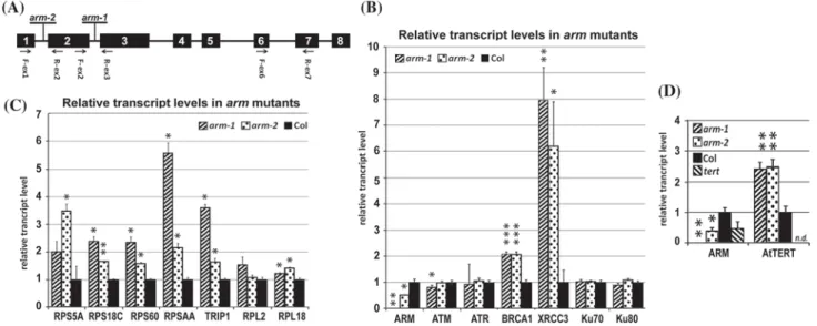
To examine the role of ARM in planta , we analyzed homozygous Arabidopsis T-DNA inser tion lines SALK_063839C (arm-1, intron 2) and SALK_150486C (arm-2, intron 1) (Fig. 3a). RT-qPCR results using exon 6- and 7-specific primers confirmed only arm-1 as a null allele, whereas the arm-2 allele caused only a partial decrease (~ 50%) in the level of the ARM transcript when compared to Col-0 wild-type plants (Fig. 3b). Sequencing of the RT-PCR product overlapping the T-DNA insertion
site in arm-2 mutants revealed that the T-DNA was spliced out, resulting in a correct splice junction. Both arm lines displayed normal vegetative growth and no detectable morphological differences were observed compared to soil-grown wild-type (Col-0) plants in three subsequent generations of homozygous arm−/− mutants (Supplemen- tal Fig. S3, see Supplemental material for details).
To investigate the telomere phenotype we analyzed tel- omere length in the third generation (G3) of homozygous mutant plants using terminal restriction fragment (TRF) analysis. Although telomeres in both arm-1/arm-1 and arm-2/arm-2 G3 plants were slightly longer when com- pared to those of wild-type plants (Supplemental Fig.
S4), a paired Student t-test indicated that these changes were not significant (the two-tailed P values equal 0.1175 and 0.0751 for arm-1 and arm-2, respectively). Telomer- ase activity was tested in 7-day-old seedlings of the G3 generation of homozygous arm-1 and arm-2 lines using a telomere repeat amplification protocol (TRAP). No changes in telomerase activity were observed using the conventional or quantitative TRAP assays (Supplemental Fig. S5). These results suggest that ARM is not essential for maintaining telomere length and telomerase activity.
ARM may be involved in translation initiation
We and other groups (Dokladal et al. 2015; Schrump- fova et al. 2016; Zhou et al. 2016) recently suggested that
Fig. 3 Analyses of gene expression in mutant plants. a Position of the T-DNA insertions within the ARM gene and primer sites used for genotyping and RT-qPCR are shown. b Transcript levels of genes involved in DNA repair, and c ribosomal protein and translation- related genes were affected in the arm-1 and arm-2 T-DNA inser- tion lines. d Transcript levels of AtTERT were increased in arm-1 and arm-2 lines, and transcript levels of the ARM gene were decreased in
a tert mutant. The level of transcripts of indicated genes (b–d) were calculated relative to those in wild-type (Col-0) leaves (b, c) or 7-day- old seedlings (d) using the ΔΔCt method and ubi10 as a reference gene. Relative transcript levels were calculated as a median of tran- script levels; two-tailed P values were calculated using an unpaired t-test (*p < 0.05; **p < 0.001; ***p = 0.0001). n.d. not detected
http://doc.rero.ch
Arabidopsis telomerase and its interacting partners, the RRM and the SMH family protein AtTRB1, may be involved in the regulation of ribosomal and translation-related genes.
However, data from tert−/− mutants (Amiard et al. 2014) showed no changes in transcript levels of ribosomal protein genes. To test a possible involvement of ARM protein in these processes, we measured transcript levels of selected representatives of ribosomal protein genes, that were upreg- ulated in rrm mutants (Dokladal et al. 2015), in arm mutant lines (Fig. 3c). We found significant changes in transcript abundance of four genes encoding components of the small ribosomal subunit (RPS5A, RPS18C, RPS60 and RPSAA):
a two to sixfold increase in transcript levels in the arm-1 line. RPS5A and RPSAA also showed a greater than two- fold increase in transcript levels in the arm-2 line. Genes encoding components of the large ribosomal subunit (RPL2, RPL18) displayed less than a twofold increase in transcript levels in mutant plants. We further analyzed transcript abun- dance of the translation initiation factor TRIP1 (TGF-beta receptor interacting protein 1) encoding a component of the 43S preinitiation complex. A greater than threefold increase in TRIP1 transcript levels was detected in the arm-1 line, supporting a possible link between ARM and the formation of a translation preinitiation complex.
To investigate a possible involvement of the ARM pro- tein in the regulation of translation-related genes, we fur- ther tested the putative promoter of the RPSAA gene that showed the highest transcriptional increase in the arm-1 line.
We fused the firefly luciferase (fluc) coding sequence with sequences 500 or 1000 bp upstream from the translation start of RPSAA and co-transfected these reporter constructs, with either a 35S::ARM effector construct or with a 35S::GUS reference construct, into Arabidopsis protoplasts. Fire- fly luciferase expression was decreased by the 35S::ARM effector construct in protoplasts co-transfected with RPSAA- 1000::fluc but not with the RPSAA-500::fluc reporter con- struct (Fig. 4). Thus, the RPSAA sequence 1000 bp upstream from the translation start seemed to be necessary to affect transcription.
ARM is involved in regulation of recombination-related genes
A direct link between telomerase activity and DNA repair pathways has not yet been established. However, these pro- cesses coexist, e.g. during chromosomal healing in response to DNA damage (Fojtova et al. 2002; Jankowska et al. 2015).
Once a double-stranded break (DSB) occurs, the cell must choose between homologous recombination (HR) and non- homologous end joining (NHEJ) to fix the break. These pathways are often in competition with each other. How- ever, natural HR occurs during DNA replication (S phase of the cell cycle) and established models for alternative
lengthening of telomeres also include the HR pathway (see Pickett and Reddel 2015; Trapp et al. 2011, for review). It is not clear whether plant telomerase and DNA repair pro- teins are collaborators or antagonists, or at what level their actions are regulated. We therefore analyzed the transcript accumulation profiles of key DSB repair genes, ATM and ATR , and representative genes related to both major DSB repair pathways in homozygous arm-1 and arm-2 mutant lines. ATM, ATR, Ku70, and Ku80, genes with established functions in telomere homeostasis and NHEJ (Amiard et al.
2011), did not show greater than twofold change in relative transcript abundance in mutant compared to that of wild- type plants (Fig. 3b, Supplemental Table S3). Transcript lev- els of BRCA1 and XRCC3, factors involved in HR (see Trapp et al. 2011; Yoshiyama et al. 2013, for review), were signifi- cantly higher in both arm T-DNA insertion lines (Fig. 3b), suggesting a possible role of ARM in the regulation of HR- related genes. Interestingly, XRCC3 transcripts were greatly increased in young leaves, but not in buds and seedlings of arm-1 mutants (Table 1, Supplemental Table S3). To investigate direct involvement of the ARM protein in tran- scriptional regulation of the XRCC3 gene, we performed a transient expression assay using luciferase expression vec- tors. Reporter constructs with the firefly luciferase coding sequence fused with sequences 500 or 1000 bp upstream from translation start of XRCC3 were co-transfected either
Fig. 4 ARM overexpression effects on XRCC3 and RPSAA promoter activation. Luciferase reporter assay shows that expression of the 35S::ARM (“effector”) construct decreases expression of RPSAA and XRCC3 promoter (“reporter”) constructs. Sequences 500 and 1000 bp upstream from the translation start fused with firefly luciferase were sufficient in the case of XRCC3 and RPSAA, respectively. A 35S::GUS construct was used as a reference. Arabidopsis protoplasts were transfected with reporter and effector/reference-encoding plas- mids and with a plasmid encoding Renilla luciferase that serves as a normalization control. Both firefly (fluc) and Renilla (rluc) luciferase activities were measured 16 h after transfection. Relative promoter activation was calculated as (fluc/rluc)effector:(fluc/rluc)reference. Two- tailed P values were calculated using an unpaired t-test (*p < 0.05;
**p < 0.001)
http://doc.rero.ch
with the 35S::ARM effector or the 35S::GUS reference con- structs into Arabidopsis protoplasts. Calculation of relative promoter activation clearly demonstrated that the XRCC3 reporters were down-regulated when ARM was over- expressed, and sequences within 500 bp upstream from the translation start of XRCC3 were sufficient for this suppres- sion (Fig. 4). We therefore focused further experiments on mRNA abundance of Arabidopsis HR-related genes (Singh et al. 2010). In total, nine genes displayed a greater than twofold increase in transcript levels in both mutant lines, and two additional genes showed increased transcript abundance only in the homozygous arm-1 line (Table 1). Affected genes were found among members of the RecA/RAD51 family, genes for ssDNA binding proteins, and the MUS81–EME1 resolvase complex. Interestingly, in contrast to RAD51 paralogs that function as recombination mediators, the level of recombinase RAD51 transcripts was significantly decreased in leaves and buds but not in seedlings of the homozygous arm-1 line (Table 1, Supplemental Table S3).
Several affected genes are also important for mitochondrial DNA repair, but transcript levels of other genes from these pathways were not changed in arm-1 mutants (Supplemental Table S3). In addition, we observed that AtTERT transcript levels were not affected in young leaves and buds. How- ever a > twofold increase was detected in seedlings of the arm-1 mutant line (Fig. 3d, Supplemental Table S3). No significant change in ARM transcript levels was detected in
seedlings of tert−/− plants (Fig. 3d), which is in agreement with microarray data of Amiard et al. (2014). In conclu- sion, transcript levels of AtTERT and of several other genes involved in the HR pathway were affected in arm mutants, suggesting involvement of ARM in their transcriptional or posttranscriptional regulation.
ARMC6 binds TRF2 and human telomerase
Searching for Arabidopsis ARM protein functions, we ana- lyzed its putative human homolog ARMC6. To test putative interactions of ARMC6 with other proteins, we ran pilot experiments in vitro. We detected weak positive protein–pro- tein interactions between TRF2 and ARMC6 (Supplemental Fig. S6a). Expression of ARMC6 protein in a rabbit reticu- locyte lysate (RRL) system was low. However, we observed the same result using an ARMC6 construct cloned into two different vectors (pTriEx4 and pDEST17). Arabidopsis ARM protein interacts with AtTERT. Thus, we investigated if ARMC6 was able to interact with human telomerase. We reconstituted telomerase activity using constructs of hTERT and hTR (Bachand et al. 2000) expressed in a RRL. The c-myc tagged construct of ARMC6 was able to immunopre- cipitate telomerase activity (Supplemental Fig. S6b). The same result was observed for TPP1 protein that is a known interactor with hTERT (Zaug et al. 2010) and was used as a positive control. Whether these results indicate a functional
Table 1 Relative transcription levels of homologous recombination-related genes affected in homozygous arm mutants
SD standard deviation
a More than twofold change in transcript level (2−ddCt) is highlighted
AGI number Gene name Leavesa Budsa
arm-1 arm-2 arm-1
2−ddCt SD 2−ddCt SD 2−ddCt SD
DNA damage complex III
AT4G21070 BRCA1 2.09 0.09 2.08 0.09 0.88 0.11
RecA/RAD51 family
AT2G19490 RecA(2) 2.47 0.09 2.66 0.30 0.77 0.08
AT3G10140 RecA(3) 2.02 0.11 2.05 0.07 0.98 0.08
AT5G20850 RAD51 0.47 0.07 0.79 0.17 0.44 0.05
AT2G28560 RAD51B 3.05 0.40 3.86 0.16 0.62 0.03
AT2G45280 RAD51C 2.08 0.24 2.60 0.28 0.39 0.01
AT3G22880 DMC 2.11 0.18 1.56 0.04 0.44 0.14
AT5G57450 XRCC3 7.96 1.25 6.20 1.7 0.94 0.14
MUS81–EME1 complex
AT2G21800 EME1(A) 2.80 0.36 3.21 0.06 2.35 0.15
Single strand DNA binding
AT4G11060 MTSSB 2.00 0.21 2.70 0.25 0.59 0.15
AT3G18580 SSB 2.15 0.13 2.31 0.13 1.61 0.11
DNA helicases
AT2G01440 RecG 2.01 0.12 1.82 0.06 1.71 0.16
http://doc.rero.ch
similarity between human and plant ARM proteins needs further investigation.
Discussion
Plant ARM repeat proteins represent a functionally heter- ogeneous group, formally unified only by the presence of armadillo/β-catenin-like repeats serving as protein–protein interaction domains (see Tewari et al. 2010, and references therein). Our original hypothesis about putative involvement of Arabidopsis ARM protein in non-telomeric functions of telomerase (Lee et al. 2012) was inspired by the human model. hTERT forms a transcription regulatory complex with β-catenin and a chromatin remodeling factor BRG1, thereby providing a link to the Wnt signaling pathway (Park et al. 2009). The putative Arabidopsis homolog of BRG1 is the Snf2-like ATPase chromatin remodeling 12 (CHR12) (see Dona and Mittelsten Scheid 2015 for review). Unfor- tunately, we could not test its protein–protein interactions because CHR12 (1102 amino acids) was not expressed either in the Y2H or BiFC systems (not shown). Animal catenins can localize at the plasma membrane, where they bind the cytoplasmic tail of cadherin (Aberle et al. 1994; Ozawa et al.
1989), and in the nucleus, where β-catenin can affect expres- sion of Wnt target genes (Behrens 2000). Cytosolic β-catenin must be stabilized by the concerted action of several kinases and scaffold proteins before it enters the nucleus (see Coates 2003; Tewari et al. 2010, for review). We observed that GFP-ARM protein localizes in the nucleus, cytoplasm, and plasma membrane of plant cells (Fig. 1c, Supplemental Fig.
S2). We do not know whether the Arabidopsis ARM protein needs specific modifications before it can enter the nucleus;
however, variable ARM localization may indicate a mul- tifunctional role such as that shown by human β-catenin.
Posttranslational modifications of the ARM protein may also explain the discrepancy between the negative co-IP result and the positive BiFC result for ARM–TRFL9 interaction.
Interestingly, pollen-specific Arabidopsis Armadillo Repeat Only 1 (ARO1) protein, that regulates cell growth and actin organisation, also shows a sub-cellular localization similar to that of β-catenin (Gebert et al. 2008).
We have previously characterized the interaction between the ARM protein and the CTE domain of AtTERT predomi- nantly in the cytoplasm of tobacco BY-2 protoplasts (Lee et al. 2012). Here we further investigated protein–protein interactions and determined the telomere phenotype using arm T-DNA insertion mutant lines (Supplemental Fig. S4).
Our analyses showed no significant changes in telomere lengths and telomerase activity, suggesting that the ARM protein is not essential for telomere maintenance, and that the observed interaction with telomerase may reflect possi- ble non-telomeric functions. Tissue-specific ARM expression
correlates partially with that of AtTERT (Fig. 2). Thus, while investigating further possible telomerase-linked roles of ARM, we focused on (i) protein–protein interactions with proteins identified previously as telomere/telomerase inter- actors in vivo and in vitro and (ii) mRNA levels of genes presumably affected in arm mutants. We demonstrated that ARM interacts with TRF-like family I proteins and the chromatin remodeling protein CHR19 both in yeast and in planta. The protein parts of Arabidopsis telomerase holo- enzyme, AtTERT and POT1a, interact with CHR19, mem- bers of the TRF-like family I proteins, and with SMH pro- teins (summarized on Fig. 1f). Interestingly, ARM, TRB1, and CHR19 (but none of the TRF-like family I proteins) were found among proteins that co-purified with Arabi- dopsis TERT and POT1a using tandem affinity purification (Majerska et al. 2017). These interactions further link tel- omerase and ARM with transcriptional regulation because of reported functions of chromatin modulators for SMH proteins and chromatin remodeling protein CHR19 (see Dona and Mittelsten Scheid 2015; Prochazkova Schrump- fova et al. 2016, for review). In addition to a transcriptional regulation function, putative mammalian/yeast homologs of CHR19 promote DNA end resection (Costelloe et al.
2012), the first step common to all homologous recombina- tion repair reactions. Thus, it is tempting to speculate that AtTERT–CHR19 and/or TRB1–CHR19 interactions might be important, e.g., for telomere repeat addition de novo. In conclusion, dual functions reported for the interaction part- ners of ARM and/or AtTERT in telomere maintenance and transcriptional regulation support the hypothesis establish- ing an interaction network of ARM, AtTERT, POT1a, and other proteins (Fig. 1f) involved in non-telomeric pathways, but this requires further investigation.
It is unclear which of the human armadillo proteins rep- resents a functional homolog of the Arabidopsis ARM pro- tein. Human β-catenin contains twelve armadillo repeats and it has been suggested that, with the exception of glyco- gen synthase kinase-3 (GSK-3) that destabilizes cytosolic β-catenin, there are no Wnt signaling pathway homologues in Arabidopsis (Jonak and Hirt 2002). Moreover, we identi- fied a putative human homolog, ARMC6, containing four armadillo repeats (accession Q6NXE6, http://www.unipr ot.org), among proteins co-purified with the human shel- terin protein TRF2 (Giannone et al. 2010). ARMC6 protein does not have any known biological function and its interac- tion with hTRF2 has not been tested. Putative homologs of ARMC6 were predicted in silico in evolutionarily diverse groups including animals, plants, green algae, chromalveo- lates and excavates, but not in fungi (see Tewari et al. 2010 for review). In our pilot study (see Supplemental Material for details), we confirmed an interaction between TRF2 and ARMC6 in vitro (Supplemental Fig. S6A). ARMC6 protein also pulls-down human telomerase reconstituted
http://doc.rero.ch
in vitro (Supplemental Fig. S6B), resembling the Arabi- dopsis ARM–TERT interaction described here. It remains to be shown whether there are any additional biochemical or functional similarities among these putative Arabidopsis and human homologs.
Non-telomeric functions of telomerase in animals are mediated by TERT-dependent alteration of gene expression (reviewed in Majerska et al. 2011). In the search for ARM biological functions and possible non-telomeric functions of AtTERT, we investigated the mRNA levels of several gene groups in arm mutants. Initially, we tested genes with telo- box-containing promoters because binding to such genomic loci was demonstrated for SMH proteins (Schrumpfova et al.
2016; Zhou et al. 2016), and also because TRF-like family I proteins can bind telomeric repeats in vitro, similarly to SMH proteins (Karamysheva et al. 2004; Schrumpfova et al.
2004). Increased mRNA levels of ribosomal genes under the control of telobox-containing promoters were also observed in mutant lines with a disrupted RRM gene that encodes another AtTERT-interacting protein (Dokladal et al. 2015).
We observed significant changes in transcription of some (but not all) ribosomal genes under the control of telobox- containing promoters in arm-1 and arm-2 T-DNA insertion lines (Fig. 3c). Affected genes represented a subset of those encoding proteins involved in the formation of a translation preinitiation complex, and we detected down-regulation of the putative promoter of the RPSAA gene, suggesting that ARM can modulate its expression in plant cells. In the particular case of the RPSAA promoter, the telobox-like sequence is present within the 500 bp sequence that was not affected by ARM (Fig. 4). These results also show that the presence of the telobox regulatory motif is not a critical determinant for specific gene expression, confirming results of previous studies (Dokladal et al. 2015; Regad et al. 1994;
Schrumpfova et al. 2016; Tremousaygue et al. 2003; Zhou et al. 2016). Secondly, we investigated genes involved in DNA repair pathways and found significant changes in expression of HR-related genes (Fig. 3b; Table 1). Analyses of gene expression in arm mutant plants showed that most genes influenced by disruption of the ARM gene were up- regulated in leaves, the XRCC3 gene being the most affected one (Table 1). However, the same genes were down-regu- lated or not affected in floral buds (Supplemental Table S3), a complex tissue with the highest natural levels of ARM and AtTERT transcripts (Fig. 2), indicating more complex or spe- cialized roles of ARM in a tissue-specific manner. The only exception was a gene encoding EME1a, part of the MUS81- EME1 resolvase recognizing specific Holliday-junction structures (Geuting et al. 2009), which was similarly up- regulated in leaves and buds of arm mutants (Table 1).
Investigation of other genes implicated in specific meiotic functions showed that in some cases, only mild deregulation occurred in buds (Supplemental Table S3). This observation
awaits further study. It should be noted that telomeres were slightly longer in arm mutants. Thus, we cannot exclude the possibility that slight defects in telomere length might be sufficient to induce expression of HR-related genes. A putative ARM function as a transcriptional repressor was supported by our results demonstrating down-regulation of the XRCC3 promoter construct in a luciferase assay in Arabi- dopsis protoplasts (Fig. 4). We speculate that the down- regulation of the RPSAA and XRCC3 promoter constructs most likely depends on ARM recognition of some additional DNA binding factor(s) and recruitment of a transcription repressor(s). These factors could represent completely dif- ferent subsets of transcriptional regulators, including those unrelated to telomerase or TRB/TRFL-I proteins.
In conclusion, our results suggest that ARM can func- tion as a modulator of gene expression, possibly through its interaction with nuclear proteins involved in transcriptional regulation. Thus, we suggest that ARM can participate in non-telomeric functions of telomerase, and can also perform its own telomerase-independent functions.
Acknowledgements We would like to thank our colleagues Jiří Fajkus and Miloslava Fojtová (Institute of Biophysics, Czech Republic) for helpful comments on the manuscript draft, Andrej Hurný (IST Austria) for help with the luciferase reporter assay, Chantal Autexier (McGill University, Montreal, Quebec, Canada) and Ctirad Hofr (Masaryk University Brno, Czech Republic) for human protein constructs, Jan Paleček (Masaryk University Brno, Czech Republic) for vectors, and Bořivoj Vojtěšek (Masaryk University Brno, Czech Republic) for anti- HA antibodies. This work was supported by the Grant Agency of the Czech Republic (18-07027S) and by institutional support.
Author contributions LD performed experiments except RT-qPCR analysis of pollen samples (ND and DH), EB contributed in pheno- type analyses, ES analysed data, L-YL and SG were involved in ARM localization experiments, SG and ES designed study, LD, SG and ES wrote the paper.
Compliance with ethical standards
Conflict of interest The authors have no conflict of interest to declare.
References
Aberle H, Butz S, Stappert J, Weissig H, Kemler R, Hoschuetzky H (1994) Assembly of the cadherin-catenin complex in vitro with recombinant proteins. J Cell Sci 107(Pt 12):3655–3663
Alonso JM et al (2003) Genome-wide insertional mutagenesis of Arabidopsis thaliana. Science 301:653–657
Amiard S, White C, Gallego ME (2011) Recombination proteins and telomere stability in plants. Curr Protein Pept Sci 12:84–92 Amiard S, Da Ines O, Gallego ME, White CI (2014) Responses to
telomere erosion in plants. PLoS ONE 9:e86220. https ://doi.
org/10.1371/journ al.pone.00862 20
Bachand F, Kukolj G, Autexier C (2000) Expression of hTERT and hTR in cis reconstitutes and active human telomerase ribonucleo- protein. RNA 6:778–784
http://doc.rero.ch
Behrens J (2000) Control of beta-catenin signaling in tumor develop- ment. Ann N Y Acad Sci 910:21–33
Blasco MA (2005) Mice with bad ends: mouse models for the study of telomeres and telomerase in cancer and aging. EMBO J 24:1095–
1103. https ://doi.org/10.1038/sj.emboj .76005 98
Broccoli D, Smogorzewska A, Chong L, de Lange T (1997) Human telomeres contain two distinct Myb-related proteins, TRF1 and TRF2. Nat Genet 17:231–235. https ://doi.org/10.1038/ng109 7-231
Chong L, van Steensel B, Broccoli D, Erdjument-Bromage H, Hanish J, Tempst P, de Lange T (1995) A human telomeric protein. Sci- ence 270:1663–1667
Citovsky V et al (2006) Subcellular localization of interacting proteins by bimolecular fluorescence complementation in planta. J Mol Biol 362:1120–1131. https ://doi.org/10.1016/j.jmb.2006.08.017 Coates JC (2003) Armadillo repeat proteins: beyond the animal king-
dom. Trends Cell Biol 13:463–471
Collins K (2006) The biogenesis and regulation of telomerase holoen- zymes. Nat Rev Mol Cell Biol 7:484–494. https ://doi.org/10.1038/
nrm19 61
Costelloe T et al (2012) The yeast Fun30 and human SMARCAD1 chromatin remodellers promote DNA end resection. Nature 489:581–584. https ://doi.org/10.1038/natur e1135 3
Culligan KM, Robertson CE, Foreman J, Doerner P, Britt AB (2006) ATR and ATM play both distinct and additive roles in response to ionizing radiation. Plant J 48:947–961. https ://doi.org/10.1111/
j.1365-313X.2006.02931 .x
da Costa e Silva O, Klein L, Schmelzer E, Trezzini GF, Hahlbrock K (1993) BPF-1, a pathogen-induced DNA-binding protein involved in the plant defense response. Plant J 4:125–135
de Lange T (2005) Shelterin: the protein complex that shapes and safe- guards human telomeres. Genes Dev 19:2100–2110. https ://doi.
org/10.1101/gad.13460 05
Dokladal L, Honys D, Rana R, Lee LY, Gelvin SB, Sykorova E (2015) cDNA library screening identifies protein interactors potentially involved in non-telomeric roles of arabidopsis telomerase. Front Plant Sci 6:985. https ://doi.org/10.3389/fpls.2015.00985 Dona M, Mittelsten Scheid O (2015) DNA damage repair in the con-
text of plant chromatin. Plant Physiol 168:1206–1218. https ://doi.
org/10.1104/pp.15.00538
Dupl’akova N, Dobrev PI, Renak D, Honys D (2016) Rapid separa- tion of Arabidopsis male gametophyte developmental stages using a Percoll gradient. Nat Protoc 11:1817–1832. https ://doi.
org/10.1038/nprot .2016.107
Duplakova N, Renak D, Hovanec P, Honysova B, Twell D, Honys D (2007) Arabidopsis gene family profiler (aGFP)—user-oriented transcriptomic database with easy-to-use graphic interface. BMC Plant Biol 7:39. https ://doi.org/10.1186/1471-2229-7-39 Dvorackova M, Fojtova M, Fajkus J (2015) Chromatin dynamics of
plant telomeres and ribosomal genes. Plant J 83:18–37. https ://
doi.org/10.1111/tpj.12822
Fajkus J, Kovarik A, Kralovics R (1996) Telomerase activity in plant cells. FEBS Lett 391:307–309
Fitzgerald MS, McKnight TD, Shippen DE (1996) Characterization and developmental patterns of telomerase expression in plants.
Proc Natl Acad Sci USA 93:14422–14427
Fojtova M, Fulneckova J, Fajkus J, Kovarik A (2002) Recovery of tobacco cells from cadmium stress is accompanied by DNA repair and increased telomerase activity. J Exp Bot 53:2151–2158 Fojtova M, Peska V, Dobsakova Z, Mozgova I, Fajkus J, Sykorova E
(2011) Molecular analysis of T-DNA insertion mutants identi- fied putative regulatory elements in the AtTERT gene. J Exp Bot 62:5531–5545. https ://doi.org/10.1093/jxb/err23 5
Fulcher N, Riha K (2015) Using centromere mediated genome elimina- tion to elucidate the functional redundancy of candidate telomere
binding proteins in Arabidopsis thaliana. Front Genet 6:349. https ://doi.org/10.3389/fgene .2015.00349
Gebert M, Dresselhaus T, Sprunck S (2008) F-actin organization and pollen tube tip growth in Arabidopsis are dependent on the gametophyte-specific Armadillo repeat protein ARO1. Plant Cell 20:2798–2814. https ://doi.org/10.1105/tpc.108.06102 8
Geuting V, Kobbe D, Hartung F, Durr J, Focke M, Puchta H (2009) Two distinct MUS81-EME1 complexes from Arabidopsis pro- cess Holliday junctions. Plant Physiol 150:1062–1071. https ://
doi.org/10.1104/pp.109.13684 6
Giannone RJ, McDonald HW, Hurst GB, Shen RF, Wang Y, Liu Y (2010) The protein network surrounding the human telomere repeat binding factors TRF1, TRF2, and POT1. PLoS ONE 5:e12407. https ://doi.org/10.1371/journ al.pone.00124 07 Gohring J, Fulcher N, Jacak J, Riha K (2014) TeloTool: a new tool for
telomere length measurement from terminal restriction fragment analysis with improved probe intensity correction. Nucleic Acids Res 42:e21. https ://doi.org/10.1093/nar/gkt13 15
Greider CW, Blackburn EH (1985) Identification of a specific tel- omere terminal transferase activity in Tetrahymena extracts. Cell 43:405–413
Greider CW, Blackburn EH (1987) The telomere terminal transferase of Tetrahymena is a ribonucleoprotein enzyme with two kinds of primer specificity. Cell 51:887–898
Heinekamp T, Kuhlmann M, Lenk A, Strathmann A, Droge-Laser W (2002) The tobacco bZIP transcription factor BZI-1 binds to G-box elements in the promoters of phenylpropanoid pathway genes in vitro, but it is not involved in their regulation in vivo.
Mol Genet Genomics 267:16–26. https ://doi.org/10.1007/s0043 8-001-0636-3
Jankowska M et al (2015) Holokinetic centromeres and efficient tel- omere healing enable rapid karyotype evolution. Chromosoma 124:519–528. https ://doi.org/10.1007/s0041 2-015-0524-y Janouskova E et al (2015) Human Rap1 modulates TRF2 attraction to
telomeric DNA. Nucleic Acids Res 43:2691–2700. https ://doi.
org/10.1093/nar/gkv09 7
Jonak C, Hirt H (2002) Glycogen synthase kinase 3/SHAGGY-like kinases in plants: an emerging family with novel functions. Trends Plant Sci 7:457–461
Karamysheva ZN, Surovtseva YV, Vespa L, Shakirov EV, Shippen DE (2004) A C-terminal Myb extension domain defines a novel family of double-strand telomeric DNA-binding proteins in Arabi- dopsis. J Biol Chem 279:47799–47807. https ://doi.org/10.1074/
jbc.M4079 38200
Kuchar M, Fajkus J (2004) Interactions of putative telomere-binding proteins in Arabidopsis thaliana: identification of functional TRF2 homolog in plants. FEBS Lett 578:311–315. https ://doi.
org/10.1016/j.febsl et.2004.11.021
Lee LY et al (2012) Screening a cDNA library for protein-protein inter- actions directly in planta. Plant Cell 24:1746–1759. https ://doi.
org/10.1105/tpc.112.09799 8
Lugert T, Werr W (1994) A novel DNA-binding domain in the Shrunken initiator-binding protein (IBP1). Plant Mol Biol 25:493–506
Majerska J, Sykorova E, Fajkus J (2011) Non-telomeric activities of telomerase. Mol Biosyst 7:1013–1023. https ://doi.org/10.1039/
c0mb0 0268b
Majerska J et al (2017) Tandem affinity purification of AtTERT reveals putative interaction partners of plant telomerase in vivo.
Protoplasma 254:1547–1562. https ://doi.org/10.1007/s0070 9-016-1042-3
Nagaoka S, Takano T (2003) Salt tolerance-related protein STO binds to a Myb transcription factor homologue and confers salt tolerance in Arabidopsis. J Exp Bot 54:2231–2237. https ://doi.org/10.1093/
jxb/erg24 1
http://doc.rero.ch
Nelson BK, Cai X, Nebenfuhr A (2007) A multicolored set of in vivo organelle markers for co-localization studies in Arabidopsis and other plants. Plant J 51:1126–1136. https ://doi.org/10.1111/
j.1365-313X.2007.03212 .x
Ogrocka A, Sykorova E, Fajkus J, Fojtova M (2012) Developmen- tal silencing of the AtTERT gene is associated with increased H3K27me3 loading and maintenance of its euchromatic environ- ment. J Exp Bot 63:4233–4241. https ://doi.org/10.1093/jxb/ers10 7 Ozawa M, Baribault H, Kemler R (1989) The cytoplasmic domain of
the cell adhesion molecule uvomorulin associates with three inde- pendent proteins structurally related in different species. EMBO J 8:1711–1717
Park JI et al (2009) Telomerase modulates Wnt signalling by associa- tion with target gene chromatin. Nature 460:66–72. https ://doi.
org/10.1038/natur e0813 7
Pfaffl MW (2004) Quantification strategies in real-time PCR. In: Bustin SA (ed) A-Z of quantitative PCR. International University Line, La Jolla, CA, pp 87–112
Pickett HA, Reddel RR (2015) Molecular mechanisms of activity and derepression of alternative lengthening of telomeres. Nat Struct Mol Biol 22:875–880. https ://doi.org/10.1038/nsmb.3106 Prochazkova Schrumpfova P, Schorova S, Fajkus J (2016) Tel-
omere- and telomerase-associated proteins and their functions in the plant cell. Front Plant Sci 7:851. https ://doi.org/10.3389/
fpls.2016.00851
Regad F, Lebas M, Lescure B (1994) Interstitial telomeric repeats within the Arabidopsis thaliana genome. J Mol Biol 239:163–169.
https ://doi.org/10.1006/jmbi.1994.1360
Rossignol P, Collier S, Bush M, Shaw P, Doonan JH (2007) Arabi- dopsis POT1A interacts with TERT-V(I8), an N-terminal splic- ing variant of telomerase. J Cell Sci 120:3678–3687. https ://doi.
org/10.1242/jcs.00411 9
Schrumpfova P, Kuchar M, Mikova G, Skrisovska L, Kubicarova T, Fajkus J (2004) Characterization of two Arabidopsis thaliana myb-like proteins showing affinity to telomeric DNA sequence.
Genome 47:316–324. https ://doi.org/10.1139/g03-136
Schrumpfova PP, Vychodilova I, Dvorackova M, Majerska J, Dokladal L, Schorova S, Fajkus J (2014) Telomere repeat binding proteins are functional components of Arabidopsis telomeres and inter- act with telomerase. Plant J 77:770–781. https ://doi.org/10.1111/
tpj.12428
Schrumpfova PP, Vychodilova I, Hapala J, Schorova S, Dvoracek V, Fajkus J (2016) Telomere binding protein TRB1 is associated with promoters of translation machinery genes in vivo. Plant Mol Biol 90:189–206. https ://doi.org/10.1007/s1110 3-015-0409-8 Sharma M, Pandey A, Pandey GK (2014) beta-Catenin in plants and
animals: common players but different pathways. Front Plant Sci 5:143. https ://doi.org/10.3389/fpls.2014.00143
Simaskova M et al (2015) Cytokinin response factors regulate PIN- FORMED auxin transporters. Nat Commun 6:8717. https ://doi.
org/10.1038/ncomm s9717
Singh SK, Roy S, Choudhury SR, Sengupta DN (2010) DNA repair and recombination in higher plants: insights from comparative genomics of Arabidopsis and rice. BMC Genom 11:443. https ://
doi.org/10.1186/1471-2164-11-443
Surovtseva YV, Shakirov EV, Vespa L, Osbun N, Song X, Shippen DE (2007) Arabidopsis POT1 associates with the telomerase RNP and is required for telomere maintenance. EMBO J 26:3653–3661.
https ://doi.org/10.1038/sj.emboj .76017 92
Tenea GN, Spantzel J, Lee LY, Zhu Y, Lin K, Johnson SJ, Gelvin SB (2009) Overexpression of several Arabidopsis histone genes increases agrobacterium-mediated transformation and transgene expression in plants. Plant Cell 21:3350–3367. https ://doi.
org/10.1105/tpc.109.07060 7
Tewari R, Bailes E, Bunting KA, Coates JC (2010) Armadillo-repeat protein functions: questions for little creatures. Trends Cell Biol 20:470–481. https ://doi.org/10.1016/j.tcb.2010.05.003
Trapp O, Seeliger K, Puchta H (2011) Homologs of breast cancer genes in plants. Front Plant Sci 2:19. https ://doi.org/10.3389/
fpls.2011.00019
Tremousaygue D, Garnier L, Bardet C, Dabos P, Herve C, Lescure B (2003) Internal telomeric repeats and ‘TCP domain’ protein- binding sites co-operate to regulate gene expression in Arabidop- sis thaliana cycling cells. Plant J 33:957–966
Voinnet O, Lederer C, Baulcombe DC (2000) A viral movement protein prevents spread of the gene silencing signal in Nicotiana bentha- miana. Cell 103:157–167
Yanhui C et al (2006) The MYB transcription factor superfamily of Arabidopsis: expression analysis and phylogenetic comparison with the rice MYB family. Plant Mol Biol 60:107–124. https ://
doi.org/10.1007/s1110 3-005-2910-y
Yoshiyama KO, Sakaguchi K, Kimura S (2013) DNA damage response in plants: conserved and variable response compared to animals.
Biology (Basel) 2:1338–1356. https ://doi.org/10.3390/biolo gy204 1338
Zachova D et al (2013) Structure-function relationships during transgenic telomerase expression in Arabidopsis. Physiol Plant 149:114–126. https ://doi.org/10.1111/ppl.12021
Zaug AJ, Podell ER, Nandakumar J, Cech TR (2010) Functional inter- action between telomere protein TPP1 and telomerase. Genes Dev 24:613–622. https ://doi.org/10.1101/gad.18818 10
Zhou Y, Hartwig B, James GV, Schneeberger K, Turck F (2016) Com- plementary activities of TELOMERE REPEAT BINDING pro- teins and polycomb group complexes in transcriptional regulation of target genes. Plant Cell 28:87–101. https ://doi.org/10.1105/
tpc.15.00787