HAL Id: hal-03148806
https://hal-amu.archives-ouvertes.fr/hal-03148806
Submitted on 22 Feb 2021
HAL is a multi-disciplinary open access
archive for the deposit and dissemination of
sci-entific research documents, whether they are
pub-lished or not. The documents may come from
teaching and research institutions in France or
abroad, or from public or private research centers.
L’archive ouverte pluridisciplinaire HAL, est
destinée au dépôt et à la diffusion de documents
scientifiques de niveau recherche, publiés ou non,
émanant des établissements d’enseignement et de
recherche français ou étrangers, des laboratoires
publics ou privés.
Distributed under a Creative Commons Attribution - NonCommercial - NoDerivatives| 4.0
International License
Real-world experience of leadless left ventricular
endocardial cardiac resynchronization therapy: A
multicenter international registry of the WiSE-CRT
pacing system
Benjamin J. Sieniewicz, Timothy Betts, Simon James, Andrew Turley,
Christian Butter, Martin Seifert, Lucas V.A. Boersma, Sam Riahi, Petr
Neuzil, Mauro Biffi, et al.
To cite this version:
Benjamin J. Sieniewicz, Timothy Betts, Simon James, Andrew Turley, Christian Butter, et al..
Real-world experience of leadless left ventricular endocardial cardiac resynchronization therapy: A
multi-center international registry of the WiSE-CRT pacing system. Heart Rhythm, Elsevier, 2020, 17 (8),
pp.1291-1297. �10.1016/j.hrthm.2020.03.002�. �hal-03148806�
endocardial cardiac resynchronization therapy: A
multicenter international registry of the WiSE-CRT
pacing system
Benjamin J. Sieniewicz, PhD,
*
†Timothy R. Betts, MD,
‡Simon James, MBBS,
xAndrew Turley, MBChB,
xChristian Butter, MD,
{Martin Seifert, MD,
{Lucas V.A. Boersma, MD, PhD,
kSam Riahi, MD, PhD,
**
Petr Neuzil, MD,
††Mauro Bif
fi, MD,
‡‡Igor Diemberger, MD, PhD,
‡‡Pasquale Vergara, MD, PhD,
xxMartin Arnold, MD,
{{David T. Keane, PhD, FHRS,
kkPascal Defaye, MD,
***
Jean-Claude Deharo, MD, PhD,
†††Anthony Chow, MD,
‡‡‡Richard Schilling, MD, FHRS,
‡‡‡Jonathan Behar, PhD,
xxxChristopher A. Rinaldi, MD, FHRS
*
†From the *Division of Imaging Sciences and Biomedical Engineering, King’s College London, United Kingdom,†Cardiology Department, Guys and St Thomas’ NHS Foundation Trust, London, United Kingdom,‡Oxford University Hospitals NHS Foundation Trust, Oxford, United Kingdom,xThe James Cook Hospital, South Tees Hospitals NHS Foundation Trust, Middlesbrough, United Kingdom,
{Immanuel Klinikum Bernau Herzzentrum Brandenburg, Bernau, Germany,kSt. Antonius Ziekenhuis,
Nieuwegein, Utrecht, Netherlands/AUMC, Amsterdam, Netherlands, **Aalborg University Hospital, Aalborg, Denmark,††Na Homolce Hospital, Prague, Czech Republic,‡‡Policlinico S’Orsola, Bologna, Italy,xxSan Raffaele Hospital, Milan, Italy,{{University Hospital Erlangen, Department of Cardiology, Erlangen, Germany,kkSt. Vincent’s University Hospital, Dublin, Ireland, ***CHU Grenoble Alpes, Grenoble, France,†††Hopital La Timone, Marseille, France,‡‡‡St. Bartholomew’s Hospital, London, United Kingdom, andxxxKing’s College London, London, United Kingdom.
BACKGROUND Biventricular endocardial pacing (BiV ENDO) is a therapy for heart failure patients who cannot receive transvenous epicardial cardiac resynchronization therapy (CRT) or have not re-sponded adequately to CRT. BiV ENDO CRT can be delivered by a new wireless LV ENDO pacing system (WiSE-CRT system; EBR Sys-tems, Sunnyvale, CA), without the requirement for lifelong antico-agulation.
OBJECTIVE The purpose of this study was to assess the safety and efficacy of the WiSE-CRT system during real-world clinical use in an international registry.
METHODS Data were prospectively collected from 14 centers implanting the WiSE-CRT system as part of the WiCS-LV Post Market Surveillance Registry. (ClinicalTrials.govIdentifier: NCT02610673).
RESULTS Ninety patients from 14 European centers underwent im-plantation with the WiSE-CRT system. Patients were predominantly male, age 68.2 6 10.5 years, left ventricular ejection fraction 30.6% 6 8.9%, mean QRS duration 180.7 6 27.0 ms, and 40% with ischemic etiology. Successful implantation and delivery of BiV ENDO pacing was achieved in 94.4% of patients. Acute (,24 hours), 1- to 30-day, and 1- to 6-month complications rates were 4.4%, 18.8%, and 6.7%, respectively. Five deaths (5.6%) occurred within 6 months (3 procedure related). Seventy percent of patients had improvement in heart failure symptoms.
CONCLUSION BiV ENDO pacing with the WiSE-CRT system seems to be technically feasible, with a high success rate. Three procedural deaths occurred during the study. Procedural complications mandate
Dr Sieniewicz is supported by a British Heart Foundation Project Grant. Dr Betts is supported by the Oxford Biomedical Research Center. Dr Rinaldi receives research funding and/or consultancy fees from Abbott, Medtronic Inc, Boston, and LivaNova; has received speaker’s fees and honoraria from EBR Systems; and is part of the steering group for the SOLVE CRT study. All other authors have reported that they have no conflicts relevant to the contents of this paper to disclose. Aspects of this work have been previously presented at Heart Rhythm Congress (HRC), Heart Rhythm Society (HRS) Sessions, the European Heart Rhythm Association (EHRA) Congress, the Heart Failure Society of America (HFSA) meeting, and the British Society of Heart Failure (BSH) annual conference.
ClinicalTrials.govIdentifier: NCT02610673. Address reprint requests and correspondence: Dr Benjamin J Sieniewicz, Department of Imaging Sciences and Biomedical Engineering, St Thomas’ Hospital, 4th Floor, North Wing, London, SE1 7EH, United Kingdom. E-mail address:benjamin.sieniewicz@kcl. ac.uk.
1547-5271/© 2020 The Authors. Published by Elsevier Inc. on behalf of Heart Rhythm Society. This is an open access article under the CC BY-NC-ND license (http://creativecommons.org/ licenses/by-nc-nd/4.0/).
adequate operator training and implantation at centers with immedi-ately available cardiothoracic and vascular surgical support. KEYWORDS Cardiac resynchronization therapy; Endocardial pacing; Heart failure; Leadless pacing; Nonresponder
(Heart Rhythm 2020;17:1291–1297)
©
2020 The Authors. Pub-lished by Elsevier Inc. on behalf of Heart Rhythm Society. This is an open access article under the CC BY-NC-ND license (http:// creativecommons.org/licenses/by-nc-nd/4.0/).Introduction
Heart failure is a significant cause of morbidity and mortal-ity,1with disease progression resulting in adverse left ven-tricular (LV) remodeling and dyssynchronous electrical and mechanical activation.2 Cardiac resynchronization therapy (CRT) restores regional activation synchrony and enhances cardiac contractility.3 However, 30%–50% of patients do not show improvement with conventional CRT delivered from an epicardial LV lead within a tributary of the coronary sinus.4–6In addition, implantation of an epicardial LV lead is not always possible due to technical and anatomic limitations,7particularly in patients undergoing an upgrade from a pre-existing cardiac implantable electronic device because of central venous stenosis/occlusion.8
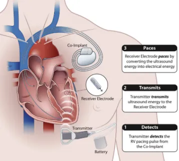
LV endocardial pacing is a potential therapy for patients who either cannot receive transvenous epicardial CRT or have not responded adequately to CRT.9 LV endocardial stimulation traditionally has been delivered via pacing leads placed transseptally, which mandates lifelong anticoagula-tion due to the risk of thromboembolic complicaanticoagula-tions. A novel wireless LV endocardial pacing system (WiSE-CRT system; EBR Systems, Sunnyvale, CA) delivers electrical stimulation to the LV endocardial surface of the heart by transducing acoustic energy from an ultrasound (US) pulse generator implanted subcutaneously in an intercostal space (Figures 1and2).10The US waves are converted to electrical stimulation energy by a small receiver electrode deployed percutaneously into the LV cavity. The receiver electrode is fully endothelialized after four weeks, avoiding the need
for long-term anticoagulation. The stimulation is triggered by right ventricular (RV) pacing, resulting in near simulta-neous (w2–5 ms) LV and RV endocardial activation. Pa-tients must be prescreened using a combination of linear and cardiac US (echocardiography) to ensure successful delivery of consistent LV endocardial pacing from the most efficient location.
The system has undergone a first-in-man study evalua-tion.11 More recently, in the nonrandomized SELECT-LV (Safety and Performance of Electrodes implanted in the Left Ventricle) study, 35 patients across 6 centers were im-planted, and promising results were seen in terms of clinical response and LV reverse remodeling.12Previous work also evaluated the possibility of identifying and targeting the optimal pacing site.13The WICS-LV Post Market Surveil-lance Registry (ClinicalTrials.gov Identifier: NCT02610673) was undertaken to assess the safety and ef fi-cacy of the WISE-CRT system in a real-world setting. The device is CE (Conformité Européenne [French for European Conformity]) marked in Europe and is indicated for patients who are unable to receive conventional CRT or who are non-responders to CRT.
Methods
Data collection
The WICS-LV Post Market Surveillance Registry prospec-tively collected data from all 14 European centers implanting the WiSE-CRT System. All patients studied provided full written consent to participate. Patient data was de-identified and collected anonymously using a uniquely identifiable study number.
Inclusion criteria
The WiSE-CRT system is CE marked for 3 approved indica-tions12: (1) patients in whom LV lead deployment was not possible or had previously failed due to anatomic constraints, high capture thresholds, or phrenic nerve stimulation; (2) pa-tients undergoing an upgrade to CRT in whom implantation of an LV lead was impractical or complex due to issues with venous access or undesirable because of previous pocket infection; and (3) patients who previously were nonre-sponders to conventional transvenous epicardial CRT. Non-responders were defined as patients who had no change or had worsening of symptoms or New York Heart Association functional class after 6 months of treatment as confirmed by the treating physician. Patients were classified as having either ischemic cardiomyopathy or nonischemic cardiomy-opathy using a combination of cardiac magnetic resonance imaging, electroanatomic mapping, coronary angiography, and clinical history.
Figure 1 The WiSE-CRT pacing system.
Endpoints
Three safety and efficacy assessments were prespecified: (1) Procedural success, requiring successful implantation of all WiSE-CRT components and confirmation of biventricular pacing on a postimplant 12-lead electrocardiogram. (2) Safety of the system, evaluating acute (,24 hours), interme-diate (24 hours–1 month), and long-term (1–6 months) com-plications. Adjudication was performed by the local principal investigator at each site. (3) Clinical response to biventricular endocardial pacing, assessing the proportion of patients who experienced improvement in clinical symptoms 6 months af-ter implantation.
Clinical response
Clinical response was assessed by a clinical composite score. This simple global assessment of symptoms classified each patient into 1 of 3 categories: improved, worsened, or un-changed.
Statistical analysis
Continuous variables with a gaussian distribution are given as mean6 SD. Significance testing on continuous, normally
distributed paired data was performed using 2-tailed paired Student t tests. Significance testing on continuous, non-normally distributed paired data was performed using the Wilcoxon signed rank test. Significance testing on contin-uous, non-normally distributed unpaired data was performed using the Mann-Whitney U test. If both independent and dependent factors were categorical, significance testing was performed using thec2test. Odds ratios were calculated us-ing binary logistic regression. If both independent and depen-dent factors were continuous, odds ratios and significance testing were performed using linear regression. P,.05 was considered significant. Analysis was performed using PASW Statistics 24 (SPSS Inc, Chicago, IL).
Results
Patient characteristics
A total of 90 patients from 14 European centers were im-planted with the WiSE-CRT system (Table 1). Patients were predominantly male (80.0%) (mean age 68.26 10.5 years; mean left ventricular ejection fraction 30.6% 6 8.9%). Mean QRS duration was 180.7 6 27.0 ms, and 40.0% of patients had an ischemic etiology. In terms of
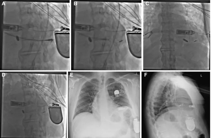
Figure 2 A: The WiSE-CRT delivery catheter across the aortic valve, with the pacing electrode advanced to the tip of the catheter and about to be deployed in the myocardium. The battery and ultrasound (US) pulse generator also are visible. B: The WiSE-CRT electrode affixed to the myocardium after release. C: Poster-oanterior view of the Micra transcatheter pacing system (TPS) (Medtronic Inc, Minneapolis, MN) and the WiSE-CRT US pulse generator and pacing electrode in situ. D: Right anterior oblique view of the Micra TPS and WiSE-CRT US pulse generator, battery, and pacing electrode. E: Posterior chest radiograph showing the WiSE-CRT system in situ in combination with the Micra TPS. F: Lateral chest radiograph showing the WiSE-CRT system in situ in combination with the Micra TPS.
indication, 48.9% of patients had failed conventional transve-nous CRT implant, 37.8% were deemed a complex upgrade, and 13.3% previously were nonresponders to epicardial CRT. Four patients (4.4%) withdrew from the registry after undergoing implantation with the system, so no follow-up data for these patients were recorded. The majority of patients (n5 82) were implanted via the femoral artery using a retro-grade transaortic approach as previously described.10In a mi-nority of patients in whom issues with arterial patency, tortuosity, or aortic valve disease/replacement precluded a retrograde approach (n 510), a novel transseptal approach was used.14
Procedural success
Confirmation of biventricular pacing
Biventricular pacing was confirmed by postimplantation 12-lead electrocardiogram in 85 of 90 patients (94.4%). In 5 cases (5.6%), consistent capture of the LV endocardial pac-ing electrode could not be achieved. Failure of the screenpac-ing process to exclude 2 unsuitable patients was later confirmed: thefirst patient had comorbid chronic obstructive pulmonary disease with significant lung encroachment affecting the US signal between the subcutaneous transmitter array and LV endocardial pacing electrode; and the second patient had a distance between the LV pacing electrode to US array that was too large (.13 cm) to allow achievement of consistent
capture. One patient never received an LV pacing electrode due to periprocedural tamponade, which led to the implant procedure being aborted. One patient experienced subopti-mal device functionality after the US transmitter displaced due to improper initialfixation, and revision with a new US transmitter resolved the issue. In thefinal patient, the elec-trode was believed to be implanted in myocardial scar, and delivery of LV stimulation resulted in intermittent capture.
Safety and complications
Acute complications
Procedural acute complications (,24 hours) occurred in 4 patients (4.4%) (Table 2). Two patients experienced cardiac tamponade after electrode placement, and 1 of these patients died 4 days later (see section on Intermediate complications). Two patients had complications related to US transmitter array/battery placement (1 pneumothorax, 1 pleural effu-sion). Neither patient required intervention, and both resolved with conservative management. No electrode embo-lizations occurred.
Intermediate complications
Intermediate complications (24 hours–1 month) occurred in 17 patients (18.8%), including 1 patient death that occurred 4 days postimplant, which was attributed to an acute LV perforation that resulted in cardiac tamponade. The most common adverse events were related to femoral arterial ac-cess in 4 patients (4.4%): 1 femoral hematoma that resolved with conservative management and 3 femoral artery pseu-doaneurysms, 2 of which required surgical intervention. Four patients (4.4%) had transmitter/battery pocket hema-tomas, and 3 patients (3.3%) had transmitter/generator pocket infection. Three patients (3.3%) had postprocedural lower respiratory tract infection, and 2 patients had postprocedural acute kidney injury.
Table 1 Patient characteristics
No. of patients 90
Age (y) 68.26 10.5
Male 72 (80.0)
ICM etiology 36 (40.0) NYHA functional class
I 1 (1.1) II 33 (36.7) III 56 (62.2) IV 0 (0) Echocardiographic data LVEF (%) 30.66 8.9 LVESV (ml) 130.46 78.5 LVEDV (ml) 185.76 93.0 ECG Atrial arrhythmia 47 (52.2) QRS duration (ms) 180.76 27.0 RV paced morphology 81 (90.0) LBBB morphology 6 (6.7) BiV paced morphology 3 (3.3) Indication
Failed LV lead implant 44 (48.9) Complex upgrade 34 (37.8) Failure to respond to CRT 12 (13.3)
Values are given as n (%) or mean6 SD unless otherwise indicated. BiV5 biventricular; ECG 5 electrocardiography; ICM 5 ischemic cardio-myopathy; LBBB5 left bundle branch block; LV 5 left ventricle; LVEDV 5 left ventricular end-diastolic volume; LVEF5 left ventricular ejection frac-tion; LVESV5 left ventricular end-systolic volume; NYHA 5 New York Heart Association; RV5 right ventricle.
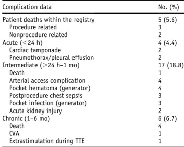
Table 2 Patient safety outcome data
Complication data No. (%) Patient deaths within the registry 5 (5.6)
Procedure related 3 Nonprocedure related 2 Acute (,24 h) 4 (4.4) Cardiac tamponade 2 Pneumothorax/pleural effusion 2 Intermediate (.24 h–1 mo) 17 (18.8) Death 1
Arterial access complication 4 Pocket hematoma (generator) 4 Postprocedure chest sepsis 3 Pocket infection (generator) 3 Acute kidney injury 2 Chronic (1–6 mo) 6 (6.7)
Death 4
CVA 1
Extrastimulation during TTE 1
CVA5 cerebrovascular accident; TTE 5 transthoracic echocardiography
Chronic complications
Chronic complications (1–6 months) occurred in 6 patients, including 4 deaths (2 believed to be procedure/device related). One patient with persistent atrialfibrillation treated with apixaban suffered a cerebellar infarct 5 months postim-plant. Given that the receiver electrode endothelializes within 4–6 weeks, this complication was not believed to be procedure or device related. Stimulation of the WiSE-CRT system during echocardiography was observed in 1 patient at the 6-month follow-up visit. The receiver elec-trode had been positioned in an LV apical position. The sonographer observed a self-terminating run of ectopic ven-tricular extrasystoles while obtaining an apical 4-chamber view. The run of ectopic beats stopped immediately after the probe was withdrawn, and the patient reported no symptoms.
Learning curve and success/complications
Most of the complications (76%) occurred within a center’s first 10 cases, suggesting an initial learning curve when using this technology.
Patient deaths
Five patients (5.6%) died within 6 months of implantation. Three deaths (3.3%) were adjudicated to be procedure/device related, 2 of which were related to LV perforation. Details of patient deaths are given in theSupplemental Information— Patient Deaths.
CRT response
Four patients who were lost to follow-up were excluded from the chronic response analysis. All 86 patients who received CRT at 6 months had data evaluating their clinical response. Sixty patients (69.8%) reported an improved clin-ical composite score; 12 (14.0%) had no change in compos-ite score; and 4 (4.7%) had a worsened composcompos-ite score. The group of 86 patients included 5 who experienced inconsis-tent LV endocardial stimulation and 5 who died during follow-up. Comprehensive echocardiographic study of LV volumes to assess remodeling was available for 43 patients and can be reviewed in the Supplemental Information— Echocardiographic RemodelingandSupplemental Table 1.
Discussion
The WICS-LV Post Market Surveillance Registry is the largest experience to date of this novel technology to achieve leadless LV endocardial pacing. The registry was designed to assess the procedural success, safety, and long-term efficacy of leadless LV endocardial pacing in a real-world setting of 14 European centers.
The principal findings were as follows. (1) The WiSE-CRT system achieved procedural success, with biventricular endocardial pacing confirmed in .94% of patients. (2) Device/procedural-related adverse events occurred in a sig-nificant number of patients. Three procedural deaths (3.3%) occurred; 4.4% of patients had a procedural complication
within 24 hours of the procedure; 18.8% had a complication between 24 hours and 1 month after the procedure; and 6.7% had a complication between 1 and 6 months after the proced-ure. (3) At 6 months, the system was associated with a favor-able clinical response rate of 70%.
Comparison with previous studies
The WiSE-CRT Post Market Surveillance Registry repre-sents the largest evaluation of this novel pacing technology and establishes that real-world use of the WiSE-CRT LV endocardial pacing system is effective. It has a complication profile and response rate similar to those seen in the SELECT-LV study,12a prospective multicenter nonrandom-ized trial that assessed the safety and performance of the WiSE-CRT system in 35 patients. Similar to the current study, SELECT-LV reported successful implantation of the system in 97% of cases. Similar rates of clinical response of 84.8% and 70% of patients reporting an improvement in clinical composite score were observed in the SELECT-LV study and the WiSE-CRT registry, respectively.
Both studies identified a significant complication rate. No episodes of cardiac tamponade were observed in the SELECT-LV trial; however, 2 patients (2.2%) were identi-fied in the WiSE-CRT registry and subsequently died. Serious acute (,24 hours) procedure/device-related events occurred in 4 patients (8.6%) in the SELECT-LV study compared to 4.4% in the WiSE-CRT registry. A serious procedure/device-related event occurred between 24 hours and 1 month after the procedure in 22.3% of patients in the SELECT-LV trial, with a similar rate of events (18.8%) in the WiSE-CRT registry. In the SELECT-LV trial 1 procedure-related death (2.8%) following VF at the time of electrode placement was reported, whereas in the WiSE-CRT registry 5 patient deaths (5.6%) occurred within a 6-month period, 3 (3.3%) of which were believed to be proced-ure related. The rate of perforation in the current registry has improved compared to the initial iteration of the device, which had an 18% tamponade rate and led to redesign of the delivery catheter.10
A lead-based system for endocardial CRT was previously analyzed in the ALSYNC (ALternate Site Cardiac Re-sYNChronization) study.9 This prospective study enrolled 138 patients who either had a failed attempt at conventional transvenous epicardial CRT or were unsuitable to undergo bi-ventricular resynchronization pacing. The primary study objective was to investigate the safety at 6 months of deliv-ering LV endocardial pacing via a lead placed across the atrial septum. The implant success rate was 89.4%, which is consistent with our registryfindings. Freedom from com-plications meeting the definition of primary endpoint was 82.2% at 6 months. Fourteen transient ischemic attacks (9 pa-tients [6.8%]), 5 nondisabling strokes (5 papa-tients [3.8%]), and 23 deaths (17.4%) were reported. No death resulted from a primary endpoint complication. At 6 months, New York Heart Association functional class improved in 59% of pa-tients. Therefore, the current registry has a death rate at
follow-up lower than that observed in the ALSYNC study and a significantly reduced risk of cerebrovascular events.
Vascular access complications
The most common procedural complications were related to femoral arterial access, as the WiSE-CRT system was de-signed to allow deployment of the pacing electrode in the LV cavity via retrograde aortic access from the femoral ar-tery. This procedure requires large-bore 12F femoral arterial access and closure, skills not commonly required by prac-ticing electrophysiology/complex device specialists who tend to implant this system. Three approaches have been devised to reduce the rate of femoral arterial access compli-cations. First, vascular closure devices were used at 12 cen-ters to minimize bleeding and ensure effective arterial closure at the end of the procedure. Eleven centers used the Perclose ProGlide, Abbott Vascular (Santa Clara, California) vascular suture system. One center used the Prostar XL (Abbott Vascular) percutaneous vascular surgical system to assist vascular hemostasis. Vascular closure systems have been shown to reduce time to hemostasis.15
Second, several operators have used pre- and periproce-dural imaging to guide arterial access. Five centers used real-time US guidance to identify the femoral artery and visu-alize guidewire deployment. At one center, a contralateral femoral puncture was performed in order to facilitate an ipsi-lateral femoral angiogram before obtaining large-bore femoral access. Third, use of a transseptal approach permitting elec-trode deployment in the LV endocardium after initial femoral venous access has been shown to be possible.14In a series of 10 cases, this approach obviated the requirement for femoral arterial access, achieving successful electrode deployment without a single groin or thromboembolic complication.16 This approach may be preferable among electrophysiologists who are familiar with performing transseptal punctures.
US extrastimulation
The WiSE-CRT receiver electrode typically is not sensitive to routine US imaging, but in rare instances extrastimulation of the device is possible. In our series, this event was observed in only 1 case. However, this is an importantfinding given the widespread use of ultrasonic imaging in medicine and specifically within the field of cardiology. Apical place-ment of the LV pacing electrode seems to increase the poten-tial for extrastimulation, particularly while obtaining an apical 4-chamber view with transthoracic echocardiography, which significantly reduces the distance between the receiver electrode and the alternate US source. Other risk factors for extrastimulation include the use of high-power settings, low-frequency US (typically utilized during harmonic imag-ing), and thin body habitus. In order to avoid inadvertent extrastimulation of the pacing electrode, all patients are issued a medical device identification card stating they have a US-sensitive pacemaker. For cases in which apical deployment of the pacing electrode has been confirmed, an alternative echocardiography protocol has been developed
and shown to successfully prevent further extrastimulation. This document forms part of the clinical trial documents for the SOLVE CRT Clinical Trial (ClinicalTrials.gov Iden-tifier: NCT02922036). Echocardiography Acquisition and Transfer Manual: The SOLVE-CRT Trial (Version 5.0, 19 Dec 2019).
Study limitations
The main limitation of this current analysis is that it is a reg-istry and therefore is limited by the inherent constraints of a nonrandomized study. Importantly, neither patients nor in-vestigators were blinded, and all suitable patients enrolled in the WiCS-LV registry were implanted with the system. Although LV reverse remodeling was selected as an endpoint, other endpoints included the subjective clinical response, which may be more open to bias. A randomized, double-blinded evaluation of the WiSE-CRT system is currently underway (Stimulation Of the Left Ventricular Endocardium for Cardiac Resynchronization Therapy in Non-Responders and Previously Untreatable Patients (SOLVE CRT; ClinicalTrials.gov Identifier: NCT02922036). This study hopefully will provide an unbi-ased assessment of symptomatic and echocardiographic response associated with endocardial pacing.
Unlike conventional transvenous epicardial CRT, the WiSE-CRT system is unable to determine the percentage of effective CRT pacing. When interrogating the WiSE-CRT system using the pacing system analyzer, the number of “tracked” RV pacing signals that result in generation of a US wave can be evaluated but not whether the US signal re-sulted in successful LV endocardial capture. LV capture can only be confirmed by assessing the paced QRS morphology for successful biventricular paced morphology. Holter moni-toring can be used to confirm consistent biventricular capture when required.
Finally, comprehensive LV volume data for all patients were not available for data analysis. Comprehensive echocar-diographic study of LV volumes to assess remodeling was available for 43 patients. Fourteen patients were excluded from chronic response analysis due to failure to achieve endocardial stimulation in 5, death during follow-up in 5, and loss to follow-up in 4. LV volumetric assessment either was not performed or was of insufficient quality to allow for meaningful data analysis in 33 patients.
Conclusion
The WICS-LV Post Market Surveillance Registry is the largest series of leadless LV endocardial pacing to date. Importantly it demonstrates effective real-world use with a response rate similar to those of previous nonrandomized studies, with nearly 70% of patients reporting an improve-ment in clinical symptoms. Response rates are equivalent to those of lead-based biventricular endocardial pacing sys-tems while largely eliminating the risk of thromboembolic stroke.9Implantation of the system can be associated with a significant complication rate, but complications will occur
less frequently as operators gain more experience with the de-livery system. The risk of cardiac tamponade is in keeping with other left-sided vascular procedures such as left atrial appendage occlusion,17and given the risk of cardiac tampo-nade, which may require urgent repair, implantation should be performed at centers with on-site cardiothoracic surgical facilities. Our results suggest that endocardial CRT pacing with this novel pacing system is effective treatment for a group of high-risk patients with heart failure who either cannot receive or who have not responded to conventional CRT. Implantation of the system should be performed by adequately trained operators at centers having experience with vascular and cardiothoracic complications.
Acknowledgments
The authors would like to acknowledge Emma Perchard for her contribution to this work. The authors would like to thank Professor Pascal Defaye for providing the implantation im-ages of the WiSE-CRT system used in this article.
Appendix
Supplementary data
Supplementary data associated with this article can be found in the online version athttps://doi.org/10.1016/j.hrthm.2020. 03.002.
References
1. Petersen S, Rayner M, Wolstenholme J. Coronary Heart Disease Statistics: Heart Failure Supplement. London: British Heart Foundation; 2002.
2. Neeland IJ, Kontos MC, de Lemos JA. Evolving considerations in the manage-ment of patients with left bundle branch block and suspected myocardial infarc-tion. J Am Coll Cardiol 2012;60:96–105.
3. Nelson GS, Berger RD, Fetics BJ, et al. Left ventricular or biventricular pacing improves cardiac function at diminished energy cost in patients with dilated cardiomyopathy and left bundle-branch block. Circulation 2000;102:3053–3059.
4. Cleland JGF, Daubert JC, Erdmann E, et al. Cardiac Resynchronization-Heart Failure (CARE-HF) Study Investigators. The effect of cardiac
resynchroniza-tion on morbidity and mortality in heart failure. N Engl J Med 2005; 352:1539–1549.
5. European Society of Cardiology (ESC). European Heart Rhythm Association (EHRA), Brignole M, et al. 2013 ESC guidelines on cardiac pacing and cardiac resynchronization therapy: The Task Force on cardiac pacing and resynchroni-zation therapy of the European Society of Cardiology (ESC). Developed in collaboration with the European Heart Rhythm Association. Europace 2013; 15:1070–1118.
6. Yu C-M, Bleeker GB, Fung JW-H, et al. Left ventricular reverse remodeling but not clinical improvement predicts long-term survival after cardiac resynchroniza-tion therapy. Circularesynchroniza-tion 2005;112:1580–1586.
7. Gamble JHP, Herring N, Ginks M, Rajappan K, Bashir Y, Betts TR. Procedural success of left ventricular lead placement for cardiac resynchronization therapy. JACC Clin Electrophysiol 2016;2:69–77.
8. Abu-El-Haija B, Bhave PD, Campbell DN, et al. Venous stenosis after transve-nous lead placement: a study of outcomes and risk factors in 212 consecutive pa-tients. J Am Heart Assoc 2015;4:e001878.
9. Morgan JM, Biffi M, Gellér L, et al. ALternate Site Cardiac ResYNChronization (ALSYNC): a prospective and multicentre study of left ventricular endocardial pacing for cardiac resynchronization therapy. Eur Heart J 2016;37:2118–2127. 10. Auricchio A, Delnoy PP, Regoli F, Seifert M, Markou T, Butter C. First-in-man
implantation of leadless ultrasound-based cardiac stimulation pacing system: novel endocardial left ventricular resynchronization therapy in heart failure pa-tients. Europace 2013;15:1191–1197.
11. Auricchio A, Delnoy PPP-P, Butter C, et al. Feasibility, safety, and short-term outcome of leadless ultrasound-based endocardial left ventricular resynchroniza-tion in heart failure patients: Results of the Wireless Stimularesynchroniza-tion Endocardially for CRT (WiSE-CRT) study. Europace 2014;16:681–688.
12. Reddy VY, Miller MA, Neuzil P, et al. Cardiac resynchronization therapy with wireless left ventricular endocardial pacing: the SELECT-LV study. J Am Coll Cardiol 2017;69:2119–2129.
13. Sieniewicz BJ, Behar JM, Gould J, et al. Guidance for optimal site selection of a leadless left ventricular endocardial electrode improves acute hemody-namic response and chronic remodeling. JACC Clin Electrophysiol 2018; 4:860–868.
14. Sieniewicz BJ, Gould J, Rimington HM, Ioannou N, Rinaldi CA. Transseptal de-livery of a leadless left ventricular endocardial pacing electrode. JACC Clin Elec-trophysiol 2017;3:1333–1335.
15. Nasu K, Tsuchikane E, Sumitsuji S; PARADISE Investigators. Clinical effective-ness of the Prostar XL suture-mediated percutaneous vascular closure device following PCI: results of the Perclose AcceleRated Ambulation and DISchargE (PARADISE) Trial. J Invasive Cardiol 2003;15:251–256.
16. James S, Rinaldi CA, Turley AJ, et al. First-in-man implantation of a leadless endocardial left ventricular pacing system (WiSE-CRT) utilising a trans-septal approach. EP Europace 2018;20(Suppl 4):iv16.
17. Schmidt B, Betts TR, Sievert H, et al. Incidence of pericardial effusion after left atrial appendage closure: the impact of underlying heart rhythm—data from the EWOLUTION study. J Cardiovasc Electrophysiol 2018;29:973–978.