ORIGINAL ARTICLE
Whole-body CT in polytrauma patients: effect of arm
positioning on thoracic and abdominal image quality
Christoph Karlo&Ralph Gnannt&Thomas Frauenfelder&Sebastian Leschka&
Martin Brüesch&Guido A. Wanner&Hatem Alkadhi
Received: 26 January 2011 / Accepted: 4 March 2011 / Published online: 7 April 2011 # Am Soc Emergency Radiol 2011
Abstract The purpose of this study is to assess the influence of different arm positioning techniques on thoracic and abdominal image quality and radiation dose of whole-body trauma CT (wbCT). One hundred and fifty polytrauma patients (104 male, mean age 47±19) underwent wbCT with arms elevated above the head (group A, n=50), alongside the abdomen (group B, n=50), and on a pillow ventrally to the chest with both arms flexed (group C, n=50). Two blinded, independent observers measured image noise and rated image quality (scores 1–3) of the liver, aorta, spleen, spine, and lower lungs. Radiation dose parameters were noted, and the abdomens’ anterior–posterior diameter and scan lengths were measured. Interreader agreements for image noise (r= 0.86; p<0.001) and subjective image quality (k=0.71–0.84) were good. Noise was lower (p<0.05), image quality of the liver, aorta, spleen, and spine was higher, and radiation dose lower in group A than in groups B and C (p<0.001, each). Image quality of the spleen, liver, and aorta were higher in
group C than in group B (p<0.05, each). No significant differences in scan length (p=0.61) were found among groups. Abdominal anterior–posterior diameter correlated significantly with noise (r=0.82; p<0.01) and dose (r=0.47; p<0.001). Estimated effective radiation doses were signifi-cantly (p<0.001) higher in groups B (21.2 mSv) and C (21.9 mSv) as compared to A (16.1 mSv). In wbCT for polytrauma patients, positioning of the arms above the head results in better image quality and lower radiation dose. Placing the flexed arms on a large pillow ventrally to the chest significantly improves image quality as compared to positioning alongside the abdomen.
Keywords Whole-body computed tomography . Trauma . Arm positioning . Radiation dose
Introduction
Multi-detector-row computed tomography (CT) is today considered the most important imaging technique in the diagnostic work-up of polytrauma patients [1–5]. This can be explained by recent technical improvements in CT scanner hard- and software delivering high-quality images of various body parts within a short time [6–10]. While several studies so far have demonstrated the technical feasibility of whole-body CT introducing various protocols and contrast media application techniques [2, 11, 12], the exact role and clinical value of a whole-body CT in terms of patient outcome as compared to a CT of dedicated body regions is still a matter of debate. A recent multi-center trial suggested that the integration of whole-body CT into early trauma care significantly increases the probability of survival in polytrauma patients when compared to those undergoing non-whole-body CT [3].
C. Karlo
:
R. Gnannt:
T. Frauenfelder:
H. Alkadhi (*) Institute for Diagnostic and Interventional Radiology, University Hospital Zurich,Raemistrasse 100, 8091 Zurich, Switzerland e-mail: [email protected] S. Leschka
Institute of Radiology, General Hospital St. Gall, St. Gallen, Switzerland
M. Brüesch
Institute of Anaesthesiology, University Hospital Zurich, Zurich, Switzerland
G. A. Wanner
Department of Surgery, Division of Trauma Surgery, University Hospital Zurich,
The downside of whole-body CT represents the relative-ly high radiation exposure, which has led to further debates about the value of this approach especially in younger patients [2]. One aspect in regard to radiation exposure represents the positioning of the arms during whole-body CT [13,14]. The effect of arm positioning on radiation dose has been recently evaluated by Brink et al. [13] and Bayer et al. [14]. Both groups demonstrated that placing the arms above the head results in a reduction of radiation exposure and an improvement in image quality. However, a consid-erable number of patients are not able to position their arms above the head due to traumatic injuries of the shoulder girdle or due to multiple intensive care installations. In addition, positioning of the arm above the head might be time consuming, resulting in a delay in diagnosis and patient management with potentially fatal consequences [7,
15, 16]. Finally, arm positioning alongside the torso has been shown to reduce image quality with beam-hardening artifacts in the dorsal regions of the liver, spleen, and kidneys [4,12–15,17], eventually rendering the CT study non-diagnostic.
Thus, there is interest in a refinement of arm positioning techniques in polytrauma patients who are not able to raise their arms, with the aim to maintain the diagnostic yield of the examination.
The purpose of this study was to evaluate image quality and radiation dose estimates of patients undergo-ing whole-body CT with different arm positionundergo-ing. In addition, we introduce a positioning technique with arms being flexed in the elbow and placed over a large pillow ventrally on the chest.
Materials and methods
Patients and positioning during CT
Between June 1st and August 6th 2010, a total of 142 polytrauma patients (46 female, 104 male, mean age 47± 19, age range 20–90) were referred to our hospital’s emergency room to receive a whole-body CT study. Of those, 42 were studied with either the left or right arm next to the body due to upper extremity injuries, leaving 100 patients who were scanned with either both arms above the head (n=50, group A) or both arms alongside the body (n= 50, group B). From August 7th on, we revised the positioning of the arms of the polytrauma patients under-going whole-body CT by placing a large pillow (height of the pillow=15 cm) ventrally on the body at the level of the lower chest. Both arms were flexed in the elbow, and the forearms were positioned next to each other on top of the pillow. This positioning technique was intended for patients in whom re-positioning of the arms between the head/
cervical spine CT and chest/abdominal CT was considered too time consuming, potentially delaying diagnosis and therapy, or for patients where raising both arms was contraindicated by medical reasons. Care was taken to place the arms not on the level of the upper abdomen but on the chest. Positioning of the arms like that was performed during bedding of the patient on the CT table and during preparation of the scan by the technologist, causing no additional delay in the diagnostic work-up.
Figure 1 illustrates the arm positions above the head, alongside the body, and on the pillow ventrally to the chest. For reasons of comparability to the other groups, we also included in this study 50 consecutive patients with the new arm positioning approach (group C), scanned between August 7th and September 30th. The study was approved by the local ethics board; written informed consent was waived.
Data acquisition
All examinations were performed using a dual-source CT scanner (Somatom Definition; Siemens Healthcare, Forchheim, Germany). All patients were placed in supine position and feet first on the CT table.
Our institutional whole-body CT trauma protocol, which was applied in all 150 patients, consists of the following components: (1) a non-enhanced CT scan of the head; (2) a non-enhanced CT scan of the neck, from the base of the skull to the level of the second thoracic vertebra; (3) a contrast-enhanced CT scan of the thorax, from the level of the sixth cervical vertebra to the base of the lung; and (4) a contrast-enhanced scan of the abdomen from the level of the dome of the diaphragm to the level of the lesser trochanter. Except for the patients in group A, where the arms were placed above the head after the CT scan of the head and neck, all arm positioning remained the same during the whole examinations of patients in groups B and C.
The following scan and reconstruction parameters were kept identical for all thoraco-abdominal data acquisitions in all examinations: tube voltage 120 kV; reference tube current–time product of 200 mAs for the thorax and of 250 mAs the abdomen, using attenuation-based tube current modulation (CareDose 4D; Siemens); pitch 1.5; slice colli-mation 32 mm×0.6 mm, slice acquisition 64 mm×0.6 mm, gantry rotation time 330 ms; reconstruction slice thickness, 2 mm; reconstruction increment, 1.6 mm.
In all patients, a 100-ml bolus of iso-osmolar, non-ionic iodinated contrast material [300 mg iodine/ml, Iopromide (Ultravist 300; Bayer Schering Pharma)] followed by a saline flush of 40 ml was injected into an antecubital vein at a flow rate of 3 ml/s. Six seconds after the attenuation in the descending thoracic aorta reached the predefined threshold of 120 Hounsfield Units (HU), the thoracic data acquisition
was initiated. This delay allowed the patient to receive breathing instruction (if possible). After a further delay of 45 s, the abdominal, portal-venous enhanced phase was acquired.
All reconstructed images were archived in the hospital’s picture archive and communication system (PACS) for further image analysis and documentary purposes.
Data analysis
All data were anonymized and displayed in random order to two radiologists experienced in trauma imaging. They were blinded to each other’s results and to the arm positioning of all patients. This was achieved by cropping all images of the thorax and abdomen to display only the anatomical
Fig. 1 a Representative topograms illustrating the three arm posi-tioning techniques used in this study (from left to right): group A (arms raised above the head), group B (arms positioned alongside the body), and group C (arms located on a pillow ventrally to the chest). b Left—photograph showing the arm positioning technique in group C,
with both arms flexed in the elbow and located on a pillow ventrally to the chest, thus sparing the upper abdomen from possible beam-hardening artifacts. c Right—axial CT image in lung window settings illustrating the positioning of the arms on a pillow (asterisk) ventrally to the chest
regions of interest by a third radiologist who was not involved in image analysis.
Objective image quality
Both radiologists placed a region of interest (ROI) with a diameter of 1 cm in the right lobe of the liver at the level of the right portal vein in all patients (i.e., liver segment VI or VII), excluding focal liver lesions and major hepatic vessels, as previously shown [13]. The mean of the standard deviation of the CT number measurement in Hounsfield Units (HU) determined the image noise. In addition, the anterior-to-posterior diameter (in centimeters, from skin to skin) of all patients at the level of the right portal vein in the median line was measured to obtain a realistic measure of the patients body constitution at the level of the upper abdomen. Subjective image quality
Both observers were independently asked to assess the subjective image quality of the liver, spleen, aorta, the thoraco-lumbar spine (i.e., from the sixth thoracic to the third lumbar vertebra), and the base of the lungs on a three-point grading scale as follows:
▪ grade 1=excellent image quality, no artifacts; ▪ grade 2=diagnostic image quality, minor artifacts; ▪ grade 3=non-diagnostic image quality, severe artifacts. Reasons for non-diagnostic image quality were noted as being caused by artifacts from the arms, from installations and/or trauma board, image noise, or from motion (patient movement or breathing).
Representative imaging examples demonstrating the range of image quality scores in the liver are given in Fig.2. Radiation dose estimates
For each whole-body CT study, the dose–length product (DLP) was collected from the protocol that summarizes all relevant data generated from the CT scanner. The DLP represents the integrated radiation dose imparted by all sections of a CT examination [18]. The effective dose was estimated by using a method proposed by the European Working Group for Guidelines on Quality Criteria in CT and was derived by using the dose–length product and a conversion coefficient k [18]. The conversion coefficient is averaged between male and female models in Monte Carlo simulations [18]. This technique is used to determine doses to specific organs by simulating the X-ray absorption and scattering in various tissues with use of a mathematical model. Therefore, the effective dose is an estimate of the dose to patients during an ionizing radiation procedure and enables one to perform a direct comparison with other
sources of radiation exposure by measuring the total amount of energy entering the body and taking into account the different sensitivities of the irradiated organs [18]. Since a combination of thoracic (kthor=0.014 mSv/[mGy×cm]) and
abdominal (kabd=0.015 mSv/[mGy×cm]) acquisitions was
performed, the mean of both region-specific conversion coefficients (k=0.0145 mSv/[mGy×cm]) was used. The method of calculating the effective dose has been shown to have reasonable robustness and be highly consistent [19]. Examination time
Examination time was calculated as the difference between the time of acquisition of the most cranial image of the CT
Fig. 2 Subjective image quality scores 1 (excellent, A), 2 (moderate, B), and 3 (non-diagnostic, C) as illustrated by representative axial CT images through the liver
of the neck and the time of acquisition of the most caudal image of the CT of the abdomen, at the level of the lesser trochanter. The time needed to scan the head and mid-face was not included in these calculations since head and mid-face were performed with similar protocols in all three groups.
Scan length
The scan length was calculated as the difference between the table position of the most cranial image and the table position of the most caudal image of the thoracic and abdominal data sets in each patient. The total scan length was calculated by adding the scan lengths of the thoracic and abdominal data acquisitions.
Statistical analysis
Categorical variables were expressed as frequencies and percentages. Numerical variables were expressed as means± standard deviations. Variables were assessed for normal distribution using the Kolmogorov–Smirnov test.
Interreader agreements concerning subjective image quality assessments were analyzed with kappa statistics and interpreted as follows: κ values of 0.00 to 0.20 were considered to indicate poor agreement;κ values of 0.21 to 0.40, fair agreement; κ values of 0.41 to 0.60, moderate agreement;κ values of 0.61 to 0.80, high agreement; and κ values of 0.81 to 1.00, excellent agreement.
Interreader agreements regarding objective image quality assessments (i.e., image noise measurements) were assessed using intraclass correlation coefficients (ICC).
One-way Anova and Kruskal–Wallis analyses were
performed to test for statistically significant differences in patient’s age, a-p diameter, DLP, gender, objective image quality, and examination times among the three groups, whereas Mann–Whitney analyses were performed to test for statistically significant differences in DLP, examination times as well as subjective and objective image quality scores between the three groups (i.e., group A vs. B, group B vs. C, and group A vs. C).
Pearson correlation analyses were performed to test the correlation between the a-p diameter and the objective image quality in all three groups separately. Spearman rank order correlation was used to test the correlate between the a-p diameter and the subjective image quality in all three groups separately.
A p value <0.05 was considered to indicate statistical significance.
All statistical analyses were conducted using commer-cially available software (SPSS, release 18.0; SPSS, Chicago, IL, USA).
Results
Patient demographics
There were no significant differences in age, gender, and a-p diameter among all three patient groups (each, p>0.5). Imaging findings
Imaging findings included fractures of the spine, pelvis or ribs (n=46), traumatic lesions of the liver or spleen (n=32), aortic rupture (n=2), and lung contusions or lacerations (n= 14). Traumatic lesions of the skull or brain were present in 32 patients. In 24 patients, no traumatic lesions were detected.
Objective image quality
The interreader agreement was excellent for image noise measurements in the right liver lobe (r=0.92; p<0.001). Thus, the mean of measurements from both readers was used for further statistical analyses.
Mean image noise was 18±4 HU (range, 11–27 HU) in group A, 21±6 HU (range, 14–49 HU) in group B, and 20± 5 HU (range, 10–35 HU) in group C. Significant differences were found between groups A and B (p<0.005), between groups A and C (p<0.05) but not between groups B and C (p=0.24). A significant correlation between image noise and a-p diameter was found in all groups (r=0.42–0.82, p<0.05). Subjective image quality
Interreader agreements for subjective image quality assess-ments ranged from high to excellent with kappa (κ) values of 0.84 (liver), 0.71 (spleen), 0.75 (aorta), 0.81 (spine), and 0.75 (base of the lungs). Subjective image quality results are summarized in Table 1.
In group A, excellent image quality of all evaluated anatomical structures was present in 42% (21/50) of the patients while diagnostic image quality was found in 50% (25/50). Four patients (8%) were graded as having a non-diagnostic image quality of the liver (n=4), lungs (n=3), and spleen (n=3) due to severe artifacts caused by patient movement.
In group B, excellent image quality of all evaluated anatomical structures was present in 2% (1/50) of the patients while diagnostic image quality was found in 76% (38/50). Eleven patients (22%) showed a non-diagnostic image quality of the liver (n=4) and spleen (n=7) due to severe beam hardening artifacts from the arms (n=7) or patient movement (n=4).
In group C, excellent image quality of all evaluated anatomical structures was present in 10% (5/50) of the
patients while diagnostic image quality was found in 72% (36/50). Non-diagnostic image quality was found in 20% of the patients (10/50), most often in the liver (n = 6) and spleen (n = 4), and was caused by beam hardening artifacts from the arms (n = 7) and patient movement (n = 3).
The subjective image quality of the spleen, liver, aorta, and spine was graded significantly higher in group A when compared to group B (each, p < 0.001) and group C (each, p < 0.001) as shown in Fig. 3. Additionally, subjective image quality of the spleen (p < 0.05), liver (p < 0.05), and aorta (p < 0.05) was graded significantly higher in group C when compared to group B. No significant differences in subjective image quality were found for the bases of the lungs between groups A and B (p = 0.20), between groups A and C (p = 0.12), and between groups B and C (p = 0.76) as well as for the spine between groups B and C (p = 0.84).
Subjective image quality and a-p diameter
The correlations between the a-p diameter and the subjec-tive image quality scores were not statistically significant for all examined locations in all groups (p=0.14–0.85).
Estimated radiation dose
All radiation dose parameters are summarized in Table 2. The DLP and the estimated effective radiation dose were significantly lower in group A when compared to groups B and C (p < 0.001, each), but not between groups B and C (p = 0.27).
Examination time and scan length
Mean examination time was 390 s in group A, 400 s in group B, and 354 s in group C. Mean scan length was 83.9 cm in group A, 83.1 cm in group B, and 85.9 cm in
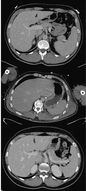
Fig. 3 Axial CT images demonstrating excellent image quality with both arms located on the pillow ventrally to the chest (a), moderate image quality with both arms placed alongside the body (b; note the artifacts in the left liver lobe), and excellent image quality with both arms elevated above the head (c)
Table 1 Subjective image quality scores
Scores Groups p value
A B C Lung 0.252 1 30 23 22 2 19 27 27 3 1 0 1 Spleen <0.001 1 43 10 22 2 5 38 24 3 2 1 3 Spine <0.001 1 47 29 28 2 3 21 22 3 0 0 0 Aorta <0.001 1 46 16 27 2 4 31 21 3 0 3 2 Liver <0.001 1 35 1 8 2 13 39 36 3 2 10 6
1 excellent image quality, 2 moderate image quality, 3 non-diagnostic image quality
group C. There were no significant differences in scan lengths (p=0.61) and examination times (p=0.44) among all groups.
Discussion
Whole-body CT is able to provide a fast and accurate diagnostic work-up of traumatic injuries [1–5]. However, it must be considered that many trauma patients are fairly young, and care needs to be taken in designing and adapting the CT scanning protocols to minimize and optimize radiation exposure [20–23]. One issue related to radiation exposure of whole-body CT is the positioning of
the patient’s arms [13, 14]. Some authors suggested
elevating the arms above the head after scanning of the head and cervical spine, before performing thoraco-abdominal CT [4, 12, 14, 18], whereas others suggest leaving the arms alongside the body for time gain [7,15,16,
24] and prevention of iatrogenic injuries [25]. The downside of arm positioning alongside the patients torso is—besides an increase in radiation exposure—the decrease in image quality which particularly affects posterior parts of the liver and spleen [4,12–15,17].
The results from our study indicate that in case the arms cannot be raised above the head, image quality of the liver and spleen is significantly better when positioning the arms on a pillow ventrally to the chest as compared to positioning of arms alongside the body, thus sparing the upper abdomen from possible beam hardening artifacts.
Nguyen et al. [12] reported a decreased image quality of the liver, spleen, and kidneys among patients whose arms were positioned alongside the body than among those whose arms were elevated. Brink et al. [13] reported an increased image quality when positioning the arms above the head as opposed to positioning either one or both arms alongside the body. Hoppe et al. [16] showed that image quality increases when repositioning the arms above the head after head and neck CT, prior to the thoraco-abdominal data acquisition. Loupatatzis et al. [26] reported that image quality deteriorated to a major extent if the arms were positioned alongside the body during trauma CT.
Our study is in line with the studies mentioned above, by indicating the best image quality in patients with arms
raised above the head, while image quality significantly decreased when positioning the arms alongside the body. Our study furthermore introduces an arm positioning technique ventrally to the lower chest, on top of a large pillow, which might be an alternative for patients who are unable or who have contraindications to elevate their arms, providing an improved image quality particularly for the organs of the upper abdomen.
Although we were able to achieve better image quality with this novel arm positioning technique compared to placing the arms alongside the body, we observed the radiation dose to be significantly higher when the arms were on the pillow (29.8%, 6.0 mSv) or alongside the body (30.2%, 6.1 mSv), compared to positioning of the arms above the head. Brink et al. [13] reported an additional dose of 8 mSv when placing the arms alongside the body as compared to raising the arms above the head, and proposed the latter arm repositioning as a tool for optimizing the dose–benefit ratio of CT in polytrauma patients. Bayer et al. [14] showed that radiation dose significantly increases if the arms were placed alongside the body and not elevated above the head, reporting average effective dose values of 24.7 mSv (arms alongside the body) and 19.2 mSv (arms above the head), respectively. Having evaluated the data of 276 whole-body trauma CT scans, Baskerville et al. [27]
reported mean CTDIvol values from 77 to 115 mGy for
thoraco-abdominal data acquisitions. Finally, Inaba et al. [22] reported average radiation dose values of 20.7 mSv for whole-body CT. Estimated effective radiation doses of our CT protocol for the chest and abdomen estimated 16.1 mSv for patients in group A, 21.2 mSv in group B, and 21.9 mSv in group C, indicating an average increase of 5.4 mSv (corresponding to 34%) when not raising the arms above the head.
Hoppe et al. [16] reported a slightly increased scan time after repositioning the patients from head-first to feet-first on the CT table when compared to a non-repositioning protocol. In contrast, and similar to the results from Bayer et al. [14] and Nguyen et al. [12], the examination times in our study were not significantly different when re-positioning the arms above the head compared to when leaving the arms alongside the torso or on a pillow ventrally to the chest. This most probably is caused by differences in injury severity among the patients of all groups due to our
Groups
A B C p value
Scan timea(s; mean±SD) 390±173 400±230 354±150 0.44 Scan length of thoraco-abdominal CT (cm; mean±SD) 839.3±57.1 831.6±55.3 858.8±57.9 0.61 Estimated effective radiation dose (mSv; mean±SD) 16.1±5.4 21.2±7.2 21.9±5.4 <0.001 Table 2 Scan time, scan length,
and radiation dose estimates
a
Calculated from the time of acquisition of the most cranial image of the cervical spine to the time of acquisition of the most caudal image of the abdomen
non-randomized study design. Patients with less severe injury being more prevalent in group A might have been able to reposition their arms relatively fast, causing no relevant delay between cervical spine and thoracic data acquisition.
We acknowledge the following study limitations. As previously reported [13,28], the presence of typical beam-hardening artifacts in the upper abdomen might have un-blinded the arm position to the readers despite the fact that images were cropped to the region of interest before data analysis. However, due to multiple electrocardiographic leads, these artifacts were occasionally seen also in patients from other groups. Then, this study suffers from the inherent limitations of its retrospective design, including a bias in patient inclusion. Since our three groups were not randomized and injury severity was different among the groups, different impacts on examination time such as manipulations by the anesthesiologists due to patient instability and differences in injury severity might have influenced the total examination times among groups. However, differences in injury severity did not affect image quality and radiation dose estimates, being the main scope of this study. Additionally, further investigations regarding the effect of arm positioning on image quality of the head and cervical spine are required, particularly considering the use of a single-pass contrast-enhanced whole-body CT protocol.
Conclusion
Our study confirms that best image quality and lowest radiation doses can be achieved in whole-body CT of polytrauma patients with elevation of the arms above the head when performing thoraco-abdominal CT. However, if raising the arms above the head is not possible or may delay the diagnostic work-up in an inacceptable manner, positioning of the arms, flexed in the elbow, on a large pillow ventrally to the chest considerably improves image quality at similar radiation dose levels when compared to positioning the arms alongside the body.
References
1. Becker CD, Poletti PA (2005) The trauma concept: the role of MDCT in the diagnosis and management of visceral injuries. Eur Radiol 15(Suppl 4):D105–D109
2. Broder J, Warshauer DM (2006) Increasing utilization of computed tomography in the adult emergency department, 2000–2005. Emerg Radiol 13(1):25–30
3. Huber-Wagner S, Lefering R, Qvick LM et al (2009) Effect of whole-body CT during trauma resuscitation on survival: a retrospective, multicentre study. Lancet 373(9673):1455–1461 4. Leidner B, Adiels M, Aspelin P, Gullstrand P, Wallen S (1998)
Standardized CT examination of the multitraumatized patient. Eur Radiol 8(9):1630–1638
5. Novelline RA, Rhea JT, Rao PM, Stuk JL (1999) Helical CT in emergency radiology. Radiology 213(2):321–339
6. Rieger M, Sparr H, Esterhammer R et al (2002) Modern CT diagnosis of acute thoracic and abdominal trauma. Anaesthesist 51 (10):835–842
7. Ptak T, Rhea JT, Novelline RA (2003) Radiation dose is reduced with a single-pass whole-body multi-detector row CT trauma protocol compared with a conventional segmented method: initial experience. Radiology 229(3):902–905
8. Philipp MO, Kubin K, Hormann M, Metz VM (2003) Radiological emergency room management with emphasis on multidetector-row CT. Eur J Radiol 48(1):2–4
9. Kalra MK, Rizzo SM, Novelline RA (2005) Reducing radiation dose in emergency computed tomography with automatic expo-sure control techniques. Emerg Radiol 11(5):267–274
10. Kalra MK, Rizzo SM, Novelline RA (2005) Technologic innovations in computer tomography dose reduction: implications in emergency settings. Emerg Radiol 11(3):127–128
11. Latifi A, Torkzad O, Labruto F, Ullberg U, Torkzad MR (2009) The accuracy of focused abdominal CT in patients presenting to the emergency department. Emerg Radiol 16 (3):209–215
12. Nguyen D, Platon A, Shanmuganathan K, Mirvis SE, Becker CD, Poletti PA (2009) Evaluation of a single-pass continuous whole-body 16-MDCT protocol for patients with polytrauma. AJR Am J Roentgenol 192(1):3–10
13. Brink M, de Lange F, Oostveen LJ et al (2008) Arm raising at exposure-controlled multidetector trauma CT of thoracoabdominal region: higher image quality, lower radiation dose. Radiology 249 (2):661–670
14. Bayer J, Pache G, Strohm PC et al (2010) Influence of arm positioning on radiation dose for whole body computed tomog-raphy in trauma patients. J Trauma [Epub ahead of print] 15. Fanucci E, Fiaschetti V, Rotili A, Floris R, Simonetti G (2007)
Whole body 16-row multislice CT in emergency room: effects of different protocols on scanning time, image quality and radiation exposure. Emerg Radiol 13(5):251–257
16. Hoppe H, Vock P, Bonel HM, Ozdoba C, Gralla J (2006) A novel multiple-trauma CT-scanning protocol using patient repositioning. Emerg Radiol 13(3):123–128
17. Sliker CW, Mirvis SE (2007) Imaging of blunt cerebrovascular injuries. Eur J Radiol 64(1):3–14
18. Menzel H-G, Schibilla H, Teunen D (2000) European guidelines on quality criteria for computed tomography. European Commis-sion, Luxembourg
19. Morin R (1988) Monte Carlo simulation in the radiological sciences. CRC, Boca Raton
20. Brenner DJ, Hall EJ (2007) Computed tomography—an increasing source of radiation exposure. N Engl J Med 357(22):2277–2284 21. Brenner DJ, Elliston CD (2004) Estimated radiation risks
potentially associated with full-body CT screening. Radiology 232(3):735–738
22. Inaba K, Branco BC, Lim G et al (2010) The increasing burden of radiation exposure in the management of trauma patients. J Trauma [Epub ahead of print]
23. Sodickson A, Baeyens PF, Andriole KP et al (2009) Recurrent CT, cumulative radiation exposure, and associated radiation-induced cancer risks from CT of adults. Radiology 251(1):175–184 24. Wurmb TE, Quaisser C, Balling H et al (2010) Whole-body
multislice computed tomography (MSCT) improves trauma care in patients requiring surgery after multiple trauma. Emerg Med J [Epub ahead of print]
25. Benneker LM, Bonel HM, Zumstein MA, Exadaktylos AK (2007) A novel multiple-trauma CT-scanning protocol using patient repositioning may increase risks of iatrogenic injuries. Emerg Radiol 13(6):349–351, author reply 53
26. Loupatatzis C, Schindera S, Gralla J et al (2008) Whole-body computed tomography for multiple traumas using a triphasic injection protocol. Eur Radiol 18(6):1206–1214
27. Baskerville JR, Chang JH, Viator M et al (2009) Dose versus diagnosis: iatrogenic radiation exposure by multidetector compu-terised tomography in an academic emergency department with
measurement of clinically actionable results and emergently treatable findings. Emerg Med J 26(1):15–19
28. Schertler T, Glucker T, Wildermuth S, Jungius KP, Marincek B, Boehm T (2005) Comparison of retrospectively ECG-gated and nongated MDCT of the chest in an emergency setting regarding workflow, image quality, and diagnostic certainty. Emerg Radiol 12(1–2):19–29