ORIGINAL ARTICLE
FRAX
®
assessment of osteoporotic fracture probability
in Switzerland
K. Lippuner&H. Johansson&J. A. Kanis&R. Rizzoli
Received: 19 February 2009 / Revised: 30 March 2009 / Accepted: 6 April 2009 / Published online: 11 June 2009
# International Osteoporosis Foundation and National Osteoporosis Foundation 2009
Abstract
Summary A Swiss-specific FRAX® model was developed. Patient profiles at increased probability of fracture beyond currently accepted reimbursement thresholds for bone mineral density (BMD) measurement by dual X-ray absorptiometry (DXA), and osteoporosis treatment were identified.
Introduction This study aimed to determine which constella-tions of clinical risk factors, alone, or combined with BMD measurement by DXA, contribute to improved identification of Swiss patients with increased probability of fracture. Methods The 10-year probability of hip and any major osteoporotic fracture was computed for both sexes, based on Swiss epidemiological data, integrating fracture risk and death hazard, in relation to validated clinical risk factors, with and without BMD values.
Results Fracture probability increased with age, lower body mass index (BMI), decreasing BMD T-score, and all clinical risk factors used alone or combined. Several constellations of risk factor profiles were identified, indicating identical or higher absolute fracture probability than risk factors currently accepted for DXA reimbursement in Switzerland. With
identi-cal sex, age and BMI, subjects with parental history of hip fracture had as high a probability of any major osteoporotic fracture as patients on oral glucocorticoids or with a prevalent fragility fracture. The presence of additional risk factors further increased fracture probability.
Conclusions The customised FRAX® model indicates that a shift from the current DXA-based intervention paradigm, toward a fracture risk continuum based on the 10-year probability of any major osteoporotic fracture may improve identification of patients at increased fracture risk.
Keywords Clinical risk factors . FRAX® . Hip fracture . Osteoporotic fracture . 10-year fracture probability
Introduction
Osteoporotic fractures are one of the leading causes of morbidity in men and women living in industrialised countries. Switzerland belongs to the countries at highest risk for osteoporotic fractures [1,2]. Life expectancy at birth is amongst the highest worldwide [3], and Switzerland ranks second worldwide with regard to the proportion of elderly in its population [4]. Furthermore, the number of persons older than 65 years is expected to double between the years 2005 and 2050 [5]. Based on health economic modelling, ageing of the population is expected to lead to a massive increase in health expenditure due to osteoporotic fractures in coming decades if current diagnostic and treatment behaviour remains unchanged [6]. Thus, the reality of osteoporosis in Switzerland today may be considered as a paradigm for the future of other industrialised countries.
The current gold standard for the diagnosis of osteopo-rosis is the measurement of bone mineral density (BMD) by dual X-ray absorptiometry (DXA). The WHO proposed an K. Lippuner (*)
Osteoporosis Policlinic, Inselspital,
Bern University Hospital and University of Bern, 3010 Bern, Switzerland
e-mail: kurt.lippuner@insel.ch H. Johansson
:
J. A. KanisWHO Collaborating Centre for Metabolic Bone Diseases, University of Sheffield Medical School,
Sheffield, UK R. Rizzoli
Division of Bone Diseases, WHO Collaborating Centre for Osteoporosis Prevention, Department of Rehabilitation and Geriatrics, University Hospital and Medical Faculty of Geneva, Geneva, Switzerland
operational definition of osteoporosis as a BMD that lies 2.5 SD or more below the average mean value of young healthy women (T-score≤−2.5 SD), accepting by inference
the same definition for men [7, 8], more recently made
explicit [9]. This definition has been readily accepted by most regulatory agencies and used as a cut-off for limiting treatment access. In Switzerland, bisphosphonates are the mainstay of drug therapy for osteoporosis and are generally reimbursed if the patient has documented osteoporosis
defined as a BMD T-score ≤−2.5 SD measured by DXA
and/or a fracture. Reimbursement of DXA examination is mainly restricted to patients with clinically overt osteopo-rosis or a fracture caused by a low-energy trauma. It is also possible for those with hypogonadism, gastrointestinal dis-eases leading to malabsorption, primary hyperparathyroidism and chronic users of glucocorticoids. Therefore, currently reimbursed DXA indications exclude patients presenting with other well-known major risk factors for osteoporosis, such as a parental history of hip fracture, tobacco and/or alcohol abuse and rheumatoid arthritis.
Prospective epidemiological studies have shown that, although fracture risk increases with decreasing BMD, many fractures occur in subjects with a BMD T-score value above the operational threshold [10–12]. The recent Swiss Osteo-Care survey measured BMD by DXA at the lumbar spine, the total hip and/or the femoral neck in 1,152 patients presenting with a fragility fracture at an emergency ward. This study found that 46% of patients had osteoporosis, 35% had osteopenia (−2.5<T-score≤−1 SD) and 19% had normal BMD (T-score>−1.0 SD) [12]. Thus, 54% of all patients with a fragility fracture have bone mass above the diagnostic threshold for osteoporosis.
Recently, the use of clinical risk factors has been shown to enhance the performance of BMD in the prediction of hip and osteoporotic fractures in men and women [13]. In order to identify the major clinical risk factors for osteopo-rotic fracture, the data from nine prospective primary cohorts and 11 prospective validation cohorts, including more than 275,000 persons corresponding to 1.4 million person-years with more than 22,711 reported fractures, were analysed [13]. The validation analysis included the results from the Swiss SEMOF-cohort [14]. In addition to any prior fragility fracture that occurred after age 50, age, sex, body mass index and additional risk factors were considered. These included prior use of glucocorticoids, secondary osteopo-rosis, rheumatoid arthritis, a parental history of hip fracture, current cigarette smoking, and alcohol intake of three or more units/day. These factors were identified as clinical predictors of osteoporotic fracture probability, independent-ly of BMD [13]. Taking into account local epidemiological data, the impact of these risk factors on the 10-year absolute probability of fracture can allow for country-specific prediction of individual fracture probability, based on the
individual risk factor profile. This case-finding algorithm, known as FRAX®, has been developed in collaboration with the WHO and has been customised to the epidemiology of several countries including the UK [1], the USA [15] and Japan [16].
In an earlier publication, all of the elements required to populate a Swiss-specific FRAX® model were validated [17]. The aim of the present study was to determine which constellations of clinical risk factors, alone or in combina-tion, and with or without a BMD measurement by DXA, would contribute to improved identification of patients with increased probability of fracture in the Swiss environment.
Methods
The effect of BMD, gender and age on the 10-year absolute probability of hip and any major osteoporotic fracture (hip, vertebral, distal radius and proximal humerus) by 5-year age groups has been previously reported [17]. Baseline data included the incidence of hospitalisation for fractures and osteoporotic fractures in the year 2000 as published by the Swiss Federal Office of Statistics [2] and results from the prospective Swiss OsteoCare survey [12] standardised for hip fractures. These values were extrapolated for the determi-nation of the total number of major clinical osteoporotic fractures (hospitalised and non-hospitalised). Additional baseline data included death risk and incidence tables for Switzerland in 1999 published by the WHO [18] and femoral neck BMD T-scores derived from the National Health and Nutrition Examination Survey (NHANES) III data for Caucasian women aged 20–29 years [19].
The clinical risk factors identified in the nine prospective validated cohorts [13] were applied to the Swiss epidemio-logical data using the methodology previously described for the development of the FRAX® fracture probability assess-ment model in the UK [20]. Briefly, BMI, as mathematically derived from height and weight, BMD T-scores at the femoral neck, and age between 50 and 90 years were used as continuous variables. The following clinical risk factors, consistently reported in all primary cohorts, were used as dichotomous variables: current cigarette smoking, alcohol intake of three or more units daily, rheumatoid arthritis, other causes of secondary osteoporosis, current and prior use of glucocorticoids, previous fragility fracture, including mor-phometric vertebral fractures discovered by chance on an X-Ray, and parental history of hip fracture. ‘Use of gluco-corticoids’ depicts either current or previous treatment with oral glucocorticoids with an exposure period of≥3 months at a dose of≥5 mg daily of prednisolone or equivalent doses of other glucocorticoids. The effects of all these factors on the 10-year probability of fracture were modelled for hip and major clinical osteoporotic fractures, with and without BMD
for both sexes. For each model, fracture and death were computed as continuous hazard functions using a Poisson regression as previously reported [20].
Importantly, rheumatoid arthritis was considered sepa-rately from other causes of secondary osteoporosis. The presence of rheumatoid arthritis was shown to increase fracture probability independently of BMD and glucocorti-coid intake [21]. Other forms of secondary osteoporosis, such as hypogonadism or premature menopause (<45 years) [22–24], inflammatory bowel diseases [25–28], immobili-sation due to spinal cord injury [29] and thyroid disorders [30], are generally associated with increased fracture probability; however, whether these are independent of BMD remains controversial. Therefore, for FRAX® mod-elling purposes, other causes of secondary osteoporosis were attributed the same level of risk as rheumatoid arthritis in the absence of a BMD value and no additional risk if a BMD value was available. In the presence of rheumatoid arthritis and another cause of secondary osteoporosis, the risks allocated are those for rheumatoid arthritis only. BMD refers to the femoral neck BMD as measured by DXA in men or women. For the purpose of this manuscript, BMI was set at 25 kg/m2and age at 65 years unless otherwise indicated.
Results
Effect of BMI and BMD on fracture probability
At any age, the absolute 10-year fracture probability was
higher in men and women with lower BMI values (Fig.1).
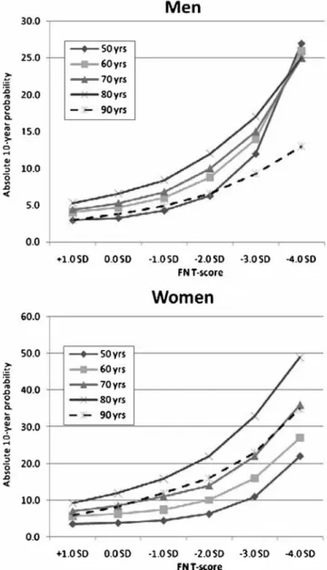
In addition, the contribution of a low BMI on fracture pro-bability was higher in elderly compared to younger patients. At any BMI value, 90-year old patients had a lower absolute 10-year fracture probability than 80-year old patients, the effect being more pronounced in men than in women. This reflects the fact that in the FRAX® model, death hazard from all causes and fracture probability are competing events. As shown in Fig. 2, absolute 10-year fracture probabilities increased exponentially with decreasing T-score values. The effect of BMD on fracture probability again decreased in very old men and women, indicating that, with advanced age, there is a higher probability of dying from any other competing cause rather than sustaining an osteoporotic fracture. Fracture probability in the presence of single risk factors Fracture probability was always found to be higher in women than in men. This was consistent across any given age, BMI value, T-score value, and for any single risk factor. In the absence of any validated risk factor selected for FRAX® modelling, the 10-year probability of a major
osteoporotic fracture at age 65 years and at a BMI of 25 kg/m2 (base case) was 5.6% in men and 9.5% in women. The data
presented in Table 1 indicate that the presence of risk
factors increased this base case probability. Large increases in fracture probability were observed in the case of a previous fragility fracture or of parental history of hip fracture. Both of these factors doubled the probability, reaching 11.0% and 18.0% in men and women,
respec-tively. At age 65, a BMD T-score of−2.5 SD measured at
the femoral neck increased 10-year fracture probability to 11.0% and 14.0% in men and women, respectively. This fracture probability further increased up to 18.0% in men Fig. 1 Absolute 10-year major fracture probability at various levels of BMI (kg/m2) and at different ages for Swiss men and women, without clinical risk factors
and 24.0% in women, depending on which associated single clinical risk factor was present.
As shown in Table2, the 10-year absolute probability of any major osteoporotic and hip fracture in the presence of a single risk factor increased with advancing age in both sexes. Between a 50-year-old man without risk factors and an 80-year-old man with a parental history of hip fracture, the individual probability of any major osteoporotic fracture increased 6.5-fold (from 3.4% to 22.0%). In women, this probability increased 10.2-fold. For equivalent scenarios, the probability of suffering a hip fracture increased 75-fold in men and 100-fold in women from base line levels.
Fracture probability was found to increase with decreas-ing BMD T-score values in both sexes, as shown in Table3
and Fig. 2. In the absence of any risk factor, the 10-year probability of a major osteoporotic fracture for a 65-year-old woman presenting with the commonly accepted treatment
threshold T-score of −2.5 SD was 14.0%. This fracture
probability was equal to or lower than that of a woman with a T-score of−2.0 SD and either rheumatoid arthritis, present or past glucocorticoid treatment, previous fragility fracture during adulthood or parental history of hip fracture. The fracture probability was also lower than that of a woman with a T-score of−1.0 SD and a parental history of hip fracture. In addition, in men without clinical risk factors and a T-score of −2.5 SD, the absolute 10-year fracture probability for any major osteoporotic fracture was 11.0%. With a parental history of hip fracture, the same level of absolute fracture probability was achieved at a T-score of−1.0 SD. However, based on the current thresholds for reimbursement, treatment with a bisphosphonate would be reimbursed in the former case, but not in the latter, despite the probability of a fracture being equal in both cases.
Fracture probability with multiple risk factors
The absolute 10-year major osteoporotic or hip fracture probability was found to increase exponentially with the number of associated risk factors. In the absence of an available BMD T-score value and in the base case (BMI of 25 kg/m2and age of 65 years), the probability of any major osteoporotic fracture was increased 8.2-fold in men presenting with all risk factors (46.0%) compared to men with no risk factors (5.6%) and 7.5-fold (71.0% and 9.5%, respectively) in women as shown in Table4. In the case of patients having all risk factors, a BMD T-score of−2.5 SD compared to no BMD value available contributed only modestly to fracture probability in women (72.0% vs. 71.0%) but considerably more in men (58.0% vs. 46.0%). These results indicate that, depending on sex and age, the relevance of a T-score value with regard to fracture probability varied.
At the age of 65 and for any major osteoporotic fracture resulting from long-term treatment with oral glucocorticoids, the lowest 10-year fracture probability corresponding to that currently reimbursed in Switzerland was 8.7% in men and 15.0% in women. As shown in
Table 5, several pairs of other risk factors gave an
equivalent or higher fracture probability level. Similarly, with the treatment threshold set at≤−2.5 SD as required for reimbursement of drug therapy of osteoporosis, the corresponding 10-year fracture probabilities were 11.0%
in men and 14.0% in women. At a T-score of −1.0 SD,
several pairs of associated clinical risk factors resulted in fracture probabilities higher than that achieved for this threshold. These were exceeded even more frequently when more than two risk factors were present in the same patient.
Fig. 2 Absolute 10-year major fracture probability by age and femoral neck BMD in Swiss men and women. BMI was set at 25 kg/m2
Discussion
The present study shows that levels of 10-year fracture probability equivalent to those currently accepted for reim-bursement of BMD measurement by DXA in Switzerland are achieved with several clinical risk factor profiles and combinations. These include risk factors not (yet) accepted for reimbursement, such as a parental history of fracture, tobacco and/or alcohol abuse and rheumatoid arthritis. This suggests that, with identical 10-year fracture probabilities, adequate diagnostic workup is not equally accessible to all patients presenting with identical fracture risk. Consequently, with
current access to osteoporosis diagnosis, too few patients at increased probability of fracture are adequately identified and subsequently treated. This is consistent with earlier reports in-dicating that a significant proportion of osteoporotic fractures occur in patients with a T-score above−2.5 SD [11,12].
Low bone mass, as measured by DXA, is an important single predictor of fracture risk [31–34], and BMD mea-sured at the femoral neck has been shown to outperform clinical risk factors alone at all ages for hip fracture prediction [13]. However, as shown by the present findings, BMD alone does not capture all determinants of fracture probability, and the consideration of additional risk factors
Osteoporotic fracturea Hip fracture
50 60 70 80 50 60 70 80
Men
No clinical risk factors 3.4 5.4 7.2 12.0 0.2 0.5 1.6 4.8
Current cigarette smoking 3.6 5.6 7.5 12.0 0.3 0.7 2.2 6.1
Alcohol intake>2 units daily 4.1 6.5 9.0 15.0 0.3 0.8 2.4 7.2
Rheumatoid arthritis 4.6 7.3 10.0 17.0 0.3 0.9 2.8 8.2
Oral glucocorticoids 5.5 8.5 11.0 16.0 0.4 1.0 3.0 7.9
Previous fragility fracture 7.3 11.0 13.0 19.0 0.7 1.5 3.4 7.3
Parental history of hip fracture 6.8 10.0 11.0 22.0 0.2 0.7 3.4 15.0
Women
No clinical risk factors 4.3 8.1 14.0 27.0 0.3 0.8 2.8 10.0
Current cigarette smoking 4.5 8.6 15.0 30.0 0.4 1.2 4.2 14.0
Alcohol intake>2 units daily 5.1 9.7 17.0 33.0 0.4 1.2 4.3 15.0
Rheumatoid arthritis 5.8 11.0 19.0 37.0 0.5 1.4 5.0 17.0
Oral glucocorticoids 7.0 13.0 22.0 40.0 0.6 1.7 5.8 19.0
Previous fragility fracture 9.1 16.0 25.0 42.0 1.1 2.3 5.9 15.0
Parental history of hip fracture 8.4 15.0 22.0 44.0 0.4 1.1 6.0 30.0
Table 2 Ten-year probability (in per cent) of a major osteopo-rotic or hip fracture in men and women according to age and the presence of a single risk factor
Rows in italics indicate clinical risk factors included in FRAX® and currently accepted for DXA reimbursement in Switzerland. BMI set at 25 kg/m2
a
Hip, clinical spine, humeral or forearm fracture
Table 1 Ten-year probability (in per cent) of a major osteoporotic or hip fracture in men and women according to the presence of a single risk factor
Without BMD T-score=−2.5 SD
Men Women Men Women
Osteoporotica Hip Osteoporotica Hip Osteoporotica Hip Osteoporotica Hip
No clinical risk factors 5.6 0.9 9.5 1.5 11.0 4.0 14.0 3.4
Current cigarette smoking 5.9 1.3 10.0 2.2 12.0 6.4 15.0 5.7
Alcohol intake>2 units daily 6.9 1.3 12.0 2.2 14.0 6.1 17.0 5.1
Rheumatoid arthritis 7.7 1.6 13.0 2.6 14.0 5.7 18.0 4.8
Oral glucocorticoids 8.7 1.8 15.0 3.1 17.0 7.0 22.0 6.2
Previous fragility fracture 11.0 2.2 18.0 3.6 18.0 6.6 22.0 5.6
Parental history of hip fracture 11.0 1.2 18.0 1.9 18.0 4.2 24.0 3.6
Rows in italics indicate clinical risk factors included in FRAX® and currently accepted for DXA reimbursement in Switzerland. BMI set at 25 kg/ m2, age set at 65 years
a
Table 4 Ten-year probability (in per cent) of a major osteoporotic or hip fracture in men and women with multiple risk factors as a function of Femoral neck BMD
Without BMD BMD T-score=−2.5 SD BMD T-score=−1.0 SD
Osteoporotic fracturea
Hip fracture Osteoporotic fracturea
Hip fracture Osteoporotic fracturea
Hip fracture
Men
No clinical risk factors 5.6 0.9 11.0 4.0 5.8 0.9
Current cigarette smoking 5.9 1.3 12.0 6.4 5.9 1.5
+Alcohol intake>2 units daily 7.3 1.9 16.0 9.6 7.4 2.3
+Rheumatoid arthritis 10.0 3.4 21.0 13.0 9.7 3.3
+Oral glucocorticoids 16.0 6.5 32.0 21.0 15.0 5.7
+Previous fragility fracture 31.0 15.0 46.0 33.0 24.0 9.3
+Parental history of hip fracture 46.0 20.0 58.0 34.0 37.0 9.8
Women
No clinical risk factors 9.5 1.5 14.0 3.4 7.9 0.7
Current cigarette smoking 10.0 2.2 15.0 5.7 8.0 1.2
+Alcohol intake>2 units daily 13.0 3.3 19.0 8.6 9.7 1.8
+Rheumatoid arthritis 18.0 5.8 25.0 12.0 13.0 2.5
+Oral glucocorticoids 29.0 12.0 39.0 21.0 20.0 4.7
+Previous fragility fracture 51.0 27.0 56.0 33.0 31.0 7.7
+Parental history of hip fracture 71.0 34.0 72.0 34.0 51.0 8.1
BMI set at 25 kg/m2, age set at 65 years
The sign“+” indicates that the individual risk factors are successively added (incremental risk)
a
Hip, clinical spine, humeral or forearm fracture
Table 3 Ten-year probability (in per cent) of a major osteoporotic fracture in men and women according to Femoral neck BMD and presence of a single risk factor
Osteoporotic fracturea T-score=
–4.0 SD T-score= –3.0 SD T-score= –2.5 SD T-score= –2.0 SD T-score= –1.5 SD T-score= –1.0 SD T-score= 0.0 SD T-score= +1.0 SD Without BMD Men
No clinical risk factors 24.0 14.0 11.0 8.6 7.0 5.8 4.5 3.8 5.6
Current cigarette smoking 31.0 17.0 12.0 9.4 7.4 5.9 4.4 3.6 5.9
Alcohol intake>2 units daily 32.0 18.0 14.0 11.0 8.7 7.1 5.4 4.5 6.9
Rheumatoid arthritis 32.0 18.0 14.0 11.0 9.1 7.5 5.8 4.9 7.7
Oral glucocorticoids 36.0 22.0 17.0 13.0 11.0 9.0 7.0 6.0 8.7
Previous fragility fracture 37.0 22.0 18.0 14.0 12.0 9.7 7.6 6.5 11.0
Parental history of hip fracture 33.0 22.0 18.0 15.0 13.0 11.0 9.0 7.9 11.0
Women
No clinical risk factors 30.0 17.0 14.0 11.0 9.0 7.9 6.6 5.6 9.5
Current cigarette smoking 37.0 20.0 15.0 12.0 9.3 8.0 6.4 5.4 10.0
Alcohol intake>2 units daily 38.0 22.0 17.0 13.0 11.0 9.5 7.8 6.6 12.0
Rheumatoid arthritis 38.0 22.0 18.0 14.0 12.0 10.0 8.4 7.2 13.0
Oral glucocorticoids 45.0 27.0 22.0 17.0 14.0 13.0 10.0 8.9 15.0
Previous fragility fracture 45.0 27.0 22.0 18.0 15.0 13.0 11.0 9.3 18.0
Parental history of hip fracture 43.0 29.0 24.0 20.0 17.0 16.0 13.0 11.0 18.0
Rows in italics indicate clinical risk factors included in FRAX®and currently accepted for DXA reimbursement in Switzerland. BMI set at 25 kg/m2, age set at 65 years
a
Hip, clinical spine, humeral or forearm fracture
improves its predictive value. Screening with DXA alone is generally not considered sensitive enough, and thus, the identification of patients with osteoporosis generally relies on case-finding strategies [1]. Therefore, FRAX® repre-sents a unique opportunity for identifying those subjects who should benefit from further diagnostic measures, includ-ing the assessment of risk factors not captured by FRAX® such as risk factors for falls, gastrointestinal malabsorption syndromes, increased biochemical markers of bone turnover and/or vitamin D insufficiency. The need for additional diagnosis by X-ray examination or a laboratory assessment of calcium phosphate metabolism can also be indicated following FRAX® assessment. Depending on the findings and the T-score value measured at the femoral neck and its deviation from the mean T-score value in a population of the same age and sex, the absolute 10-year fracture probability may assist in determining which patients would benefit from treatment. Previous analysis of population-based screening with DXA followed by alendronate treatment, in the presence of osteoporosis or of fracture and osteopenia, has been shown to be cost-effective in postmenopausal women after the age of 70 years for the Swiss healthcare system [35]. A FRAX®-based pre-identification of patients eligible for DXA is likely to improve cost-effectiveness by increasing the specificity and sensitivity of case finding.
An interesting finding of the present analysis was that in 65-year old patients with all risk factors included in the
FRAX® algorithm, a T-score of −2.5 SD did not increase
fracture probability in women, though it did in men. This could be related to the fact that BMD T-score is computed from the NHANES reference range for women. Thus, in
65-year-old women, a T-score of −2.5 SD corresponds
more or less to the average of the female population of that age with multiple clinical risk factors. In men, however, this T-score (derived from the female reference range) is lower than average for men of this age, and a T-score of -2.5 SD thus adds a significant component to fracture probability.
Setting a treatment cut-off based on a T-score alone does select patients at increased probability of fracture. However, it also categorises patients at equal or higher fracture probability as non-eligible for treatment if additional clinical risk factors are not integrated in fracture risk assessment. The determination of the individual 10-year fracture probability with FRAX® would at least ensure that patients at equal risk would have equal chances of getting appropriate treatment. Interestingly, in the field of osteoporosis, treatment access is usually restricted in accordance with the inclusion criteria of the fracture endpoint trials. In the vast majority of these studies, patients were included on the basis of low bone mass measured by DXA and/or prevalent vertebral fractures [36– Table 5 Ten-year probability (in per cent) of a major osteoporotic fracture in men and women according to femoral neck BMD and risk factors combined by pairs
Without BMD With BMD T-Score = -2.5 SD With BMD T-Score = -1.0 SD
No clinical risk factors
Current cigarette smoking
Alcohol intake > 2 units daily
Rheumatoid arthritis Oral glucocorticoids
Previous fragility fracture
Parental history of hip fracture
No clinical risk factors
Current cigarette smoking
Alcohol intake > 2 units daily
Rheumatoid arthritis Oral glucocorticoids
Previous fragility fracture
Parental history of hip fracture
No clinical risk factors
Current cigarette smoking
Alcohol intake > 2 units daily
Rheumatoid arthritis Oral glucocorticoids
Previous fragility fracture
Men
No clinical risk factors 5.6 11.0 5.8
Current cigarette smoking 5.9 12.0 5.9
Alcohol intake > 2 units daily 7.3 6.9 16.0 14.0 7.4 7.1 Rheumatoid arthritis 8.2 9.6 7.7 16.0 18.0 14.0 7.7 9.2 7.5
Oral glucocorticoids 9.1 11.0 12.0 8.7 19.0 21.0 22.0 17.0 9.1 11.0 12.0 9.0
Previous fragility fracture 12.0 13.0 15.0 17.0 11.0 20.0 22.0 23.0 27.0 18.0 11.0 12.0 12.0 15.0 9.7
Parental history of hip
fracture 11.0 13.0 14.0 16.0 20.0 11.0 19.0 22.0 23.0 27.0 29.0 18.0 11.0 13.0 14.0 17.0 18.0 11.0
Women
No clinical risk factors 9.5 14.0 7.9
Current cigarette smoking 10.0 15.0 8.0
Alcohol intake > 2 units daily 13.0 12.0 19.0 17.0 9.7 9.5 Rheumatoid arthritis 14.0 16.0 13.0 20.0 22.0 18.0 10.0 12.0 10.0
Oral glucocorticoids 17.0 19.0 21.0 15.0 24.0 27.0 28.0 22.0 13.0 15.0 16.0 13.0
Previous fragility fracture 20.0 22.0 25.0 29.0 18.0 24.0 27.0 28.0 34.0 22.0 13.0 16.0 17.0 21.0 13.0
Parental history of hip
fracture 19.0 21.0 24.0 28.0 32.0 18.0 25.0 28.0 30.0 36.0 37.0 24.0 15.0 18.0 20.0 24.0 25.0 16.0
Italic fonts indicate clinical risk factors included in FRAX® and currently accepted for DXA reimbursement in Switzerland
Boxes indicate the current fracture probability threshold for treatment reimbursement in Switzerland. Higher probabilities indicated in bold fonts BMI set at 25 kg/m2, age set at 65 years
40]. However, in the subgroup post hoc analyses performed in all these studies, there was no evidence in favour of differences in efficacy related to the presence or absence of these clinical risk factors [36–40]. Moreover, several trials indicate enhanced effectiveness of pharmacologic interven-tion in patients with higher fracture probabilities as
deter-mined by the FRAX® tool [41,42].
The clinical risk factor approach in FRAX® should be considered to be conservative. Calculated fracture proba-bilities are limited to major clinical osteoporotic fractures, i.e. those of the hip, spine, distal forearm and proximal humerus. In the year 2000 in Switzerland, these fractures represented 63% and 76% of all hospitalised fractures [2] and 63% and 71% of total (hospitalised and non-hospitalised) clinical fractures in the OsteoCare survey [12] in men and women, respectively. This study only considered clinical fractures, although the more prevalent morphometric vertebral frac-tures and deformities also did increase the risk of future fracture at any site [43]. In addition these fractures are associated with substantial increases in back pain and functional limitation due to back pain [44]. Furthermore, vertebral fractures, including asymptomatic fractures, may be responsible of a loss in height. In these patients, BMI derived from body weight (kilogram) and height (square meter) will be mathematically higher, leading to a theoretical underestimation of fracture probability. Therefore, true frac-ture incidence and derived fracfrac-ture probabilities are generally likely to be underestimated. In contrast, the validated risk factors independent of BMD used in FRAX® modelling are those that allow consistent linking with BMD T-scores, age, and BMI in nine international cohorts [13]. This suggests that still other risk factors may also independently contribute to fracture risk but are not accommodated in the FRAX® algorithm. As an example, the history of falls, propensity to falling, biochemical markers of bone turnover and vitamin D status are important determinants of fracture probability not included in FRAX®. Moreover, the magnitude of the effect on fracture probability of some of the validated risk factors used in FRAX®, for which the model assumes average exposure, may depend on dosage, quantity and/or duration of exposure. In particular, the individual circumstances of the use of glucocorticoids, tobacco smoking and alcohol consumption will affect the individual fracture probability. Currently, the FRAX® tool does not integrate a dose-dependent influence. Finally, BMD measured at other sites than the femoral neck is a proven risk factor for fractures, which is not included in FRAX®. FRAX® results also do not integrate BMD increases achieved with previous or ongoing drug therapies against osteoporosis. The present study cannot address these issues. Therefore, one of the key determinants for adequate inter-pretation of the individual 10-year probability of fracture delivered by FRAX® will remain clinical judgement.
The findings of the present study are consistent with the need for a paradigm shift in osteoporosis management. The current, solely BMD threshold-dependent prevention/ treatment concept needs to evolve to a fracture probability continuum for which new intervention thresholds, based on the 10-year probability of any major osteoporotic or hip fractures, need to be defined. FRAX® based future cost-effectiveness analyses will certainly contribute to identifying medically and economically optimised osteoporosis case-finding strategies.
Acknowledgements We are grateful to Dr. Philippe Kress, Kressmed Glattbrugg Switzerland, for his contribution to the writing of the manuscript. Mrs. Marianne Perez and Dr. Sarah Frankland are acknowledged for secretarial assistance and careful reading of the manuscript, respectively.
Funding This work was supported by an unrestricted research grant from Amgen (Europe), Zug, Switzerland.
Conflicts of interest None.
References
1. Kanis JA, Burlet N, Cooper C et al (2008) European guidance for the diagnosis and management of osteoporosis in postmenopausal women. Osteoporos Int 19:399–428
2. Lippuner K, Golder M, Greiner R (2005) Epidemiology and direct medical costs of osteoporotic fractures in men and women in Switzerland. Osteoporos Int 16(Suppl 2):S8–S17
3. Mathers CD, Murray CJL, Lopez AD et al (2001) World Health Organization Global Programme on Evidence for Health Policy Discussion Paper No. 38.http://www.who.int/healthinfo/paper38. pdf. Accessed on May 13, 2006
4. Robine JM, Paccaud F (2005) Nonagenarians and centenarians in Switzerland, 1860–2001: a demographic analysis. J Epidemiol Community Health 59:31–37
5. Bundesamt für Statistik. Szenarien zur Bevölkerungsentwicklung der Schweiz 2005-2050. http://www.bfs.admin.ch/bfs/portal/de/ index/news/publikationen.Document.83713.pdf. Accessed on May 14, 2008
6. Schwenkglenks M, Lippuner K, Hauselmann HJ et al (2005) A model of osteoporosis impact in Switzerland 2000–2020. Osteo-poros Int 16:659–671
7. (1993) Consensus development conference: diagnosis, prophylax-is, and treatment of osteoporosis. Am J Med 94:646-650 8. Kanis JA, Melton LJ 3rd, Christiansen C et al (1994) The
diagnosis of osteoporosis. J Bone Miner Res 9:1137–1141 9. Kanis JA, McCloskey EV, Johansson H et al (2008) A reference
standard for the description of osteoporosis. Bone 42:467–475 10. Kanis JA, Johnell O, Oden A et al (2000) Risk of hip fracture
according to the World Health Organization criteria for osteopenia and osteoporosis. Bone 27:585–590
11. Siris ES, Miller PD, Barrett-Connor E et al (2001) Identification and fracture outcomes of undiagnosed low bone mineral density in postmenopausal women: results from the National Osteoporosis Risk Assessment. Jama 286:2815–2822
12. Suhm N, Lamy O, Lippuner K (2008) Management of fragility fractures in Switzerland: results of a nationwide survey. Swiss Med Wkly 138:674–683
13. Kanis JA, Oden A, Johnell O et al (2007) The use of clinical risk factors enhances the performance of BMD in the prediction of hip and osteoporotic fractures in men and women. Osteoporos Int 18:1033–1046
14. Krieg MA, Cornuz J, Ruffieux C et al (2006) Prediction of hip fracture risk by quantitative ultrasound in more than 7000 Swiss women>or =70 years of age: comparison of three technologically different bone ultrasound devices in the SEMOF study. J Bone Miner Res 21:1457–1463
15. Dawson-Hughes B, Tosteson AN, Melton LJ 3rd et al (2008) Implications of absolute fracture risk assessment for osteopo-rosis practice guidelines in the USA. Osteoporos Int 19:449– 458
16. Fujiwara S, Nakamura T, Orimo H et al (2008) Development and application of a Japanese model of the WHO fracture risk assessment tool (FRAX). Osteoporos Int 19:429–435
17. Lippuner K, Johansson H, Kanis J et al (2008) Remaining lifetime and absolute 10-year probabilities of osteoporotic fracture in Swiss men and women. Osteoporos Int . doi: 10.1007/s00198-008-0779-8
18. United Nations population division (2003) World population prospects: the 2002 revision and world urban prospects. Popula-tion Division of the Dept Economic and Social Affairs of the UN Secretariat
19. Looker AC, Wahner HW, Dunn WL et al (1998) Updated data on proximal femur bone mineral levels of US adults. Osteoporos Int 8:468–489
20. Kanis JA, Johnell O, Oden A et al (2008) FRAX and the assessment of fracture probability in men and women from the UK. Osteoporos Int 19:385–397
21. Kanis JA, Johansson H, Oden A et al (2004) A meta-analysis of prior corticosteroid use and fracture risk. J Bone Miner Res 19:893–899
22. Melton LJ 3rd, Achenbach SJ, Gebhart JB et al (2007) Influence of hysterectomy on long-term fracture risk. Fertil Steril 88:156– 162
23. Melton LJ 3rd, Khosla S, Malkasian GD et al (2003) Fracture risk after bilateral oophorectomy in elderly women. J Bone Miner Res 18:900–905
24. Melton LJ 3rd, Alothman KI, Khosla S et al (2003) Fracture risk following bilateral orchiectomy. J Urol 169:1747–1750
25. Vestergaard P, Mosekilde L (2002) Fracture risk in patients with celiac Disease, Crohn's disease, and ulcerative colitis: a nation-wide follow-up study of 16, 416 patients in Denmark. Am J Epidemiol 156:1–10
26. Vestergaard P, Krogh K, Rejnmark L et al (2000) Fracture risk is increased in Crohn's disease, but not in ulcerative colitis. Gut 46:176–181
27. Loftus EV Jr, Achenbach SJ, Sandborn WJ et al (2003) Risk of fracture in ulcerative colitis: a population-based study from Olmsted County, Minnesota. Clin Gastroenterol Hepatol 1:465–473 28. Loftus EV Jr, Crowson CS, Sandborn WJ et al (2002) Long-term
fracture risk in patients with Crohn's disease: a population-based study in Olmsted County, Minnesota. Gastroenterology 123:468–475
29. Zehnder Y, Luthi M, Michel D et al (2004) Long-term changes in bone metabolism, bone mineral density, quantitative ultrasound parameters, and fracture incidence after spinal cord injury: a cross-sectional observational study in 100 paraplegic men. Osteoporos Int 15:180–189
30. Vestergaard P, Mosekilde L (2003) Hyperthyroidism, bone mineral, and fracture risk–a meta-analysis. Thyroid 13:585–593 31. Kanis JA (2002) Diagnosis of osteoporosis and assessment of
fracture risk. Lancet 359:1929–1936
32. Marshall D, Johnell O, Wedel H (1996) Meta-analysis of how well measures of bone mineral density predict occurrence of osteopo-rotic fractures. BMJ 312:1254–1259
33. Cummings SR, Black D (1995) Bone mass measurements and risk of fracture in Caucasian women: a review of findings from prospective studies. Am J Med 98:24S–28S
34. Black DM, Cummings SR, Genant HK et al (1992) Axial and appendicular bone density predict fractures in older women. J Bone Miner Res 7:633–638
35. Schwenkglenks M, Lippuner K (2007) Simulation-based cost-utility analysis of population screening-based alendronate use in Switzerland. Osteoporos Int 18:1481–1491
36. Cummings SR, Black DM, Thompson DE et al (1998) Effect of alendronate on risk of fracture in women with low bone density but without vertebral fractures: results from the Fracture Inter-vention Trial. Jama 280:2077–2082
37. Black DM, Delmas PD, Eastell R et al (2007) Once-yearly zoledronic acid for treatment of postmenopausal osteoporosis. N Engl J Med 356:1809–1822
38. Black DM, Cummings SR, Karpf DB et al (1996) Randomised trial of effect of alendronate on risk of fracture in women with existing vertebral fractures. Fracture Intervention Trial Research Group. Lancet 348:1535–1541
39. Neer RM, Arnaud CD, Zanchetta JR et al (2001) Effect of parathyroid hormone (1–34) on fractures and bone mineral density in postmenopausal women with osteoporosis. N Engl J Med 344:1434–1441
40. Ettinger B, Black DM, Mitlak BH et al (1999) Reduction of vertebral fracture risk in postmenopausal women with osteoporo-sis treated with raloxifene: results from a 3-year randomized clinical trial. Multiple Outcomes of Raloxifene Evaluation (MORE) Investigators. JAMA 282:637–645
41. Kanis JA, Johansson H, Oden A et al (2009) Bazedoxifene reduces vertebral and clinical fractures in postmenopausal women at high risk assessed with FRAX®. Bone 44:1049–1054. doi:10.1016/j. bone.2009.02.014
42. McCloskey EV, Johansson H, Oden A et al (2009) Ten-year fracture probability identifies women who will benefit from clodronate therapy—additional results from a double blind, placebo-controlled randomised study. Osteoporos Int 20:811– 817. doi:10.1007/s00198-008-0786-9
43. Pongchaiyakul C, Nguyen ND, Jones G et al (2005) Asymptom-atic vertebral deformity as a major risk factor for subsequent fractures and mortality: a long-term prospective study. J Bone Miner Res 20:1349–1355
44. Nevitt MC, Ettinger B, Black DM et al (1998) The association of radiographically detected vertebral fractures with back pain and function: a prospective study. Ann Intern Med 128:793–800