HAL Id: tel-02862796
https://tel.archives-ouvertes.fr/tel-02862796
Submitted on 9 Jun 2020HAL is a multi-disciplinary open access
archive for the deposit and dissemination of sci-entific research documents, whether they are pub-lished or not. The documents may come from teaching and research institutions in France or abroad, or from public or private research centers.
L’archive ouverte pluridisciplinaire HAL, est destinée au dépôt et à la diffusion de documents scientifiques de niveau recherche, publiés ou non, émanant des établissements d’enseignement et de recherche français ou étrangers, des laboratoires publics ou privés.
Mild Endoplasmic Reticulum Stress Protects From Cell
Death : The Role Of Autophagy
Clemence Levet
To cite this version:
Clemence Levet. Mild Endoplasmic Reticulum Stress Protects From Cell Death : The Role Of Autophagy. Cellular Biology. Université Claude Bernard - Lyon I, 2012. English. �NNT : 2012LYO10209�. �tel-02862796�
THESE DE L’UNIVERSITE CLAUDE BERNARD LYON 1
ECOLE DOCTORALE : Biologie Moléculaire Intégrative et Cellulaire
DIPLOME DE DOCTORAT Discipline : Biologie
soutenue publiquement le 25 Octobre 2012 par
Clémence LEVET
Mild Endoplasmic Reticulum Stress Protects
From Cell Death :
The Role Of Autophagy
Directeur de thèse : Pr Bertrand MOLLEREAU
JURY : Dr Magali SUZANNE, Rapporteur Dr Hervé TRICOIRE, Rapporteur
Dr Serge BIRMAN, Examinateur Dr Matthias FAURE, Examinateur Dr Sébastien GAUMER, Examinateur
2
This thesis has been performed in the “apoptosis and neurogenetic” group headed by the Pr Bertrand Mollereau in the Laboratory of Molecular Biology of the Cell at the Ecole Normale Superieure de Lyon.
This these was supported by grants from the cluster 11 HNV (Rhone Alpes) and La ligue contre le cancer.
3
Remerciements
Je souhaite remercier les membres de mon jury Magali Suzanne, Hervé Tricoire, Serge Birman, Matthias Faure et Sébastien Gaumer d’avoir accepter de juger mon travail.
Je tiens à remercier mon directeur de thèse Bertrand Mollereau pour m’avoir accueilli au sein de son équipe. Je le remercie pour la confiance qu’il m’a accordée. Je le remercie également pour son encadrement, ses conseils et sa disponibilité tout au long de ma thèse.
Je remercie également tous les membres de l’équipe « apoptose et neurogenetique » présents et passés. Je les remercie pour leur aide et leur soutient ainsi que pour leur bonne humeur.
J’adresse également mes remerciements au Laboratoire de Biologie Moléculaire de la Cellule, à la SFR BioSciences Gerland, au Platim et à la plateforme Drosotools pour m’avoir donné accès à des infrastructures et des matériaux essentiels à la réalisation de ma thèse.
Pour finir, je remercie mes parents, ma sœur ainsi que tous les membres de ma famille pour leur soutient tout au long de ma thèse.
5
Abstract
The last years have been very successful in identifying mechanisms, which control apoptosis in metazoan. However, the regulation of cell death in specific cell type remains to be determined. An excess of neuron apoptosis can lead to neurodegenerative diseases such as Huntington, Parkinson or Alzheimer diseases. Neurodegeneration is usually associated to Endoplasmic Reticulum stress (ER stress), autophagy or oxidative stress. However, the role of these mechanisms in the regulation of neurodegeneration is not clearly established. To test the role of ER stress in the regulation of neuronal death, we used several models of neurodegeneration in Drosophila and mammals. First, we have shown that the genetic induction of ER stress protected photoreceptors of the Drosophila eye from apoptosis. Then, we have shown that the protective effect of ER stress is conserved in both Drosophila and mouse models of Parkinson disease. In order to characterize the protective effect of ER stress, we have studied the activation of protective mechanisms upon ER stress. We have shown that in the Drosophila retina, ER stress can induce an anti-oxidative response and autophagy. Interestingly, autophagy is only activated in presence of both ER stress and cell death signal. We have focused on the role of autophagy in the protective effect of ER stress. We have shown that the activation of autophagy was required for the protective effect of ER stress. Thus, we have shown that ER stress response is not only involved in the reduction of misfolded protein accumulation, but can also protect neurons form cell death by activating autophagy.
Keyword: Apoptosis, endoplasmic reticulum stress, autophagy, neurodegeneration, Parkinson disease, Drosophila, mouse
7
Titre et résumé
Un stress modéré du réticulum endoplasmique protège de la mort
cellulaire : rôle de l’autophagie
Ces dernières années ont été très fructueuses pour l’identification des mécanismes fondamentaux de l’apoptose chez les métazoaires. Cependant, il reste beaucoup à apprendre sur la manière dont sont régulés les programmes de mort en fonction du type cellulaire. Un excès d’apoptose des neurones peut conduire à différentes pathologies neurodégénératives comme les maladies de Huntington, Parkinson ou Alzheimer. La neurodégénérescence est souvent associée à un stress du réticulum endoplasmique (RE), à l’autophagie et à un stress oxydatif. Cependant le rôle de ces mécanismes dans la régulation de la neurodégénérescence n’a pas été clairement établi. Afin de tester l’implication du stress du RE dans la régulation de la mort neuronale, nous avons utilisé différents modèles de neurodégénérescence chez la
Drosophila melanogaster et les mammifères. Nous avons d’abord montré que l’induction
d’un stress modéré du réticulum protégeait les photorécepteurs de l’œil de drosophile contre l’apoptose. Nous avons ensuite montré que l’effet protecteur du stress du RE était conservé dans les modèles de maladie de Parkinson chez la drosophile et la souris. Afin de caractériser l’effet protecteur du stress du réticulum, nous avons étudié l’activation de mécanismes protecteurs lors d’un stress du réticulum. Nous avons montré que dans la rétine de drosophile l’activation du stress du RE dans un contexte de mort neuronale induisait une réponse anti-oxydante et l’autophagie. Nous nous sommes plus particulièrement intéressés au rôle de l’autophagie dans le rôle protecteur du stress du RE. Nous avons montré que l’activation de l’autophagie était nécessaire à l’effet protecteur du stress du RE contre l’apoptose. Nous avons donc pu mettre en évidence que la réponse au stress du RE ne participait pas uniquement à réduire l’accumulation de protéines mal-conformées mais permettait aussi de protéger les neurones contre la mort cellulaire en activant l’autophagie.
Mots clés : Apoptose, stress du réticulum endoplasmique, autophagie, neurodégénérescence, maladie de Parkinson, drosophile, souris
9
Résumé substantiel
Ces dernières années ont été très fructueuses pour l’identification des mécanismes fondamentaux de l’apoptose chez les métazoaires [19, 20]. Cependant, il reste beaucoup à apprendre sur la manière dont sont régulés les programmes de mort en fonction du type cellulaire. Un excès d’apoptose peut conduire à différentes pathologies neurodégénératives comme les maladies de Huntington, Parkinson ou Alzheimer. Le stress du réticulum endoplasmique (RE), le stress oxydatif et l’autophagie sont associés à la neurodégénérescence [15, 21-24]. Cependant, leur importance dans la progression de ces maladies n’a pas encore été déterminée.
Le RE est un organelle par lequel transitent les protéines sécrétées et les protéines transmembranaires. Ces protéines acquièrent leur bonne conformation grâce aux protéines chaperonnes résidentes et vont s’associer en complexes. Seules les protéines correctement repliées et associées pourront sortir du RE et transiter par les voies de sécrétion. Ce mécanisme de contrôle permet de s’assurer de la qualité des protéines sécrétées ou associées à la membrane. La présence de protéines anormalement conformées induit une voie de signalisation appelée la réponse UPR (pour « Unfolded Protein Response ») [28]. La réponse UPR est une réponse adaptative au stress du RE qui est conservée au cours de l’évolution. En effet, la plupart des effecteurs de la réponse UPR identifiés chez les vertébrés ont un homologue chez la drosophile [35]. La réponse UPR chez les vertébrés est activée par un senseur du stress du réticulum : BiP. En absence de stress du RE, BiP est associé à des récepteurs transmembranaires du RE: PERK (pour « PKR-like ER kinase »), ATF6 (pour « activating transcription factor 6 ») et IRE1 (pour « inositol-requiring enzyme 1 »). Lors d’un stress du RE, BiP se dissocie de ces partenaires, il se fixe sur les protéines mal-conformées afin de jouer son rôle de chaperonne. Les voies PERK, ATF6 et IRE1 sont alors activées. Elles vont induire l’expression de chaperonnes, activer les voies de dégradation des protéines et réduire la synthèse protéique. Des études suggèrent qu’une réponse UPR intense et prolongée induit l’activation du programme d’apoptose [37]. Cependant, il n’est pas établi si la réponse UPR est la cause de la mort neuronale dans les maladies neurodégénératives. La conservation des mécanismes de mort cellulaire au cours de l’évolution nous permet d’utiliser des modèles génétiques simples, comme la drosophile, pour mieux comprendre la régulation de la mort cellulaire chez l’homme.
10
L’œil de Drosophila melanogaster est un excellent modèle pour l’étude in vivo des maladies neurodégénératives. En effet, l’expression ectopique de protéines pathologiques dans la rétine induit la dégénérescence progressive des cellules photoréceptrices (PR) avec des caractéristiques similaires à celles qui sont observées dans les maladies humaines [41]. Nous avons établi un modèle de neurodégénérescence chez la drosophile dépendant de la surexpression des gènes pro-apoptotiques reaper et Dp53 (homologue drosophile du gène p53 de mammifère). La surexpression de reaper induit l’apoptose des cellules photoréceptrices de l’œil. Un crible génétique nous a permis d’identifier les mutations du gène ninaA (homologue de la cyclophiline A humaine) comme suppresseurs de la mort cellulaire. NinaA code une chaperonne nécessaire à la maturation de la Rhodopsine-1 dans le réticulum. L’absence de NinaA provoque l’accumulation de protéines Rh1 mal conformées induisant un stress du RE et une réponse UPR dans les cellules photoréceptrices de l’œil de drosophile. Nous avons observé que la réponse UPR, qui est normalement dédiée à éviter l’accumulation des protéines mal conformées, générait des signaux anti-apoptotiques.
Nous avons montré que le stress du RE protège les neurones contre la mort cellulaire dans des modèles de maladie de Parkinson. Nous avons utilisé la surexpression de α-synucléine dans les neurones dopaminergique ainsi qu’un traitement au paraquat comme modèles de la maladie de Parkinson in vivo chez la drosophile. Nous avons montré que l’induction chimique du stress du RE avec de la tunicamycine protégeait les neurones contre la mort cellulaire et améliorait la motricité des drosophiles. De plus, l’induction d’un stress du RE par la tunicamycine protège les neurones dopaminergiques de souris in vivo contre la mort cellulaire induite par la 6-hydoxydopamine (6-OHDA). L’injection de 6-OHDA dans le cerveau des souris induit un stress oxydatif, la mort des neurones dopaminergiques ainsi que des défauts locomoteurs. Le stress du RE, induit par un prétraitement à la tunicamycine, active donc des mécanismes qui protègent les neurones contre la mort cellulaire. En particulier la réponse UPR active des mécanismes protecteurs tels que la réponse anti-oxydante et l’autophagie qui pourraient participer à l’inhibition de l’apoptose des neurones.
Les cellules exposées à un stress du RE induisent une réponse anti-oxydante qui pourrait les protéger contre la mort cellulaire. La réponse anti-oxydante est activée pour protéger les cellules contre l’apoptose induite par la présence de formes réactives de l’oxygène. La réponse anti-oxydante fait appel à différentes protéines anti-oxydante comme la Superoxyde dismutase, la Catalase, la Glutathion S transférase ou la Ferritine [45, 46]. Nous avons montré que la réponse anti-oxydante et en particulier la Ferritine étaient activées lors d’un stress du
11
réticulum. Ces protéines permettent de réduire le stress oxydatif mais leur contribution dans la protection contre la mort cellulaire lors d’un stress du RE n’est pas démontrée.
Les cellules exposées à un stress du RE induisent l’autophagie qui pourrait contribuer à les protéger de la mort cellulaire. L’autophagie permet aux cellules de survivre dans des conditions de carence alimentaire, de stress ou en présence d’organelles défectueux. Le cytoplasme et les organelles sont emprisonnés dans des vacuoles où ils seront dégradés. Il a été montré que l’autophagie pouvait être activée lors d’un stress du RE pour limiter l’expansion du RE et ainsi permettre la survie des cellules exposées à un fort stress du RE [47, 48]. Nous avons montré que l'autophagie était activée lors d’un stress du réticulum associé à un signal de mort cellulaire dans les photorécepteurs de l’œil de drosophile ainsi que dans les neurones dopaminergiques de souris. De plus, nous avons montré que l’activation de l’autophagie est requise pour l’effet protecteur du stress du RE contre l’apoptose dans la rétine de drosophile. Nous avons donc montré que l’activation de l’autophagie par un stress du RE combiné à un signal de mort cellulaire protégeait les cellules contre l’apoptose. L’implication de l’autophagie dans l’effet protecteur du stress du réticulum endoplasmique n’exclue pas un rôle potentiel de la réponse anti-oxydante qui reste cependant à être démontrée. Le stress modéré du RE ainsi que l’autophagie offrent donc des nouvelles perspectives thérapeutiques contre les maladies neurodégénératives.
13
List of Publications
- Mendes C, Levet C, Chatelain G, Dourlen P, Fouillet A, Dichtel-Danjoy ML, Gambis A, Ryoo HD, Steller H and Mollereau B. ER stress protects from neurodegeneration in Drosophila EMBO Journal (2009), 28(9):1296-307.
- Levet C*, Fouillet A*, Virgone A, Robin M, Dourlen P, Touret M, Rieusset J, Belaidi E, Ovize M, Nataf S, Mollereau B. ER stress inhibits neuronal death by promoting autophagy. Autophagy (2012), 1;8(6):915-26
- Dichtel-Danjoy ML, Ma D, Dourlen P, Chatelain G, Napoletano F, Robin M, Corbet M, Levet C, Hafsi H, Hainaut P, Ryoo HD, Bourdon JC, Mollereau B. Drosophila p53 isoforms differentially regulate apoptosis and apoptosis-induced proliferation. Cell Death and Differentiation (2012)
15
Table of Contents
Remerciements ... 3 Abstract... 5
Titre et résumé... 7
Résumé substantiel... 9
List of Publications... 13
Table of Contents... 15
Abbreviations... 19
1. Introduction ... 21
1.1. Programmed cell death ... 23
1.1.1. Cell death : morphological and biochemical characterization ... 23
1.1.2. Apoptosis ... 24
1.1.2.1. Morphological and biochemical characterization ... 24
1.1.2.2. Apoptosis: a programmed cell death ... 26
1.1.3. Caspases : the actors of apoptosis ... 27
1.1.3.1. The identification of caspases ... 27
1.1.3.2. Structure and function of caspases ... 28
1.1.3.2.1. The initiator caspases ... 30
1.1.3.2.2. The effector caspases ... 32
1.1.3.3. Effector caspases targets ... 34
1.1.4. Extrinsic and intrinsic pathways of apoptosis ... 35
1.1.4.1. The extrinsic pathway ... 35
1.1.4.2. The intrinsic pathway... 37
1.1.4.2.1. Mitochondria and apoptosis ... 37
1.1.4.2.2. BCL-2 family ... 41
1.1.5. Inhibitor of Apoptosis Proteins (IAPs) ... 44
1.1.6. IAP antagonists ... 46
1.1.6.1. IAP antagonist characteristics ... 46
1.1.6.2. IAP antagonist regulation ... 49
1.1.7. P53... 51
1.1.8. Neurodegenerative diseases ... 53
1.1.8.2. Models of neurodegeneration... 54
16
1.2.1. Autophagy, a degradation process ... 57
1.2.1.1. Autophagy characteristics ... 57
1.2.1.1.1. Autophagy, definition and identification ... 57
1.2.1.1.2. Micro-autophagy, macro-autophagy and chaperone mediated autophagy ... 57
1.2.1.1.3. Roles of autophagy... 58
1.2.1.1.4. The autophagy machinery : Atg genes ... 59
1.2.1.2. The control of autophagy by the autophagic genes ... 62
1.2.1.2.1. Initiation phase ... 62
1.2.1.2.2. Elongation phase ... 62
1.2.1.2.2.1. Atg8-phosphatidylethanolamine (PE) is required for the elongation phase 63 1.2.1.2.2.2. Atg5-Atg12-Atg16 complex is required for the elongation phase ... 64
1.2.1.2.3. Maturation phase ... 65
1.2.1.3. Autophagy : a random and a specific degradation process ... 67
1.2.1.4. Autophagy detection ... 69
1.2.1. Autophagy regulation pathways ... 71
1.2.1.1. Atg1 controls the autophagic process... 71
1.2.1.2. TOR is a key regulator of autophagy ... 72
1.2.1.3. PI3K/beclin complex induces autophagy ... 73
1.2.2. Autophagy, dual roles in cell survival and cell death ... 74
1.2.2.1. Autophagic cell death ... 74
1.2.2.2. Autophagy, a survival mechanism ... 76
1.3. Endoplasmic Reticulum ... 79
1.3.1. Identification of the ER ... 79
1.3.2. The structure of the ER ... 79
1.3.2.1. The vesicular / tubular organization ... 79
1.3.2.2. ER and other organelles ... 80
1.3.2.3. Rough and Smooth Endoplasmic reticulum : ribosome binding ... 83
1.3.3.1. Protein maturation, folding and export ... 84
1.3.3.1.1. Secreted and membrane proteins are matured in the ER ... 84
1.3.3.1.2.2. ERAD ... 91
1.3.3.2. Calcium... 92
1.3.4. ER stress and the unfolded protein response ... 94
1.3.4.1. ER stress ... 94
1.3.4.2. The UPR ... 96
1.3.4.2.1. IRE1/Xbp1 ... 96
17
1.3.4.2.3. PERK ... 102
1.3.5. UPR and cell death ... 104
1.3.5.1. UPR : an adaptive response ... 104
1.3.5.2. UPR activation can lead to cell death... 106
1.3.5.3. Switch from adaptive response to apoptosis ... 109
1.3.6. ER stress is detected in neurodegenerative diseases ... 111
2. Results ... 113
2.1. Article 1 : ER stress protects from retinal degeneration ... 115
2.2. Article 2 : ER stress inhibits neuronal death by promoting autophagy... 131
2.2.1. ER stress is protective in neurodegenerative disease ... 131
2.2.2. Autophagy mediates the protective effect of ER stress ... 132
3. Discussion ... 147
3.1. ER stress in diseases ... 149
3.1.1. ER stress and UPR activation in neurodegenerative diseases ... 149
3.1.1.1. ER stress and the regulation of neurodegeneration ... 149
3.1.1.2. UPR branches control ER stress effect in neurodegeneration ... 152
3.1.1.3. Relevance to pathology ... 154
3.2. Protective mechanisms activated by ER stress ... 155
3.2.1. Anti-oxidant response ... 155
3.2.1.1. The relationship between the anti-oxidant response and ER stress ... 155
3.2.1.2. Anti-oxidant response and neurodegeneration ... 157
3.2.2. 4EBP... 157
3.2.3. Autophagy ... 159
3.2.3.1. Autophagy and ER stress ... 159
3.2.3.2. Selective autophagy degradation... 161
3.2.3.3. How ER stress combined with an apoptotic signal can induces autophagy? ... 162
References
... 165
19
Abbreviations
4E-BP eIF4E binding protein
6-OHDA 6-hydroxydopamine
AIF Apoptosis-inducing factor
ALS Amyotrophic Lateral Sclerosis
AMD Age-related macular degeneration
Apaf1 Apoptotic protease activating factor 1 ATF4 Activating transcription factor 4 ATF6 Activating transcription factor 6
Atg gene AuTophaGy gene
Bcl2 B-cell lymphoma 2
BH Bcl-2 homology domains
BI-1 Bax inhibitor 1
BIR domain Baculovirus IAP repeats domain
C. elegans Caenorhabditis elegans
CARD Caspase-recruitment domain
ced gene Caenorhabditis elegans Death Genes
Chk2 Checkpoint kinase 2
CHOP C/EBP homologous transcription factor
PI3K Phosphoinositide 3-kinase
CNS Central Nervous System
CNX Calnexin
CRT Calreticulin
Cvt Cytoplasm-to-vacuole targeting
d4E-BP Drosophila 4E-BP
DAMM Death associated molecule related to Mch2
DCP-1 Death caspase-1
DD Death domain
Debcl Death executioner Bcl-2 homologue
DECAY Death executioner caspase related to Apopain/Yama
DED Death effector domain
dFADD Drosophila Fas-associated death domain
DIABLO Direct inhibitor of apoptosis-binding protein with low pI
DIAP Drosophila inhibitor of apoptosis
DiI 1,1′-dihexadecyl-3,3,3′,3′-tetramethylindocarbocyanine perchlorate
DISC Death-inducing signaling complex
DNA Deoxyribonucleic acid
DR Death receptor
DREDD Death related ced-3/Nedd2-like protein
DRICE Drosophila Interleukin-1β-converting enzyme
Drosophila Drosophila melanogaster
dTOR Drosophila Target of rapamycin
dTRAF Drosophila TNF-receptor-associated factor
DTT Dithiothreitol
EM Eectron microscopy
ER Endoplasmic reticulum
ERAD ER associated degradation
20
ERQC ER quality control
FADD Fas-associated death domain
FIP200 FAK family interacting protein of 200 kD
GFP Green Fluorescent protein
GH3 domain Grim Homology 3 domain
GRP Glucose-regulated protein
hid Head involution defective
Hsc3 Heat shock protein cognate 3
HSPs Heat shock proteins
HTRA2 High-temperature-requirement protein A2
Htt Huntingtin
IAPs Inhibitor of Apoptosis Proteins
IBM IAP binding motif
ICE Interleukin-1β-converting enzyme
IRE1 Inositol-requiring enzyme 1
ITA Inhibitor of T cell apoptosis
JNK c-Jun N-terminal kinases
Lamp Lysosome associated membrane protein
LC3 Microtubule-associated protein 1 light chain 3
MAMs Mitochondria-associated membranes
Mdm2 Mouse double minute 2
MOM Mitochondria outer membrane
MOMP Mitochondria outer membrane permeabilization
NF-kB Nuclear factor-kappa B
NinaA Neither inactivation nor afterpotential A
Nrf2 Nuclear Factor Erythroid 2p45-Related Factor-2
PCD Programmed cell death
PDI Protein disulfide isomerase
PE Phosphatidylethanolamine
PERK PKR-like ER kinase
PRCs Photoreceptor cells
ref(2)P Refractory to sigma P
RFP Red fluorescent protein
Rh1 Rhodopsin 1
RHG Reaper, Hid, Grim
RIDD Regulated IRE1-dependant decay
RNAi Ribonucleic acid interference
ROS Reactive oxygen species
RP Retinitis pigmentosa
Rpr Reaper
SERCA Sarco/Endoplasmic Reticulum Calcium ATPase
SMAC Second mitochondria-derived activator of caspases
SOD Superoxide dismutase
TNF Tumor necrosis factor
TOR Target of rapamycin
TORC TOR complex
TRAF Tumor necrosis factor-receptor-associated factor
UPR Unfolded protein response
XBP1 Factor X box-binding protein 1
21
23
1.1. Programmed cell death
1.1.1. Cell death : morphological and biochemical characterization
Development and homeostasis of multicellular organisms depends on the proper balance between proliferation, differentiation and cell death. The metazoan development is characterized by a fast increase of cell number. Some cells differentiate and others are eliminated via cell death to lead to functional adult tissues [4, 49]. Dying cells have been observed during embryogenesis and metamorphosis and caught the attention of the biologists during the 19th and 20th century. By the mid-20th century, it was clear that cell death was a naturally occurring process necessary for development [50]. Developmental cell death is essential for the development of most of the pluricellular organism such as insects and vertebrates [51]. Cell death has also been observed during C. elegans development, however this cell death is not essential for development. This observation considerably helped the identification of cell death defective mutant and genes required for death during development [52, 53] The specific cell death during C. elegans development suggests that the cell death process is tightly controlled at the level of the organism [54]. The term “programmed cell death” (PCD) defined cell death not as random or accidental in nature, but as a sequence of controlled steps leading to locally and temporally defined self destruction [55]. The term PCD was introduced in 1964 during the characterization of inter-segmental muscle degeneration of the silkmoth [56]. This term is now used to define temporally and locally regulated cell elimination during development or tissue homeostasis.
Different programmed cell death types occur to kill cell during development and tissue homeostasis. Three types of programmed cell death have been identified: apoptosis, autophagic cell death and necrotic cell death [57]. These three PCD are not equivalent regarding the control of cell death during development and tissue homeostasis. Apoptosis is the main PCD. It is involved both during development and the life of an organism. During development, cell death allows the elimination of transitory organs and tissues. For example the tail of the amphibian and the larval tissue of Drosophila are removed by apoptosis [19, 58]. Cell death is also involved in tissue remodeling and organ shaping such as the digit formation during mammals development or the rotation of genitalia in Drosophila
24
development [59, 60]. Almost all developmental programmed cell death requires apoptosis. Apoptosis is characterized by apoptotic corpses formation and the activation of specific effectors, the caspases. This mechanism was observed in almost all dying tissues during development and thus, it has been thus proposed that apoptosis was the only developmental PCD. However, rare exceptions have shown that developmental cell death PCD could be caspase independent [4, 61]. Two Drosophila tissues, the salivary gland and the mid-gut, are eliminated via autophagy, a caspase independent PCD [62, 63]. Autophagic cell death has also been observed in Dictyostelium [64]. However, the role of autophagic cell death during development was only observed so far in these two tissues in Drosophila and in
Dictyostelium. Apoptosis is thus the major developmental cell death. In addition to
development, apoptosis is also involved in tissue homeostasis. The PCD which occurs during tissue homeostasis is not only dependent of apoptosis it can also involve autophagic cell death and necrotic cell death [65]. Indeed, the inhibition of caspase in Apaf1 mutant mice does not suppress interdigital cell death. The interdigital death in absence of apoptosis involves necrosis [66]. In addition, necrosis also participates to interdigital cell death in mouse, T-cell elimination especially upon Toxoplasma gondii infection or in mouse neurons upon ischemia [67, 68]. To conclude, apoptosis can be considered as a developmental programmed cell death whereas necrosis and autophagy, few exceptions excluded, are restricted to tissue homeostasis control.
1.1.2. Apoptosis
1.1.2.1. Morphological and biochemical characterization
Apoptosis is a cell death mechanism. The term “Apoptosis” has been first used by Hippocrate and Galen in the first century BC as a medical term to describe “falling off of the bones” upon bone fracture and also “dropping of the scabs” [69, 70]. Then, Kerr, Wylie and Currie used the term apoptosis, meaning “falling off” as leaves from trees, to name programmed cell death [71]. Apoptosis describes the stereotypical and morphological features culminating in the controlled self-destruction of a cell [71-73]. Apoptosis was defined as a basic biological process, which allow the turnover of cells in a normal tissue and also the removal of cell in diseased tissues [74]. Upon apoptosis, dying cells present several features, which have been characterized using electron microscopy. Apoptotic cells arbor chromatin condensation and
25
DNA fragmentation [75, 76]. The nuclear changes of apoptotic cell are associated to cytoplasmic changes [58]. As the global cell size diminishes, the organelles concentrate in the cytoplasm, however, they are still intact. Moreover, the cytosqueleton of the cell is disorganized leading to plasma membrane blebbing [77]. During all this process, the apoptotic cells lose adhesion with neighboring cells [73]. Finally, cells break up into apoptotic bodies, which are eliminated by surrounding cells via phagocytosis [59, 78]. This phagocytosis is induced by changes in the plasma membrane of apoptotic cells, which are identified as a “eat me” signal by surrounding healthy cells. Upon all the steps of the apoptotic process, the plasma membrane is conserved and there is no leakage of intracellular content [75]. Because apoptotic cells are eliminated by phagocytosis before membrane rupture, apoptosis does not activate the immune response.
Apoptosis is the main programmed cell death which occurs during development and homeostasis of multicellular organism. During development, apoptosis allow the elimination of transitory organs and tissues. For example the tail of the amphibian is removed by apoptosis [19, 58]. In addition, apoptosis is required during development and all life of an organism for cellular homeostasis and the elimination of unwanted or damaged cells [79, 80]. Indeed, 107 cells are eliminated daily among the 1014 cells, which composed the human body. For example, apoptosis allows the elimination of the unwanted B and T lymphocytes. Indeed, newly synthesized lymphocytes carry T-cell receptors which can virtually recognize all kind of proteins. In the thymus, T lymphocytes that recognize auto-antigens are eliminated via apoptosis [81-83]. This clonal selection occurs in the thymus for the T lymphocytes while it takes place in germinal centers of lymphoid organs for B lymphocytes [83, 84]. Therefore apoptosis is involved in the maturation of the immune system [85]. Apoptosis is also required in other mammalian tissues such as the turnover of epithelial cell in mouse [86]. Moreover, specific cell elimination is involved during development in both vertebrates and invertebrates. For example, Drosophila leg morphogenesis requires apoptosis of cell localized at the presumptive joint. Indeed, in this model, caspases inhibition prevent apoptosis in the leg imaginal disc leading to altered leg joints in the fly [80]. Another example of specific cell apoptosis is the elimination of neurons during development to form the mature nervous system [87-89]. Thus, apoptosis is essential for organism development and also the maintenance of tissue function. Any impairment in the control of apoptosis can be deleterious. Thus apoptosis needs to be tightly controlled.
26
1.1.2.2. Apoptosis: a programmed cell death
Apoptosis is a genetically controlled cell death process. The discovery of the ced genes in C.
elegans has been an important step in the identification and characterization of genes
regulating apoptosis. As the cell lineage of C. elegans has been identified, it is an ideal model to follow cells during development and to study developmental apoptosis in a living organism [88, 90]. Genetic screens have led to the identification of apoptosis regulators in C. elegans named cell death-defective (ced) genes. Ced-3 and Ced-4 have been identified as the main executioners of cell death. Indeed ced-3 mutant has up to 20% more neural cells than wild-type animals [91]. Moreover, ced-4 mutant also presents an excess of cell survival. As the combination of ced-3 and ced-4 mutant did not have enhanced effect compared to the single mutant, this suggests that these two proteins act sequentially to induce apoptosis [91]. This hypothesis has been supported by the overexpression of ced-4 which induced ced-3 activation [92]. The apoptotic pathway has been completed in C. elegans with the identification of other cell death inducer such as Egl-1 (Egg laying defective-1). Egl-1 mutant arbors excessive cell number due to remaining cells, which were programmed to dye [93]. Egl-1 is thus a pro-apoptotic factor. The pro-pro-apoptotic effect of Egl-1 is linked to Ced-3 and Ced-4 as the overexpression of egl-1 induced a Ced-3/Ced-4 dependent cell death [93]. These results show for the first time the existence of a molecular cascade, which induces apoptosis. In addition to pro-apoptotic genes, the C. elegans screens also led to the identification of anti-apoptotic gene. Indeed, the loss of function of ced-9 increased cell death during C. elegans development. Respectively, ced-9 overexpression led to supernumerary cells in the adult C.
elegans [94]. Thus, 9 has been identified as pro-survival factor. In addition to 3, ced-4 and ced-9, other ced genes regulate apoptotic process. Ced-1, ced-2, ced-5, ced-6, ced-7, ced-8, ced-10 and ced-12 are involved in phagocytosis of apoptotic cell corpses [95]. These
proteins are required for the recognition and the degradation of apoptotic corpses by engulfing cell. Thus, the genes involved in the regulation of apoptosis in C. elegans share homology with genes in Drosophila and mammals. Ced-3 and ced-4 are respectively the homologs of the mammalian caspase-9 and apaf-1. Egl-1 is the homolog of BH3-only protein, a pro-apoptotic member of the Bcl2 family. In addition Ced-9 is the homolog of anti-pro-apoptotic members of the Bcl-2 family such as Bcl-2 or Bcl-XL. The ced genes involved in apoptotic cell corpse engulfment are conserved in Drosophila and mammals and these ced homologs are also involved in phagocytosis [96-98]. These homologies suggest that mechanism, which regulate cell death in metazoans, are conserved in evolution (Table 1).
27
1.1.3. Caspases : the actors of apoptosis
1.1.3.1. The identification of caspases
Caspases are proteases conserved throughout evolution, which are involved in apoptosis induction and execution. The first caspase, interleukin-1β-converting enzyme (ICE also known as Caspase-1), has been identified in humans [99, 100]. However, ICE has not been identified for its role in apoptosis but in the immune response, as it is required for the processing of the inactive interleukin-1-β into the pro-inflammatory cytokine [101, 102]. The role of ICE in apoptosis has been first suggested with the identification of its homolog in C.
elegans, the pro-apoptotic gene ced-3 [103]. A domain essential for ICE activity presents
43% of homology with ced-3 [104]. The high homology between ICE and ced-3 suggests that ICE could be a pro-apoptotic factor. This hypothesis has been confirmed by the overexpression of the rat ICE in cells, which activates cell death [104]. The discovery of Ced-3/ICE established the role of caspases as cell death effectors in the apoptotic pathway [104, 105]. Several members of the caspase family have been identified in metazoan. C. elegans
Gene Family Worm Fly Mouse
Caspases ced-3, csp-1, csp-2 dredd, dronc, strica, dcp-1, drice, decay, damm
caspases 1-14
Bcl-2 ced-9, egl-1 debcl-1, buffy bcl-2, bcl-xl, bcl-w, mcl-1, bax, bak, bok, bik, bid, bad, bim
APAF-1 ced-4 dark/dapaf-1/hac-1 apaf1
IAP bir-1, bir-2 diap-1, diap-2, dbruce, deterin
xiap, c-iap1, c-ap2, hILP-2, mI-iap, niap, survivin, bruce
RHG domain ? rpr, hid, grim, sickle smac/diablo, omi/htrA2
Genes involved in the engulfment process
ced-1, -2, -5, -6, -7, -10, -12
draper, MBC srec, crk-II, dock180, gulp, ABC transporter, rac1, elmo1
28
genome contains four caspases: ced-3, csp-1 (caspase homolog-1), csp-2 and csp-3 [106]. In
Drosophila, seven caspases have been identified : Dredd, Dronc, Strica, Drice, Dcp-1, Decay,
Damm [6]. Fourteen caspases have been characterized in mammals : Caspase-1 to Caspase-10 and Caspase-14 found in human and Caspase-11 to Caspase-13 found in other mammals [107]. Thus the caspases appear as a protein family conserved in evolution. The majority of the caspases are involved in apoptosis execution. However, some Caspases have non-apoptotic function [108]. Indeed, caspases can be implicated in immune response and neuron differentiation and development in mammals [108]. Caspases are also implicated in sperm differentiation and neuroblast formation in Drosophila [109, 110]. Moreover, DRONC, the
Drosophila homolog of Caspase-8 is involved in apoptosis induced proliferation [111].
1.1.3.2. Structure and function of caspases
Caspases are present in the cell as a catalytic inactive zymogen, which is activated by cleavage. Caspase zymogens contain three domains: a large subunit (~20 kDa), and a small subunit (~10 kDa), which are highly conserved throughout evolution and also a variable N-terminal domain named the prodomain (Figure 1). The variable N-N-terminal domain defines two types of caspases. The initiator caspases contain a long N-terminal domain, whereas the effector caspases contains a short N-terminal domain. The N-terminal domain of initiator caspases can be a ‘caspase-recruitment domain’ (CARD), ‘death effector domain’ (DED) or ‘pyrin domain’ (PYD). Even if caspases have different N-terminal domain, they all carry a protease activity. Most, caspases are cysteine proteases that cleave their substrates after an aspartate residue. For example, ICE cleaves inactive interleukin-1-β and requires an aspartate residue at the cleavage site [99, 101]. This specific cleavage lead to the designation of these proteins as caspases : Cysteinyl ASPartate-specific proteASE [112]. However, this protease activity is not present in the zymogen. To be activated, effector caspases have to be cleaved. The caspase activation requires specific cleavage at select aspartate residues. Activation involves proteolytic processing of the pro-enzyme between the large and the small subunit. After their cleavage, large and small subunits associate to form a heterodimer [102]. The heterodimers associate to form a heterotetramer. The heterotetramer has two protease catalytic sites, which can cleave protease targets (Figure 2).
29
Caspase family has been divided into 3 sub-groups. The 3 groups are characterized by both the function and the structure of the caspases. The first group contains the caspases, which do not have an apoptotic function. These caspases are the mammalian Caspases-1, -4, -5, -11, -12 and -13, which are involved in inflammation [107]. Indeed, as discuss previously Caspase-1 is an interleukin-1β-converting enzyme and is therefore implicated in the immune response. Caspases-4, -5, -11 and -13 have been also identified has activators of interleukin. These caspases participate to the inflammatory response and could trigger apoptosis specifically in response to pathogens. The second group of caspases contains apoptotic caspases. This group is sub-divided in two: the initiator caspases and the effector caspases. The initiator caspases are the human Caspases-2, -8, -9 and -10, the Drosophila DREDD and DRONC the respective homologs of Caspases-8 and -9 and the C. elegans Ced-3. The effector caspases are Caspase-3, -6 and -7 in human and DRICE, Dcp-1 in Drosophila. The two group of apoptotic caspases are defined by their divergence in the N-terminal domain and in their specific function in the apoptotic pathway.
Figure 1: The initiator and effector caspases in C. elegans, Drosophila, and mammals. Only
caspases with potential or demonstrated role in apoptosis are shown. The small and large subunits, and the protein and protein interaction motifs (such as CARD and DED) are shown. Note that the long pro-domain in STRICA does not contain any protein-protein interaction motif(s). The relative lengths of various domains and subunits are not drawn to size and only serve as a guide here to demonstrate the various types of apoptotic caspases found in C. elegans, Drosophila and mammals. (adapted from [6])
30
1.1.3.2.1. The initiator caspases
The initiator caspases are upstream caspases. They are characterized by a long N-terminal prodomain. This domain is variable between caspases and contains homotypic protein-protein motifs. Several N-terminal domains have been identified: “caspase-recruitment domain” (CARD), “death effector domain” (DED) or “pyrin domain” (PYD) (Figure 1). It has been proposed that these domains control caspase regulation upon apoptosis induction. These domains could therefore control the specific activation of certain caspases in presence cell death induction signals via either the intrinsic or the extrinsic pathways (these pathways will be further described in 1.1.4 Part). However, despite their specific activation, they have common downstream targets. The common feature of the initiator caspases is their ability to autoactivate and also to activate downstream caspases: the effector caspases. The autoactivation of caspases requires the formation oligomers or multimers of pro-initiator caspase and not cleavage. The best characterized multimeric complex for initiator caspase activation is the apoptosome. In this structure, the mammalian pro-caspase-9 forms aggregates in association with Apaf-1 and Cytochrome C [113]. The pro-caspase 9 is activated by the proximity with other pro-caspases within the complex. Then, the activated initiator caspases cleaves itself and effector caspases. The activation of effector caspases by initiator caspases is required for apoptosis induction.
In mammals, Caspases-8, -9 and -10 are the main initiator caspases. The effector caspases are activated by both the intrinsic and the extrinsic pathways, which respectively are mitochondria dependant and independent. Caspase-9, the Ced-3 homolog, is the only caspase
Figure 2: Processing of caspase precursor to form active enzyme. TheNH2-terminal domain, which
is highly variable in length (23 to 216 amino acids) and sequence, is involved in regulation of the caspases. Caspase zymogens are activated by proteolytic cleavage on two Aspartate. Two processed precursors assemble into a heterotetrameric active enzyme with active catalytic site. (adapted from [1])
31
involved in the mitochondria pathway. The activation of Caspase-9 involves the apoptosome. The apoptosome is composed of Caspase-9, Apaf-1 and Cytochrome C. In presence of apoptotic signal, Cytochrome C is released from the mitochondria and binds to Apaf-1. Caspase-9 is recruited in the apoptosome via its CARD domain, which mediates its interaction with Apaf-1 [113]. In this complex, Caspase-9 forms homodimers and auto-activates [114]. Caspase-9 activation is required for apoptosis of several cell types. Caspase-9 has been first identified as a member of the caspase family involved in cytotoxic T cell-mediated apoptosis [115]. Then, the analysis of caspase-9 mutant revealed a more general role of Caspase-9 in apoptosis regulation during mammal development. Indeed, mouse mutant for caspase-9 exhibits developmental defects, which leads to embryo lethality [116]. This lethality is associated to cell death deficiency. This blockage of apoptosis in caspase-9 KO mice is characterized by the absence of Caspase-3 cleavage and of lack of DNA fragmentation in cells intended to die [117]. Thus Caspase-9 appears as an upstream actor of apoptosis, which leads to the activation of other caspases. In addition to Caspase-9, effector caspases can also be activated by Caspases-8 and -10. Unlike Caspase-9, Caspases-8 and-10 are not activated in the apoptosome and their activation does not require mitochondria. They are activated in another activation platform, the death-inducing signaling complex (DISC) and are involved in the extrinsic pathway. The integration of Caspases-8 and-10 in the DISC complex requires their N-terminal DED domain. In this complex Caspases-8 and -10 form homodimers and autoactivate [118, 119]. The activated Caspase-8 and -10 leads to effector caspase activation such as Caspase-3.
The Drosophila genome contains three initiator caspases: Dronc, Dredd and Strica (Figure 1). Dronc, the homolog of Ced-3/Caspase-9, has been identified as an initiator caspase. As its mammalian homolog, Dronc activation could involve the formation of the apoptosome as suggested by the aggregation of Dronc next to the mitochondria [120]. However, the role of apoptosome in the activation of caspases is controversial and will be discuss in the 1.1.4.2.1 Part. In Drosophila, the regulation of Dronc involves mainly the inhibitor of apoptosis proteins (IAPs), which inhibit caspase activity [121] (see 1.1.5 Part). Dronc activity is required during Drosophila development in several tissues such as the larval brain and the eye imaginal disc [6, 122-124]. Moreover, as dronc mutations are lethal at the pupal stage, it suggests that Dronc has also a role in apoptosis induction upon metamorphosis [122, 124]. In addition to its role in Drosophila development and metamorphosis, Dronc also initiates cell death induced by ectopic factors such as X-ray irradiation and pro-apoptotic genes
over-32
expression such as reaper [125]. Thus, Dronc appears as a central actor of apoptosis in
Drosophila, which is activated by several signals and in different tissues. Cell death
regulation during Drosophila development appears to be mediated by a temporal and special regulation of Dronc [126]. Indeed ectopic expression of dronc leads to excess of cell death any modification of dronc expression leads to development defect. Dronc mediated apoptosis requires the activation effector caspase such as Drice or Dcp1. These effector caspases can also be activated by other initiator caspases such as Dredd and Strica. However, these caspases are less efficient for Drice activation compare to Dronc. Indeed, even if Dredd is able to activate downstream caspases, Dredd is more involved in immunity than apoptosis regulation. Strica is another Drosophila initiator caspase which can trigger apoptosis [127]. However, the downstream effectors of Strica have not been identified. In conclusion, Dronc, Dredd and Strica are all initiator caspases but Dronc is the main player of apoptosis in
Drosophila.
1.1.3.2.2. The effector caspases
The effector caspases are the downstream actors of apoptosis. The effector caspases are characterized by a small N-terminal domain and as initiator caspases contains a large and a small subunit (Figure 1). Their activation requires the cleavage of the large and the small subunit. However, unlike initiator caspases, the prodomain of the effector caspases prevents their autoactivation. Thus, the effector caspases require other proteases for their activation. The main proteases involved in effector caspase activation are the initiator caspases [128]. Thus, the two classes of caspases act in the same pathway to trigger apoptosis. Once activated the effector caspase cleave cellular substrates for the completion of apoptosis.
In mammals there are three effector caspases: Caspases-3, -6, and -7. Caspase-3 is the mammalian homolog of Ced-3. It has therefore been proposed that this protein could participate in apoptosis [129]. To test this hypothesis, caspase-3 knockout mice have been studied. These mutants exhibit developmental defects and lethality similar to caspase-9 mutants. Caspase-3 knockout mice harbor tissue hyperplasia and supernumerary cells. Thus Caspase-3 is required for developmental apoptosis [130]. The classification of Caspase-3 has an effector caspase result from the structure of its prodomain and its incapacity to autoactivate [131]. In addition to Caspase-3, Caspases-7 and -6 have also been identified as effector caspases as they carry a short pro-domain (Figure 1). It has been shown that in dying cell
33
Caspases-3, -7 and -6 can be activated and lead to apoptosis [132-134]. The fact that several effector caspases are activated upon apoptosis suggests that they could have specific function. Particularly, it has been shown that Caspases-3 and -7 had different subcellular localization [132, 134]. Indeed, Caspase-3 appeared as a cytoplasmic protein, however Caspase-7 was translocated to the endoplasmic reticulum [132]. Therefore, due to their specific localization, Caspases-3 and -7 can have different targets which lead to apoptosis. Several cytoplasmic targets of the effector caspases have been identified as discuss in 1.1.3.3 Part. However the specific targets of Caspases-7 and -6 remain to be further studied.
Drice and Dcp-1 are two effector caspases in Drosophila. Drice and Dcp1 are caspases with a short pro-domain which are respectively the homologs of Caspase-7 and Caspase-3. This homology suggests that Drice and Dcp1 are effector caspases. It has been shown that Dronc physically interacts with and cleaves Drice both in vivo and in vitro [135, 136]. However, in addition to Drice, Dronc can activate other effector caspases. Indeed, Dronc mutation is lethal at the larval stage whereas Drice mutation induces pupal lethality [122, 137]. This result suggests that Dronc activity not only relies on Drice but could involve other effector caspases such as Dcp1. Indeed, Dcp1 RNAi suppresses dronc over-expression induced apoptosis whereas dronc RNAi does not inhibit dcp1 over-expression induced cell death in the eye [138]. This experiment suggests that Dcp1 is a downstream target of Dronc. Thus, Drice and Dcp1 are effector caspases which trigger apoptosis. These caspase are involved for
Drosophila development [139, 140]. Drice or dcp1 mutant can lead to developmental lethality
[137, 141]. Drice1 mutant is lethal for 80% of pupae whereas dcp1 mutant is lethal for only 20% of the pupae [137]. In addition, drice mutant adults have defects in formation of eyes, arista and wings, which suggests that Drice inhibition alters cell death upon the development of these tissues [137]. As the penetrance of drice mutant phenotype is higher in dcp1 mutant, Drice appears as the major initiator caspase involves in development. However dcp1-drice double mutant enhances drice single mutant lethality, which shows that the two effector caspase acts in parallel in apoptosis upon development [137]. Moreover, it has been shown that, even if both Drice and Dcp1 are both activated by irradiation, Drice is a more effective inducer of irradiation induced cell death than Dcp1 [142]. These results suggest that Dcp1 is less efficient for apoptosis. Indeed, Dcp1 is not able to compensate Drice impairment. Interestingly, if dcp1 is expressed with drice promoter, Dcp1 activates apoptosis after irradiation. Thus the difference between Dcp1 and Drice regarding apoptosis induction is not due to their intrinsic activity but to their pattern and level of expression [142]. Thus it seems
34
that in some cases, apoptosis can specifically require Drice and not Dcp1 [141]. For example, it has been shown that S2 cell death is only dependent of Drice. But, upon development, both Dcp1 and Drice are required. Those results suggest that in Drosophila, apoptosis requires either Dcp1 or Drice or both effector caspases.
1.1.3.3. Effector caspases targets
Activated caspases cleave cellular substrates causing morphological changes, and ultimately leading to apoptosis. Over 1000 caspase substrates have been identified [143, 144]. There are still uncertainties concerning which substrates are functionally required in the apoptotic process. Nevertheless, some effector caspase targets can be linked to apoptosis features such as DNA fragmentation. The DNA fragmentation can be mediated by DNase activation during apoptosis. A DNase, DNA Fragmentation Factor (DFF), has been identified as a target of Caspase-3. Caspase-3 mediated cleavage of DFF produces an active DNase and thus leads to DNA fragmentation [145]. DNase activation could also be triggered by the inhibition of DNase inhibitors. Indeed, caspase-3 cleaves the inhibitor of the caspase-activated DNase (ICAD), which leads to the activation of CAD and DNA fragmentation [146, 147]. In addition to DNA fragmentation, apoptotic cell shape changes and apoptotic cells finally detach from the neighboring cell. Cell shape alteration upon apoptosis involves cytoskeleton disorganization [77]. For example, one structural component of the cell, the nuclear Lamin, is cleaved by effector caspases, which facilitates the orderly disassembly of the cell [148, 149]. Moreover, effector caspases cleave β-catenin both in Drosophila and in mammals, promoting the loss of contact of the apoptotic cell with neighboring cells and thought to facilitate cellular clearance and engulfment [150, 151].
In addition to caspases targets, which are involved in apoptosis, other targets have been identified. Indeed, even if caspases are predominantly involved in cell death, they also function in non-apoptotic processes. A variety of mammalian caspases have been demonstrated to have functions in a range of differentiation processes, including muscle specification, terminal nucleation of red blood cells, and lens epithelial cell development in the eye [152, 153]. For example, myoblast differentiation to form muscle requires the activation of caspases. Indeed, caspase inhibition prevents myofiber formation and expression of muscle-specific proteins. Interestingly, it has been shown that in the differentiating myoblasts as in apoptotic cells, there is a reorganization of the cytosqueleton. Thus both in
35
differentiating and dying cell, caspases could induce actin remodeling [152]. As mammalian caspases, the Drosophila caspases are involved in non apoptotic functions. Drosophila caspases have been involved in a myriad of cellular processes during development. That includes cell border migration, sperm differentiation or Sensory Organ Precursor (SOP) specification [137, 154, 155]. For example, caspases are required for spermatid individualization upon sperm differentiation. Caspase activation is detected in differentiating sperm, however, apoptosis is not activated. The inhibition of caspases prevents spermatid individualization defects which leads to male sterility [109]. The targets of caspases specifically involved in these mechanisms remain to be characterized.
1.1.4. Extrinsic and intrinsic pathways of apoptosis
Caspase induction is regulated by two different pathways in mammal cells depending of the origin of the death signal: the extrinsic and the intrinsic pathways. The extrinsic pathway involved cell death receptors of the tumor necrosis factor (TNF) family, which respond to extracellular apoptotic signals. The intrinsic pathway involves mitochondria and is activated in response to internal signals such as genotoxic damage, oxidative stress, heat shock and developmental signals.
1.1.4.1. The extrinsic pathway
The extrinsic pathway is a receptor-mediated pathway, which leads to caspase activation. Apoptosis can be activated be extracellular signals. These signals are sensed by receptors localized on the plasma membrane called the death receptors, which transduce signals in the cytoplasm to induce apoptosis. These receptors are members of the tumor necrosis factor (TNF) receptor family. Twenty-nine TNF receptors (TNFRs) have been identified in mammals [156]. TNFR family members are involved in different cellular processes such as inflammation and apoptosis [157]. Six members of the TNF receptor family are known to be involved in apoptosis: Fas, TNFR1, DR3, TNF-related apoptosis-inducing ligand (TRAIL)-R1 and -R2 and DR6 [158-160]. These receptors are characterized by cysteine-rich domains (CRDs). The extracellular domains of the TNFRs allow the specific binding of the ligand. In presence of the ligands, the receptors adopt a tertiary structure. When the TNFRs form trimers, they activate downstream signals in the cytoplasm [161]. The intracellular part of
36
TNFRs contains a death domain (DD), which functions as a protein-protein binding site. Indeed on their DD domain, TNFRs bind to adaptor proteins, which also have a DD. For example, Fas-associated death domain (FADD) and the TNF-receptor-associated factor (TRAF) are two adaptor proteins, which respectively bind to Fas and TNFR1. The adaptator proteins in addition to the DD also have a death effector domain (DED). This DED allows the recruitment of caspases in the complex. Indeed, caspase-8 carries a DED, which interacts with the DED of the adaptor protein. The protein interactions lead to the formation of a complex, which contains TNFR-adaptor proteins-caspases, called death-inducing signaling complex (DISC). The DISC is a protein platform where initiator caspases dimerize and autoactivate leading to activation of effector caspases such as caspase-3 and to apoptosis (Figure 3). Thus, the DISC is an effective caspase activation platform. However, the DISC also contains proteins, which prevent caspase activation. For example, cellular-FLICE inhibitory protein (c-FLIP) can be associated to FADD [162]. c-FLIP contains a DED, which competes with the binding site of caspases inhibiting their activation in the DISC complex [163]. In addition to c-FLIP, DISC mediated caspase activation can be regulated by cyclic AMP (cAMP). cAMP prevent the formation of DISC inhibiting the recruitment of adaptator proteins to TNFR1 [164]. DISC is a platform where the activation of the initiator Caspase-8 is tightly controlled.
Homologs of the extrinsic pathway have been characterized in Drosophila. Genetic screens have identified the first invertebrate TNF superfamily ligand, Eiger, and the TNFR-1 homolog, Wengen [165-167]. Although both genes are potent inducers of apoptosis, the mechanism involved seems to be distinct from the mechanism used by their mammalian counterparts. Wengen does not require the caspase-8 homolog Dredd to promote apoptosis [168]. Instead, Eiger and Wengen-induced cell death is entirely dependent on the JNK pathway [165, 167, 168]. Interestingly, the activation of JNK pathway can lead to rpr activation and thus caspase activation [168]. However, it has been shown that caspase inhibition by P35 or Dronc-dominant negative only partially suppresses Eiger induced cell death [165]. This suggests that caspase activation triggered by Eiger is not essential for cell death induction. Thus, Eiger induces mainly cell death via a caspase independent cell death. Thus as in mammals, TNF in Drosophila participates in apoptosis regulation. However, the
Drosophila TNF only has a limited function in apoptosis control via caspase regulation, rather
it regulates cell death via JNK pathway which induces cell death by caspase independent mechanisms which could be necrosis.
37
1.1.4.2. The intrinsic pathway
1.1.4.2.1. Mitochondria and apoptosis
Mammalian mitochondria are pivotal regulators of the intrinsic pathway of apoptosis. Mitochondria contain in the intermembrane space apoptogenic factors such as Cytochrome C, second mitochondria-derived activator of caspases (Smac)/direct inhibitor of apoptosis (IAP)-binding protein with low pI (Diablo), high-temperature-requirement protein A2 (Htra2)/Omi [169-176]. These factors are released to the cytoplasm upon apoptosis induction, they are. The release of these factors in the cytoplasm induces caspases activation and DNA fragmentation. The main apoptotic regulation from the mitochondria is mediated by Cytochrome C release and apoptosome formation. Apoptotic signals induces the release of Cytochrome C in the Figure 3: The apoptotic pathways in C. elegans, Drosophila and mammals. Functional homologs across species are represented by the same color and shape. Arrows represent relation between apoptotic pathway components. The dashed arrows are hypotheses that require further studies.
38
cytoplasm, which trigger the multimerisation of Apaf1 [177]. This complex recruits pro-caspase-9 forming the apoptosome. In the apoptosome, Caspase-9 is autoactivated, which leads to the activation of effector caspases. The caspase activation mediated by mitochondria and the apoptosome constitutes the intrinsic pathway of apoptosis (Figure 3). In addition to apoptosome formation, mitochondria can trigger apoptosis via other mechanisms. Caspases can be activated by Smac/Diablo and HtrA2/Omi. Indeed, they activate caspases via the inactivation of the inhibitor apoptosis proteins (IAPs). Their role in caspase regulation will be discussed later in the 1.1.5 Part.
Mitochondria outer membrane permeabilization (MOMP) is essential for the release of pro-apoptotic factors: Cytochrome C, Smac/Diablo, Htra2/Omi. MOMP can be triggered by specific pores in the mitochondrial membrane [178]. These pores are formed by proteins of the Bcl2 family such as Bax and Bak and are described as the most effective Cytochrome C transporter. Bax and Bak are pro-apoptotic members of the Bcl2 family which can form dimers. The pore can be either homodimer of Bak and Bax or heterodimer Bax/Bax. These different types of dimer are all efficient for the transport of Cytochrome C. It has been shown that Bax and Bak have redundant function. Indeed, it has been shown that the mutant mice for either Bax or Bak are healthy viable whereas the double mutant mice bax-/- bak-/- have developmental defects that can lead to perinatal death [179]. Moreover, it has been shown that in bax-/- bak-/- mouse cells, the release of Cytochrome C is inhibited upon apoptosis induction. These results indicate that Bax and Bak have redundant function in the released of Cytochrome C from mitochondria. The role of Bcl2 family in mitochondria permeabilization will be further described in the 1.1.4.2.2 Part. The addition to Bax and Bak pores, other transporter localized in the mitochondria membrane have been identified such has Voltage Dependent Anion Channel (VDAC) or Permeability transition pore (PTP). These transporters have been mostly implicated in the necrosis process via the induction of mitochondria swelling and MOM rupture [178, 180, 181]. However, several experiments also shown that these transporter can be involved in Cytochrome C, AIF or Smac/Diablo released from the mitochondria to the cytoplasm [182-185]. These transporters have therefore been related to apoptosis regulation. Via the released of pro-apoptotic compound, mitochondria is essential for apoptosis regulation in mammals [186, 187].
Mitochondria fragmentation could induce apoptosis independently of Cytochrome C. During apoptosis, mitochondria undergo various structural changes. These morphological changes are
39
due to fission and fusion processes, which could therefore play a role in apoptosis. Indeed, Dynamin-related protein 1 (Drp1) a protein involved in fission of mitochondria is linked to apoptosis. Inhibition of Drp1 blocks mitochondria fission and inhibits apoptosis [188]. Moreover, Drp1 co-localizes with Bax, a Bcl-2 family member, which suggests that Bax could mediate mitochondria fragmentation upon apoptosis. It has been shown in mammals and C. elegans that Bax overexpression promoted both mitochondrial fragmentation and Cytochrome C released [189, 190]. Interestingly these two mechanisms are independent, as Bcl-XL overexpression block Cytochrome C release but not mitochondria fragmentation [190]. Mitochondria fragmentation could be involved in the released of mitochondrial Smac/Diablo and thus leads to apoptosis.
The role of mitochondria in the regulation of apoptosis in Drosophila is controversial. Orthologs of the mitochondrial pro-apoptotic factors have been identified in Drosophila but their role in caspase activation remains unclear. The Drosophila genome encodes orthologs of Cytochrome C and members of the Bcl-2 family [109, 191-194]. The existence of all these components of the intrinsic pathway suggests that it could be involved in caspase regulation in Drosophila. The role of mitochondria in cell death induction has been studied. Mitochondria from apoptotic cells are capable to induce caspase activation and cleavage of Poly-(ADP-ribose) polymerase (PARP), a substrate for active caspases in Drosophila S2 cell [195, 196]. This result suggests that mitochondria of dying cells are associated with caspase activation. Interestingly, Dronc, a Drosophila effector caspase, has been shown to localize near mitochondria, suggesting that an apoptosome may form in contact to mitochondria [136]. However, opposite results have been shown in vitro. In this experiment, mitochondria from apoptotic S2 cells were incapable to induce caspase activation, suggesting that Drosophila mitochondria do not play a role in caspase activation [197]. These contradictory results failed to determine the precise role of mitochondria in caspases regulation. To determine a potential role of mitochondria in apoptosis induction, the role of Cytochrome C has been studied. First, the activity of Drosophila Cytochrome C has been tested. Purified Drosophila Cytochrome C is able to replace human Cytochrome C in activating caspases through Apaf-1, which suggest that Cytochrome C function is conserved in evolution [198]. In addition, Cytochrome C can activate Drosophila caspases in embryonic lysates [196]. Even if, Cytochrome C is able to activate caspases, it has to be released from the mitochondria upon cell death induction to activate caspases in the cytoplasm. It has been observed that upon apoptosis induced by reaper over-expression, Cytochrome C is released from mitochondria [199]. However, it has also
![Table 1: Apoptotic genes conservation upon evolution (adapted from [4])](https://thumb-eu.123doks.com/thumbv2/123doknet/14252458.488386/28.892.83.813.116.539/table-apoptotic-genes-conservation-evolution-adapted.webp)


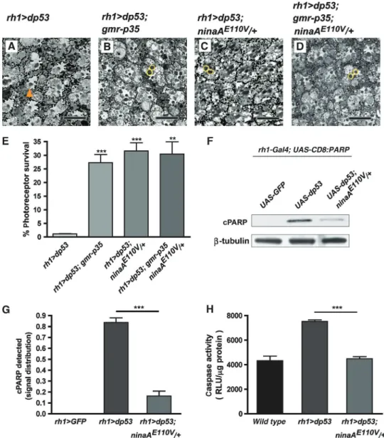
![Figure 7 ER stress triggers upregulation of the fer2lch and d4E-BP genes. (A–H) Horizontal retinal cryosections stained for nuclear b -galactosidase activity from flies carrying the transposable P-element line inserted in fer2lch (Pz[ry, lacZ] fer2lch035 )](https://thumb-eu.123doks.com/thumbv2/123doknet/14252458.488386/127.892.194.703.121.768/triggers-upregulation-horizontal-cryosections-galactosidase-activity-carrying-transposable.webp)