Publisher’s version / Version de l'éditeur:
Journal of Pharmacology And Experimental Therapeutics, 315, July 2, pp.
563-570, 2005
READ THESE TERMS AND CONDITIONS CAREFULLY BEFORE USING THIS WEBSITE. https://nrc-publications.canada.ca/eng/copyright
Vous avez des questions? Nous pouvons vous aider. Pour communiquer directement avec un auteur, consultez la
première page de la revue dans laquelle son article a été publié afin de trouver ses coordonnées. Si vous n’arrivez pas à les repérer, communiquez avec nous à PublicationsArchive-ArchivesPublications@nrc-cnrc.gc.ca.
Questions? Contact the NRC Publications Archive team at
PublicationsArchive-ArchivesPublications@nrc-cnrc.gc.ca. If you wish to email the authors directly, please see the first page of the publication for their contact information.
NRC Publications Archive
Archives des publications du CNRC
This publication could be one of several versions: author’s original, accepted manuscript or the publisher’s version. / La version de cette publication peut être l’une des suivantes : la version prépublication de l’auteur, la version acceptée du manuscrit ou la version de l’éditeur.
For the publisher’s version, please access the DOI link below./ Pour consulter la version de l’éditeur, utilisez le lien DOI ci-dessous.
https://doi.org/10.1124/jpet.105.087528
Access and use of this website and the material on it are subject to the Terms and Conditions set forth at
Differential Regulation of Cystic Fibrosis Transmembrane Conductance
Regulator by Interferon gamma in Mast Cells and Epithelial Cells
Kulka, Marianna; Dery, Rene; Nahirney, Drew; Duszyk, Marek; Befus, A.
Dean
https://publications-cnrc.canada.ca/fra/droits
L’accès à ce site Web et l’utilisation de son contenu sont assujettis aux conditions présentées dans le site LISEZ CES CONDITIONS ATTENTIVEMENT AVANT D’UTILISER CE SITE WEB.
NRC Publications Record / Notice d'Archives des publications de CNRC:
https://nrc-publications.canada.ca/eng/view/object/?id=98787079-6caa-4084-931a-1ae692a96347
https://publications-cnrc.canada.ca/fra/voir/objet/?id=98787079-6caa-4084-931a-1ae692a96347
Differential Regulation of Cystic Fibrosis Transmembrane
Conductance Regulator by Interferon ␥ in Mast Cells and
Epithelial Cells
Marianna Kulka,
1Rene Dery, Drew Nahirney, Marek Duszyk, and A. Dean Befus
Pulmonary Research Group, University of Alberta, Edmonton, Alberta, Canada
Received April 5, 2005; accepted July 26, 2005
ABSTRACT
Cystic fibrosis transmembrane conductance regulator (CFTR) is a cAMP-dependent chloride channel in epithelial cells; recently, we identified it in mast cells. Previous work that we confirmed showed that interferon ␥ (IFN␥) down-regulated CFTR expres-sion in epithelial cells (T84), but by contrast, we found that IFN␥ up-regulated CFTR mRNA and protein expression in rat and human mast cells. IFN␥ up-regulation of CFTR in mast cells was inhibited by p38 and extracellular signal-regulated kinase (ERK) kinase inhibitors but not a Janus tyrosine kinase (JAK)2 inhibitor, whereas in T84 cells IFN␥-mediated down-regulation of CFTR was JAK2-dependent and ERK- and
p38-indepen-dent. Furthermore, IFN␥ down-regulation of CFTR in T84 epi-thelial cells was STAT1-dependent, but up-regulation of CFTR in mast cells was STAT1-independent. Thus, differential regu-latory pathways of CFTR expression in mast cells and epithelial cells exist that depend upon either p38/ERK or JAK/STAT pathways, respectively. Surprisingly, IFN␥ treatment of mast cells inhibited Cl⫺efflux, in contrast to up-regulation of CFTR/
mRNA and protein expression. However, down-regulation of Cl⫺ flux correlated with IFN␥-mediated inhibition of mediator
secretion. This and other work suggests that the effect of IFN␥ on CFTR expression in mast cells is important for their function.
The cystic fibrosis transmembrane conductance regulator (CFTR) is a cAMP-dependent Cl⫺ channel that controls
transepithelial electrolyte transport, fluid flow, and ion con-centrations in the intestine, lungs, pancreas, and sweat glands (Gibson et al., 2003). Over 1200 disease-associated mutations in the cystic fibrosis gene have been reported to the Cystic Fibrosis Genetic Analysis Consortium database (www.genet.sickkids.on.ca/cftr/). Approximately 70% of pa-tients with the disease have a deletion of phenylalanine at amino acid position 508 (⌬F508) that severely decreases CFTR expression in the plasma membrane and compromises permeability to Cl⫺. CFTR expression is temporally and
spa-tially complex and is regulated by many factors, including
cytokines (Besancon et al., 1994; Baudouin-Legros et al., 2005).
In mast cells, several specific Cl⫺conductances have been
identified and linked with degranulation. After antigen stim-ulation of rat peritoneal mast cells (PMC), there is an in-crease in Cl⫺uptake (Romanin et al., 1991; Friis et al., 1994).
Cl⫺ channel blockers such as
5-nitro-2-(3-phenylpro-pylamino) benzoic acid inhibit both mast cell Cl⫺current and
degranulation (Romanin et al., 1991), whereas diphe-nylamine-2-carboxylate blocks Fc⑀RI-stimulated degranula-tion and forskolin-induced Cl⫺current in PMCs (Kulka et al.,
2002a). Moreover, mast cell-stabilizing compounds cromolyn and nedocromil inhibit mast cell degranulation as well as Cl⫺
ion flux (Alton and Norris, 1996). We have identified CFTR and voltage-gated chloride channel (ClC) family members ClC-2, 3, 4, 5, and 7 in rat mast cells (Kulka et al., 2002a,b), and others have identified ClC3, 5, and 7 in human mast cells (Duffy et al., 2001; Bradding et al., 2003). Thus, given that CFTR in mast cells may be important for their functions, we have studied the regulation of CFTR expression in mast cells.
This work was funded by grants to A.D.B. from the Canadian Institutes of Health Research (CIHR) and to M.D. from the Canadian Cystic Fibrosis Foundation. M.K. was supported by a student voucher from CIHR.
1Current address: Allergy-Immunology Division, Feinberg School of Medi-cine, Northwestern University, Chicago, IL.
Article, publication date, and citation information can be found at http://jpet.aspetjournals.org.
doi:10.1124/jpet.105.087528.
ABBREVIATIONS: CFTR, cystic fibrosis transmembrane conductance regulator; PMC, rat peritoneal mast cell(s); ClC, voltage-gated Cl⫺channel; IFN, interferon; JAK, Janus tyrosine kinase; STAT, signal transduction and activator of transcription; MAPK, mitogen-activated protein kinase; ERK, extracellular signal-regulated kinase; TNF, tumor necrosis factor; AG-490, ␣-cyano-(3,4-dihydroxy)-N-benzylcinnamide tyrphostin B42; SB202190, C20H14FN3O; U0126, 1,4-diamino-2,3-dicyano-1,4-bis(2-aminophenylthio)-butadiene); PMA, phorbol 12-myristate 13-acetate; HTB, HEPES Tyrode’s buffer; MC, mast cell; RCMC, rat cultured mast cell(s); FBS, fetal bovine serum; LAD2, Laboratory of Allergic Diseases mast cell line 2; PBS, phosphate-buffered saline; PAGE, polyacrylamide gel electrophoresis; JNK, c-Jun NH2-terminal kinase; FAM, 6-carboxyfluorescein; BSA, bovine serum albumin; PCR, polymerase chain reaction; MQAE, N-(ethoxycarbonylmethyl)-6-methoxyquinolinium bromide; IL, interleukin.
THEJOURNAL OFPHARMACOLOGY ANDEXPERIMENTALTHERAPEUTICS Vol. 315, No. 2
Copyright © 2005 by The American Society for Pharmacology and Experimental Therapeutics 87528/3056048
JPET 315:563–570, 2005 Printed in U.S.A.
563
at Univ Of Prince Edw Island on June 26, 2009
jpet.aspetjournals.org
In epithelial cells, IFN␥ down-regulates expression of CFTR resulting in a significant decrease in CFTR-mediated Cl⫺current (Besancon et al., 1994). IFN␥ is a member of a
family of inducible secretory proteins produced largely by activated T lymphocytes and natural killer cells (Schroder et al., 2004). IFN␥ modulates gene expression by activating Janus tyrosine kinase (JAK), resulting in signal transducer and activator of transcription (STAT) 1 binding and phos-phorylation. Phosphorylated STAT1 dimerizes and translo-cates into the nucleus where it binds to ␥-activated sequence elements and initiates transcription (Schroder et al., 2004). In addition to the JAK/STAT pathway, IFN␥ activates other signal-transduction proteins such as p38 mitogen-activated protein kinases (MAPKs) and extracellular signal-regulated kinase (ERK) 1/2 MAPK (Ramana et al., 2002). With regard to mast cells, IFN␥ can inhibit proliferation, TNF-mediated cytotoxicity, cell differentiation, and mediator release (Kir-shenbaum et al., 1988; Bissonnette and Befus, 1990; Holliday et al., 1994). We hypothesized that IFN␥ may down-regulate CFTR expression in mast cells by a JAK/STAT1-dependent pathway, as in epithelial cells. However, in contrast to epi-thelial cells, we found that IFN␥ up-regulated CFTR expres-sion in both rat and human mast cells and in a JAK/STAT1 independent manner. Surprisingly, Cl⫺flux measurements
indicate that IFN␥ treatment of mast cells reduces Cl⫺flux
despite the up-regulation of CFTR levels.
Materials and Methods
Materials. The JAK2 inhibitor AG-490 was obtained from Cal-biochem (San Diego, CA), the p38 inhibitor SB202190 and the ERK inhibitor U0126 were from Cell Signaling Technology Inc. (Beverly, MA). Phorbol 12-myristate 13-acetate (PMA) was obtained from Sig-ma-Aldrich (St. Louis, MO). Stem cell factor, TNF, and IFN␥ (rat and human) were purchased from PeproTech (Rocky Hill, NJ).
Rats and PMC Isolation. Male Sprague-Dawley rats (300 –350 g; Charles River, St. Constant, QC, Canada) were housed in a patho-gen-free viral antibody-free facility. Rats were sacrificed by cervical dislocation under anesthesia, and PMCs were isolated as described previously (Kulka et al., 2002a). Briefly, 20 ml of HEPES Tyrode’s buffer (HTB) containing 137 mM NaCl, 5.5 mM glucose, 2.7 mM KCl, 0.5 mM NaH2PO4, 1 mM CaCl2, 12 HEPES, pH 7.2, and 1% BSA) was injected into the peritoneal cavity and massaged gently for 30 sec; the peritoneal cavity was opened, the buffer collected and kept at 4°C. After centrifugation at 200g for 5 min, the cell pellet was resuspended in 5 ml of HTB, layered on top of a 30/80% Percoll gradient, centrifuged at 500g for 20 min, and the MCs were collected from the pellet. PMCs were ⬎98% pure and ⬎96% viable as mea-sured by trypan blue exclusion.
Nippostrongylus brasiliensis Sensitization. Sprague-Dawley
rats were sensitized to N. brasiliensis by a single subcutaneous injection of 3000 L3 larvae (Befus et al., 1982). The adult worms were expelled after 10 days, but the mast cells remained sensitized with worm antigen-specific IgE for several weeks. The rats were used for experiments 30 to 40 days postinfection.
Cell Culture. The rat cultured MC (RCMC) line 1.11.2 (kindly provided by B. Chan and A. Froese, Winnipeg, MB Canada) was cultured in RPMI 1640 medium containing 5% FBS (Invitrogen, Carlsbad, CA), 100 U/ml penicillin, 100 g/ml streptomycin, and 10 mM HEPES (Sigma-Aldrich). The recently established human mast cell line Laboratory for Allergic Diseases (LAD) 2 (Kirshenbaum et al., 2003) (a generous gift from Drs. Kirshenbaum, Akin, and Met-calfe, National Institutes of Health, Bethesda, MD), was cultured in serum-free media (StemPro-34; Invitrogen) supplemented with 2 mML-glutamine, 100 U/ml penicillin, 50 g/ml streptomycin, and
100 ng/ml stem cell factor. The T84 epithelial cell line was cultured in F-12/Dulbecco’s modified Eagle’s medium (Invitrogen) containing 5% FBS, 100 U/ml penicillin, and 100 g/ml streptomycin. All cells were incubated in a humidified atmosphere of 5% CO2in air at 37°C. Western Blot. Cells were washed with PBS and 1 ⫻ 106cells were lysed in buffer containing loading dye solution (lithium dodecyl sulfate) sample buffer (Invitrogen), 10% -mercaptoethanol (Sigma-Aldrich), 0.1 M dithiothreitol (Sigma-Aldrich) and protease inhibitor cocktail (Roche Diagnostics, Indianapolis, IN). Whole cell lysates (30 g) were separated on 4 to 12% Bis-Tris SDS-PAGE gels (Invitrogen) and transferred onto nitrocellulose membranes. The membranes were blocked with 3% milk in Tris-buffered saline-0.05% Tween for 1 h and then probed with primary antibodies against CFTR (clone H-182) and STAT1 (Santa Cruz Biotechnology Inc., Santa Cruz, CA), phosphoSTAT1 (BD Transduction Laboratories, Chicago, IL), phos-pho-stress-activated MAPK/c-Jun NH2-terminal kinase (JNK) (Thr183/Tyr185; Cell Signaling Technology Inc.), phospho-p38 MAPK (Thr180/Tyr182; Cell Signaling Technology Inc.), and phos-pho ERK1/2 (Thr202/Tyr204; Cell Signaling Technology Inc.), or anti-actin (Sigma-Aldrich) in 4% BSA/PBS for 1 h at room temper-ature. The membranes were washed with TBS-Tween three times and then incubated with the horseradish peroxidase-linked second-ary antibody (sheep anti-rabbit; Jackson ImmunoResearch Labora-tories, West Grove, PA, or goat anti-mouse; Santa Cruz Biotechnol-ogy Inc.) for 1 h. The membranes were developed with chemiluminescence reagent (Invitrogen) for 1 min and exposed to chemiluminescence film for 1 to 5 min.
Confocal Microscopy. CFTR was localized in sham-treated or IFN␥ treated (80 ng/ml, 24 h) rat PMCs and human T84. After incubation, T84 cells were detached by 10-min incubation with tryp-sin/EDTA at room temperature and 50,000 PMCs or T84 were then cytocentrifuged onto Superfrost plus charged slides using a Shandon cytospin 2 (Fisher Scientific, Mississauga, ON, Canada) at 5g for 6 min in PBS containing 20% FBS. Cells were then air-dried overnight and fixed in 75% acetone/25% absolute ethanol for 15 min at ⫺20°C. Nonspecific binding sites were blocked by incubation in blocking buffer (PBS containing 3% BSA and 10% normal goat serum) for 2 h at room temperature. Slides were then incubated with mouse anti human CFTR primary antibody (MA1-935; Affinity Bioreagents, Golden, CO) at 1/50 dilution in blocking buffer for 2 h at room temperature. After three washes in PBS, specific antibody binding was detected with Alexa 568-conjugated goat anti-mouse IgM (Mo-lecular Probes, Eugene, OR) at 1/2000 dilution in blocking buffer for 1 h at room temperature. Cell images were obtained using an Olym-pus FV1000 laser scanning confocal microscope (Carsen Group, Markham, ON, Canada) with 400⫻ magnification.
Quantitative Real-Time Polymerase Chain Reaction. Our previous study established that mast cells express CFTR mRNA (Kulka et al., 2002a). In the current study, RNA was isolated as described previously (Gilchrist et al., 1997) and quantitative real-time PCR assay was performed using gene-specific fluorescently labeled primers and a 7700 sequence detector (Applied Biosystems, Foster City, CA). All primers and reagents were obtained from In-vitrogen. Primers were designed using the LUX primer design tool and are listed along with their Invitrogen reference code (Table 1). Each primer set consisted of one labeled [6-carboxyfluorescein (FAM) fluorescent reporter at the 5⬘ end] and one unlabeled primer. Low-ercase nucleotides in the labeled primer sequence represent hairpin-generating segments of the labeled primer. Data were collected dur-ing the annealdur-ing/extension phase of PCR and analyzed usdur-ing the comparative Ctmethod (Nazarenko et al., 2002).
36ClⴚFlux Measurements. Changes in [Cl⫺] were measured by incubating 1 ⫻ 106cells/ml with 8.7 mM Na36Cl (MP Biomedicals, Aurora, OH) in flux buffer (137 mM NaCl, 4 mM KCI, 1 mM MgSO4, 1 mM CaCl2, 20 mM HEPES, 1 mg/ml BSA, and 1 mg/ml glucose) at 37°C for 30 min (Friis et al., 1994). The incubation was terminated by transferring 100 l of the cell suspension onto 120 l of silicone oil in long, thin Eppendorf tubes. The tubes were centrifuged at 18,000g
564
Kulka et al.at Univ Of Prince Edw Island on June 26, 2009
jpet.aspetjournals.org
for 30 s and then placed into a freezing methanol bath. The bottom of each tube was cut off and placed into a scintillation vial with 48 mM NaOH. Each vial was vortexed for 1 min after which 5 ml of scintillation fluid was added and placed in a Beckman scintillation counter.36Cl⫺uptake was calculated based on the specific activity of 36Cl⫺in the extracellular medium, calculated as the sum of extra-cellular Cl⫺and added36Cl⫺(in nanomoles) divided by the radioac-tivity of the added36Cl⫺(in cpm). All values of36Cl⫺uptake were corrected for36Cl⫺ trapped in the extracellular space, which was determined by measuring cpm immediately after 36Cl⫺ addition (50 ⫾ 10.2 cpm).
N-(Ethoxycarbonylmethyl)-6-methoxyquinolinium Bromide
(MQAE) Measurements. Fluorescence measurements were per-formed in HTB. Gluconate, Br⫺, or I⫺buffers were identical to HTB except that 137 mM NaCl was replaced by equivalent amounts of sodium gluconate, NaBr, or NaI, respectively. One million cells per milliliter were incubated with 5 mM of the Cl⫺-sensitive dye MQAE (Molecular Probes, Eugene, OR) in 1 ml of HTB for 30 min at 37°C. Cells were washed twice and resuspended in 50 l of HTB. MQAE is quenched by Cl⫺ anions and thus, when Cl⫺ leaves the cell, it dissociates from MQAE and fluorescence increases. MQAE fluores-cence was excited at 350 nm, and the emission was measured at 450 nm with a PTI spectrofluorimeter (Photon Technology International, London, ON, Canada), using Felix software (version 1.42). All exper-iments were performed at 37°C. To produce a driving force for Cl⫺ efflux, the cells were added to 1 ml of gluconate buffer and MQAE fluorescence was monitored for up to 10 min. Cl⫺efflux was calcu-lated as the initial rate of change of MQAE fluorescence after addi-tion of cells to the gluconate buffer. For quantitative analysis, the data collected in the first 60 s were fitted using linear regression, and the slope was used as a measure of Cl⫺ efflux. All traces were normalized to initial baseline reading (buffer, no cells). In some experiments, 10 worm equivalents/ml of N. brasiliensis antigen was added to the cell suspension, and changes in fluorescence were mon-itored for up to 10 min.
Statistics. All data are presented as mean of at least three inde-pendent experiments with standard error of the mean (S.E.M.). Where indicated, data were analyzed using a paired t test for sample means, analysis of variance, or the Tukey-Kramer multiple compar-isons test.
Results
Interferon ␥ Up-Regulates CFTR mRNA and Protein Expression in Rat and Human Mast Cells. PMA, TNF,
and IFN␥ down-regulate CFTR expression in epithelial cells (Nakamura et al., 1992; Shen et al., 1993; Besancon et al., 1994). To determine whether CFTR in mast cells was simi-larly regulated, RCMC were treated with PMA, TNF, or IFN␥ for 24 h and CFTR expression was identified by West-ern blotting as we have done previously (confirmed using isotype controls for flow cytometry, Western blot, and immu-nohistochemistry; Kulka et al., 2002a). As expected, TNF and IFN␥ decreased CFTR expression in T84 cells (Fig. 1A). Sur-prisingly, however, TNF and IFN␥ up-regulated CFTR ex-pression in RCMC. PMA had no detectable effect on CFTR
expression in RCMC or T84 (Fig. 1B). Because IFN␥ up-regulates STAT1 expression (Hu et al., 2002), membranes were stripped and reprobed with anti-STAT1. As expected, STAT1 protein (visible as a double band, representing STAT1␣ and STAT1) was up-regulated in both RCMC and T84 by PMA, TNF, and IFN␥.
Fig. 1. Western blot analysis of CFTR and STAT1 expression in un-treated (lane 1), PMA-un-treated (lane 2), TNF-un-treated (lane 3), and IFN␥-treated (lane 4) T84 (A) and RCMC (B). T84 cells were IFN␥-treated with PMA (10 ng/ml), human recombinant TNF (10 ng/ml), or IFN␥ (10 ng/ml) for 24 h. RCMC were treated with PMA (10 ng/ml) or rat recombinant TNF (10 ng/ml) or IFN␥ (10 ng/ml) for 24 h. Cell lysates were resolved on an 4 to 12% SDS-PAGE and blotted with anti-CFTR, anti-STAT1, or anti-actin antibody. Western blots shown represent three independent experiments. The effect of 24 h IFN␥ treatment (80 ng/ml) on CFTR expression was characterized in T84 cells (C) and rat peritoneal mast cells (D), using confocal laser scanning microscopy (bar on the figure represents 10 m). TABLE 1
List of quantitative real-time PCR primers used in experiments
Product GenBank Accession No. Primer Sequence Invitrogen Order Code
Human CFTR (forward; unlabeled) NM_000492 5⬘-GCATACTGCTGGGAAGAAGCAA-3⬘ hCFTR_1017RL_973FU Human CFTR (reverse; FAM- labeled) NM_000492 3⬘-gactcgACATAGGCTGCCTTCCGAGtC-5⬘ hCFTR_1017RL
Rat CFTR (forward) XM_347232 5⬘-CGCAGGTTCTCAGTGGACGA-3⬘ rat CFTR_1986RL_1976FU Rat CFTR (reverse; FAM-labeled) XM_347232 3⬘-cacctgACTGTTTGGCTTTGTTCCAGGtG-5⬘ rat CFTR_1986RL Human/rat -actin (forward) NM_001101 5⬘-GACGAGGCCCAGAGCAAGA-3⬘ actin_304RL_257FU Human/rat -actin (reverse; FAM-labeled) NM_001101 3⬘-caactgTCTCCATGTCGTCCCAGtTG-5⬘ actin_304RL
at Univ Of Prince Edw Island on June 26, 2009
jpet.aspetjournals.org
Confocal analysis of CFTR expression in T84 cells showed a largely cytoplasmic distribution and as expected from the results of Western blot analysis, the intensity of CFTR stain-ing was decreased after IFN␥ treatment (80 ng/ml, 24 h; Fig. 1C). By contrast, CFTR expression in rat PMC was increased after IFN␥ treatment and seemed to be associated with gran-ules (Fig. 1D). Studies of nonpermeabilized cells identified some CFTR in a plasma membrane-like distribution on T84, but there was no obvious CFTR with such a distribution on PMC (not shown).
To further characterize up-regulation of CFTR in mast cells, CFTR mRNA expression after IFN␥ dose-response and time-course treatments was analyzed in RCMC, human LAD2, and T84 (Fig. 2). Quantitative PCR analysis con-firmed that IFN␥ (10 ng/ml) significantly (p ⬍ 0.05) up-regulated CFTR mRNA expression in RCMC by 3 h after treatment, and the magnitude of this up-regulation was 39 ⫾ 13% at 12 h. In LAD2 MC, the up-regulation of CFTR was statistically significant by 8 h of IFN␥ treatment and by 12 h was 54 ⫾ 17% greater than in untreated cells (Fig. 2A). Significant up-regulation of CFTR was induced in mast cells within 8 h with as little as 1 ng/ml (RCMC) or 10 ng/ml (LAD2) of IFN␥ (Fig. 2B).
By contrast, IFN␥ significantly decreased CFTR mRNA expression in T84 cells within 3 h (Fig. 2, A and B) and by 12 h the magnitude of decrease was 49 ⫾ 4% at 12 h of treatment. The IFN␥ effect was dose-dependent such that 1, 10 and 100 ng/ml IFN␥ decreased CFTR mRNA expression in T84 cells by 23, 50, and 83% after 8 h (all statistically sig-nificant decreases compared with the untreated group).
Western blot analysis showed that by 6 h, CFTR expres-sion was increased compared with untreated RCMC and by
24 h, CFTR expression was significantly up-regulated (Fig. 2C). In T84 cells, a decrease in CFTR protein expression was observable at 6 h and remained low at 24 h (Fig. 2D). In both T84 and RCMC, STAT1 protein was up-regulated after 6 h of treatment and remained elevated up to 24 h.
IFN␥ Up-Regulation of CFTR Is Inhibited by MAP Kinase Inhibitors. To determine which IFN␥ signaling
pathways were activated in mast cells compared with epithe-lial cells, RCMC were treated with IFN␥, and whole cell lysates were analyzed for phosphorylated STAT1, p38, ERK, and JNK (Fig. 3). IFN␥ activated STAT1, ERK, and p38, but not JNK phosphorylation. STAT1 was activated at 5 min and remained activated for up to 30 min. ERK2 (bottom band) was constitutively activated, but phosphorylation of both ERK1 (top) and ERK2 (bottom) was induced after 5 min. p38 was activated at 15 min, later than STAT1 or ERK.
To determine whether JAK/STAT1, p38 or ERK signaling pathways were involved in IFN␥-mediated up-regulation of CFTR, RCMC, LAD2, and T84 were treated with IFN␥ in the presence of a JAK2 inhibitor, AG-490; a p38 kinase inhibitor, SB202190; and an ERK MAPK inhibitor, U0126. In RCMC and LAD2, AG-490 did not affect IFN␥-mediated up-regula-tion of CFTR protein expression, but both SB202190 and U0126 partially inhibited IFN␥-mediated up-regulation of CFTR (Fig. 4). By contrast, in T84 cells, AG-490 blocked IFN␥-mediated down-regulation of CFTR. Membranes were stripped and reblotted with anti-STAT1 to compare regula-tion of another IFN␥-responsive protein. STAT1 up-regula-tion in RCMC was sensitive to AG-490 but in human LAD2 cells, STAT1 up-regulation was inhibited by AG-490, SB202190, and U0126. T84 cells were similar to the RCMC in that STAT1 up-regulation was only blocked by AG-490 but
Fig. 2. Quantitative PCR analysis of CFTR expression in RCMC, LAD2, and T84 after different times and doses of IFN␥ treatment (A and B). Asterisks represent statistical signifi-cance as determined by Student’s t test compared with untreated sample (time 0) in each case (p ⬍ 0.05). The dose used in A was 10 ng/ml, and in B treatment was for 8 h. Western blot analysis of CFTR, STAT, and actin expression in RCMC and T84 after a time course (hours) of 10 ng/ml IFN␥ treatment (C and D) (n ⫽ 3).
566
Kulka et al.at Univ Of Prince Edw Island on June 26, 2009
jpet.aspetjournals.org
not SB202190 and U0126. Densitometry analysis of three independent blotting experiments confirmed that AG-490 did not affect IFN␥-mediated up-regulation of CFTR protein ex-pression in both LAD2 and RCMC (Fig. 4B; p ⬍ 0.05).
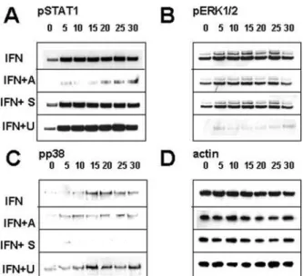
To confirm the actions of AG-490, SB202190, and U0126, RCMC were treated with IFN␥ (10 ng/ml) in the presence of these inhibitors, and STAT1, p38, and ERK1/2 phosphoryla-tion was assessed by Western blotting (Fig. 5). As expected, AG-490 but not SB202190 or U0126 inhibited STAT1 consti-tutive and IFN␥-induced phosphorylation (Fig. 5A). U0126 inhibited ERK1/2 constitutive and IFN␥-induced phosphory-lation (Fig. 5B). SB202190 inhibited IFN␥ induced p38 phos-phorylation (Fig. 5C).
IFN␥ Inhibits Both Constitutive and Antigen-In-duced ClⴚFlux in Mast Cells. To determine the effect of
IFN␥ on Cl⫺flux in resting and antigen-IgE-activated mast
cells, we used two methods: measurement of36Cl⫺uptake
and assessment of Cl⫺sensitive fluorescence using MQAE.
Studies with 36Cl⫺ have shown that IFN␥ treatment
de-creases Cl⫺uptake of PMC (Fig. 6A; p ⬍ 0.01). A time course
of PMC Cl⫺uptake shows that IFN␥ did not have an effect at
the earlier treatment points (less than 2 h) but decreased Cl⫺
uptake at 20 and 24 h (Fig. 6B). Similar results were ob-tained with36Cl⫺uptake measurements in RCMC (data not
shown).
Fluorescence measurements were performed with mast
Fig. 4. A, IFN␥-mediated up-regulation of mast cell CFTR protein requires activation of JAK2 and/or p38 and ERK. RCMC, LAD2, and T84 were treated with IFN␥ (10 ng/ml) with or without JAK2 inhibitor (AG-490; 30 mg/ml), p38 inhibitor (SB202190; 30 mg/ml), or ERK MAPK inhibitor (U0126; 30 mg/ml) for 24 h. Representative of three inde-pendent experiments. B, densitometry summary of data in Fig. 4. RCMC were treated with IFN␥ (10 ng/ml) with or without JAK2 inhibitor (AG-490; 30 mg/ml), p38 inhibitor (SP202190; 30 mg/ml), or ERK inhibitor (U0126; 30 mg/ml) for 24 h, and expression of CFTR (black columns) and STAT1 (gray columns) was analyzed by Western blot (n ⫽ 3 independent experiments; error bars represent S.E.M.). As-terisks represent statistical significance as determined by Student’s t test (p ⬍ 0.05).
Fig. 3. Western blot analysis of STAT1 and MAPK activation after IFN␥ treatment. RCMC were treated with 10 ng/ml IFN␥ for indicated times, and cell lysates were probed with antibodies to phosphorylated STAT1, p38, ERK, or JNK. Blots were stripped and probed with anti-actin to show equal loading. Representative of three independent experiments.
Fig. 5. RCMC were treated with IFN␥ (10 ng/ml) alone (IFN) or pre-treated with AG-490 (IFN⫹A; 30 mg/ml), SB202190 (IFN⫹S; 30 mg/ml), or U0126 (IFN⫹U; 30 mg/ml) for 10 min and then stimulated with IFN␥ (10 ng/ml) and for 1 to 30 min. Cell lysates were analyzed for phosphor-ylation of STAT1 (A), ERK1/2 (B), or p38 (C), and -actin (D) was as-sessed as a loading control. Representative of three independent experi-ments.
at Univ Of Prince Edw Island on June 26, 2009
jpet.aspetjournals.org
cells loaded with MQAE in HTB solution and chloride efflux was measured after placing cells in gluconate buffer. Figure 6C shows that IFN␥ treatment significantly reduced Cl⫺flux
in sensitized PMC not challenged with antigen (p ⬍ 0.05; n ⫽ 8 and 11, control and IFN␥-treated cells, respectively). After antigen challenge (10 worm equivalents/ml) the magnitude of the IFN␥-mediated depression in Cl⫺ efflux was reduced
(Fig. 6D). Although antigen challenge in the absence of IFN␥ treatment showed a trend toward reduced Cl⫺ efflux, this
was not statistically significant (p ⬎ 0.5; n ⫽ 3). Identical results were obtained in both PMC and RCMC under condi-tions when the cells were loaded in gluconate buffer and placed in HTB to measure Cl⫺influx (data not shown).
Measurements of halide permeabilities indicated that Br⫺
was more permeable that Cl⫺ and I⫺ in PMC cells [Br⫺
(1.34) ⬎ Cl⫺(1.00) ⱖ I⫺(0.68); n ⫽ 3 in each set]. Similar
results were obtained with RCMC [Br⫺(1.19) ⱖ Cl⫺(1.00) ⬎
I⫺ (0.61); n ⫽ 3 in each set). The halide permeability
se-quence, Br⫺ ⱖ Cl⫺ ⬎ I⫺, is characteristic of CFTR Cl⫺
channels (Illek et al., 1999) and suggests that CFTR channels are an important component of Cl⫺flux in mast cells.
Discussion
This is the first study demonstrating that CFTR expres-sion is regulated differently in epithelial and nonepithelial cells. Moreover, we show that IFN␥-induced up-regulation of CFTR in MC involved MAPK signaling pathways, whereas IFN␥-induced down-regulation of CFTR in epithelial cells involved JAK/STAT pathways. Paradoxically, we also show that despite IFN␥ up-regulation of CFTR in mast cells, IFN␥ treatment depressed mast cell Cl⫺ flux in multiple assay
systems.
It is now well established that CFTR gene expression is regulated in a complex, cell- and stimulus-specific manner
that may involve both transcriptional and posttranscrip-tional mechanisms. For example, TNF decreases CFTR mRNA in human colonic epithelial cells but not in airway epithelial cells, whereas IL-1 increases it only in airway epithelial cells (Baudouin-Legros et al., 2005). Although stimulation of CFTR gene expression by IL-1 involves acti-vation of the CFTR promoter (Brouillard et al., 2001), down-regulation of CFTR by TNF and IFN␥ involves mainly post-transcriptional mechanisms (Baudouin-Legros et al., 2005). The results of this study show that in mast cells both TNF and IFN␥ increase CFTR mRNA, but whether this process affects CFTR gene transcription and/or mRNA stability is presently unknown. However, the fact that IFN␥ increased CFTR protein levels to a greater extent than the mRNA suggests that IFN␥ treatment may increase mRNA stability rather than CFTR gene transcription.
IFN␥ modulation of gene expression is mediated by both STAT1-dependent and independent pathways (Gil et al., 2001). Our results show that IFN␥ activates STAT1, ERK1/2, and p38 but not JNK, suggesting that these pathways are also induced in mast cells. Using inhibitors to JAK/STAT, ERK, and p38, we determined that in both rat and human mast cells, CFTR up-regulation is JAK/STAT independent but requires activation of the MAPK pathways mediated by ERK and p38. In T84 cells, IFN␥-mediated down-regulation of CFTR is inhibited by AG-490 but is unaffected by the p38 and ERK inhibitors. STAT1 expression, by comparison, is up-regulated by IFN␥ in both mast cells and T84 epithelial cells and is inhibited by AG-490, suggesting that STAT1 up-regulation is a positive feedback mechanism that sensi-tizes mast cells to IFN␥ as previously observed in human macrophages (Duffy et al., 2001). In human LAD2 cells, IFN␥-mediated up-regulation of STAT1 is also sensitive to ERK and p38 inhibitors, perhaps indicating the importance
Fig. 6. IFN␥ decreases C1⫺flux. Rat peritoneal mast cells
were purified (⬎95%) and treated with IFN␥ at various doses (A; 24 h) or for various times (B; 80 ng/ml). After treatment, mast cells were washed and incubated with
36C1⫺for 30 min. Mast cells were spun through oil, and
radioactivity of cell pellets was measured in a scintillation counter (n ⫽ 5 independent experiments; p ⬍ 0.01 com-pared with untreated). Effects of IFN␥ on C1⫺flux in PMC
as measured by MQAE (C and D). Effects of IFN␥ (80 ng/ml, 24-h pretreatment) on C1⫺efflux from sensitized rat
peritoneal mast cells. After IFN␥ pretreatment, cells were loaded with the C1⫺-sensitive dye MQAE, and the driving
force for C1⫺ efflux involved placing them in gluconate
buffer with or without antigen challenge (10 worm equiv-alents/ml; n ⫽ 8 –11 independent experiments; p ⬍ 0.01).
568
Kulka et al.at Univ Of Prince Edw Island on June 26, 2009
jpet.aspetjournals.org
of MAPK pathways in IFN␥ signaling. Therefore, IFN␥ acti-vates at least two pathways in mast cells—the JAK/STAT pathway responsible for up-regulation of STAT1 and the p38/ERK pathway(s) that is responsible for up-regulation of CFTR. Although ERK activation is involved in CFTR up-regulation, PMA activates ERK but does not up-regulate CFTR (Fig. 1). This suggests that ERK activation requires activation of other molecules, perhaps p38, for up-regulation of CFTR mRNA.
The exact role of CFTR in mast cell function is unknown and is the subject of other work in our laboratory. To date, we have established that diphenylamine-2-carboxylate, a drug known to not only inhibit CFTR but also to have other activ-ities, blocks Fc⑀RI-stimulated degranulation of PMC (Kulka et al., 2002a). Moreover, knockdown of CFTR expression by antisense oligonucleotides in the human mast cell line HMC-1 reduces Cl⫺flux, adhesion to fibronectin and calcium
ionophore A23187-induced degranulation and IL-6 produc-tion (A. Schwingshackl and R. Dery, unpublished data). Our working hypothesis is that CFTR in mast cells is an impor-tant component of Cl⫺flux and perhaps of other activities, as
recognized for epithelial cells (Rowe et al., 2005).
In turn, the role of mast cells in cystic fibrosis is unclear. Recently, mast cells have been recognized as important play-ers in innate and acquired immune responses (Marshall, 2004). Moreover, there are increased numbers of mast cells in nasal polyps from cystic fibrosis patients compared with non-cystic fibrosis patients and many show signs of activation in cystic fibrosis (Henderson and Chi, 1992). Differences have also been found in mast cell numbers in human fetal trachea between cystic fibrosis and noncystic fibrosis specimens (Hubeau et al., 2001). Interestingly, mast cell numbers and mast cell-specific genes and others genes associated with innate immunity are up-regulated in the intestine in CFTR null mice that show a severe intestinal phenotype (Norkina et al., 2004). Thus, the role of mast cells in cystic fibrosis warrants further investigation.
The finding of IFN␥-mediated increase in CFTR expression and decrease in Cl⫺flux could be explained in several ways.
For example, if IFN␥ treatment leads to cell depolarization, this would tend to reduce Cl⫺flux under our experimental
conditions, perhaps by channels other than CFTR. Alter-nately, IFN␥ may modulate expression of other proteins in-volved in Cl⫺ flux, e.g., soluble N-ethylmaleimide-sensitive
factor attachment protein receptor proteins, which inhibit CFTR activity by decreasing channel open probability (Cormet-Boyaka et al., 2002). It is also possible that although IFN␥ increases CFTR expression in mast cells, this may not involve maturation of CFTR and its translocation to the plasma membrane, where it could be fully functional. Indeed, our confocal studies of CFTR expression support the hypoth-esis that the increase in CFTR expression in MC is mainly observed intracellularly, most likely in association with gran-ules (Fig. 1D). In addition, studies of the biosynthetic pro-cessing and intracellular trafficking of CFTR indicate that CFTR undergoes constitutive endocytosis and recycling (Pic-ciano et al., 2003). Thus, IFN␥ treatment could affect the balance between CFTR degradation and recycling back to the plasma membrane, reducing the effective amount of CFTR in the plasma membrane.
The role of IFN␥-mediated up-regulation of CFTR in mast cell physiology is difficult to determine. Further studies are
required to characterize the functional effects of increased CFTR on mast cell functions such as Cl⫺transport,
degran-ulation, and mediator release in response to stimuli such as allergens. Furthermore, the transcription factors involved in CFTR up-regulation in mast cells also must be examined to provide insight into regulation of the CFTR promoter. The mechanisms that modulate CFTR gene expression through extracellular and intracellular signals may ultimately pro-vide targets for therapy in cystic fibrosis where CFTR expres-sion is abnormal.
Acknowledgments
We thank Lynelle Watt for help in the preparation of this manu-script and Dr. Dean D. Metcalfe for helpful advice. James Dooley provided skilled technical support for the confocal analyses.
References
Alton EW and Norris AA (1996) Chloride transport and the actions of nedocromil sodium and cromolyn sodium in asthma. J Allergy Clin Immunol 98:S102–S105; discussion S105–S106.
Baudouin-Legros M, Hinzpeter A, Jaulmes A, Brouillard F, Costes B, Fanen P, and Edelman A (2005) Cell-specific posttranscriptional regulation of CFTR gene ex-pression via the influence of MAP kinase cascades on the 3⬘UTR part of the transcripts. Am J Physiol, in press.
Befus AD, Pearce FL, Gauldie J, Horsewood P, and Bienenstock J (1982) Mucosal mast cells. I. Isolation and functional characteristics of rat intestinal mast cells.
J Immunol 128:2475–2480.
Besancon F, Przewlocki G, BaroI I, Hongre AS, Escande D, and Edelman A (1994) Interferon-␥ downregulates CFTR gene expression in epithelial cells. Am J Physiol
267:Cl398 –Cl404.
Bissonnette EY and Befus AD (1990) Inhibition of mast cell-mediated cytotoxicity by IFN␣/ and -␥. J Immunol 145:3385–3390.
Bradding P, Okayama Y, Kambe N, and Saito H (2003) Ion channel gene expres-sion in human lung, skin and cord blood derived mast cells. J Leukoc Biol
73:614 – 620.
Brouillard F, Bouthier M, Leclerc T, Clement A, Baudouin-Legros M, and Edelman A (2001) NF-B mediates up-regulation of CFTR gene expression in Calu-3 cells by interleukin-1. J Biol Chem 276:9486 –9491.
Cormet-Boyaka E, Di A, Chang SY, Naren AP, Tousson A, Nelson DJ, and Kirk KL (2002) CFTR chloride channels are regulated by a SNAP-23/syntaxin 1A complex.
Proc Natl Acad Sci USA 99:12477–12482.
Duffy SM, Leyland ML, Conley EC, and Bradding P (2001) Voltage-dependent and calcium-activated ion channels in the human mast cell line HMC-1. J Leukoc Biol
70:233–240.
Friis UG, Johansen T, Hayes NA, and Foreman JC (1994) IgE-receptor activated chloride uptake in relation to histamine secretion from rat mast cells. Br J
Pharmacol 111:1179 –1183.
Gibson RL, Burns JL, and Ramsey BW (2003) Pathophysiology and management of pulmonary infections in cystic fibrosis. Am J Respir Crit Care Med 168:918 – 951.
Gil MP, Bohn E, O’Guin AK, Ramana CV, Levine B, Stark GR, Virgin HW, and Schreiber RD (2001) Biologic consequences of Stat1-independent IFN signaling.
Proc Natl Acad Sci USA 98:6680 – 6685.
Gilchrist M, MacDonald AJ, Neverova I, Ritchie B, and Befus AD (1997) Optimiza-tion of the isolaOptimiza-tion and effective use of mRNA from rat mast cells. J Immunol
Methods 201:207–214.
Henderson WR Jr and Chi EY (1992) Degranulation of cystic fibrosis nasal polyp mast cells. J Pathol 166:395– 404.
Holliday MR, Banks EM, Dearman RJ, Kimber I, and Coleman JW (1994) Interac-tions of IFN␥ with IL-3 and IL-4 in the regulation of serotonin and arachidonate release from mouse peritoneal mast cells. Immunology 82:70 –74.
Hu X, Herrero C, Li WP, Antoniv TT, Falck-Pedersen E, Koch AE, Woods JM, Haines GK, and Ivashkiv LB (2002) Sensitization of IFN␥ Jak-STAT signaling during macrophage activation. Nat Immunol 3:859 – 866.
Hubeau C, Puchelle E, and Gaillard D (2001) Distinct pattern of immune cell population in the lung of human fetuses with cystic fibrosis. J Allergy Clin
Immunol 108:524 –529.
Illek B, Tam AWK, Fischer H, and Machen TE (1999) Anion selectivity of apical membrane conductance of Calu 3 human airway epithelium. Pflugers Arch 437: 812– 822.
Kirshenbaum AS, Akin C, Wu Y, Rottem M, Goff JP, Beaven MA, Rao VK, and Metcalfe DD (2003) Characterization of novel stem cell factor responsive human mast cell lines LAD 1 and 2 established from a patient with mast cell sarcoma/ leukemia; activation following aggregation of Fc⑀RI or Fc␥RI. Leuk Res 27:677– 682.
Kirshenbaum AS, Worobec AS, Davis TA, Goff JP, Semere T, and Metcalfe DD (1988) Inhibition of human mast cell growth and differentiation by interferon ␥-1. Exp
Hematol 26:245–251.
Kulka M, Gilchrist M, Duszyk M, and Befus AD (2002a) Expression and functional characterization of CFTR in mast cells. J Leukoc Biol 71:54 – 64.
Kulka M, Schwingshackl A, and Befus AD (2002b) Mast cells express chloride channels of the ClC family. Inflamm Res 51:451– 456.
Marshall JS (2004) Mast-cell responses to pathogens. Nat Rev Immunol 4:787–799.
at Univ Of Prince Edw Island on June 26, 2009
jpet.aspetjournals.org
Nakamura H, Yoshimura K, Bajocchi G, Trapnell BC, Pavirani A, and Crystal RG (1992) Tumor necrosis factor modulation of expression of the cystic fibrosis trans-membrane conductance regulator gene. FEBS Lett 314:366 –370.
Nazarenko I, Lowe B, Darfler M, Ikonomi P, Schuster D, and Rashtchian A (2002) Multiplex quantitative PCR using self-quenched primers labeled with a single fluorophore. Nucleic Acids Res 30:e37.
Norkina O, Kaur S, Ziemer D, and De Lisle RC (2004) Inflammation of the cystic fibrosis mouse small intestine. Am J Physiol 286:G1032–G1041.
Picciano JA, Ameen N, Grant BD, and Bradbury NA (2003) Rme-1 regulates the recycling of the cystic fibrosis transmembrane conductance regulator. Am J
Physiol 285:C1009 –C1018.
Ramana CV, Gil MP, Schreiber RD, and Stark GR (2002) Stat-1dependent and -independent pathways in IFN-␥-dependent signaling. Trends Immunol 23:96 – 101.
Romanin C, Reinsprecht M, Pecht I, and Schindler H (1991) Immunologically
acti-vated chloride channels involved in degranulation of rat mucosal mast cells.
EMBO (Eur Mol Biol Organ) J 10:3603–3608.
Rowe SM, Miller S, and Sorscher EJ (2005) Cystic fibrosis. N Engl J Med 352:1992– 2001.
Schroder K, Hertzog PJ, Ravasi T, and Hume DA (2004) Interferon-␥: an overview of signals, mechanisms and functions. J Leukoc Biol 75:163–189.
Shen BQ, Barthelson RA, Skach W, Gruener DC, Sigal E, Mrsny RJ, and Widdi-combe JH (1993) Mechanism of inhibition of cAMP-dependent epithelial chloride secretion by phorbol esters. J Biol Chem 268:19070 –19075.
Address correspondence to: Dr. A. Dean Befus, Room 550A HMRC, Pul-monary Research Group, Department of Medicine, University of Alberta, Ed-monton, AB, Canada, T6G 2S2. E-mail: dean.befus@ualberta.ca
570
Kulka et al.at Univ Of Prince Edw Island on June 26, 2009
jpet.aspetjournals.org