HAL Id: hal-02635206
https://hal.inrae.fr/hal-02635206
Submitted on 27 May 2020
HAL is a multi-disciplinary open access
archive for the deposit and dissemination of sci-entific research documents, whether they are pub-lished or not. The documents may come from teaching and research institutions in France or abroad, or from public or private research centers.
L’archive ouverte pluridisciplinaire HAL, est destinée au dépôt et à la diffusion de documents scientifiques de niveau recherche, publiés ou non, émanant des établissements d’enseignement et de recherche français ou étrangers, des laboratoires publics ou privés.
in the CBEL protein from the oomycete Phytophthora
parasitica
Thomas Martinez, Hélène Texier, Virginie Nahoum, Claude Lafitte, Gianluca
Cioci, Laurent Heux, Bernard Dumas, Michael O’Donohue, Elodie Gaulin, C.
Dumon
To cite this version:
Thomas Martinez, Hélène Texier, Virginie Nahoum, Claude Lafitte, Gianluca Cioci, et al.. Probing the functions of carbohydrate binding Modules in the CBEL protein from the oomycete Phytophthora parasitica. PLoS ONE, Public Library of Science, 2015, 10 (9), �10.1371/journal.pone.0137481�. �hal-02635206�
Probing the Functions of Carbohydrate
Binding Modules in the CBEL Protein from the
Oomycete
Phytophthora parasitica
Thomas Martinez1,2,3,4,5, Hélène Texier3,4,5,6, Virginie Nahoum7,8, Claude Lafitte1,2,
Gianluca Cioci3,4,5, Laurent Heux9, Bernard Dumas1,2, Michael O’Donohue3,4,5, Elodie Gaulin1,2☯, Claire Dumon3,4,5☯*
1 Université Toulouse 3, UPS, Laboratoire de Recherche en Sciences Végétales, 24 chemin de Borde Rouge, BP42617, Auzeville, F-31326, Castanet-Tolosan, France, 2 CNRS, Laboratoire de Recherche en Sciences Végétales, 24 chemin de Borde Rouge, BP42617, Auzeville, F-31326, Castanet-Tolosan, France, 3 Université de Toulouse; INSA, UPS, INP, LISBP, 135 Avenue de Rangueil, F-31077 Toulouse, France, 4 CNRS, UMR5504, F-31400 Toulouse, France, 5 INRA, UMR792 Ingénierie des Systèmes Biologiques et des Procédés, F-31400 Toulouse, France, 6 Cinabio ADISSEO France SAS, Hall Gilbert Durand 3, 135 avenue de Rangueil, 31077 Toulouse, France, 7 Université de Toulouse, UPS, IPBS, Toulouse, F-31077, France, 8 Institut de Pharmacologie et de Biologie Structurale (IPBS), Centre National de la Recherche Scientifique (CNRS), Toulouse, F-31077, France, 9 CERMAV, CNRS, Grenoble, France
☯ These authors contributed equally to this work. *claire.dumon@insa-toulouse.fr
Abstract
Oomycetes are microorganisms that are distantly related to true fungi and many members of this phylum are major plant pathogens. Oomycetes express proteins that are able to inter-act with plant cell wall polysaccharides, such as cellulose. This interinter-action is thought to be mediated by carbohydrate-binding modules that are classified into CBM family 1 in the CAZy database. In this study, the two CBMs (1–1 and 1–2) that form part of the cell wall gly-coprotein, CBEL, from Phytophthora parasitica have been submitted to detailed characteri-zation, first to better quantify their interaction with cellulose and second to determine whether these CBMs can be useful for biotechnological applications, such as biomass hydrolysis. A variety of biophysical techniques were used to study the interaction of the CBMs with various substrates and the data obtained indicate that CBEL’s CBM1-1 exhibits much greater cellulose binding ability than CBM1-2. Engineering of the family 11 xylanase from Talaromyces versatilis (TvXynB), an enzyme that naturally bears a fungal family 1 CBM, has produced two variants. The first one lacks its native CBM, whereas the second contains the CBEL CBM1-1. The study of these enzymes has revealed that wild type TvXynB binds to cellulose, via its CBM1, and that the substitution of its CBM by oomycetal CBM1-1 does not affect its activity on wheat straw. However, intriguingly the addition of CBEL during the hydrolysis of wheat straw actually potentiates the action of TvXynB variant lacking a CBM1. This suggests that the potentiating effect of CBM1-1 might not require the formation of a covalent linkage to TvXynB.
a11111
OPEN ACCESS
Citation: Martinez T, Texier H, Nahoum V, Lafitte C, Cioci G, Heux L, et al. (2015) Probing the Functions of Carbohydrate Binding Modules in the CBEL Protein from the OomycetePhytophthora parasitica. PLoS ONE 10(9): e0137481. doi:10.1371/journal. pone.0137481
Editor: Els JM van Damme, Ghent University, BELGIUM
Received: May 20, 2015 Accepted: August 17, 2015 Published: September 21, 2015
Copyright: © 2015 Martinez et al. This is an open access article distributed under the terms of the
Creative Commons Attribution License, which permits unrestricted use, distribution, and reproduction in any medium, provided the original author and source are credited.
Data Availability Statement: All relevant data are within the paper.
Funding: The funder provided support in the form of salaries for HT, but did not have any additional role in the study design, data collection and analysis, decision to publish, or preparation of the manuscript. The specific roles of this author is articulated in the "author contributions" section.
Competing Interests: The authors declare that this commercial affiliation along with any other relevant declarations relating to employment does not alter
Introduction
Although plant cell walls constitute the most abundant source of renewable carbon on Earth, the industrial extraction of their constituent sugars is difficult, because the polysaccharides are chemically complex, structured and interlinked, and are embedded in a matrix of lignin, which is a high molecular weight, amorphous polyphenolic polymer [1]. To overcome these difficul-ties, microorganisms that degrade plant cell walls in natural ecosystems have developed a num-ber of strategies, which include plant cell wall-specific enzymes and related proteins that together surmount the many natural obstacles that confer recalcitrance to plant cell wall [2].
Carbohydrate binding modules (CBMs) are a diverse group of protein modules that are mainly found appended to carbohydrate-acting enzymes (CAZymes). Within the Carbohy-drate Active enzyme database (http://www.cazy.org/), CBMs are classified into 71 families (as of July 2015) based on amino acid sequence similarity [3]. Among the reported targets of CBMs, several have been shown to bind to crystalline cellulose, while others bind to amor-phous cellulose, chitin,β-1,3-glucans, xylan, mannan, galactan or starch, with association con-stants ranging from 103M–1to 106M–1[4,5]. In this context, the main proposed role for CBMs is to bind to polysaccharides, thus increasing effective enzyme concentration and enhancing enzyme activity [6,7]. Furthermore, it was also demonstrated that CBMs can poten-tiate enzyme activity by targeting polymers other than the substrate of the catalytic domain [8]. This study suggests that the possession of a CBM would confer an advantage for polysaccharide degrading enzymes in the context of complex substrate (e.g. lignocellulosic biomass).
The family 1 carbohydrate-binding module (CBM1) is mostly found in fungal cellulose-degrading enzymes [9] where they are thought to efficiently direct cellulose binding to crystalline cellulose structure [10]. Several studies have shown that CBM1 alone disrupts the crystalline structure of cellulose and facilitates hydrolysis by rendering the substrate more accessible [11– 13]. Accordingly, the removal of a CBM1 from fungal cellulases frequently leads to reduced ligand affinity and to diminished hydrolytic activity on insoluble substrates [7,14–16]. CBM1s are also detected in fungal xylanases (i.e GH10 and GH11), either at the N- or C-terminus [9]. The presence of CBM1s in fungal xylanases does not always appear to be beneficial for the degra-dation of soluble and insoluble xylans [17]. However, the exact role of this module in the hydro-lysis of lignocellulosic substrates is still poorly understood and requires further investigation.
Structural studies have revealed that all known CBMs display one of three binding site topologies (type A, B or C) [5]. Regarding type A CBMs, which are all small domains (32 to 36 aa long), structural analysis of the family 1 CBMs belonging to the cellobiohydrolase I from Trichoderma reesei has revealed that these binding domains display a wedge-shaped structure that is characterized by a flat hydrophobic surface composed of three aromatic residues (Y5, Y31 and Y32) that, along with polar residues (Q7 and N29), form the polysaccharide binding interface [18]. Moreover, CBM1s contain four cysteine residues forming two disulfide bridges that are required for the correct folding of the domain [18]. Overall, it is thought that these two structural features are common to all fungal CBM1s [9].
Although most CBM1s are of fungal origin, some are produced by oomycetes [19], fungal-like, filamentous microorganisms that are actually more closely related to the heterokont algae (i.e., diatoms, brown algae) than to true Fungi. The Oomycota phylum contains more than 800 species that occupy terrestrial or aquatic environments and includes some of the most devastat-ing plant pathogens (e.g. the causal agent of potato blight Phytophthora infestans or the sudden oak death pathogen Phytophthora ramorum) [20]. Unlike the cell-wall degrading enzymes from true Fungi, many of those isolated from oomycetal species do not possess CBM1s [19,
21]. On the other hand, in oomycetal species CBM1s have been detected as single domains, or as part of multidomain proteins [21,22]. Regarding the latter, these are often proteins that are
their adherence to PLOS ONE policies on sharing data and materials.
composed of CBM1s that are appended to a so-called PAN/Apple module, a protein domain that can fulfil diverse biological functions by mediating protein-protein or protein-carbohy-drate interactions [23–25]. Significantly, although CBM1s of oomycetal origin display some specific features, they also share conserved cysteines and aromatic residues with the CBM1s of fungal origin [19].
The cellulose-binding elicitor lectin (CBEL) from Phytophthora parasitica represents the best described oomycetal CBM1-containing protein. This non-enzymatic cell wall glycoprotein harbors two homologous, but non-identical copies of CBM1-PAN/Apple association, with the two repeats being separated by a threonine/proline-rich linker region [26]. This glycoprotein is described as an elicitor due to its capacity to trigger plant immune responses [27]. In vitro, CBEL has been shown to bind to tobacco cell walls and crystalline cellulose (i.e. Avicel) [27]. Previous findings show that CBEL is a lectin that acts as a structural protein in the cellulosic cell wall of Phytophthora and mediates the attachment of the microorganism to host surfaces [28]. Structural prediction using in silico modelling has shown that oomycetal CBM1s harbour five cysteines, four of these being engaged in two disulphide bridges, and has suggested that amino acids F25, Y51 and Y52 in CBM1-1 and Y162, F187 and Y188 in CBM1-2 are surface-exposed and confer cellulose binding ability to CBEL [19]. Furthermore, experimental work has revealed that the affinity for cellulose and the elicitor activity of CBEL are both significantly diminished when Y52 and Y188 (located in CBM1-1 and CBM1-2 respectively of the glycopro-tein) were mutated to alanine [27]. However, apart from this study it is clear that so far CBM1s from oomycetes have been much less characterised than their fungal counterparts.
In this study, we set out to better characterize CBEL and in particular it’s CBM1s. The moti-vation underlying this study was a desire to better understand the polysaccharide binding capa-bilities of the oomycetal CBM1s and to determine whether these could be useful to enhance the degradation of lignocellulosic biomass. To reach these aims, the CBEL-associated CBM1s were characterized using a variety of techniques and were also linked to a fungal xylanase using a protein engineering strategy. Overall, the data described in this work confirm the polysaccha-ride binding ability of the oomycetal CBM1s and reveal the potential of these protein modules for the development of biomass degradation strategies.
Materials and Methods
General material and chemicals
Unless otherwise stated, all chemicals were of analytical grade and purchased from Sigma– Aldrich (St. Louis, MO, USA). Restriction enzymes, Taq DNA polymerase and their corre-sponding buffers were obtained from New England Biolabs (Ipswich, MA, USA). Oligonucleo-tides were synthesized by Eurogentec (Angers, France). Avicel PH-101 (Ref. 11365) and birchwood xylan (BWX) (Ref. X0502) were purchased from Sigma Aldrich. Cellohexaose, sugar beet arabinan, barley mixed-linkage glucan and wheat arabinoxylan were purchased from Megazyme (Wicklow, Ireland). Wheat straw (Apache variety), harvested in 2007 in southern France, was obtained from ARD (Pomacle, France) and milled to 0.5 mm as previ-ously described [29] and cellulose nanocrystals from cotton linters (in a 2% w/w aqueous sus-pension) were kindly prepared by Laurent Heux (CERMAV, Grenoble, France) using an established method [30]. The resulting nanoparticles displayed lath form (200 ± 60 nm, length, 20 ± 10 nm, width and 7 nm height).
Heterologous expression of proteins in E. coli
Cloning ofTvXynB in E.coli. XynB from Talaromyces versatilis (GenBank AJ489605.1) was cloned into pET28a between NdeI and HindIII (generating pET-TvXynB), using PCR to
amplify the two exons of the XynB gene, exon1 primers E1fo5’-ATACTTCATATGGCTGAGG CGATCAACTACAACC-3’ and E1re 5’-CTGTAGGTGATGGGTTAGCATCACCTGGTTGCCA ACC-3’, and exon 2 primers were E2fo 5’-GGCAACCAGGTGATGCTAACCCCATCACCTAC AGCGGC-3’ and E2re 5’-ATTCAAAGCTTCTTGGCACTGGCTGTAGTAAGCG-3’. Using the previously prepared construction as template DNA, TvXynBΔCBM was obtained by PCR, amplifying only the xylanase-encoding sequence (nucleotides 1 to 693) of the previously cloned
XynB sequence using primers E1fo and E2SSCBM:5’-ATTCAAAGCTTCAGCAGCACCGGTG
CCTCC-3’ and cloned into pET28 (generating pET-TvXynBΔCBM). The sequence encoding the first copy of CBM1 (designated CBM1-1) from the Phytophthora parasitica CBEL (amino acids 23 to 55) was amplified by PCR using the CBEL cDNA [27] as the template. The ampli-con was cloned into pET28a- TvXynBΔCBM using HindIII and XhoI, thus creating a new cas-sette encoding the fusion protein TvXynB-CBM1-1.
Protein expression inE. coli. CBEL from Phytophthora parasitica (GenBank ID: X97205) and variants thereof (CBELY52A_Y188A, CBELY52A, CBELY188A) were expressed in E. coli strain
BL21 DE3 as previously described [27]. Briefly, protein expression was induced by adding iso-propylthio-β-D-1-thiogalactopyranoside (500 μM final) to a bacterial culture (incubated at 37°C with shaking) when an absorbance (600nm) reading of 0.5 (approximately 3 h after inoc-ulation) was achieved. The culture was pursued for a further 4 h at 37°C and then the cells were recovered by centrifugation (5000 g, 20 min, 4°C) and lyzed to extract protein inclusion bodies. Urea-solubilized inclusion bodies were dialyzed at 4°C against 10 L of sodium acetate buffer (100 mM, pH 5.2) and applied to a CM-Sepharose chromatography column equilibrated in the same buffer. Proteins were separated by applying a NaCl gradient (0 to 1 M) and eluted pro-teins were combined and neutralized by exhaustive dialysis (100 volumes) at 4°C for 48 h against water. The proteins TvXynB, TvXynBΔCBM and TvXynB-CBM1-1 were expressed in E. coli strain Tuner DE3 following a similar procedure, except that expression was induced using a lower concentration of IPTG (200μM final) and growth was pursued for a further 16 h at 16°C after induction. After recovery of a cell pellet by centrifugation (5000 g, 20 min, 4°C) and cell lysis by sonication (10 min, using pulses of 0.5 s), the cell supernatant containing the soluble protein fraction was applied to a cobalt affinity chromatography column (Talon) in 20 mM Tris-Hcl pH 8.0 300 mM NaCl buffer (Talon buffer). His-tagged proteins were eluted using a gradient of imidazole (0 to 200 mM) and then all fractions containing the recombinant protein were combined and dialyzed against 100 volumes of 20mM Tris-HCl, pH 7.0 buffer at 4°C for 24 h. The purified enzymes were adjudged homogeneous after examination of a SDS-PAGE and stored at 4°C. Protein concentrations were determined by measuring absor-bance at 280 nm and applying the Lambert–Beer equation. Theoretical molar extinction coeffi-cients were calculated using ProtParam online software [31].
Solid state depletion assays. Adsorption assays to Avicel cellulose were performed in 0.1 mM sodium acetate buffer, pH 5. Proteins at concentrations in the range 0–25 μM were incu-bated under continuous agitation (1100 rpm, thermomixer Biorad) for 16 h at 25°C with 2.5% Avicel, before recovery of a supernatant and a solid fraction using centrifugation (10 min, 10, 000 g, 22°C). Upon removal of the supernatant, the solid fraction was once again submitted to centri-fugation in order to ensure that the entire liquid fraction was recovered and then the concentra-tion of the unbound, soluble protein fracconcentra-tion (i.e. in the supernatant) was determined using the Bradford method (Protein Reagent Assay, Bio-Rad). The bound protein concentration was then deduced by subtracting the unbound concentration from the initial protein concentration. The binding parameters (KDand Bmax) were calculated by fitting the data to a single-site Langmuir
isotherm using Sigma-plot software (version 12.0). Each experiment was performed in triplicate. Fluorescence spectroscopy experiments. Fluorescence spectra were acquired at 25°C using a Cary Eclipse Agilent technology spectrofluorimeter and 5 mm × 5 mm quartz cuvettes.
The excitation and emission wavelengths were 295 and 300–500 (scan) nm respectively and slit widths were 5 nm. To perform the experiments, a buffered protein solution 7μM protein (final concentration) in 50 mM HEPES, pH 7.5 was placed in a cuvette with 0, 0.05, 0.1, 0.5, 1 and 1.5% w/v of final concentration of a suspension of cellulose nanocrystals. Spectra were recorded at 2 nm intervals until no further spectral changes were detected. At this point it was assumed that binding was complete. All titrations were performed in triplicate. When a wavelength shift of intensity Emaxwas observed, the barycentric mean wavelength of the integral between 300 to
500 nm was calculated as previously described [32]. The dissociation constant KDand Bmax
were calculated from the curve by fitting the data to a one site binding (hyperbola) model con-tained in the Sigma-plot software suite (version 12.0).
Isothermal titration calorimetry (ITC). Isothermal titration calorimetry (ITC) experi-ments were conducted at 25°C in 50 mM HEPES, pH 7.4 using a Microcal ITC200 instrument (GE Healthcare, Little Chalfont, UK). To ensure minimal buffer mismatch, prior to the experi-ment the proteins were dialyzed against the buffer and the ligand molecules were directly solu-bilized in it. Experiments consisted of a series of 20 x 2μl injections of cellohexaose (1.6 to 3 mM) into a protein solution (CBEL or CBELY52A_Y188Aat 80 to 110μM) contained in the
ther-mostatic cell (initial delay of 60 s, duration of 4 s and spacing of 100 s). ITC experiments were systematically performed in duplicate. A titration of the ligand in the sample cell containing only buffer was subtracted from the actual binding experiment before data analysis. The cor-rected binding isotherms were fitted to a two sequential binding sites model using non-linear least squares analysis to obtain the value of the equilibrium binding constant (KA), and
enthalpy changes (ΔH) associated with ligand binding.
Measurement of xylanase activity. All enzyme activity measurements were performed in triplicate by monitoring the release of reducing sugars, which were quantified using the stan-dard DNS method. During the reaction aliquots (100μL) were removed at regular intervals and immediately mixed with an equal volume of 3,5-dinitrosalicylic acid (DNS) reagent [33]. Afterwards, the absorbance (540 nm) of each sample was measured and compared to a stan-dard curve prepared using a xylose solution at different concentrations in order to determine concentration of reducing xylose equivalents. No correction was made for the fact that the actual product of hydrolysis is mainly xylo-oligosacharides.
Activity ofTvXynB and derivatives on birchwood xylan. First, to determine the optimal operating conditions for recombinant TvXynB, enzyme activity was measured on 5 g/l birch-wood xylan (BWX) at 40°C using 50 mM citrate-phosphate buffers of pH ranging from 2.5 to 5. Once the optimal pH for activity was determined, the reaction temperature was varied, while operating at the optimal pH (3.0). To measure activities, the DNS method was employed and reactions were performed as described in the xylanase activity section unless stated otherwise. Activities were expressed in units, where 1 unit is defined as the quantity of enzyme required to release 1μmole of reducing xylose equivalent per minute. Once the optimal conditions for TvXynB were determined, enzyme kinetics were studied by performing reactions in 50 mM cit-rate-phosphate buffer, pH 3.0 at 40°C using eight different concentrations of BWX in the range 0.5–25 g. l−1. Reactions were monitored using the DNS and initial velocities were plotted as a function of substrate concentration using SigmaPlot Ver12.0 enzyme kinetics package. Data were processed using the non-linear regression algorithm and values of kcatand apparent Km
were obtained. Thermostability was monitored by preincubating the enzymes and variants (100 mM) at 40°C, 45°C, 50°C and 55°C for up to 24 h in 50 mM citrate-phosphate buffer, pH 3.0. Residual xylanase activity in each case was then assayed as described above all experiments were performed in triplicate.
Determination of activity on wheat straw. Xylanase activity on milled wheat straw (WS) was determined as described above (at 40°C in 50 mM citrate-phosphate buffer, pH 3.0) using
2% (w/v) WS and XynB, XynBΔCBM or XynB-CBM1-1 at final protein concentrations of 0, 0.1, 0.25 0.5, 1 and 2.5 mg of protein per g of substrate. After 24-h incubation under continuous agitation, the reaction mixture was centrifuged to remove solid matter (10 min, 10,000 x g, RT) and the amount of reducing sugars released into the supernatant was quantified by DNS method., Experiments were performed in triplicate and student’s t-test was realized using p-value< 0.05, and t-test compared to TvXynB, the wild type reference. Enzyme activity was expressed in units as described above.
Monitoring synergy between CBEL andTvXynB variants. In order to detect any syner-gistic effects, procured by combining CBEL with TvXynB or one of its derivatives at 0.1 mg/g of WS, activities were measured using WS substrate as described above, except that CBEL was added to a final concentration of 10 mg/g of WS and reaction were performed in 2 mL eppen-dorf tubes at 40°C for 24 h with continuous agitation (using a Biorad Thermomixer operating at 1,400 rpm). Control experiments that only contained CBEL were also performed. For analy-sis, the reaction mixture was centrifuged (10,000 x g for 10 min) and the amount of reducing sugars in the supernatant was quantified using the DNS assay. Experiments were performed in triplicate, and student’s t-test was realized using p-value < 0.05, and t-test compared compared to TvXynBΔCBM reference. Activities were expressed in units as described above.
Results and Discussion
CBEL is a glycoprotein from Phytophthora parasitica that is composed of two CBM1s and two non-catalytic modules designated PAN/APPLE and are known to bind to crystalline cellulose in vitro (Fig 1). A previous mutagenesis study has provided evidence that Y52 and Y188 located on the surface of CBM1-1 and CBM1-2 respectively are important for cellulose binding, since the double mutant CBELY52-Y188was deprived of binding ability [27]. Since the binding
prop-erties of CBEL were studied exclusively with regard to elicitor activity, we decided to better characterize and quantify the binding properties of CBEL, using the double mutant CBE-LY52-Y188and the single mutants: CBELY52Aand CBELY188A.
CBEL-crystalline cellulose binding capacity
Solid state depletion assay. A solid state depletion assay using Avicel was performed in order to quantify CBEL binding to this crystalline cellulose. In this assay, wild type CBEL dis-played ability to bind the ligand, while use of the double mutant, CBELY52A_Y188A,failed to
reveal any detectable binding, consistent with the previous finding that the aromatic residues confer binding ability to CBEL (Table 1). Similarly, a variant of CBEL bearing a single mutation in its CBM1-1 (CBELY52A) also displayed 15-fold decreased binding to Avicel. This is in sharp
contrast to the binding ability displayed by the other single mutant CBELY188A(mutated in
CBM1-2), which was similar to that of wild type CBEL (Table 1). Interestingly, this latter obser-vation suggests that CBM1-1 is the main determinant of cellulose binding in CBEL and that Y52 plays a key in this function.
Fluorescence spectroscopic analysis of CBM-ligand interactions. To further investigate the binding properties of the CBEL variants, the interaction of these with cellulose nanocrystals
Fig 1. Schematic representation of CBEL protein domains. CBM1s and PAN/Apple are numbered from the N- to the C-terminus of the protein. CBM1s and PAN/Apple domains are symbolized by grey and white boxes respectively. The linker is represented by a black line.
was studied using fluorescence spectrophotometry. The advantage of using cellulose nanocrys-tals was that these are water soluble and mimic the cellulose surface [34,35]. In these experi-ments, the calculated value for KDwas 2.5 g.L-1for CBEL, but binding was not detected in the
case of CBELY52A_Y188A, a result that is once again consistent with the other data reported in
this study (Table 2). Likewise, as before the binding ability of the single mutant CBELY52Awas
found to be severely reduced with the KDvalue being increased approximately 40-fold, while
that of CBELY188Awas only increased 6-fold (Table 2). Together, these data support the view
that CBEL binding to cellulosic compounds is mainly driven by CBM1-1 and that the binding ability of CBM1-2 is much lower. In this respect it is noteworthy that the binding ability of CBM1-1 to Avicel is comparable to that of fungal CBM1s and that residue Y52 in CBM1-1 appears to be a functional homolog of Y32 in the CBM1 of T. reesei cellobiohydrolase I, the lat-ter being known to be a cellulose binding delat-terminant [36,37]. However, the results also raise a new question concerning the possible specificity and thus the biological role of CBM1-2.
CBEL-cellooligosaccharide binding capacity
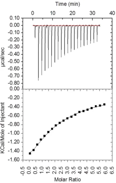
In order to obtain detailed information on binding abilities of CBEL, isothermal titration calo-rimetry (ITC) experiments (Fig 2) were done in the presence of cellohexaose, the longest solu-ble cello-oligosaccharide commercially availasolu-ble. Upon analysis of the ITC data, and based on the previous results it appeared that the CBEL binding to cellohexaose titration could be best analysed using a two sequential binding sites model, with higher (KD1 = 52μM, ΔH1 = -1967
kcal mol-1) and lower affinity (KD2 ~ 800μM, ΔH1 = -9156 kcal mol-1) binding sites (Fig 2).
Although the affinity observed is weak, it is consistent with the affinity observed for cellulases with cellohexaose and with other CBM families [4,38]. In the case of CBELY52-Y188, under the
experimental conditions, we were unable to observe any binding to cellohexaose (data not
Table 1. Characterization of CBEL andTvXynB binding to Avicel by solid state depletion assay.
Protein Bmax KD (μmol.g-1)1 (μmol.L-1)2 CBELWT 0.094± 0.004 4.3± 0.4 CBELY188A 0.125± 0.003 6.3±0.5 CBELY52A 0.29± 0.07 65.3± 20.6 CBELY52A_Y188A N.D. N.D. TvXynB 0.42± 0.02 0.6± 0.1 TvXynB-CBM1-1 0.092± 0.002 2.1± 0.2 TvXynBΔCBM N.D. N.D.
1Bmax is expressed inμmol of protein per g of substrate. 2
KDis expressed asμmol.L-1of proteins. doi:10.1371/journal.pone.0137481.t001
Table 2. Characterization of CBEL binding to cellulose nanocrystals by fluorescence spectroscopy.
Protein KD(% w/v) CBELWT 0.24± 0.07 CBELY188A 1.6± 0.3 CBELY52A 10.4± 3.7 CBELY52A_Y188A N.D N.D: no binding detected. doi:10.1371/journal.pone.0137481.t002
shown), which supports the hypothesis that the residues Y52 and Y188 are key determinants of CBEL’s sugar binding ability.
The action of CBEL on wheat straw
Some fungal CBM1s display the ability to disrupt the surface of cellulose via a non-catalytic mechanism [11–13]. Therefore, to investigate whether CBEL could exhibit the ability to alter cellulose structure in a similar manner, recombinant CBEL was incubated with wheat straw. CBEL induced the release of a small, but significant amount of reducing sugars (905μM), com-pared to the BSA control (103μM). Although the activity of PAN/Apple domains on the sub-strate could not be excluded in this assay, this result supports the view that CBM1s from CBEL somehow modify cell wall polysaccharides. However, we were unable to pursue and complete this study, because it proved impossible to produce CBM1-1 on its own in E. coli in order to
Fig 2. Binding interaction between CBEL and cellohexaose as investigated by isothermal titration calorimetry (ITC). The upper panel shows the thermogram representing the heat of binding. The lower panel shows the titration curve. Fitting procedure was performed using a two sequential binding site model. doi:10.1371/journal.pone.0137481.g002
evaluate its impact on wheat straw. Nevertheless, it was previously shown using a partially folded CBM1-1 synthetic peptide, that it triggers plant immune responses [27,39]. To further pursue our investigation of action of CBEL on wheat straw, we also combined CBEL with a commercial cellulolytic cocktail. However, in this case the action of the cocktail was not accen-tuated, implying that the cocktail alone was able to access the polysaccharides that were tar-geted by CBEL in the previous experiment (data not shown).
Appendage of CBM1-1 onto a fungal endoxylanase
Addition of xylanases to cellulose mixture has been reported to enhance glucose release by increasing the accessibility of the cellulose to cellulases through the removal of hemicellulose [40,41]. Moreover the precise role of CBM1s appended to fungal xylanases in the hydrolysis of lignocellulose is unclear, even if it is known that CBMs potentiate enzyme action on plant cell walls by targeting polymers that are in close vicinity to the partner enzyme’s substrate [8]. In this context we investigated whether oomycetal CBM1s can potentiate the action of a fungal xylanase on wheat straw. Therefore a, chimeric enzyme, TvXynB-CBM1-1, was created by replacing the CBM1 of the Talaromyces versatilis xylanase B, TvXynB [42] with CBEL CBM1-1. Likewise, for the completeness of the study, a truncated mutant was created, TvXynBΔCBM, which lacks a CBM1. Both proteins were successfully expressed in a soluble form in E. coli and could be purified to near homogeneity. TvXynB and TvXynBΔCBM recombinant proteins dis-played apparent molecular weights of 27.8 and 26 kDa, which are consistent with the expected molecular weight.
Properties ofTvXynB and its variants. Biochemical characterization of TvXynB and the variants revealed that the three enzymes displayed very similar properties, which were actually rather consistent with the properties of the TvXynB expressed in Pichia pastoris, [26]. Impor-tantly, all of the enzymes were optimally active in the pH range 3.0–3.5 with a maximum activ-ity at pH 3.0, which is relatively low, but not unusual for fungal endoxylanases [43], and at 55°C. However, at 55°C the enzymes described in this study were less stable (loss of 25% activ-ity after 30 minutes) than TvXynB expressed in P. pastoris [26]. Therefore, experiments were actually performed at 40°C, thus ensuring that the enzymes would remain stable for at least 24h (data not shown). This difference in thermostability of recombinant TvXynB expressed in E. coli and P. pastoris might be due to glycosylation, which was proven to be extensive in P. pas-toris, but totally absent in E. coli. Regarding specific activity, all three enzymes TvXynB, TvXynBΔCBM and TvXynB-CBM1-1 exhibited similar behaviour (1515 ± 162 IU. mg-1 pro-tein) on birchwood xylan (BWX) (Table 3). Similarly, kinetic analysis revealed almost identical values for kcatand apparent Km(Table 3), confirming that neither the presence of the wild type
CBM1 nor that of the CBEL-derived domain modulated the activity of the catalytic domain on BWX. This result is consistent with previous findings that indicate that CBM1s generally target cellulose, even when they are appended to catalytic modules that do not hydrolyze cellulose [38,44]. This conclusion was supported by depletion assays, which revealed that both the wild type TvXynB and the variant TvXynB-CBM1-1 were able to bind to Avicel, whereas the
Table 3. Optimal reaction conditions and kinetic parameters ofTvXynB and mutants on BWX.
pH—Temp. Km kcat kcat/KM Activity
(g/L-1) (s-1) (s-1.g.L-1) (UI/mg)
TvXynB 3–55 14.7± 3.0 734.2± 75.6 50.01 1516± 162
TvXynBΔCBM 3–55 14.3± 2.5 728.3± 62.3 51.07 1814± 155
TvXynB-CBM1-1 3–55 15.0± 3.6 786.2± 99.6 52.34 1690± 214
TvXynBΔCBM did not (Table 1). Nevertheless, binding to Avicel was not identical, since the wild type TvXynB bound more strongly than TvXynB-CBM1-1. Moreover, gel retardation assays using the wild type enzyme and TvXynB-CBM1-1 failed to reveal interactions of any of the proteins with sugar beet arabinan, wheat arabinoxylan or mixed-linkageβ-(1, 3) (1, 4)-D -glucan (data not shown).
Wheat straw hydrolysis byTvXynB and its variants, and the influence of CBEL. Using wheat straw as the substrate, hydrolytic properties of the TvXynB-CBM1-1 were further
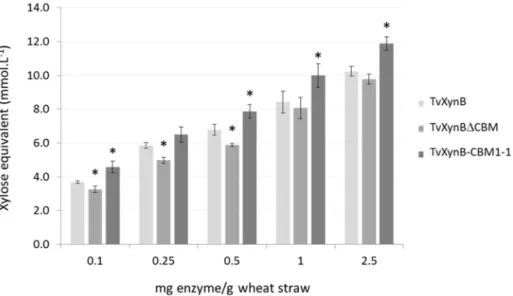
inves-tigated, comparing performance with that of the wild type enzyme and TvXynBΔCBM respec-tively. After 24-h hydrolysis, the amount of reducing sugars released by TvXynBCBM1-1 was generally higher than that observed with TvXynB. At low enzyme loadings this increase in activity was roughly 10% but was more significant (16, 18 and 16%) at higher (0.5, 1 and 2.5 mg/g) loadings respectively (Fig 3). Therefore, this confirms that CBM1-1 can at least restore wild type activity in XynΔCBM and at best can enhance activity beyond wild type levels. Pre-sumably this effect can be attributed to the binding of CBM1-1 to cellulose, which is in intimate contact with cell wall arabinoxylans.
To complete this study, wheat straw hydrolysis was also performed using a combination of either TvXynBΔCBM, TvXynBCBM1-1, or TvXynB, and CBEL (Fig 4). This revealed that the reaction procured a yield of soluble reducing sugars that was similar to the one involving TvXynB-CBM1-1. If one assumes that the PAN/Apple domains play no role here, this observa-tion is both intriguing and confusing, since it might imply that potentiaobserva-tion of the activity of the xylanase module by the CBM1-1 does not require a covalent linkage. Alternatively, the binding of CBEL (via its CBM1-1) to cellulose might provoke amorphogenesis that in turn leads to the non-specific solubilisation of components that are not covalently bound in the cell wall. To pursue this investigation, and better understand the underlying phenomena, it will be necessary to renew efforts to obtain an isolated recombinant form of CBM1-1.
Fig 3. Wheat straw hydrolysis byTvXynB xylanase and its derivatives. Black, grey and white bars represent TvXynB-CBM1-1, TvXynB and TvXynBΔCBM respectively. Increasing amounts of TvXynB, TvXynBΔCBM and TvXynB-CBM1-1 (0.1 to 0.5 mg enzyme/g substrate) were incubated with 0.02 g of wheat straw. The amount of solubilized reducing extremities was measured after 24h of hydrolysis at 40°C, pH 3.0 using the DNS assay. A control without enzyme was included and all experiments were conducted in triplicate. Asterisks indicate significant differences (p-value<0.05, t-test compared to TvXynB, the wild type reference).
Conclusions
This study provides new insight into the cellulose binding properties of CBM1s from the oomycetal protein CBEL and reveals that CBM1-1 is probably the major determinant of CBEL’s interaction with cellulose. Moreover, this work has demonstrated that the CBM1-1 from TvXynB somehow potentiates the action of the xylanase, although exactly how this is achieved remains unclear. Indeed, although appending the CBM1-1 to the catalytic module of TvXynB enhanced activity compared to the equivalent enzyme lacking a CBM, a similar result could be obtained by combining TvXynBΔCBM with free CBEL. Therefore, although this study has shown that CBM1-1 could be interesting for the creation of chimeric enzymes, it also tantalizingly raises a number of new questions that require further investigation.
Acknowledgments
The authors would like to thank Dr. P. Roblin (Soleil-Synchroton, Saclay, France) for scientific discussions and M. Maestracci and O. Guais for kindly providing the TvXynB gene. The equip-ment used for isothermal titration calorimetry is part of the Integrated Screening Platform of Toulouse (PICT, IBiSA). Dr. V. Gervais is gratefully acknowledged for assistance with the ITC experiment.
Author Contributions
Conceived and designed the experiments: TM HT BD MOD EG CD. Performed the experi-ments: TM HT VN CL LH. Analyzed the data: TM HT VN GC BD MOD EG CD. Contributed reagents/materials/analysis tools: TM HT VN CL GC LH EG CD. Wrote the paper: TM VN GC LH BD MOD EG CD.
Fig 4. Complementation ofTvXynB with CBEL in Wheat straw hydrolysis. TvXynBΔCBM (0.1 mg enzyme/g substrate) was incubated with or without CBEL (10 mg.g-1of wheat straw) at pH 3.0 and 40°C under agitation (1400 rpm) during 24h. TvXynB-CBM1-1 without CBEL is shown as control. Experiments were conducted in triplicate. Asterisks indicate significant differences (p-value<0.05, t-test compared to TvXynBΔCBM reference).
References
1. Jorgensen H, Kristensen JB, Felby C. Enzymatic conversion of lignocellulose into fermentable sugars: challenges and opportunities. Biofuel, Bioprod. Bioref. 2007: 1–16.
2. Mäkelä M, Donofrio N, de Vries R. Plant biomass degradation by fungi. Fungal Genet Biol. 2014:2–9. doi:10.1016/j.fgb.2014.08.010PMID:25192611
3. Lombard V, Golaconda Ramulu H, Drula E, Coutinho PM, Henrissat B. The carbohydrate-active enzymes database (CAZy) in 2013. Nucleic Acids Res. 2014;D490–5. doi:10.1093/nar/gkt1178PMID:
24270786
4. Montanier C, van Bueren AL, Dumon C, Flint JE, Correia MA, Prates JA, et al. Evidence that family 35 carbohydrate binding modules display conserved specificity but divergent function. Proc Natl Acad Sci U S A. 2009: 3065–70. doi:10.1073/pnas.0808972106PMID:19218457
5. Gilbert HJ, Knox JP, Boraston AB. Advances in understanding the molecular basis of plant cell wall polysaccharide recognition by carbohydrate-binding modules. Curr Opin Struct Biol. 2013: 669–77. doi:
10.1016/j.sbi.2013.05.005PMID:23769966
6. Reinikainen T, Ruohonen L, Nevanen T, Laaksonen L, Kraulis P, Jones TA, et al. Investigation of the function of mutated cellulose-binding domains of Trichoderma reesei cellobiohydrolase I. Proteins. 1992: 475–82. PMID:1438185
7. Carrard G, Koivula A, Soderlund H, Beguin P. Cellulose-binding domains promote hydrolysis of differ-ent sites on crystalline cellulose. Proc Natl Acad Sci U S A. 2000: 10342–7. PMID:10962023
8. Hervé C, Rogowski A, Blake AW, Marcus SE, Gilbert HJ, Knox JP. Carbohydrate-binding modules pro-mote the enzymatic deconstruction of intact plant cell walls by targeting and proximity effects. Proc Natl Acad Sci U S A. 2010:15293–8. doi:10.1073/pnas.1005732107PMID:20696902
9. Várnai A, Mäkelä MR, Djajadi DT, Rahikainen J, Hatakka A, Viikari L. Carbohydrate-binding modules of fungal cellulases: occurrence in nature, function, and relevance in industrial biomass conversion. Adv Appl Microbiol. 2014:103–65. doi:10.1016/B978-0-12-800260-5.00004-8PMID:24767427
10. Guillén D, Sanchez S, Rodriguez-Sanoja R. Carbohydrate-binding domains: multiplicity of biological roles. Applied Microbiol Biotech. 2010:1241–9.
11. Boraston AB, Bolam DN, Gilbert HJ, Davies GJ. Carbohydrate-binding modules: fine-tuning polysac-charide recognition. Biochem J. 2004:769–81.
12. Xiao Z, Gao PJCGJW, Zhang T.H., Liu Y.S. J., Qu Y, Wang T. Cellulose-binding domain of endogluca-nase III from Trichoderma reesei disrupting the structure of cellulose. J Biotechnology Letters. 2001:711–5.
13. Gao PJ, Chen GJ, Wang TH, Zhang YS, Liu J. Non-hydrolytic disruption of crystalline structure of cellu-lose by cellucellu-lose-binding domain and linker sequence of cellobiohydrolase I from Penicillium janthinel-lum. Act Bioch Sin 2001:13–8.
14. Carrard G, Linder M. Widely different off rates of two closely related cellulose-binding domains from Tri-choderma reesei. Eur J Biochem. 1999:637–43. PMID:10411622
15. Tomme P, Van Tilbeurgh H, Pettersson G, Van Damme J, Vandekerckhove J, Knowles J, et al. Studies of the cellulolytic system of Trichoderma reesei QM 9414. Analysis of domain function in two cellobiohy-drolases by limited proteolysis. Eur J Biochem. 1988:575–81.
16. Linder M, Teeri T. The roles and function of cellulose-binding domains. J Biotech. 1997:15–28. 17. van Gool MP, van Muiswinkel GC, Hinz SW, Schols HA, Sinitsyn AP, Gruppen H. Two GH10
endo-xylanases from Myceliophthora thermophila C1 with and without cellulose binding module act differ-ently towards soluble and insoluble xylans. Bioresour Technol. 2012:123–32.
18. Kraulis PJ, Clore G, Nilges T, Jones G, Petterson J, Knowles A, et al. Determination of the three-dimen-sional solution structure of the C-terminal domain of cellobiohydrolase I from Trichoderma reesei: a study using nuclear magnetic resonance and hybrid distance geometry-dynamical simulated anneal-ing. J Biochemistry. 1989:7241–57.
19. Larroque M, Barriot R, Bottin A, Barre A, Rougé P, Dumas B, et al. The unique architecture and function of cellulose-interacting proteins in oomycetes revealed by genomic and structural analyses. BMC Genomics. 2012:605–09. doi:10.1186/1471-2164-13-605PMID:23140525
20. Judelson HS. Dynamics and innovations within oomycete genomes: insights into biology, pathology, and evolution. Eukaryot Cell. 2012:1304–12. doi:10.1128/EC.00155-12PMID:22923046
21. Blackman LM, Cullerne DP, Hardham AR. Bioinformatic characterisation of genes encoding cell wall degrading enzymes in the Phytophthora parasitica genome. BMC Genomics. 2014:785. doi:10.1186/ 1471-2164-15-785PMID:25214042
22. Larroque M, Ramirez D, Lafitte C, Borderies G, Dumas B, Gaulin E. Expression and purification of a biologically active Phytophthora parasitica cellulose binding elicitor lectin in Pichia pastoris. Protein Expr Purif. 2011: 217–23
23. Tordai H, Banyai L, Patthy L. The PAN module: the N-terminal domains of plasminogen and hepatocyte growth factor are homologous with the apple domains of the prekallikrein family and with a novel domain found in numerous nematode proteins. FEBS Lett. 1999:63–7.
24. Brown PJ, Gill AC, Nugent PG, McVey JH, Tomley FM. Domains of invasion organelle proteins from apicomplexan parasites are homologous with the Apple domains of blood coagulation factor XI and plasma pre-kallikrein and are members of the PAN module superfamily. FEBS Lett. 2001:31–8. 25. Brecht S, Carruthers VB, Ferguson DJ, Giddings OK, Wang G, Jakle U, et al. The toxoplasma
microne-mal protein MIC4 is an adhesin composed of six conserved apple domains. J Biol Chem. 2001:4119– 27.
26. Villalba-Mateos F, Rickauer M, Esquerré-Tugayé MT. Cloning and characterization of a cDNA encod-ing an elicitor of Phytophthora parasitica var. nicotianae that shows cellulose-bindencod-ing and lectin-like activities. Mol Plant-Microbe Interac. 1997:1045–53.
27. Gaulin E, Drame N, Lafitte C, Torto-Alalibo T, Martinez Y, Ameline-Torregrosa C, et al. Cellulose bind-ing domains of a Phytophthora cell wall protein are novel pathogen-associated molecular patterns. Plant Cell. 2006:1766–77. PMID:16766692
28. Gaulin E, Jauneau A, Villalba F, Rickauer M, Esquerre-Tugaye M, Bottin A. The CBEL glycoprotein of Phytophthora parasitica var. nicotianae is involved in cell wall deposition and adhesion to cellulosic substrates. J Cell Sci. 2002:4565–75. PMID:12415001
29. Song L, Laguerre S, Dumon C, Bozonnet S, O'Donohue MJ. A high-throughput screening system for the evaluation of biomass-hydrolyzing glycoside hydrolases. Bioresour Technol. 2010:8237–43. doi:
10.1016/j.biortech.2010.05.097PMID:20579873
30. Fumagalli M, Ouhab D, Boisseau SM, Heux L. Versatile gas-phase reactions for surface to bulk esterifi-cation of cellulose microfibrils aerogels. Biomacromolecules. 2013:3246–55. doi:10.1021/bm400864z
PMID:23889256
31. Gasteiger E, Gattiker A, Hoogland C, Ivanyi I, Appel RD, Bairoch A. ExPASy: The proteomics server for in-depth protein knowledge and analysis. Nucleic Acids Res. 2003:3784–8. PMID:12824418
32. Lakey JH, Massotte D, Heitz F, Dasseux J-L, Faucon J-F, Parker MW, et al. Membrane insertion of the pore-forming domain of colicin A. The FEBS Journal. 1991:599–607.
33. Miller G.L, Use of dinitrosalicylic acid reagent for determination of reducing sugar. Anal Chem. 1959:426–8.
34. Lehtiö J, Sugiyama J, Gustavsson M, Fransson L, Linder M, Teeri TT. The binding specificity and affin-ity determinants of family 1 and family 3 cellulose binding modules. Proc Natl Acad Sci U S A. 2003:484–9. PMID:12522267
35. Elazzouzi-Hafraoui S, Nishiyama Y, Putaux JL, Heux L, Dubreuil F, Rochas C. The shape and size dis-tribution of crystalline nanoparticles prepared by acid hydrolysis of native cellulose. Biomacromole-cules. 2008:57–65. PMID:18052127
36. Linder M, Mattinen ML, Kontteli M, Lindeberg G, Stahlberg J, Drakenberg T, et al. Identification of func-tionally important amino acids in the cellulose- binding domain of Trichoderma reesei cellobiohydrolase I. Prot Sci 1995:1056–64.
37. Beckham GT, Matthews JF, Bomble YJ, Bu L, Adney WS, Himmel ME, et al. Identification of amino acids responsible for processivity in a Family 1 carbohydrate-binding module from a fungal cellulase. J Phys Chem B. 2010:1447–53. doi:10.1021/jp908810aPMID:20050714
38. Brás JL, Cartmell A, Carvalho AL, Verzé G, Bayer EA, Vazana Y, et al. Structural insights into a unique cellulase fold and mechanism of cellulose hydrolysis. Proc Natl Acad Sci U S A. 2011:5237–42. doi:10. 1073/pnas.1015006108PMID:21393568
39. Dumas B, Bottin A, Gaulin E, Esquerré-Tugayé MT. Cellulose-binding domains: cellulose associated-defensive sensing partners? Trends Plant Sci. 2008:160–4. doi:10.1016/j.tplants.2008.02.004PMID:
18329320
40. Kumar R, Wyman CE. Effect of enzyme supplementation at moderate cellulase loadings on initial glu-cose and xylose release from corn stover solids pretreated by leading technologies. Biotechnol Bioeng. 2009:457–67. doi:10.1002/bit.22068PMID:18781688
41. Hu J, Arantes V, Saddler J. The enhancement of enzymatic hydrolysis of lignocellulosic substrates by the addition of accessory enzymes such as xylanase: is it an additive or synergistic effect? Biotec Biof. 2011:36–8.
42. Lafond M, Guais O, Maestracci M, Bonnin E, Giardina T. Four GH11 xylanases from the xylanolytic fun-gus Talaromyces versatilis act differently on (arabino)xylans. Appl Microbiol Biotechnol. 2014:6339–52. doi:10.1007/s00253-014-5606-xPMID:24664446
43. Polizeli ML, Rizzatti AC, Monti R, Terenzi HF, Jorge JA, Amorim DS. Xylanases from fungi: properties and industrial applications. Appl Microbiol Biotechnol. 2005:577–91. PMID:15944805
44. De La Mare M, Guais O, Bonnin E, Weber J, Francois JM. Molecular and biochemical characterization of three GH62α-l-arabinofuranosidases from the soil deuteromycete Penicillium funiculosum. Enzyme Microb Technol. 2013:351–8. doi:10.1016/j.enzmictec.2013.07.008PMID:24034435