HAL Id: hal-03095526
https://hal.archives-ouvertes.fr/hal-03095526
Submitted on 4 Jan 2021HAL is a multi-disciplinary open access archive for the deposit and dissemination of sci-entific research documents, whether they are pub-lished or not. The documents may come from teaching and research institutions in France or abroad, or from public or private research centers.
L’archive ouverte pluridisciplinaire HAL, est destinée au dépôt et à la diffusion de documents scientifiques de niveau recherche, publiés ou non, émanant des établissements d’enseignement et de recherche français ou étrangers, des laboratoires publics ou privés.
Fabrication of 2D monolayers and 3D spheroids of cells
in acoustic levitation
Nathan Jeger-Madiot, Niclas Setterblad, Lousineh Arakelian, Valérie
Vanneaux, Mauricio Hoyos, Jérôme Larghero, Jean-Luc Aider
To cite this version:
Nathan Jeger-Madiot, Niclas Setterblad, Lousineh Arakelian, Valérie Vanneaux, Mauricio Hoyos, et al.. Fabrication of 2D monolayers and 3D spheroids of cells in acoustic levitation. 36th International Symposium on Microscale Separation and Bioanalysis, Sep 2020, Saint-Malo, France. �hal-03095526�
Fabrication of 2D monolayers and 3D spheroids of cells in acoustic
levitation
Nathan Jeger-Madiot1, Niclas Setterblad2, Lousineh Arakelian2, Valérie Vanneaux2,
Mauricio Hoyos2, Jérôme Larghero2, and Jean-Luc Aider1 1Laboratoire Physique et Mécanique des Milieux Hétérogènes (PMMH) 2Institut Universitaire d’Hématologie de l’Hôpital Saint-Louis
E-mail: nathan.jeger-madiot@espci.fr / mauricio.hoyos@espci.fr / jean-luc.aider@espci.fr
Introduction
Today, three-dimensional (3D) cell cultures tend to replace 2D conventional method because of their more relevant tissue-mimicking characteristics. Indeed, the 3D cell architecture (spheroïd, organoïd, etc) and the microenvironment is closer to In Vivo physiological behaviour [1, 2]. The main difficulties remain in creating a scaffold compatible with the targeted cells and tissues. Bioprinting is one of the great objectives for tissue engineering. For instance, stereolithography is a 3D printing technology where the freestanding object is built layer by layer with a photosensitive polymer resin through the projection of a UV image in the top plane. In recent promising work, stereolithography has been applied to create 3D hydrogel structures to guide cells like hepatocytes [3]. Nevertheless, ideally, scaffold-free methods are needed. We propose a new method combining microfluidic channels and the acoustic radiation force (ARF) to structure and to control contactless the shape of stem-cells aggregates.
2D stratification and shaping
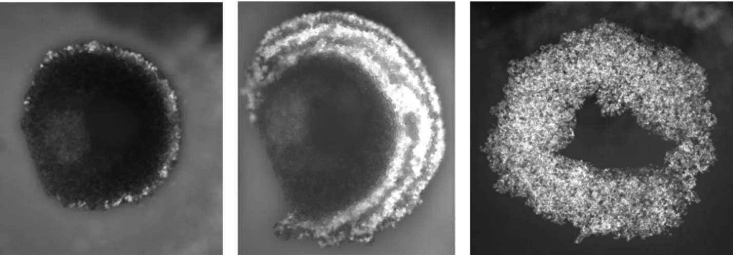
In a standing wave cavity, the cells can be trapped in a central cluster. After a short transient period and using the proper parameters, the cluster becomes a circular monolayer levitating in the surrounding fluid (Fig. 1a). The control of the medium flow and of the ARF allows various manipulations. For example, it is possible to structure the circular monolayer with different 2D stratifications of particles or cells into the acoustic nodal plane. The result is a cluster with type 1 particles or cells at its centre, surrounded by a ring of type 2 particles or cells (Fig. 1b). Other layers can be added, leading to a concentric multi-annular monolayer.
Multilayer aggregation: toward 3D construction
The 2D approach can be extended to 3D. Indeed, the height of the geometry of the cavity or the acoustic frequency can be modified in order to increase the number of pressure nodes and then the corresponding number of cell aggregates. It leads to a superposition of monolayers distributed along the height of the cavity (Fig. 2a).
Changing dynamically the acoustic frequency, activating or turning off the ultrasounds, using more complex cavities allows more refined manipulations of the clusters. For instance, it becomes possible to move an aggregate from one node to another or let the aggregates sediment at the bottom of the cavity to form a multilayer aggregate over the substrate.
Long term experiment with stem cells
To confirm these concepts on cells manipulation and cells culture in acoustic levitation, long term experiments with Mesenchymal stem Cells (MSCs) have been carried out. MSCs have been maintained in acoustic levitation during 24h. Without contact with a classical petri dish and in acoustic levitation, MSC behavior tends to shape spheroids. Then, we compare the viability and the behavior of the cells cluster with a classical cells culture. The MSCs have been found alive and functional and could grow and divide in a normal way.
Conclusion
With our set of microfluidic chips, we show different ways to manipulate, structure and grow stem cells over a long period of time, from a few hours to a few days. One of the advantages of such systems are the material biocompatibility, the ease of construction and the versatility. As this approach is intrinsically “scaffold-free”, it is important to find ways to organize spatially the cells. Our experiments open the path to such manipulations.
Figure 2: Example of multi-nodes aggregation. (a) Cavity with 7 acoustic nodes of 10 µm red polystyrene particles. (b) Cavity with 13 acoustic nodes of hollow glass microspheres
References
[1] Y. Fang et R. M. Eglen, « Three-Dimensional Cell Cultures in Drug Discovery and Development », Slas discovery:
Advancing Life Sciences R&D, 22(5), 456-472. https://doi.org/10.1177/1087057117696795
[2] V. van Duinen, S. J. Trietsch, J. Joore, P. Vulto, et T. Hankemeier, « Microfluidic 3D cell culture: from tools to tissue models », Curr. Opin. Biotechnol., vol. 35, p. 118‑126, déc. 2015.https://doi.org/10.1016/j.copbio.2015.05.002
[3] «Photopatterning of hydrogel scaffolds coupled to filter materials using stereolithography for perfused 3D culture of hepatocytes », J.A. Neiman, R. Raman, V. Chan, M.G. Rhoads, M.S. Raredon, J.J. Velazquez, R.L. Dyer, R. Bashir, P.T.Hammond, L.G. Griffith, Biotechnology and bioengineering 112 (2015) 777. https://doi.org/10.1002/bit.25494
Figure 1: Examples of different patterns obtained with ultrasound and flow control. (a) Circular monolayer of 10 µm polystyrene particles. (b) 2D stratification of 10µm red and green fluorescent particles. (c) Ring monolayer of endothelial cells