Publisher’s version / Version de l'éditeur:
Acta Crystallographica, B34, pp. 1741-1744, 1978
READ THESE TERMS AND CONDITIONS CAREFULLY BEFORE USING THIS WEBSITE. https://nrc-publications.canada.ca/eng/copyright
Vous avez des questions? Nous pouvons vous aider. Pour communiquer directement avec un auteur, consultez la
première page de la revue dans laquelle son article a été publié afin de trouver ses coordonnées. Si vous n’arrivez pas à les repérer, communiquez avec nous à PublicationsArchive-ArchivesPublications@nrc-cnrc.gc.ca.
Questions? Contact the NRC Publications Archive team at
PublicationsArchive-ArchivesPublications@nrc-cnrc.gc.ca. If you wish to email the authors directly, please see the first page of the publication for their contact information.
NRC Publications Archive
Archives des publications du CNRC
This publication could be one of several versions: author’s original, accepted manuscript or the publisher’s version. / La version de cette publication peut être l’une des suivantes : la version prépublication de l’auteur, la version acceptée du manuscrit ou la version de l’éditeur.
Access and use of this website and the material on it are subject to the Terms and Conditions set forth at
Choline O-sulphate
Post, Michael L.
https://publications-cnrc.canada.ca/fra/droits
L’accès à ce site Web et l’utilisation de son contenu sont assujettis aux conditions présentées dans le site LISEZ CES CONDITIONS ATTENTIVEMENT AVANT D’UTILISER CE SITE WEB.
NRC Publications Record / Notice d'Archives des publications de CNRC:
https://nrc-publications.canada.ca/eng/view/object/?id=45b416db-8ee4-4ed9-b9db-45a8e488b2b4
https://publications-cnrc.canada.ca/fra/voir/objet/?id=45b416db-8ee4-4ed9-b9db-45a8e488b2b4
P E R C H L O R O B E N Z Y L I D E N E C Y C L O H E X A - 2 , 5 - D I E N E 1741
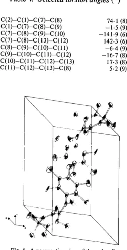
Table 4. Selected torsion angles (o)
C(2)-C(1)-C(7)-C(8) C(1)-C(7)-C(8)-C(9) C(7)-C(8)-C(9)-C(10) C (7)-C (8)-C ( 13)-C (12) C (8)-C (9)-C ( 10)-C (11) C(9)-C(10)-C(l l)-C(12) C(10)-C(I l)-C(12)-C(13) C(I 1)-C(12)-C(13)-C(8) 74. l (8) --1.5 (9) --141.9 (6) 142.3 (6) - 6 . 4 (9) --16.7 (8) 17.3 (8) 5.2 (9) ~
~
b~N~~
...
~'~)~
\
~
Fig. 4. A perspective view of the unit cell.
providing the crystals, and to Enraf-Nonius, who kindly supplied us with a CAD-4 automatic diffrac- tometer.
References
AHMED, F. R., HALL, S. R., PIPPY, M. E. & HUBER, C. P.
(1966). NRC Crystallographic Programs for the
IBM/360 System. National Research Council, Ottawa, Canada.
BALLESTER, M., REIRA, J., CASTAI~ER, J., BADIA, C. &
MONSO, J. M. (1971). J. Am. Chem. Soc. 93 (3), 2215-
2225.
BALLESTER, M., RIERA, J. & DE LA FUENTE, C. (1970).
Unpublished.
BROWN, G. M. & STRYDOM, O. A. W. (1974). Acta Cryst.
B30, 801-804.
GALl, S. (1975). Crystal and Molecular Structures of
Perchlorinated Organic Compounds. Thesis. Univ. of Barcelona, Spain.
GALi, S., MmAVITLLES, C. & FONT-ALTABA, M. (1975).
A cta Cryst. B 31, 2510-2512.
HOWELLS, E. R., PHmLWS, D. C. & ROGERS, D. (1950).
A cta Cryst. 3, 21 0-214.
International Tables for X-ray Crystallography (1962). Vol. III. Birmingham: Kynoch Press.
JOHNSON, C. K. (1965). ORTEP. Report ORNL-3794. Oak
Ridge National Laboratory, Tennessee.
MAIN, P., WOOLFSON, M. M., LESSINGER, L., GERMAIN, G.
& DECLERCQ, J. P. (1974). MULTAN 74. A System of
Computer Programs for the Automatic Solution of Crystal Structures from X-ray Diffraction Data. Univs. of York, England, and Louvain-la-Neuve, Belgium.
PEDERSEN, I. F. (1975). Acta Cryst. B31,2931-2933.
Acta Cryst. (1978). B34, 1741-1744
Choline O-Sulphate*
BY MICHAEL L. POSTt
Division o f Biological Sciences, National Research Council o f Canada, Ottawa K1A 0R6, Canada (Received 18 November 1977; accepted 23 December 1977)
Abstract. CsH13NO48 , M r = 183.22, monoclinic,
P 2 J c , a = 8.391 (2), b = 8-674 (2), c = 11.317 (2) A,
fl =- 97.29 (1) °, D m = 1.50, D e = 1.49 g c m -3, Z = 4,
R = 0.034 on 1240 observed data. The molecule exists
as a zwitterion with a gauche conformation in the
choline chain; electrostatic stability is achieved through intra- and intermolecular interactions.
* Issued as NRCC No. 16585.
+ Present address: Division of Chemistry, NRCC.
Introduction. ( C H s ) 3 N C H 2 C H 2 O S O 3 was prepared by the method of Stevens & Vohra (1955), and good air- stable crystals were grown by vapour-diffusion methods in an ethanol/water system. Preliminary photographic work showed that the cell dimensions and space group were in agreement with those previously reported (Okaya, 1966); the presence of pseudo- symmetry was indicated with the majority of the/-odd reflections weak in intensity.
A crystal (0-48 × 0.36 × 0-10 mm) was mounted for cell refinement and data collection with b parallel to
1742 C H O L I N E O-SULPHATE the tp axis of a Picker card-automated diffractometer
equipped with a scintillation detector and pulse-height discriminator. Intensity data were recorded throughout the range 20 < 130 ° with Ni-filtered Cu Kt~ radiation (2 = 1.54178 A) employing the 0--20 scanning mode operating at 2 ° min -~ in 20, with scan widths from 1-8 to 2.4 ° in 20, dependent upon 20. Background counts of 10 s duration were recorded at the higher scan limit. Of 1387 independent reflections, 147 had/net < pre- determined threshold criteria (0.1 x background or 80 counts), and these were excluded during refinement of the structure. Scaling, with respect to three check reflections, Lorentz, polarization and absorption cor- rections [/~(Cu Ka) = 32.80 cm -11 were applied and
Fo'S derived. At a later stage a correction was made for
secondary extinction.
The position of the - O - S O 3 group was determined from a sharpened Patterson map and the remaining heavy atoms were chosen in a difference Fourier synthesis. Ten of the eleven non-H atoms were related by a pseudo mirror plane coincident with the c glide. The whole molecule then required a shift of ] along z to bring it to the correct position, the originally incorrect placement being a result of ambiguity due to pseudo- symmetry. Refinement was accomplished by block- diagonal least squares. The strong pseudosymmetry present in the original model slowly relaxed during the early cycles, such that the /-odd reflexions began to receive a realistic contribution. From that point, the refinement proceeded smoothly, with non-H atoms thermally anisotropic, and H atoms, all of which were found in a difference Fourier synthesis, being allotted isotropic thermal motion. Convergence was attained at an R of 0.034 and an Rw* of 0.046 for 1240 reflections (R = 0-039 for all data) and no shift was >0. | 3 a in the last cycle. A final difference map showed no physically interpretable features, the largest peaks being <0.2 e A -a. The function minimized throughout was ~ w(IFol - IFcl) 2 with weights, w, in the form: w = 1.0 if F o <
* R w = [ Y w ( I F o l - IFct)z/x'' w F 2 l '/2.
Table 1. Final positional coordinates (x 104) for the
non-hydrogen atoms, with estimated standard deviations in parentheses x y z S(1) 2871 (1) 2414 (1) 762 (1) O(1) 4356 (2) 1943 (2) 1752 (1) 0(2) 1569 (2) 2740 (3) 1419 (2) 0(3) 2664 (2) 1038 (2) 67 (1) 0(4) 3404 (3) 3710 (2) 159 (2) N(1) 7837 (2) 2689 (2) 2753 (1) C(l) 4795 (2) 3091 (2) 2653 (2) C(2) 6354 (2) 2680 (2) 3387 (2) C(3) 9278 (3) 2737 (4) 3686 (2) "(2(4) 7877 (3) 4062 (3) 1960 (2) C(5) 7936 (2) 1275 (2) 2015 (2)
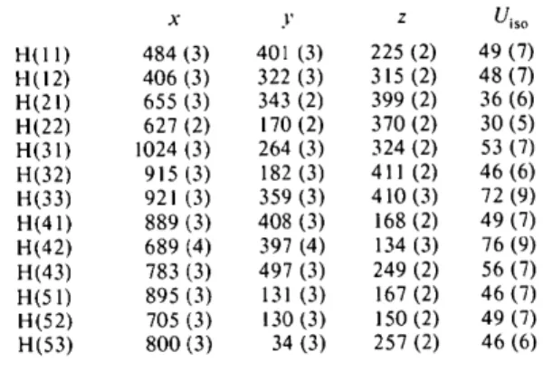
Table 2. Final hydrogen-atom positional coordinates (× 103) and isotropic thermal parameters I(A 2 × 10³),
with estimated standard deviations in parentheses
x y z Uis o H{I I) 484 (3) 401 (3) 225 (2) 49 (7) H(12) 406 (3) 322 (3) 315 (2) 48 (7) H(21) 655 (3) 343 (2) 399 (2) 36 (6) H(22) 627 (2) 170 (2) 370 (2) 30 (5) H(31) 1024 (3) 264 (3) 324 (2) 53 (7) H(32) 915 (3) 182 (3) 411 (2) 46 (6) H(33) 921 (3) 359 (3) 410 (3) 72(9) H(41) 889 (3) 408 (3) 168 (2) 49 (7) H(42) 689 (4) 397 (4) 134 (3) 76 (9) H(43) 783 (3) 497 (3) 249 (2) 56 (7) H(51) 895 (3) 131 (3) 167 (2) 46 (7) H(52) 705 (3) 130 (3) 150 (2) 49 (7) H(53) 800 (3) 34 (3) 257 (2) 46 (6) 12.7, w ~/2 = 12.7/F o if F o > 12.7. Scattering factors were taken from International Tables f o r X-ray
Crystallography (1974) and Stewart, Davidson &
Simpson (1965), and all computations were carried out with the NRC program set (Ahmed, Hall, Pippy & Huber, 1973). Final atomic parameters are presented in Tables 1 and 2.*
Discussion. The conformation of the choline chain in various classes of small molecules has been studied in the solid state (Sundaralingam, 1968; Baker, Chothia, Pauling & Petcher, 1971; Viswamitra, Seshadri, Post & Kennard, 1975; Jagner & Jensen, 1977, and references therein), in solution (Dufourcq & Lussan, 1972; Aslanian, Balkanski & Lautie, 1977; Koyama, Toda & Kyogoku, 1977), and by theoretical calculations (Pullman, 1976). Attention has also been directed toward macromolecules containing the choline moiety,
* Lists of structure factors, anisotropic thermal parameters and bond lengths and angles for the hydrogen atoms have been deposited with the British Library Lending Division as Supple- mentary Publication No. SUP 33316 (10 pp.). Copies may be obtained through The Executive Secretary, International Union of Crystallography, 13 White Friars, Chester CH l l NZ, England.
,)
Fig. 1. A perspective view of choline O-sulphate. Non-hydrogen atoms are represented by 50% probability thermal ellipsoids and hydrogen atoms by spheres of arbitrary radius.
C H O L I N E O - S U L P H A T E 1743 O c _....,, ° P"..---6-¢
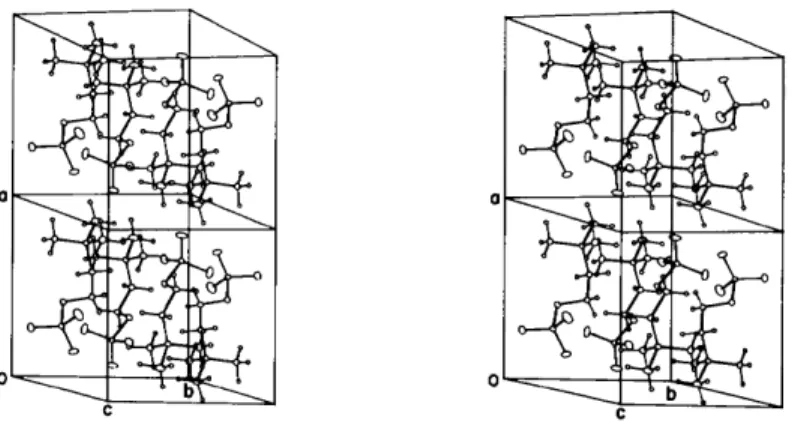
Fig. 2. Stereoscopic view of the contents of two unit cells.
e.g. the biomembrane constituents such as phospho- lipids (Sundaralingam, 1972; Dufourcq & Lussan, 1972; Gupta & Govil, 1972; McAlister, Yathindra &
Sundaralingam, 1973), which contain phosphoryl-
choline as a head-group. Generally, the diverse physical approaches lead to the identification of certain favoured
conformers, and particularly the indication of a gauche
arrangement of the O - C - C - - N bonds, as is found in the present structure.
The constitution of choline O-sulphate ensures that no hydrogen bonding is possible and that the molecule possesses net zero charge - in contrast to the great majority of structures previously reported. For crystal- line choline O-sulphate, weaker forces of association in the lattice will predominate, with a concomitant low perturbation of molecular conformation. It is likely that the structural features reported here resemble closely those which exist for the molecule in some solvents.
The struqture (Fig. l) is zwitterionic with formal
chain r e p r e s e n t a t i o n ~ * N - C - C - O - e S ~ , but with
charge almost certainly distributed over atoms bonded
at N and S. The choline moiety exhibits a gauche
con form ation, with a torsion angle N ( 1)--C ( 2 ) - C ( 1 ) - O(1) of 66.1 ° ]other torsion angles: C(3)--N(1)--
C ( 2 ) - C ( 1 ) = 162.9°; C ( 2 ) - C ( 1 ) - O ( 1 ) - S ( 1 ) =
- 1 6 9 . 6 ° ; C ( 1 ) - O ( I ) - S ( 1 ) - O ( 2 ) = - 5 7 . 3 ° ] , and intramolecular distances N( 1)... O(1) and C ( 5 ) . . . O(1) o f 3.065 (2) and 3.037 (2) A, respectively, each atom pair imparting partial electrostatic stabilization. Bond lengths and angles for the 'heavy' atoms (Table 3) are as expected. The distorted tetrahedral geometry around
S(1), with all O ( 1 ) - S ( 1 ) - O ( n ) angles less than sp 3
values, has been previously observed for O-sulphate esters (Fries & Sundaralingam, 1971). The average C--H distance is 0.95 A with an average associated angle of 109 ° .
Lattice packing (Fig. 2) is partly achieved through a number of head-to-tail approaches of the zwitterion, the closer of these being across centres of symmetry with a cell translation in the a [ 0 ( 3 ) . . . C ( 5 ) , 3.087 (2)
Al
and b ] 0 ( 4 ) . . . C ( 4 ) , 3.160 ( 2 ) A l directions. Additionally,Table 3. Bond lengths (/k) and angles ( 0 ) f o r the
non-hydrogen atoms
Estimated standard deviations are in parentheses.
S(l)-O(1) 1.619 (1) C(1)-C(2) 1.501 (3) S(l)-O(2) 1.425 (2) N(I)-C(2) 1.513 (3) S(1)-O(3) 1.427 (2) N(1)-C(3) 1.501 (3) S(1)-O(4) 1.417 (2) N(1)-C(4) 1.494 (3) O(I)-C(1) 1.439 (3) N(I)-C(5) 1.493 (3) O(1)-S(1)-O(2) 105.2 (l) C(I)-C(2)-N(I) 116.6 (2)
O(1)-S(1)-O(3) I01.5 (l) C(2)-N(I)-C(3) 107.7 (2)
O(1)-S(1)-O(4) 105.7 (1) C(2)-N(l)-C(4) l l 1.8 (2) O(2)-S(1)-O(3) 113.9 (l) C(2)-N(1)-C(5) I 11.6 (2) O(2)-S(1)-O(4) 113.8 (1) C(3)-N(l)-C(4) I08.8 (2) O(3)-S(1)-O(4) 114.9 (l) C(3)-N(1)-C(5) 108.6 (2) S(l)-O(l)-C(l) 114.9 (1) C(4)-N(1)-C(5) 108.1 (2) O(1)-C(1)-C(2) 110.8 (2)
there are some longer approaches between 0 ( 2 ) and a- translated --N(CH3) 3 groups [ 0 ( 2 ) . . . N(1), C (4), C(5); at 3.645 (2), 3.430 (2) and 3.447 (2) ,/k respectively]. All these interactions are of an electrostatic character such that, overall, the charge on the molecule is accommodated both inter- and intramolecularly. Other contacts in the lattice, none of which are unusually short, are of the van der Waals type.
I thank Dr Martin Young, of this laboratory, for supplying the material and the National Research Council of Canada for the award of a Research Associateship.
References
AHMED, F. R., HALL, S. R., PIPPY, M. E. & HUBER, C. P.
(1973). NRC Crystallographic Programs for the
IBM/360 System. Accession numbers 133-147 in J. Appl.
Cryst. 6, 309-346.
ASLANIAN, D., BALKANSKI, M. • LAUTIE, A. (1977)../. Am.
Chem. Soc. 99, 1974-1976.
BAKER, R. W., CHOTHIA, C. H., PAULING, P. & PETCHER, T.
J. (197 l). Nature (London), 230, 439-445.
1744 C H O L I N E O - S U L P H A T E
FRIES, D. C. & SUNDARALINGAM, M. (1971). Acta Cryst.
B27, 401-410.
GUPTA, S. P. & GOVIL, G. (1972). FEBS Lett. 27, 68-70.
International Tables for X-ray Crystallography (1974). Vol. IV. Birmingham: Kynoch Press.
JAGNER, S. (~ JENSEN, B. (1977). Acta Cryst. B33, 2757-
2762.
KOYAMA, Y., TODA, S. & KYOGOKU, Y. (1977). Chem. Phys.
Lipids, 19, 74-92.
MCALISTER, J., YATHINDRA, N. & SUNDARALINGAM, M.
(1973). Biochemistry, 12, 1189-1195.
OKAYA, Y. (1966). Acta Cryst. 21, A124-A125.
PULLMAN, B. (1976). Proceedings of the Eighth Jerusalem
Symposium on Quantum Chemistry and Biochemistry,
edited by B. PULLMAN, pp. 55-80. Holland: Reidel.
STEVENS, C. M. (~ VOHRA, P. (1955). J. Am. Chem. Soc. 77,
4935-4936.
STEWART, R. F., DAVIDSON, E. R. & SIMPSON, W. T. (1965).
J. Chem. Phys. 42, 3175-3187.
SUNDARALINGAM, M. (1968). Nature (London), 217, 35-37.
SUNDARALINGAM, M. (1972). Ann. NYAcad. Sci. 195, 324-
355.
VISWAMITRA, M. A., SESHADRI, T. P., POST, M. L. &
KENNARD, O. (1975). Nature (London), 258, 497-501.
Acta Cryst. (1978). B34, 1744-1746
2-Hydroxy-3-phenyl-3-pyrrolidinylpropionamide
BY A. KmFEL
Lehrstuhl, ffir Mineralogie und Kristallographie am Mineralogischen Institut der Universit?it Bonn, Poppelsdorfer Schloss, D 5300 Bonn, Federal Republic o f Germany
(Received 16 December 1977; accepted 14 January 1978)
Abstract. C13HlsN202, monoclinic, P2~/c, a =
7.844 (3), b = 16.619 (7), c = 20.802 (12) /k, fl =
103-2 (3) ° , V = 2640 (2) /k 3 , Z = 8, D x = 1.179 g
cm -3. The compound is an erythro phenylisoserine
derivative. One molecule of the asymmetric unit forms an N . - - O hydrogen-bonded dimer with its inversion image; the second is connected to two different dimers by O . . . O hydrogen bonds. The result is a network of molecules within a layer parallel to (100) of thickness dl00 = 7.636 /k. The layers are held together by packing forces only.
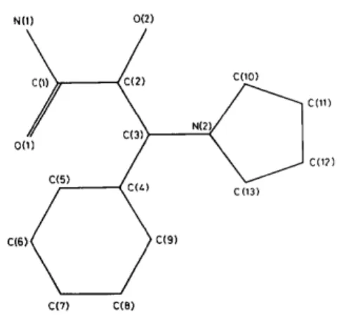
Introduction. The title compound (Fig. 1) was syn- thesized by Dr W. Tack and Professor Zymalkowski (Pharmazeutisches Institut der Universit~.t Bonn). A crystal, 0.2 × 0.1 x 0.1 mm, was selected for the diffraction experiments. Intensity measurements were carried out in the co mode on an automatic Syntex P21 four-circle diffractometer with Mo K(t radiation (2 = 0.71069 ,&) monochromatized by a graphite crystal. 3406 reflexions were recorded, resulting in a set of 2921 unique reflexions, of which 887 were regarded as unobserved (I < 30).
No absorption correction was applied (,u = 0.90
cm-1). The structure was solved with M U L T A N
(Germain, Main & Woolfson, 1971), which fixed the positions of 33 non-hydrogen atoms in the asymmetric unit. The completion of the structure solution was achieved by Fourier methods and calculation of H
N0)
0(2)
C(2)
C(7)
C(8)
COO)
N ( 2 ~
C(11)
CO?)
C(13)
Fig. 1. The numbering scheme.
atom positions. Refinement was by full-matrix least
squares with weights w = I/a2(F) and anisotropic
temperature factors. The H atoms were allocated the isotropic temperature factors of their carrier atoms, but their positions were refined. Owing to the large number of parameters (416) refinement had to take place in overlapping cycles. An isotropic extinction factor g
{Zachariasen, 1963; F c = kFo[1 + fl(20)glc]} was
included in the list of variables (final value 7 x 10⁻⁷). With six reflexions excluded from refinement the final R