HAL Id: inserm-00925284
https://www.hal.inserm.fr/inserm-00925284
Submitted on 7 Jan 2014
HAL is a multi-disciplinary open access
archive for the deposit and dissemination of
sci-entific research documents, whether they are
pub-lished or not. The documents may come from
teaching and research institutions in France or
abroad, or from public or private research centers.
L’archive ouverte pluridisciplinaire HAL, est
destinée au dépôt et à la diffusion de documents
scientifiques de niveau recherche, publiés ou non,
émanant des établissements d’enseignement et de
recherche français ou étrangers, des laboratoires
publics ou privés.
on alveolarization in rat lungs: morphometric and gene
expression analysis.
Elodie Zana-Taieb, Laura Butruille, Marie-Laure Franco-Montoya, Emmanuel
Lopez, Flore Vernier, Isabelle Grandvuillemin, Danièle Evain-Brion, Philippe
Deruelle, Olivier Baud, Christophe Delacourt, et al.
To cite this version:
Elodie Zana-Taieb, Laura Butruille, Marie-Laure Franco-Montoya, Emmanuel Lopez, Flore Vernier,
et al.. Effect of two models of intrauterine growth restriction on alveolarization in rat lungs:
morpho-metric and gene expression analysis.. PLoS ONE, Public Library of Science, 2013, 8 (11), pp.e78326.
�10.1371/journal.pone.0078326�. �inserm-00925284�
Effect of Two Models of Intrauterine Growth Restriction
on Alveolarization in Rat Lungs: Morphometric and Gene
Expression Analysis
Elodie Zana-Taieb1,2,3,9*, Laura Butruille4, Marie-Laure Franco-Montoya5, Emmanuel Lopez1,2,3, Flore Vernier1,2, Isabelle Grandvuillemin6, Danie`le Evain-Brion1,2,9, Philippe Deruelle4,
Olivier Baud2,7,10,11, Christophe Delacourt2,5,8,9, Pierre-Henri Jarreau1,2,3,9
1Institut National de la Sante´ Et de la Recherche Me´dicale (INSERM) U767, Paris, France, 2 PremUp, Paris, France, 3 Service de Me´decine et Re´animation ne´onatales de Port-Royal, Groupe hospitalier Cochin, Broca, Hoˆtel-Dieu, Assistance Publique – Hoˆpitaux de Paris, Paris, France, 4 Unite´ environnement pe´rinatal et croissance, EA4489, Faculte´ de Me´decine, Poˆle recherche, IFR 114,Universite´ Lille Nord de France, Lille, France, 5 Institut National de la Sante´ Et de la Recherche Me´dicale (INSERM) U955 IMRB Equipe 04, Cre´teil, France, 6 Institut National de la Sante´ Et de la Recherche Me´dicale (INSERM) UMR 1076, Faculte´ de Pharmacie, Universite´ de la Me´diterrane´e. Marseille, France, 7 Institut National de la Sante´ Et de la Recherche Me´dicale (INSERM) U676, Paris, France, 8 Service de Pneumologie Pe´diatrique, Hoˆpital Necker-Enfants Malades, Assistance Publique – Hoˆpitaux de Paris, Paris, France, 9 Universite´ Paris Descartes, Paris, France, 10 Service de Re´animation et Pe´diatrie ne´onatales, Hoˆpital Robert Debre´, Assistance Publique – Hoˆpitaux de Paris, Paris, France, 11 Universite´ Paris Diderot, Paris, France
Abstract
Intrauterine growth restriction (IUGR) in preterm infants increases the risk of bronchopulmonary dysplasia, characterized by arrested alveolarization. We evaluated the impact of two different rat models (nitric oxide synthase inhibition or protein deprivation) of IUGR on alveolarization, before, during, and at the end of this postnatal process. We studied IUGR rat pups of dams fed either a low protein (LPD) or a normal diet throughout gestation and pups of dams treated by continuous infusion of Nv-nitro-L-arginine methyl ester (L-NAME) or its diluent on the last four days of gestation. Morphometric parameters, alveolar surface (Svap), mean linear intercept (MLI) and radial alveolar count (RAC) and transcriptomic analysis were determined with special focus on genes involved in alveolarization. IUGR pups regained normal weight at day 21 in the two treated groups. In the LPD group, Svap, MLI and RAC were not different from those of controls at day 4, but were significantly decreased at day 21, indicating alveolarization arrest. In the L-NAME group, Svap and RAC were significantly decreased and MLI was increased at day 4 with complete correction at day 21. In the L-NAME model, several factors involved in alveolarization, VEGF, VEGF-R1 and –R2, MMP14, MMP16, FGFR3 and 4, FGF18 and 7, were significantly decreased at day 4 and/or day 10, while the various factors studied were not modified in the LPD group. These results demonstrate that only maternal protein deprivation leads to sustained impairment of alveolarization in rat pups, whereas L-NAME impairs lung development before alveolarization. Known growth factors involved in lung development do not seem to be involved in LPD-induced alveolarization disorders, raising the question of a possible programming of altered alveolarization.
Citation:Zana-Taieb E, Butruille L, Franco-Montoya M-L, Lopez E, Vernier F, et al. (2013) Effect of Two Models of Intrauterine Growth Restriction on Alveolarization in Rat Lungs: Morphometric and Gene Expression Analysis. PLoS ONE 8(11): e78326. doi:10.1371/journal.pone.0078326
Editor:William B. Coleman, University of North Carolina School of Medicine, United States of America ReceivedMay 21, 2013; Accepted September 11, 2013; Published November 21, 2013
Copyright: ß2013 Zana-Taieb et al. This is an open-access article distributed under the terms of the Creative Commons Attribution License, which permits unrestricted use, distribution, and reproduction in any medium, provided the original author and source are credited.
Funding:This work has been supported by a Legs Poix grant of la Chancellerie des Universite´s de Paris, 2006 and by the Air Liquide Foundation, 2011. The funders had no role in study design, data collection and analysis, decision to publish, or preparation of the manuscript.
Competing Interests:The authors declare that co-author Prof. Olivier Baud is a PLOS ONE Editorial Board member, which does not alter the authors’ adherence to all PLOS ONE policies on sharing data and materials.
* E-mail: elodie.zana-taieb@cch.aphp.fr
Introduction
Bronchopulmonary dysplasia (BPD) is a chronic lung disease affecting preterm infants and characterized by arrested alveolar-ization in the developmental program. Lungs affected by BPD exhibit fewer and larger alveoli and decreased pulmonary microvascular development [1]. Several recent reports indicate that intrauterine growth restriction (IUGR), defined as failure of the fetus to achieve the expected weight for a given gestational age [2], could contribute to the development of BPD [3]. Most cases of IUGR are due to placental insufficiency, which decreases placental and umbilical blood flow and therefore placental transfer of glucose, essential amino acids and oxygen to the fetus [4]. IUGR is an important health problem, responsible for an increased risk of
morbidity and perinatal mortality [4] and has long-term conse-quences, such as metabolic syndrome, usually attributed to ‘‘fetal programming’’. Some studies have also suggested that compro-mised fetal growth can lead to lung dysfunction during infancy [3,5], childhood [5,6], and adulthood [7,8]. However, the mechanistic basis for a relationship between IUGR and BPD and the possibility of fetal programming of lung development have not been elucidated, as the effects of experimental models of IUGR on lung development depend on the model studied, especially timing, nature of the insult and species [9].
Various models affecting the quality of intrauterine environ-ment and leading to IUGR have been developed, such as low-protein diet (LPD), uterine artery ligation, administration of N-omega-nitro-L-arginine methyl ester (L-NAME, a non-specific
NO synthase inhibitor), and gestational hyperoxia or hypoxia [10]. The effects of these models on lung development have not been previously reported. Diaz et al. in a model of IUGR induced by L-NAME, suggested that postnatal catch-up growth may completely correct lung development disorders present at birth in IUGR rat pups, [11], whereas Maritz et al., inducing IUGR by umbilico-placental embolization in sheep, showed that structural alterations in the lung induced by IUGR were apparent at 8 weeks and still present 2 years after birth, indicating that IUGR may result in permanent changes [12,13].
To investigate the relationship between IUGR and alveolariza-tion, a key feature of BPD, we studied two models of IUGR in rodents: one induced by maternal protein restriction throughout gestation [10,14] and the other induced by L-NAME from day 17 until the end of gestation [15]. Morphometric analysis was performed on lung tissue from rat pups at three key time-points of alveolar development in the rat, from postnatal day 4 (P4) to P21, a period characterized by a sharp rise in gas exchange surface area [16]. Alveologenesis is a highly integrated process that implies cooperative interactions between interstitial, epithelial, and vascular compartments of the lung involving several key control-molecules as various transcription factors, growth factors and matrix-remodeling enzymes [16]. To study the molecular basis of IUGR-induced altered alveolarization in these two models, gene expression analysis for 13 key genes involved in alveolarization and angiogenesis was therefore performed on P10.
Our results demonstrate that only maternal protein deprivation leads to sustained impairment of alveolarization in rat pups, whereas L-NAME impairs lung development only before alveo-larization, with full recovery thereafter at P21. Known factors of lung development do not seem to be involved in IUGR-induced alveolarization disorders in either of the two models.
Materials and Methods Animals and diets
All experiments were carried out in compliance with INSERM ethical rules and the recommendations of the National Research Council’s Guide for the Care and Use of Laboratory Animals. Experiments were designed to minimize the number of animals
needed and the discomfort during experimental procedures. On days 4, 10 and 21, rat pups were killed by an intraperitoneal overdose of pentobarbital sodium (70mg/g body weight) and then
exsanguinated by aortic transection. The ‘‘Charles River Labora-tories Ethics Committee’’ (10/17/11 and 07/28/12) approved the low-protein diet protocol. French Ministry of Agriculture animal use accreditation (no. 04860) has been granted to the DHURE laboratory in Lille for experimentation with rats.
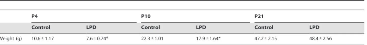
Table 1.Weight gain: at P4, P10 and P21 of rat pups in the low protein diet-induced intrauterine growth restriction group and control.
P4 P10 P21
Control LPD Control LPD Control LPD Weight (g) 10.661.17 7.660.74* 22.361.01 17.961.64* 47.262.15 48.462.56 Values are expressed as mean 6 SEM.
*p,0.05 between control and LPD group; two-tailed Mann-Whitney test. n = 16–20 rat pups per group. doi:10.1371/journal.pone.0078326.t001
Table 2.Weight gain: at P4, P10 and P21 of rat pups in L-NAME-induced intrauterine growth restriction group and control.
P4 P10 P21
Control L-NAME Control L-NAME Control L-NAME Weight (g) 10.860.47 8.660.87* 22.161.05 19.661.41* 40.964.44 45.863.9*
The significance for each bar is indicated by *p,0.05, control vs. L-NAME; two-tailed Mann-Whitney test. Values are expressed as mean 6 SEM. n = 5–10 rat pups per group.
doi:10.1371/journal.pone.0078326.t002
Figure 1. Light microscopic appearance of the lung in control and low protein diet rat pups at P4 (A and B), P10 (C and D), and P21 (E and F) of postnatal life.Photographs of the alveolar region, taken at the same magnification, are presented for each group. doi:10.1371/journal.pone.0078326.g001
Female Sprague Dawley rats from Charles River (l’Abresle, France) for the LPD model and Janvier (LeGenest St Isle, France) for the L-NAME model were mated with a male. Day 1 of pregnancy (E1) was determined by the presence of spermatozoa in vaginal smears. Pregnant rats were housed individually with free access to food and water under cyclic controlled light. All dams littered spontaneously. Day of birth was defined as P0. At P4, P10 and P21, pups were weighed before being killed and their lungs were harvested for determination of morphometric parameters or were frozen.
LPD-induced IUGR. Dams were randomly divided into two groups and fed with different diets from the day of conception until P2. A 22% protein diet was used for the Control group, and an isocaloric 9% protein diet was used for
the LPD group [14]. Caloric deficiency in the LPD group was compensated by carbohydrates. After birth, litters were equal-ized to 10 pups.
L-NAME-induced IUGR. Dams were fed with normal diet and, on day 17 of gestation, randomly assigned to receive L-NAME or saline until the end of gestation. An Alzet osmotic pump (Direct Corp., Palo Alto, CA) was placed subcutaneously on the rat’s back after general anesthesia with IsofluraneH and was used to infuse either L-NAME (SIGMA ALDRICH N5751, 98% powder diluted to a final dilution of 50 mg/dL) or saline vehicle. The infusion continued until the end of gestation. To avoid a potential effect of the product delivered via breast milk, equalized litters of 10 pups were adopted on P2 by dams free of any treatment or pump.
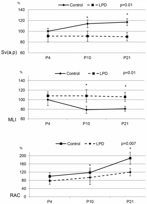
Figure 2. Morphometric analysis in control and low protein diet rat pups.Sv(a,p), Mean Linear Intercept (MLI) and Radial alveolar count (RAC) at P4, P10, and P21 were expressed as percentage of mean control value. Significance for each bar is indicated by p values, Control vs. LPD; two-tailed Mann-Whitney test (p,0.05). N = 5 animals per group.
doi:10.1371/journal.pone.0078326.g002
Lung analysis
Morphometry. A cannula adapted to the size of the trachea was selected. The cannula, filled with 4% paraformaldehyde (PFA), was inserted into the trachea and used to inflate the lungs with PFA at a pressure of 20 cmH2O. Lungs were then kept
inflated by tracheal occlusion and lung volume was evaluated using the fluid displacement method [17]. Lung volume was calculated on the basis of the measured weight increase and the specific gravity of PFA (,1 g/cm3). Five animals selected at random from each group were studied at each time-point.
Alveolar surface density (Sv(a,p)) was determined using point counting and mean linear intercept (MLI) methods described by Weibel [18]. Absolute surface area (Sa) per lung was calculated by multiplying surface density by lung volume. Radial alveolar count (RAC) was performed according to Emery’s method [19].
Quantitative real-time PCR. mRNA expression of genes closely associated with alveolarization i.e. PDGF-A, MMP14 and 16, FGF7 and 18 and their receptors FGFR3 and 4 and angiogenesis i.e. Tie 1 and 2, VEGF and its receptors, VEGFR1 and 2, and adrenomedullin (ADM) were quantified at P4, P10 and P21. Total RNA was extracted from the entire lung tissue using RNeasy Mini RNA extraction kit (Qiagen, Chatsworth, CA). Purified RNA was quantified using NanoDropTM Spectropho-tometer technology (Thermo Fischer Scientific, Wilmington, Delaware). Total RNA was converted into cDNA using 500 ng of total RNA, Superscript III reverse transcriptase, and random hexamer primers (Invitrogen). Real-time PCR was performed on an ABI Prism 7000 (Applied Biosystems, Courtaboeuf, France) using the following protocol: initial denaturation (10 min at 95uC), then a two-step amplification program (15 s at 95uC followed by 1 min at 60uC) repeated 40 times, and finally a dissociation process. Each reaction consisted of a cDNA equivalent of 0.5mg of
total RNA, 12.5ml SYBR Green PCR Master Mix and forward
and 0.9 or 0.3 mM of reverse primers in a 25ml reaction volume.
Levels of mRNA expression were normalized to a housekeeping gene (18 s mRNA) and expressed as relative value using the DDCt method relative to a calibrator sample.
Primers were designed using Primer Express 3.0 (Applied Biosystems, Foster city, CA). Primer pairs are listed in Table S1.
Statistical analysis
Multiple group comparisons were performed by Kruskal-Wallis analysis, and two-group comparisons were performed by Mann-Whitney U test using GraphPad Prism software (GraphPad software INC, San Diego, CA). A p value,0.05 was considered significant.
Results
Weight and growth
Body weight was significantly lower in the LPD group compared to the control group on P4 and P10, but not on P21 with total catch-up of growth (Table 1). Body weight was also significantly lower in the L-NAME group (Table 2) on P4 and P10 and was slightly higher than that of the control group on P21.
Morphometric analysis
Maternal LPD resulted in significantly decreased alveolarization on P21 (Fig. 1), at the end of the process, resulting in simplified lung structure with enlarged airspaces and fewer secondary septa, whereas no difference was observed on P4 (Fig. 1 and Table S2). Morphometric analysis showed a significant 30% increase of MLI on P10 and P21 and a significant decrease of Sv(a,p) in the same proportions, while no significant difference was observed on P4 in the LPD group compared to controls. On P21, RAC was decreased by 35% in the LPD group (Fig. 2).
Maternal injection of L-NAME resulted in significantly decreased alveolarization on P4, at the beginning of the alveolarization process, but not at P21. Lungs from animals exposed to L-NAME exhibited enlarged alveoli on P4, but not on P10 and P21 (Fig. 3 and Table S3). Mean linear intercept was increased by 60% and Sv(a,p) and RAC was decreased in the same proportion on P4, while no between-group differences were observed thereafter (Fig. 4).
PCR results
Gene expression in LPD-induced IUGR. mRNA levels of factors involved in alveologenesis were assessed on P4, P10 and P21 in rat pups. FGF7, FGF18, FGFR3 and -R4, MMP14 and 16 and PDGF-A expressions were not modified in the LPD group compared to the control group regardless of the time-points considered (Fig. 5). mRNA levels of genes involved in angiogenesis were assessed at the same time-points. In the LPD group, Tie 1 and 2, VEGF and its VEGFR1 and 2 receptors and adrenome-dullin gene expressions were not modified regardless of the time-points considered (Fig. 6).
Gene expression in L-NAME-induced IUGR. mRNA levels of factors involved in alveologenesis and angiogenesis were assessed on P4, P10 and P21. On P4, MMP14, MMP16, FGFR4 and PDGF expressions were significantly decreased in the L-NAME group compared to the control group. Fibroblast growth factor 7, FGF18, FGF-R3 and -R4 and MMP14 expressions were significantly decreased in the L-NAME group compared to the control group on P10. Matrix metalloprotease 14, MMP16 and FGFR3 expressions were significantly increased on P21 in the L-NAME group compared to the control group (Fig. 7). Expression of VEGF, a gene involved in angiogenesis, was significantly
Figure 3. Light microscopic appearance of the lung in control and L-NAME rat pups at P4 (A and B), P10 (C and D), and P21 (E and F) of postnatal life.Photographs of the alveolar region, taken at the same magnification, are presented for each group.
decreased in the L-NAME group compared to the control group on P4 and P10 (Fig. 8). Moreover, on P4, the expression of VEGF receptors, VEGF-R1 and –R2 was significantly decreased in the L-NAME group. On P21, adrenomedullin expression was signifi-cantly increased in the L-NAME group compared to the control group.
Discussion
This study, comprising complete structural analysis of lung tissue from rat pups at three key time-points of the alveolarization process, from the start of alveolarization on day 4, to the end of alveolarization on day 21 [16,20], demonstrated that LPD-induced IUGR in rats LPD-induced arrested alveolarization similar to that observed in BPD. In this model, alveolarization was normal at the beginning of the alveolarization process, but became
progressively impaired and markedly decreased at the end of the process. To our knowledge, this is the first study in rodents showing progressive impairment of alveolarization induced by conditions leading to IUGR. In contrast, the L-NAME model appears to be inappropriate to study long-term consequences of IUGR for lung development, as alveolarization was impaired at P4 but subsequently returned to normal by P21. This study also demonstrates major differences between the two IUGR models in terms of the expression of key genes known to be involved in alveolarization and lung angiogenesis.
In the L-NAME model, morphometric analysis revealed significant differences in alveolarization at the beginning of the alveolarization process, on P4, with correction on P21. These results are similar to previously published results in this model showing decreased alveolarization on P0 with a catch-up on P7 and P14 [11]. Although a different method was used to deliver
L-Figure 4. Morphometric analysis in control and L-NAME rat pups.Sv(a,p), Mean Linear Intercept (MLI) and Radial alveolar count (RAC) in control and L-NAME rat pups at P4, P10, and P21 were expressed as percentage of mean control value.. The significance for each time-point is indicated by p values, control vs. L-NAME; two-tailed Mann-Whitney test (p,0.05). n = 5 animals per group.
doi:10.1371/journal.pone.0078326.g004
NAME in our study [11], daily gavage versus subcutaneous pump, our study confirms that, in parallel to postnatal catch-up growth, lung development disorders present at birth in IUGR pups may be completely corrected in this model. The L-NAME model therefore appears to be inappropriate to evaluate the consequences of IUGR on alveolarization. A reversible effect of L-NAME model has been previously demonstrated: L-NAME infusion from embryonic day (E) 14 to E18 of gestation resulted in normal fetal and placental growth on E21. However, Neerhof showed that perfusion of L-NAME from day 14 to 18 of gestation only resulted in normal fetal and placental growth on P21, suggesting a reversible phenomenon [21]. Recent studies have also failed to demonstrate any long-term metabolic consequences in this model [15], questioning the value of this model to study the medium-term and long-term consequences of IUGR.
The experimental settings of the two models used in this study clearly varied in terms of the exposure window (last 4 days of gestation vs. throughout gestation) and the pathophysiological cause of low birth weight (Nitric Oxide synthase inhibition vs. malnutrition). L-NAME treatment administered at E17 to induce IUGR in rodents was first described 20 years ago [22]. Trials of earlier administration of L-NAME: from day 1 to day 18, resulted in a decreased number of implantation sites, decreased litter and placenta weights and an increased number of maternal deaths [23]. When administered at E12, L-NAME induced fetal resorptions and hind limb necrosis, which were not observed when it was administered after E15 [24]. Consequently, admin-istration of L-NAME throughout gestation induces effects other than IUGR and, in order to induce IUGR, L-NAME treatment is therefore classically started in the last third of gestation. Although
Figure 5. Effect of low protein diet-induced IUGR on mRNA expression of factors involved in alveolarization at day 4, 10 and 21. Expression of PDGF, MMP14 and 16, FGF7 and 18 and their receptors FGFR3 and 4 in neonatal rat lungs. n = 9–10 per group. mRNA expression was assessed by real-time PCR. The significance for each bar is indicated by *: p,0.05. Control vs. LPD; two-tailed Mann-Whitney test. Values are expressed as mean 6 SEM.
exposure time to each treatment could explain partly the differences observed between the 2 models, this study design nevertheless allows comparison of the pulmonary consequences of IUGR in pups of similar weights at P4.
Several reports suggest that LPD-induced IUGR is responsible for long-term metabolic consequences that are not reversible, even in adulthood: low muscle mass [25], adult-onset glucose intoler-ance [26], adult hypertension [27], and early aging [28]. In our
study, in the LPD-induced IUGR model, all parameters quanti-fying alveolarization on P4 were equal to those observed in the control group, but indicated decreased alveolarization in the LPD group at P21: MLI, which is inversely correlated with alveolariza-tion, was equal in the 2 groups at P4 and increased in the LPD group at P21; Sv(a,p) was identical at P4, but significantly decreased at P21 in the LPD group; RAC exhibited the same pattern and was also decreased at P21. Notably, LPD group pups
Figure 6. Effect of low protein diet-induced IUGR on mRNA expression of factors involved in angiogenesis at day 4, 10 and 21. Expression of Tie 1 and 2, VEGF and its two receptors (VEGF-R1 and 2) and ADM at P10 in neonatal rat lungs. n = 9–10 per group. mRNA expression was assessed by real-time PCR. The significance for each bar is indicated by *: p,0.05. Control vs. LPD; two-tailed Mann-Whitney test. Values are expressed as mean 6 SEM.
doi:10.1371/journal.pone.0078326.g006
regained normal weight at P21 and morphometric parameters can therefore be interpreted as reflecting a real reduction of alveolarization, independently of weight gain. In a previous study, we observed that morphometric parameters were closely correlat-ed with body weight and differences between groups may be difficult to interpret when body weights are different [29]. These results suggest an arrest or at least a marked impairment of alveolar development. Consequently, this experimental model can be considered to very closely fit the characteristics of BPD, correlated with arrested alveolarization [1]. These results appear to be discordant with those previously published in a different rat strain with slightly different maternal diets, eg lower protein intake in LDP and in controls [30]. These authors did not observe any impaired alveolar development on P70, but based on a less complete morphometric analysis than that conducted in our study
and, more surprisingly, increased alveolarization in the LPD group at P70.
Our study also investigated whether the gene expression of factors involved in alveolar and vascular development was affected by IUGR. Factors involved in alveolarization have been studied by three main approaches: 1) alveolarization study when these factors were not expressed in KO models [31] or by specific inhibition [32]; 2) expression of these factors during normal alveolarization [33]; 3) changes in the expression of these factors in models of postnatal insult leading to alveolarization disorders such as hyperoxia or mechanical ventilation [32]. In some cases, attempts to correct induced alveolarization disorders by administration of these factors were investigated, but with only partial success except for VEGF therapy [34]. Alveologenesis is a process coordinated by multiple interactions involving paracrine mechanisms between
Figure 7. Effect of L-NAME-induced IUGR on mRNA expression of factors involved in alveolarization at day 4, 10 and 21.Expression of PDGF, MMP14 and 16, FGF7 and 18 and their receptors FGFR3 and 4 in neonatal rat lungs. n = 4–5 rat pups per group. mRNA expression was assessed by real-time PCR. The significance for each bar is indicated by *: p,0.05, control vs. L-NAME; two-tailed Mann-Whitney test. Values are expressed as mean 6 SEM.
lung fibroblastic, epithelial, microvascular and extracellular matrix (ECM) components [16]. The factors studied here have been extensively described in this process. The role of antenatal events in the pathogenesis of alveolarization disorders has been well documented, especially in antenatal inflammation models [35]. However, alterations of factors involved in alveolarization have been less extensively studied in models and the results are fairly discordant. Intra-amniotic injection of group B streptococcus in macaques induced abnormal alveolar development with altered expressions of MMP2 and angiopoietin only [36]. Overall, the results are fairly discordant, as factors and their modifications vary from one model to another, highlighting the complexity of
processes in which each factor is involved at a precise timing with precise interactions with other factors.
In the L-NAME model, factors previously shown to be involved in alveolarization, MMP14, FGFR3 and 4, FGF18 and 7, were significantly decreased on P4 and/or P10, but this decrease was not associated with impaired alveolarization at P10. Interestingly, L-NAME, given from E10 to E20 in pregnant Sprague-Dawley rats via an osmotic pump, decreased MMP14 expression in the uterine vessel wall on E20 [37]. Whether this effect on MMP14 is also observed in the fetus and during the first days of life has yet to be studied. Surprisingly, MMP14 expression was significantly increased in the L-NAME group on P21, at a time when the
Figure 8. Effect of L-NAME-induced IUGR on mRNA expression of factors involved in angiogenesis at day 4, 10 and 21.Expression of Tie 1 and 2, VEGF and its two receptors (VEGFR1 and 2) and ADM in neonatal rat lungs. n = 4–5 rat pups per group. mRNA expression was assessed by real-time PCR. The significance for each bar is indicated by *: p,0.05, control vs. L-NAME; two-tailed Mann-Whitney test. Values are expressed as mean 6 SEM.
doi:10.1371/journal.pone.0078326.g008
alveolarization process is theoretically completed and the de-creased alveolarization observed at P4 has been corrected much earlier as observed in our study at P10 and in other studies at P7 [11]. Similar surprising results were observed for VEGF expres-sion. VEGF increased during alveolarization, and decreased when alveolarization was impaired. VEGF inhibition decreased alveo-larization and VEGF administration restored alveoalveo-larization, even after the end of the alveolarization process [34]. In our study, antenatal L-NAME treatment induced a sustained decrease of VEGF and its receptor expressions (P4 and/or P10) during the alveolarization process and in the period during which alveolar-ization can be assumed to have been restored in this model. Finally, none of the factors usually described as essential for alveolarization were modified in the LPD model. These results raise the following question: when the alveolarization process is impaired antenatally, does it respond to regulation factors other than those involved in the normal process and in postnatal models of impaired alveolarization in rat pups?
It has now been clearly established that the developing fetus perceives the environment during specific windows of sensitivity and optimizes future metabolic responses by reprogramming its genome. This reprogramming promotes early survival and reproductive success, but potentially causes a predisposition to disease in later life [10], that only develops when an additional stress occurs subsequently (preterm birth, obesity or high-fat diet). The hypothesis of the fetal or ‘‘early’’ origin of adult disease was originally proposed by Barker, who stated that environmental factors, particularly nutrition, act in early life to program risks for metabolic syndrome, hypertension, insulin resistance and obesity. Lung consequences of IUGR have now also been clearly demonstrated with an increased incidence of BPD [3] and long-term impairment of lung function [8]. Our results suggest that a compromised intrauterine environment could disrupt normal fetal lung development by pathways other than those described in classical models of altered alveolarization. When a modification of programming occurs, involvement of growth factors is inconstant as demonstrated for other organs in IUGR models. Microarray analysis in LPD-induced IUGR in rat showed that only 5 of the 54 crucial factors for kidney development were significantly modified [14]. Proteomic studies in IUGR piglets have shown that, in addition to a significant reduction in small intestine, liver and
skeletal muscle masses, IUGR was also associated with an alteration of proteins known to regulate cellular signalling and intermediary metabolism, resulting in increased proteolysis and reduced polypeptide synthesis [38]. However, the consequences on lung development specifically related to intrauterine growth restriction need to be more thoroughly investigated.
In conclusion, our results demonstrate that two models of IUGR, L-NAME and LPD, have very different impacts on lung development and gene expression patterns of key factors involved in lung development. Only the LPD model closely fits with IUGR-induced alveolarization disorder. The absence of involvement of key factors of alveolarization despite abnormal lung development raises the issue of the role of other regulators. Morbidity induced by IUGR should be analyzed in terms of its pathophysiological cause rather than low birth weight per se and LPD-induced IUGR may therefore constitute a useful tool for an experimental approach to IUGR-induced BPD.
Supporting Information
Table S1 PCR primers for quantitative real-time PCR. (DOC)
Table S2 Morphometric analysis of the lung of control and low protein diet-induced intrauterine growth re-striction groups. Significance for each time-point is indicated by symbols; two-tailed Mann-Whitney test (p,0.05). Values are expressed as mean 6 SEM. n = 5 animals per group.
(DOC)
Table S3 Morphometric analysis of the lung of control and L-NAME- induced intrauterine growth restriction groups. Significance for each time-point is indicated by symbols; two-tailed Mann-Whitney test (p,0.05). Values are expressed as mean 6 SEM. n = 5 animals per group.
(DOC)
Author Contributions
Conceived and designed the experiments: EZT EL CD PHJ DEB. Performed the experiments: EZT MLF LB FV IG. Analyzed the data: EZT EL CD PHJ. Contributed reagents/materials/analysis tools: EZT PHJ. Wrote the paper: EZT EL PD CD OB PHJ.
References
1. Jobe AH, Bancalari E (2001) Bronchopulmonary dysplasia. Am J Respir Crit Care Med 163: 1723–1729.
2. Halliday HL (2009) Neonatal management and long-term sequelae. Best Pract Res Clin Obstet Gynaecol 23: 871–880.
3. Zeitlin J, El Ayoubi M, Jarreau PH, Draper ES, Blondel B, et al. (2010) Impact of fetal growth restriction on mortality and morbidity in a very preterm birth cohort. J Pediatr 157: 733–739 e731.
4. Rosenberg A (2008) The IUGR newborn. Semin Perinatol 32: 219–224. 5. Greenough A, Yuksel B, Cheeseman P (2004) Effect of in utero growth
retardation on lung function at follow-up of prematurely born infants. Eur Respir J 24: 731–733.
6. Doyle LW (2006) Respiratory function at age 8–9 years in extremely low birthweight/very preterm children born in Victoria in 1991–1992. Pediatr Pulmonol 41: 570–576.
7. Barker DJ, Godfrey KM, Fall C, Osmond C, Winter PD, et al. (1991) Relation of birth weight and childhood respiratory infection to adult lung function and death from chronic obstructive airways disease. Bmj 303: 671–675. 8. Edwards CA, Osman LM, Godden DJ, Campbell DM, Douglas JG (2003)
Relationship between birth weight and adult lung function: controlling for maternal factors. Thorax 58: 1061–1065.
9. Briana DD, Malamitsi-Puchner A (2012) Small for gestational age birth weight: impact on lung structure and function. Paediatr Respir Rev.
10. McMillen IC, Robinson JS (2005) Developmental origins of the metabolic syndrome: prediction, plasticity, and programming. Physiol Rev 85: 571–633. 11. Diaz V, Lebras-Isabet MN, Denjean A (2005) Effect of Nomega-nitro-L-arginine
methyl ester-induced intrauterine growth restriction on postnatal lung growth in rats. Pediatr Res 58: 557–561.
12. Maritz GS, Cock ML, Louey S, Joyce BJ, Albuquerque CA, et al. (2001) Effects of fetal growth restriction on lung development before and after birth: a morphometric analysis. Pediatr Pulmonol 32: 201–210.
13. Maritz GS, Cock ML, Louey S, Suzuki K, Harding R (2004) Fetal growth restriction has long-term effects on postnatal lung structure in sheep. Pediatr Res 55: 287–295.
14. Buffat C, Boubred F, Mondon F, Chelbi ST, Feuerstein JM, et al. (2007) Kidney gene expression analysis in a rat model of intrauterine growth restriction reveals massive alterations of coagulation genes. Endocrinology 148: 5549–5557.
15. Butruille L, Mayeur S, Moitrot E, Storme L, Knauf C, et al. (2013) Maternal hypertension induced by NO blockade does not program adult metabolic diseases in growth-restricted rat fetuses. Metabolism.
16. Bourbon J, Boucherat O, Chailley-Heu B, Delacourt C (2005) Control mechanisms of lung alveolar development and their disorders in bronchopul-monary dysplasia. Pediatr Res 57: 38R–46R.
17. Franco ML, Waszak P, Banalec G, Levame M, Lafuma C, et al. (2002) LPS-induced lung injury in neonatal rats: changes in gelatinase activities and consequences on lung growth. Am J Physiol Lung Cell Mol Physiol 282: L491– 500.
18. Weibel ER (1966) [Morphometric studies on the growth of gas exchange capacity of the rat lung]. Helv Physiol Pharmacol Acta 24: C56–59. 19. Emery JL, Mithal A (1960) The number of alveoli in the terminal respiratory
unit of man during late intrauterine life and childhood. Arch Dis Child 35: 544– 547.
20. Burri PH, Dbaly J, Weibel ER (1974) The postnatal growth of the rat lung. I. Morphometry. Anat Rec 178: 711–730.
21. Neerhof MG, Synowiec S, Khan S, Thaete LG (2011) Pathophysiology of chronic nitric oxide synthase inhibition-induced fetal growth restriction in the rat. Hypertens Pregnancy 30: 28–36.
22. Yallampalli C, Garfield RE (1993) Inhibition of nitric oxide synthesis in rats during pregnancy produces signs similar to those of preeclampsia. Am J Obstet Gynecol 169: 1316–1320.
23. Fernandez Celadilla L, Carbajo Rueda M, Munoz Rodriguez M (2005) Prolonged inhibition of nitric oxide synthesis in pregnant rats: effects on blood pressure, fetal growth and litter size. Arch Gynecol Obstet 271: 243– 248.
24. Tiboni GM, Giampietro F, Di Giulio C (2003) The nitric oxide synthesis inhibitor Nomega-nitro-L-arginine methyl ester (L-NAME) causes limb defects in mouse fetuses: protective effect of acute hyperoxia. Pediatr Res 54: 69–76.
25. Toscano AE, Manhaes-de-Castro R, Canon F (2008) Effect of a low-protein diet during pregnancy on skeletal muscle mechanical properties of offspring rats. Nutrition 24: 270–278.
26. Ozanne SE (1999) Programming of hepatic and peripheral tissue insulin sensitivity by maternal protein restriction. Biochem Soc Trans 27: 94–97. 27. Petry CJ, Ozanne SE, Wang CL, Hales CN (1997) Early protein restriction and
obesity independently induce hypertension in 1-year-old rats. Clin Sci (Lond) 93: 147–152.
28. Martin-Gronert MS, Tarry-Adkins JL, Cripps RL, Chen JH, Ozanne SE (2008) Maternal protein restriction leads to early life alterations in the expression of key molecules involved in the aging process in rat offspring. Am J Physiol Regul Integr Comp Physiol 294: R494–500.
29. Mehats C, Franco-Montoya ML, Boucherat O, Lopez E, Schmitz T, et al. (2008) Effects of phosphodiesterase 4 inhibition on alveolarization and hyperoxia toxicity in newborn rats. PLoS One 3: e3445.
30. Alejandre Alcazar MA, Morty RE, Lendzian L, Vohlen C, Oestreicher I, et al. (2011) Inhibition of TGF-beta signaling and decreased apoptosis in IUGR-associated lung disease in rats. PLoS One 6: e26371.
31. Weinstein M, Xu X, Ohyama K, Deng CX (1998) FGFR-3 and FGFR-4 function cooperatively to direct alveogenesis in the murine lung. Development 125: 3615–3623.
32. Vadivel A, Abozaid S, van Haaften T, Sawicka M, Eaton F, et al. (2010) Adrenomedullin promotes lung angiogenesis, alveolar development, and repair. Am J Respir Cell Mol Biol 43: 152–160.
33. Franco-Montoya ML, Boucherat O, Thibault C, Chailley-Heu B, Incitti R, et al. (2011) Profiling target genes of FGF18 in the postnatal mouse lung: possible relevance for alveolar development. Physiol Genomics 43: 1226–1240. 34. Thebaud B, Ladha F, Michelakis ED, Sawicka M, Thurston G, et al. (2005)
Vascular endothelial growth factor gene therapy increases survival, promotes lung angiogenesis, and prevents alveolar damage in hyperoxia-induced lung injury: evidence that angiogenesis participates in alveolarization. Circulation 112: 2477–2486.
35. Thomas W, Speer CP (2011) Chorioamnionitis: important risk factor or innocent bystander for neonatal outcome? Neonatology 99: 177–187. 36. McAdams RM, Vanderhoeven J, Beyer RP, Bammler TK, Farin FM, et al.
(2012) Choriodecidual infection downregulates angiogenesis and morphogenesis pathways in fetal lungs from macaca nemestrina. PLoS One 7: e46863. 37. Hale SA, Weger L, Mandala M, Osol G (2011) Reduced NO signaling during
pregnancy attenuates outward uterine artery remodeling by altering MMP expression and collagen and elastin deposition. Am J Physiol Heart Circ Physiol 301: H1266–1275.
38. Wang J, Chen L, Li D, Yin Y, Wang X, et al. (2008) Intrauterine growth restriction affects the proteomes of the small intestine, liver, and skeletal muscle in newborn pigs. J Nutr 138: 60–66.