HAL Id: tel-03246573
https://tel.archives-ouvertes.fr/tel-03246573
Submitted on 2 Jun 2021HAL is a multi-disciplinary open access archive for the deposit and dissemination of sci-entific research documents, whether they are pub-lished or not. The documents may come from teaching and research institutions in France or abroad, or from public or private research centers.
L’archive ouverte pluridisciplinaire HAL, est destinée au dépôt et à la diffusion de documents scientifiques de niveau recherche, publiés ou non, émanant des établissements d’enseignement et de recherche français ou étrangers, des laboratoires publics ou privés.
Proteins with RBM(ring-building motif)-like domain
involved in bactaria sporulation
Bowen Liu
To cite this version:
Bowen Liu. Proteins with RBM(ring-building motif)-like domain involved in bactaria sporulation. Biomolecules [q-bio.BM]. Université Grenoble Alpes [2020-..], 2020. English. �NNT : 2020GRALV053�. �tel-03246573�
THÈSE
Pour obtenir le grade de
DOCTEUR DE L’UNIVERSITE GRENOBLE ALPES
Spécialité : BIOLOGIE STRUCTURALE ET NANOBIOLOGIE Arrêté ministériel : 25 mai 2016Présentée par
Bowen LIU
Thèse dirigée par Cécile MORLOT et co-encadrée par Thierry VERNET
préparée au sein de l’Institut de Biologie Structurale dans l'École Doctorale Chimie & Sciences du Vivant
Protéines avec un domaine de
type RBM impliquées dans la
sporulation bactérienn
e
RBM-like proteins involved in
bacterial sporulation
Thèse soutenue publiquement le 09 Novembre 2020, devant le jury composé de :
Mme Cécile BREYTON
Directrice de recherche CNRS, IBS, Grenoble (Présidente)
Mme. Véronique BROUSSOLLE
Directrice de recherche INRA, Avignon (Rapportrice)
M. Romé VOULHOUX
Directeur de recherche CNRS, LCB, Marseille (Rapporteur)
Mme. Stéphanie RAVAUD
Maître de conférence Université Lyon 1, MMSB, Lyon (Examinatrice)
M. Juan FONTECILLA-CAMPS
Ingénieur-chercheur CEA, IBS, Grenoble (Examinateur)
Mme Simonetta GRIBALDO
Directrice de recherche CNRS, Institut Pasteur, Paris (Examinatrice)
Mme Cécile MORLOT
As I finished this thesis and approaching the next milestone in my life, I am really willing to give my sincere gratitude to those who have kept supporting me.
First of all, I would like to thank my supervisor Cécile Morlot with all my heart. Cécile is such an intellectual and serious scholar who always gives me a lot of instruction and inspiration about my project. She also checked and approved each part in my thesis elaborately with her utmost patience. I am deeply grateful for her help in completing my thesis.
I would also thanks to Caroline Mas (EMBL), Carlos CONTRERAS-MARTEL (Bacterial Pathogenesis Group, IBS), Emmanuelle Neumann and Daphna Fenel (EM platform, IBS), our collaborators Christopher Rodrigues (University of Sydney, Australia), Adriano Henriques (ITQB, Portugal), Laure Bellard, Anne-Marie Villard and all my other colleagues in pneumococcus group at IBS for the helpful suggestions and technical support of my project.
In addition, Cécile and my group leader Dr. Thierry Vernet give me a lot of concern and care, especially when I firstly arrived in France and when I was isolated at home due to the Covid-19 epidemic. That helps me through the most tough period. I would also thanks to all my colleagues in pneumococcus group at IBS. In the past three years, they have given me not only support of work but also their care about my daily life. It’s my pleasure to study and work with you.
Thanks for Dr. Cécile BREYTON, Dr. Veronique BROUSSOLLE, Dr. Romé VOULHOUX, Dr. Stéphanie RAVAUD, Dr. Juan FONTECILLA-CAMPS and Dr. Simonetta GRIBALDO for accepting to participate in my thesis defense. I look forward to communicating with you in the thesis defense.
I owed the great appreciation for the financial support of China Scholarship during my Ph.D. This makes my dream of studying abroad come true.
Last but not least, thank my family and my girlfriend on the other side of the earth. Their constant encouragement and emotional support are the most powerful emotional anchor for me to complete my study.
Specialized secretion systems found in Gram-negative bacteria allow the transport of molecules across their double-membrane cell envelope. Components of these nanomachines include ring-forming proteins from the PrgK and PrgH families, which are part of the inner membrane platform in III secretion systems, or the InvG and GspD secretins from Type-III and Type-II secretion systems, respectively. Homo-oligomerization of these proteins involves a domain called RBM for "Ring-Building Motif". Despite low sequence identity, RBM domains display a conserved wedge-shaped fold composed of a three-stranded β-sheet packed against two α-helices.
Because the cell envelope of Gram-positive bacteria possess a single membrane, double-membrane spanning machineries are not necessary for secretion. During spore formation in Gram-positive bacteria however, the mother cell engulfs the developing spore, encasing it with a double membrane. Communication between the two cells involves a large multi-protein complex called the SpoIIIA-SpoIIQ complex. The global architecture and function of this new machinery remains mysterious but its components display structural similarities with essential constituents of specialized secretion systems. In particular, some of the SpoIIIA-SpoIIQ proteins possess RBM-like domains and one of them, called SpoIIIAG, forms large oligomeric rings that display remarkable similarities and differences with PrgK and PrgH rings from Type-III secretion systems. Ring formation by SpoType-IIIAG provides evidence that the SpoType-IIIA-SpoIIQ complex might serve as a secretion machinery between the mother cell and forespore but assembly of a transenvelope channel requires oligomerization of other SpoIIIA-SpoIIQ proteins.
To get further insights into the capacity of RBM-containing SpoIIIA-SpoIIQ proteins to form rings, I produced, purified and characterized full-length membrane and truncated soluble forms of these proteins. This part of my work showed that the RBM domain alone in SpoIIIAG is not sufficient to promote oligomerization in vitro, and that additional secondary structures observed in non-canonical RBM domain is not what prevents them from forming rings in vitro.
Intriguingly, RBM domains were also found in proteins that are not related to the SpoIIIA-SpoIIQ complex and raised the hypothesis that other putative secretion systems might form during sporulation. In order to investigate this, I studied the structure and oligomerization ability of one of these proteins, which is called YhcN and is likely involved in spore germination.
The crystal structure of YhcN revealed the presence of a non-canonical RBM domain and the protein did not show any oligomerization ability.
Altogether, my work questions the ring-forming function associated with RBM domains and suggests that some of these domains might have evolved to fulfill different roles.
Les systèmes de sécrétion spécialisés des bactéries Gram-négatives permettent le transport de molécules à travers leur enveloppe cellulaire à double membrane. Les composants de ces nanomachines comprennent des protéines de formation de cycle des familles PrgK et PrgH, qui font partie de la plate-forme de la membrane interne dans les systèmes de sécrétion de type III, ou les sécrétines InvG et GspD des systèmes de sécrétion de type III et de type II, respectivement. L'homo-oligomérisation de ces protéines implique un domaine appelé RBM pour "Ring-Building Motif". Malgré une faible identité de séquence, les domaines RBM présentent un pli cunéiforme conservé, composé d'une feuille β à trois brins et de deux hélices α.
Comme l'enveloppe cellulaire des bactéries Gram-positives possède une seule membrane, les machines à double membrane ne sont pas nécessaires pour la sécrétion. Cependant, lors de la formation des spores chez les bactéries Gram-positives, la cellule mère engloutit la spore en développement, l'enveloppant d'une double membrane. La communication entre les deux cellules implique un grand complexe multiprotéique appelé le complexe SpoIIIA-SpoIIQ. L'architecture globale et la fonction de cette nouvelle machinerie restent mystérieuses mais ses composants présentent des similitudes structurelles avec les constituants essentiels des systèmes de sécrétion spécialisés. En particulier, certaines des protéines SpoIIIA-SpoIIQ possèdent des domaines de type RBM et l'une d'entre elles, appelée SpoIIIAG, forme de grands anneaux oligomériques qui présentent des similarités et des différences remarquables avec les anneaux PrgK et PrgH des systèmes de sécrétion de type III. La formation d'anneaux par la SpoIIIAG fournit la preuve que le complexe SpoIIIA-SpoIIQ pourrait servir de mécanisme de sécrétion entre la cellule mère et le pré-spore, mais l'assemblage d'un canal trans-enveloppe nécessite l'oligomérisation d'autres protéines SpoIIIA-SpoIIQ.
Pour mieux comprendre la capacité des protéines SpoIIIA-SpoIIQ contenant la RBM à former des anneaux, j'ai produit, purifié et caractérisé des formes solubles de ces protéines sur toute la longueur de la membrane et sous forme tronquée. Cette partie de mon travail a montré que le domaine RBM seul dans la SpoIIIAG n'est pas suffisant pour promouvoir l'oligomérisation in vitro, et que les structures secondaires supplémentaires observées dans le domaine RBM non canonique ne sont pas ce qui les empêche de former des cycles in vitro.
De façon intrigante, des domaines RBM ont également été trouvés dans des protéines qui ne sont pas liées au complexe SpoIIIA-SpoIIQ et ont soulevé l'hypothèse que d'autres systèmes de sécrétion putatifs pourraient se former pendant la sporulation. Afin d'étudier cela, j'ai étudié la structure et la capacité d'oligomérisation de l'une de ces protéines, qui est appelée YhcN et est probablement impliquée dans la germination des spores. La structure cristallographique de YhcN a révélé la présence d'un domaine RBM non canonique et la protéine n'a montré aucune capacité d'oligomérisation.
Dans l'ensemble, mes travaux remettent en question la fonction de formation d'anneau associée aux domaines RBM et suggèrent que certains de ces domaines pourraient avoir évolué pour remplir différents rôles.
TXSS: type X secretion system
RBM: ring-building motif
OM: outer membrane
OMP: outer membrane platform
IM: inner membrane
IMP: inner membrane platform
A-Q complex: SpoIIIA-SpoIIQ complex
TMS: transmembrane-segment
EM: electron microscopy
CMC: critical micelle concentration
DDM: n-dodecyl β-D-maltoside
Ni-NTA: nickel-nitrilotriacetic acid
SUMO: small ubiquitin-like modifier
SEC-MALLS: size exclusion chromatography-multi angle laser light scattering
MST: microscale thermophoresis
PDB: protein data bank
Acknowledgement Abstract
Acronyms and abbreviation Introduction
I.Specialized secretion across bacterial membranes ... 1
A. Generalities about secretion through bacterial membranes ... 1
B. Secretion systems in Gram-negative bacteria ... 4
1.Type-I secretion systems ... 6
2.Type-II secretion systems ... 8
3.Type-III secretion systems ...11
4.Type-Ⅳ secretion systems ...14
C. Ring-building motifs in specialized secretion systems ...17
1.Presence of ring-building motifs in specialized secretion systems ...17
2.Oligomerization of ring-building motifs in specialized secretion systems ...21
II.The bacterial sporulation ...23
A. Generalities about bacterial sporulation ...23
B. The sporulation process in Bacillus subtilis ...25
1.Morphological and biochemical sporulation landmarks ...26
2.The sporulation-specific transcription factors ...33
C. The SpoIIIA-SpoIIQ complex in Bacillus subtilis ...35
1.The SpoIIIAA ATPase ...37
2.SpoIIIAB : the putative ATPase anchor ...39
3.SpoIIIAC and SpoIIIAD: putative pilins or export components? ...41
4.SpoIIIAE: permease or export component? ...43
5.SpoIIIAF: a putative ring-forming protein ...45
6.SpoIIIAG: THE ring-forming component ...47
7.SpoIIIAH and SpoIIQ: the bridging proteins ...51
8.GerM ...54
9.Function of the A-Q complex ...57
D. The SpoIIIA-SpoIIQ complex in Clostridium difficile ...59
III. Objectives and rationale of the experimental approaches ...62
A. Plasmids and oligonucleotides used during my PhD ...66 B. RBM-like plasmid construction ...67 C. Production of recombinant proteins ...68 D. Purification of soluble recombinant proteins ...68 E. Screening of detergents for solubilization and purification of membrane
proteins ...69 F. Large-scale purification of membrane recombinant proteins ...71 G. SEC-MALLS analysis ...72 H. Negative-stain EM analysis ...72 I. Mass spectrometry analysis ...73 J. Microscale thermophoresis ...74 K. Protein crystallization and X-ray diffraction data collection ...74 L. Structure determination and refinement ...76 Result ...80 A. Biophysical and structural characterization of GerM ...80 1. Context and personal contribution to the GerM study. ...80 2. Biophysical study of the interaction between GerM and other A-Q proteins ....84 B. Structural characterization of the sporulation protein GerM from Bacillus subtilis. (published in 2018) ...92 C. Determination of structural determinants for the (putative) oligomerization of RBM-containing proteins from the A-Q complex ... 103
1. Role of the transmembrane segments ... 103 2. Role of the soluble regions ... 111 D. Biophysical and structural study of YhcN ... 116 1. Context and personal contribution to the YhcN study. ... 116 2. Purification and crystallization of YhcN ... 118 3. Structural characterization of YhcNA24-E189 ... 122
4. Potential YhcN structural homologues ... 128 5. Biophysical characterization of truncated YhcN constructs ... 130 Discussion ... 134 A. RBM-containing proteins unrelated to the A-Q complex ... 134 1. RBM domains found in specialized secretion systems ... 134
2. The RBM-like domain of YhcN ... 144 B. RBM-containing proteins within the A-Q complex ... 146 1. Do SpoIIIAG form a single or a double ring? ... 146 2. Do SpoIIIAF, SpoIIIAH and GerM oligomerize? ... 151 C. Hypotheses regarding the capacity of RBM-containing proteins to oligomerize.
156
D. Hypotheses regarding the function of the A-Q complex ... 158 Valorization ... 166
1
I.
Specialized secretion across bacterial membranes
A. Generalities about secretion through bacterial membranes
The transport of molecules out of the bacterial cell requires protein complexes assembling in the membrane(s) of bacteria, allowing the secretion of endogenous molecules (Green and Mecsas 2016). Transport across the bacterial membrane is involved in an array of processes such as development, movement, conjugation, adhesion to host cells and surfaces, virulence, host symbiosis, as well as bacterial competition.
Based on their structure, function and specificity, secretion systems have been categorized into several classes. Some of the transport systems are only found in a small number of species while others are conserved in many bacterial species. Table 1 summarizes some of the major features of the secretion systems that have been described so far.
2 Table1. Classes of bacterial specialized secretion systems.
Secretion Apparatus
Secretion Signal Steps Folded Substrates Number of Membranes Gram (+) or Gram (−) T1SS C-terminus 1 No 2 Gram (−)
T2SS Unknown 2 Yes 1 Gram (−)
T3SS N-terminus 1-2 No 2-3 Gram (−) T3SS flagellar protein export apparatus N-terminus 1 No 1 Both T4SS C-terminus 1 No 2-3 Gram (−) T5SS N-terminus 2 No 1 Gram (−)
T6SS Unknown 1 Unknown 2-3 Gram (−)
T7SS C-terminus 1 Yes 1-3 Gram (+)
Sec N-terminus 1 No 1 Both
Tat N-terminus 1 Yes 1 Both
Sortase N-terminus (Sec) C-terimnus (cws)
2 Yes 1 Gram (+)
3
The selectivity for the transported molecule vary from one system to another. It can be based on the size and/or the nature of the molecule. Some systems will thus transport a broad array of substrates while others will be specific to only one or a few molecules.
Depending on the secretion system, the secreted substrate(s) can either remain associated with the bacterial membrane, or be released into the extracellular environment. In some cases, the substrate(s) will be injected into a eukaryotic or bacterial cell.
The transport happens across a single or a double bacterial membrane. In that regard, a major difference between Gram-negative and Gram-positive bacteria regarding the secretion of substances is the composition of their cell envelope. Gram-positive bacteria are surrounded by a plasma membrane and a thick layer of peptidoglycan (a polymer that is made of glycan chains cross-linked by peptide chains, and that confers the cell shape), while the cell envelope of Gram-negative bacteria contains an inner (plasma) membrane, a thin layer of peptidoglycan and an outer membrane. In Gram-negative bacteria, macromolecular systems allowing transport across the cell envelope will thus have to span the two membranes. This mechanism can happen in one or two steps (Green and Mecsas 2016, Costa et al. 2015).
The proteins that I have studied during my Ph.D. are membrane-anchored proteins specifically produced during bacterial sporulation. Although most spore-forming bacteria are Gram-positive, the developing spore is surrounded by two membranes (see chapter Ⅱ), which are reminiscent of the inner and outer membranes found in Gram-negative bacteria. Intriguingly enough, my proteins of interest display weak sequence identity but obvious structural similarities with components of specialized secretion systems found in Gram-negative bacteria. For this reason, I have focused the following sections on these particular secretion systems.
4
B. Secretion systems in Gram-negative bacteria
In Gram-negative bacteria, transport across the inner and outer membranes is carried out by secretion systems that can be divided into three groups (Costa et al. 2015).
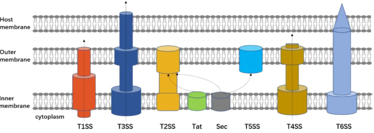
A first group includes protein complexes than span the inner membrane only. It includes the Sec system, which transports most of the secreted proteins across the inner membrane (Lycklama and Driessen 2012). Secretion across the cytoplasmic membrane can also be carried out by the Twin-arginine translocation (Tat) system, which allows fully folded proteins to pass through (Palmer and Berks 2012). The Sec and Tat pathways are the most highly conserved mechanisms for protein secretion and are present in all domains of life (bacteria, archaea and eukarya). Proteins delivered to the periplasmic space by these two systems can be subsequently transported across the outer membrane by another secretion system (such as the Type-II, Type- V and TypeIX secretions systems) (Fig. 1).
A second group includes protein complexes that span the outer membrane only, such as the Type-V secretion system (Leo, Grin, and Linke 2012). By contrast with other secretion systems that span the inner membrane, T5SS does not require a transport energy source. This self-sufficient autotransporter transports proteins through the outer membrane through a β-barrel transmembrane machinery (Meuskens et al. 2019, Bernstein 2019) (Fig. 1).
Finally, a third group includes protein nanomachines that span both the inner and outer membranes. So far, five systems have been identified in this last group, and categorized as
Type-I, Type-II, Type-III, Type-IV and Type-VI secretion systems (T1SS, T2SS, T3SS, T4SS and T6SS, respectively), based on their structural components and function (Costa et al.
2015, Green and Mecsas 2016). These double-membrane-spanning secretion systems are large multiprotein complexes allowing the secretion of specific molecules, including proteins and DNA, from the bacterial cytoplasm to the extracellular environment. Most systems exclusively secrete unfolded or partially unfolded proteins. Only T2SS and T6SS can secrete folded and partially folded proteins. So far, only T4SS has been shown to translocate DNA. Translocation, which is driven by ATP hydrolysis, happens in one (For T1SS, T3SS, T4SS and T6SS) or two steps (For T2SS). When proteins are translocated in two separate steps by T2SS, they are first
5
delivered to the cytoplasm through the Sec or the Tat pathway. Finally, the secreted substrate(s) is(are) either released in the extracellular space (For T1SS, T2SS and T4SS) (Fig. 1A) or injected into a target cell (For T3SS and T6SS).
The common structural feature of Type-I to Type-VI secretion systems is an oligomeric ring formed by outer membrane proteins, resulting in a β-barrel structure that allows secretion of substrates through the outer membrane. These systems otherwise display major differences in their structure, assembly and secretion mechanisms.
In 2008, bioinformatics analyses (HHPRED analyses) based on pairwise comparison of the primary sequence and on secondary structure predictions have identified a group of proteins produced during bacterial sporulation (The SpoIIIA proteins, see chapter Ⅱ) that would have secondary structures organized similarly to those of components found in T1SS, T2SS, T3SS and T4SS (Camp and Losick 2008, Meisner et al. 2008). I will thus focus the next sections on these specific double-membrane-spanning nanomachines (Fig. 1), which are the most pertinent transport systems relative to my Ph.D. I will summarize the state-of-the-art regarding the structure and function of these systems, with a particular emphasis on components that share structural similarities with my proteins of interest.
Figure 1. Schematic diagram of specialized secretion systems in Gram-negative bacteria. T1SS,
T3SS, T4SS and T6SS transport the substrates through the inner membrane (IM) and outer membrane (OM) into the extracellular space in one step. T3SS and T6SS inject the substrate(s) into the host membrane. T2SS and T5SS rely on the Sec or Tat pathway to realize a two-step secretion.
6 1.
Type-I secretion systems
Type-I Secretion Systems (T1SS) are present in a large number of Gram-negative bacteria, especially in pathogens of plants and animals. Since the discovery of the first T1SS substrate (the hemolysin A) in 1979, many other virulence factors have been shown to be secreted through T1SS (Noegel et al. 1979). The size of the substrates range from 10 kDa (e.g. the bacteriocins) to 1 MDa (e.g. the gigantic adhesins). Their function is also diverse, and include digestive enzymes, adhesins, heme-binding proteins and toxins. Bacteria can have several T1SS, specific of one or a few substrates.
T1SS are composed of three different membrane proteins. An ATP-binding cassette (ABC) transporter protein anchored in the inner membrane and an outer membrane factor (OMF) from the TolC family anchored in the outer membrane. Finally, a membrane fusion protein (MFP) crosses the periplasmic space and bridges the ABC transporter to the OMF (Fig. 2A) (Green and Mecsas 2016, Kanonenberg et al. 2018, Costa et al. 2015).
The ABC transporter is responsible for substrate recognition and ATP hydrolysis. The crystal structure of the ABC transporter PrtD from Aquifex aeolicus and MacB from Escherichia coli K-12 show that it forms a homodimer, each subunit containing 6 N-terminal transmembrane (TM) helices and a C-terminal nucleotide binding domain (NBD) (Fig. 2B, C) (Morgan, Acheson, and Zimmer 2017, Fitzpatrick et al. 2017). The N-terminal region of the homodimer forms an occluded channel spanning almost the entire TM region. The C-terminal NBDs locate in the cytoplasm and are responsible for ATP binding and hydrolysis.
In Escherichia coli K-12 T1SS, MacA (MFP) connect the MacB dimer (ATP-binding protein) and TolC (OMF), forms a hexamer with a central channel that allows substrate translocation (Fitzpatrick et al. 2017) (Fig. 2B). Finally, the TolC (OMF) trimer generates another channel that spans the outer membrane, through which the substrate passes. Most of the substrates possess a C-terminal signal that is recognized by the ABC transporter/MFP complex. The unfolded protein is then directly transported from the cytosol to the extracellular space, the secretion being energized by ATP hydrolysis performed by the ABC transporter. This is the
7 generally considered “one-step” translocation mechanism.
Interestingly, bioinformatic analyses performed with the CLUSTALW sequence alignment algorithm suggest that PrtD shares similarities with SpoIIIAE, which is involved in Bacillus subtilis sporulation. While the sequence identity between PrtD and SpoIIIAE is weak (13% sequence identity, 17% sequence similarity) (Fig. 2D), they both possess an equivalent number and organization of transmembrane helices (see section ⅡC). SpoIIIAE might thus share 3D structural and functional similarities with ABC transporters from T1SS.
Figure 2. Structural model of T1SS. (A) Basic structure diagram of T1SS. (B) Ribbon
representation of the structure of the MacA-MacB-TolC pump in Escherichia coli K-12 (PDB code: 5NIK). (C) Ribbon representation of the structure of the ABC transporter from A. aeolicus (PDB code 5L22). (D) Alignment of protein sequences of PrtD from A. aeolicus and SpoIIIAE (AE) from B. subtilis. Conserved residues are in red boxes; similar residues are shown by red letters boxed in blue.
8 2. Type-II secretion systems
Type-II secretion systems (T2SS) are widespread in Gram-negative bacteria and mediate
extracellular delivery of a variety of protein substrates, some of which contribute to the virulence of bacterial pathogens and niche colonization. Most substrates are enzymes, such as proteases, lipases and phosphatases (Korotkov, Sandkvist, and Hol 2012).
T2SS substrates are first delivered by the Sec (Pugsley, Kornacker, and Poquet 1991) or the Tat system (Voulhoux et al. 2001) to the periplasmic space, where they will fold completely. They are then recognized (through still unresolved recognition determinants) by the T2SS apparatus, which will translocate them into the extracellular space via a "piston" mechanism based on the extension and retraction of a pseudopilus (Korotkov and Sandkvist 2019).
T2SS consists of 12-15 different proteins that belong to four subassemblies: the
outer-membrane complex or secretin complex (GspD/PulD family of proteins), the inner-membrane platform (GspC, GspF, GspL and GspM), the secretion ATPase (GspE), and
the pseudo-pilus (GspG, GspH, GspI, GspJ, GspK and GspO) (Fig. 3A) (Korotkov and Sandkvist 2019, Green and Mecsas 2016, Gu et al. 2017, Lopez-Castilla et al. 2017).
The GspE ATPase forms a hexamer that resides in the cytoplasm and connects GspF and GspL components of the inner membrane platform. The inner membrane platform is embedded in the plasma membrane and extends into the periplasm. So far, no structure of the inner membrane platform of a T2SS has been observed at high resolution, but by analogy with
electron cryo-tomography (ECT) studies of T4P, it could be made of interconnected
cytoplasmic and periplasmic rings. Together with the GspE ATPase, which provides energy to power the system, the inner membrane platform assembles the pseudopilus (Fig. 3A). This filamentous sub-structure is mainly composed of multimers of the major pseudopilin subunit GspG and possibly of minor pseudopilins GspH, I, J and K. GpsG precursors are inserted in the inner membrane through a short terminal pre-peptide anchor. After removal of this N-terminal membrane anchor by the GspO peptidase, the pilin subunit then remains associated with the membrane through its hydrophobic tail (Pugsley and Dupuy 1992, Dupuy et al. 1992).
9
During the assembly of the pseudopilus, the hydrophobic tails are gradually extracted from the inner membrane and pack together to form the inner core of the fiber, while the globular domains interact laterally to form the pseudopilus surface (Kohler et al. 2004, Campos et al. 2010, Nivaskumar and Francetic 2014) (Fig. 3A). Polymerization of the pseudopilus is the mechanism through which the folded substrate will be pushed through the pore formed by the outer membrane complex.
The outer-membrane component is the secretin. This protein forms a homomultimeric ring-like channel (Fig. 3B) through which folded substrates are translocated from the cytoplasm to the extracellular space. The secretin has a long N-terminal region made of two to four small variable domains called N-subdomains (N0 to N3) (Yan et al. 2017). N-subdomains share a
highly conserved wedged arrangements that are composed of a two α-helices packed against
a three-stranded anti-parallel β-sheet. This small mixed α/β-modular domain, which was
termed the "ring-building motif" (RBM), is also found in ring-forming proteins from T3SS (see section B3) (Spreter et al. 2009). Packing of N-subdomains from adjacent protomers form a highly stable periplasmic ring that connects the inner-membrane complex (Korotkov et al. 2011) (Fig. 3C). The central region of the protein is a secretin domain that forms a double β-barrel structure partially inserted in the outer membrane. Each β-β-barrel contains 48 (for dodecamers) to 60 (for pentadecamers) anti-parallel β-strands. The secretin channel has an outer diameter of 110 to 170 Å and a small pore at the center of the inner barrel (Nouwen et al. 2000, Chami et al. 2005, Tosi et al. 2014, Hay et al. 2018, Yin, Yan, and Li 2018, Yan et al. 2017, Chernyatina and Low 2019). At the C-terminus of the protein, the S domain seems to act as a hook by grabbing β-strands of the neighboring protomer, which likely enhances the stability of the outer β-barrel (Fig. 3B) (Yan et al. 2017). In the closed state, a central gate, and a possible additional cap gate, block translocation of the substrates (Fig. 3D). During secretion, the N3 domain might be pushed back by the substrate and/or the pseudopilus, then the central gate and cap gate can be pushed outwards to allow opening of the channel and substrate release(Fig. 3C, D).
10 Figure 3. Structural modele of T2SS. (A) Schematic diagram of the topology and location of core components of T2SS. (B) Cryo-EM structure of the homo-multimeric GspD secretin complex from V.
cholera (PDB code: 5WQ8). One of the 15 protomers is colored in red. The position of the rings made
by the N1, N2, N3 and secretin domains of the protein are indicated, and the S domain is circled in black.
(C) Low-resolution cryo-EM structure of the N-terminal rings of the GspD channel. The gaps between
N3 and N2 rings show that the interaction between them is weak. (D) Schematic diagrams of the GspD channel in a closed state (left) and an open state (right). During secretion, the cap and central gates (in yellow) might open around the linker or glycine (marked as G) regions, and the N3 constriction sites might be pushed back in order to let the substrate pass through. Panel C-D are from Yan& Li et al., 2017.
11 3. Type-III secretion systems
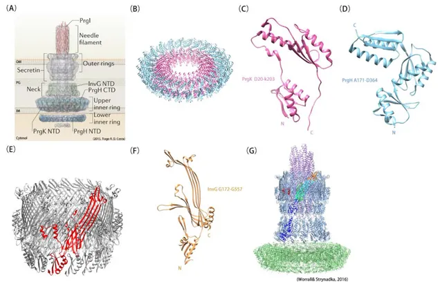
Type-III secretion systems (T3SS) were discovered in 1993 by G.P. Salmond (Salmond and Reeves 1993). Then in the last few decades, T3SS were found in a large number of Gram-negative bacterial pathogens and have been called "injectisomes", or "needle and syringe"-like apparati because of their structure (Buttner 2012). They transport a wide variety of unfolded protein substrates (called "effectors") across the inner and outer membranes in a one-step mechanism, most likely through the inner conduit of their needle component. Many of the T3SS substrates are injected into a host cell to serve as virulence factors.
More than 20 conserved proteins constitute the T3SS apparatus, which is made of two main parts (Burkinshaw and Strynadka 2014, Abrusci et al. 2014) : a double-membrane-spanning
complex composed of stacked rings and a needle-like filament that protrudes from the
bacterial surface (Fig. 4A). The transenvelope complex (or basal body) includes the export
apparatus, which is made of inner-membrane (IM) and outer-membrane (OM) rings, and
an ATPase complex in the cytosol (Abrusci et al. 2014). The needle will deliver the toxin into the host (Fig. 4A).
Within the basal body, two homomeric rings are embedded in the inner membrane (Fig. 4B). These rings contain 24 protomers from the PrgK/EscJ and PrgH/EscD protein families and display external diameters of 18 nm and 27 nm, respectively (Bergeron et al. 2015). PrgK/EscJ family members are made of two globular domains (Fig. 4C) and a C-terminal transmembrane segment. The two globular domains form two juxtaposed rings at the periplasmic surface of the inner membrane and despite low sequence identity between them or between orthologous domains (less than 30%), they share a conserved RBM fold (see section C). Members of the PrgH/EscD family proteins are composed of a N-terminal cytoplasmic globular domain, a TM segment and three periplasmic RBM domains (Fig. 4D). The three RBM domains of PrgH form homomeric rings that surround the PrgK ring(Spreter et al. 2009, Worrall et al. 2016).
Like T2SS, T3SS possess a secretin to allow the passage through the outer membrane. Secretins from T3SS belong to the InvG/EscC family of proteins, in which orthologues can
12
display rather high sequence identity (more than 60%). Like in T2SS, T3SS secretins form 12-to-15-mer rings, and possess a variable number of N-subdomains (displaying the RBM fold), a secretin and an S domain (Fig. 4E, F). In Salmonella enterica serovar Typhimurium pathogenicity island 1 (SPI-1), the InvG secretin contacts the third RBM domain of PrgH through its first N-subdomain (Fig. 4G). Although the asymmetry between the inner (PrgK and PrgH, 24-mers) and outer (InvG, 15-mer) rings appeared surprising at first, the near-atomic cryo-electron microscopy (cryo-EM) structure of the Salmonella SPI-1 injectisome obtained recently confirmed that five subunits of InvG are positioned on the top of height PrgK/PrgH subunits (Worrall et al. 2016). In the closed state, the internal diameter of the secretin measures about 15 Å while in the open state, it extends to 75 Å to allow the passage of the needle (Hu et al. 2018).
The needle complex is a helical filament emanating from the rod complex (PrgJ), which is itself encased at the center of the PrgK ring. The needle is made of more than 100 copies of
PrgI, which adopts a helix-turn-helix motif that polymerizes along both helices. The PrgI
needle is a tube that is 30-70-nm long and 8-nm wide, with an internal helical secretion channel that displays a maximal inner diameter of about 15 Å, allowing the passage of unfolded substrates (Hu et al. 2018). The polymerized needle extends through the secretin pore into the extracellular space. If the substrate must be delivered into a host cell, a translocon complex will assemble at the tip of the needle upon contact with the target cell. The translocon will form a pore in the host cell membrane, through which effectors will be injected.
Note : Cholesterol-dependent cytolysins (CDCs) such as streptolysin O and pneumolysin, which are secreted through the Sec apparatus, assemble into large oligomeric rings that form pores in cholesterol-containing membranes of target eukaryotic cells (Tilley et al. 2005). CDCs have sometimes been described as the “functional equivalent of T3SS in Gram-positive bacteria” or as “injectisomes in Gram-positive bacteria” although they share neither protein sequence similarities nor phylogenetic relationships with any components of T3SS. Furthermore, the sizes of the pores formed by CDCs (30 nm) and T3SS (2 nm) are different. More fundamentally, because effectors are first secreted across the bacterial cell envelope by the Sec apparatus,
13
CDCs are not strictly bacterial protein secretion systems but mechanisms for protein translocation across the eukaryotic plasma membrane. Finally, there is no evidence that a continuous protected channel that mediates protein transit from one cell to another (which is a fundamental feature of T3SS) is also found in CDCs. Therefore, the only common feature shared by T3SS and CDCs is that effectors are translocated into a eukaryotic host cell. So far, the only authentic Gram-positive type III secretion system is the flagellar protein export apparatus."
Figure 4. Structural model of T3SS. (A)Model of the major subassemblies of the T3SS. Adapted from Tiago R.D Costa et al., 2015. (B) Cryo-EM model of the two 24-mer rings made by the periplasmic domains of PrgK (in pink) and PrgH (in cyan) from S. typhimurium (PDB code: 5TC). (C) Ribbon representation of PrgKD20-K203, showing the two RBM domains. (D) Ribbon representation of PrgH A171-D364, showing the three periplasmic RBM domains. (E) Cryo-EM structure of the InvG OM ring from S.
typhimurium (PDB code 5TCQ). One of the 15 protomers is colored in red. (F) Ribbon representation of
a InvGG171-G557 protomer showing the secretin and the S domain. (G) Cryo-EM model of PrgH171-364 (in
green), PrgK20-203 (in green), InvG34-557 (in blue and cyan), and PrgI3-80 (in purple) components of T3SS
from S. typhimurium (PDB code 6DUZ). One monomer encompassing InvG34-557 is colored according to
structural domains : N0-N3 domains (in blue), outer β-sheet (in cyan), inner β-sheet (in green), secretin domain lip (in orange) and S domain (in red) (Hu et al. 2018). The InvG secretin contacts the third RBM domain of PrgH through its first N-subdomain. Panel G is from Worrall& Strynadka et al., 2016.
14 4. Type-Ⅳ secretion systems
Widely present in both Gram-positive and Gram-negative bacteria, Type-Ⅳ secretion systems (T4SS) are versatile secretion systems that are related to the secretion of single protein, protein-protein, DNA and DNA-protein complexes into bacterial or eukaryotic target cells (Fronzes, Christie, and Waksman 2009, Cascales and Christie 2003). They have a unique ability among other secretion systems to translocate DNA and their main function is to mediate conjugation of plasmids, including some harboring antibiotic resistance genes (Costa et al. 2015).
In a canonical T4SS (based on the VirB/D system of Agrobacterium tumefaciens), 12 components (named as VirB1-11 and VirD4) assemble and power a channel that spans the inner membrane, the outer membrane and also the recipient cell membrane, mediating direct transfer of the substrate into the target cell cytoplasm (Sgro et al. 2018). T4SS are composed of two main assembly units: a core-outer-membrane (Core-OM) complex that spans both the inner and outer membranes, and an inner-membrane (IM) complex embedded in the inner membrane (Fig. 5A). These complexes assemble a pilus that will extend into the extracellular space. The pilus is made of a polymer of the major VirB2 pilin and the tip minor pilin VirB5 (Aly and Baron 2007).
Previous structural studies showed that the core-OM complex of canonical T4SS consist of 14 heterotrimers of VirB7, VirB9 and VirB10 proteins (Sgro et al. 2018). The core complex is located in the periplasm and is embedded into the inner membrane and outer membrane through the N- and C-termini of VirB10 (Sgro et al. 2018, Fronzes, Christie, and Waksman 2009, Fronzes et al. 2009). VirB10 forms the OM pore and is surrounded by VirB7 and VirB9 (Fig. 5B) (Sgro et al. 2018, Chandran et al. 2009). Interestingly, VirB7 lipoproteins from Xanthomonales contain an additional C-terminal globular domain, similar to N-subdomains found in T2SS secretins, and displaying the RBM fold (Sgro et al. 2019). This N0/RBM1
domain wraps around the VirB9 and VirB10 layers (Fig. 5B).
15 VirB8, 14 copies of the VirB10 N-terminus, and 3 ATPases (VirB4, VirB11 and VirD4). The ATPases are thought to provide energy for substrate unfolding and transfer through the T4SS channel. VirB4, which is the most conserved ATPase, localizes on the cytosolic side of the inner membrane. It forms two distinct barrel-like pedestals, each of them containing a VirB4 hexamer and interacting with other IM complex components (Fig. 5C) (Redzej et al. 2017).
The translocation mechanism through T4SS remains unclear. The VirB11 ATPase was suggested to act as a molecular switch between a pilus biogenesis mode and a secretion mode. In a first phase, binding of VirB11 to the VirB4 ATPase would promote pilus extension. Binding of the pilus tip pilin VirB5 to the target cell would lead to VirB11 release and its association with the VirD4 ATPase (Trokter et al. 2014). Then the DNA or protein substrate would first bind VirD4, which would act as a gate at the base of the IM complex. VirD4 would then transfer the substrate to VirB11, which would deliver the substrate the channel formed by the IM complex. The mechanism through which the substrate is transferred to the OM complex and is secreted remains unresolved. The pilus might serve as the conduit for substrate translocation or as a contacting device. In the latter case, it would just allow the two cells to be close enough to allow substrate secretion via a pilus-independent mechanism (Babic et al. 2008).
Bioinformatic analyses suggest that VirB4 from Thermoanaerobacter pseudethanolicus shares sequence similarity with the SpoIIIAA ATPase involved in B. ubtilis sporulation (12% sequence identity, 18% sequence similarity).
16 Figure 5. Structural model of T4SS. (A) Model of the major subassemblies of the T4S. Adapted
from Tiago R.D Costa et al., 2015. (B) Cryo-EM structure of the OM core-complex from Xanthomonas
citri T4SS (PDB: 6GYB). Fourteen protomers of the C-terminal N0/RBM1 domain of VirB7 (in red) wrap around the 14-mer ring of VirB9 (in green), itself surrounding the 14-mer oligomer of VirB10 (in blue). (C) Schematic diagram of the T4SS structure in side (left) and bottom (right) views. The VirB4 (in yellow) and VirD4 (in blue) ATPases are respectively shown as hexamers and dimers connected to the VirB3 (in dark green), VirB6 (in green), and VirB8 (in light green) components of the inner membrane platform. The VirB7, VirB9 and VirB10 components of the core-OM complex are shown in different shades of grey. Adapted from Redzej& Waksman et al., 2017.
17
C. Ring-building motifs in specialized secretion systems
1. Presence of ring-building motifs in specialized secretion systems
Although specialized secretion systems are very different in their composition, architecture and assembly mechanism, some of their components share a common fold called the "ring-building motif" (RBM) (Spreter et al. 2009). This motif, as it is able to fold by itself (Bergeron et al. 2015), defines a new family of domains, which lacks detectable sequence identity but share similar arrangement of secondary structures : two helices stacked against a three-stranded β-sheet.
RBM domains are found in ring-forming proteins that located within IM or OM complexes of T2SS, T3SS and T4SS. (Fig. 6):
• the 1st and 2nd globular domains of PrgK/EscJ from the IM complex of T3SS. These
domains will be called PrgK RBM1 and RBM2 in this manuscript (Fig. 6A)
(Burkinshaw and Strynadka 2014, Schraidt and Marlovits 2011, Yip et al. 2005, Hu et al. 2018, Bergeron et al. 2015, Bergeron et al. 2013, Hu et al. 2019).
• the 2nd to 4th globular domains of PrgH from the IM complex of T3SS. These domains
will be called PrgH RBM1 to RBM3 in this manuscript (Fig. 6A) (Spreter et al. 2009,
Bergeron et al. 2013, Bergeron et al. 2015, Burkinshaw and Strynadka 2014, Hu et al. 2018, Hu et al. 2019).
• the 1st to 3rd N-subdomains of the InvG/EscC secretin in T3SS. These domains are
usually called N1, N2 and N3 in InvG; they will be called InvG RBM1 to RBM3 in
this manuscript. These domains are usually called N1 and N2 in EscC; they will be
called EscC RBM1 and RBM2 in this manuscript (Fig. 6B) (Spreter et al. 2009,
Schraidt and Marlovits 2011, Bergeron et al. 2013, Worrall et al. 2016, Hu et al. 2018, Hu et al. 2019).
• the 1st to 4th N-subdomains of the GspD/PulD secretin in T2SS. These domains are
usually called N0, N1, N2, N3; they will be called GspD or PulD RBM1 to RBM4 in
18 2019).
• an additional C-terminal domain found in Xanthomonales orthologues of the VirB7 component of the core-OM complex from T4SS. This domain is usually called N0; it
will be called VirB7 RBM1 in this manuscript (Fig. 6D) (Sgro et al. 2018).
The presence of RBM domains in various systems allowing transport through the inner and outer bacterial membranes could reflect a yet-unexplored evolutionary relationship between them (Souza et al., 2011).
19 Figure 6. RBM domains in different secretion systems. (A) The left panel shows the cryo-EM
structure of the PrgK-PrgH periplasmic rings from S. typhimurium T3SS (Prgk is in pink, PrgH is in light blue, PDB code: 5TCP). The right panels show the ribbon representation of PrgKD20-K203 and PrgH A171-D364, with the RBM domains labeled. (B) Cryo-EM structure of the InvG OM ring from S. typhimurium
T3SS with one monomer shown in red (PDB code: 6DV3). The right panel shows the ribbon representation of InvGG34-G557, with RBM1 to RBM3 domains labeled. (C) The left panel shows the
cryo-EM structure of the GspD secretin channel from V. cholerae T2SS with one monomer shown in red (PDB code: 5WQ8). The right panel shows the ribbon representation of GspDG97-M646, with RBM2 to RBM4
domains labeled. (D) The left panel shows the cryo-EM structure of the OM core-complex from X. citri T4SS (PDB code: 6GYB, VirB7 is in red, VirB9 is in green and VirB10 is in blue). The right panel shows the ribbon representation of the VirB7 C-terminal RBM domain. (E) Structure and topology model of the RBM3 domain of GspD from V. cholerae T2SS. α-helices are in green, β-sheets are in orange. (F) Structure and topology model of the RBM2 domain of PrgK from S. typhimurium T3SS.
20 Despite low primary sequence conservation (lower than 30%), RBM domains share an overall similar architecture made of two α-helices packing against a three-stranded antiparallel β-sheet. They are divided into two groups based on secondary structure connectivity: RBMs from the T3SS PrgK/PrgH group display an α-β-β-α-β arrangement (Fig. 6F) while RBMs from the N-subdomain secretin group display a β-α-β-β-α arrangement (Fig. 6E). Structural RBM models from the two groups have been obtained from X-ray crystallography or electron microscopy studies; the corresponding PDB entries, main characteristics and references are listed in Table 9 (see discussion). For the PrgK/PrgH group, they include EscJ RBM1-2 from E.
coli T3SS, PrgK RBM1-2 and PrgH RBM1-3 from S. typhimurium T3SS (Yip et al. 2005, Spreter
et al. 2009, Schraidt and Marlovits 2011, Bergeron et al. 2013, Bergeron et al. 2015, Worrall et al. 2016, Hu et al. 2018, Hu et al. 2019). Within the N-subdomain secretin group of T3SS, they include EscC RBM1-2 (N1-2) from E. coli T3SS and InvG RBM1-3 (N1-3) from S. typhimurium
T3SS (Spreter et al. 2009, Schraidt and Marlovits 2011, Bergeron et al. 2013, Worrall et al. 2016, Hu et al. 2018, Hu et al. 2019). Within the N-subdomain secretin group of T2SS, they include GspD RBM2-4 (N1-3) from E. coli T2SS, GspD RBM2-4 (N1-3) from V. cholerae, PulD
RBM1-4 (N0-3) from K. pneumoniae T2SS, and XcpQ RBM3-4 (N2-3) from Pseudomonas
aeruginosa T2SS (Yan et al. 2017, Hay et al. 2018, Chernyatina and Low 2019). Within the N-subdomain secretin group of T4SS, they include VirB7 RBM1 (N0) from X. citri T4SS (Sgro et
21 2. ligomerization of ring-building motifs in specialized secretion systems
On the basis of molecular modelling, a broadly conserved ring-packing arrangement had been predicted for these α/β RBM domains (Yip et al. 2005), and the term RBM for "ring-building motif" had been proposed by the group of N. Strynadka (Spreter et al. 2009). The hypothesis that the wedge-shaped fold of RBM domains triggers ring-like oligomerization was later supported by studies in which PrgK and PrgH were located in purified T3SS from S. typhimurium (using immunogold labeling combined with single-particle EM). In these studies,
24-mer oligomeric ring models of the two proteins (based on the crystal structure of EscJ for PrgK, and on the crystal structure of PrgH) were docked into corresponding cryo-EM maps (Spreter et al. 2009, Schraidt and Marlovits 2011, Schraidt et al. 2010).
Similar studies were performed with the N-subdomains of EscC from E. coli T3SS or InvG from S. typhimurium T3SS (Spreter et al. 2009, Schraidt et al. 2010, Schraidt and Marlovits 2011). These studies eventually revealed the 24:24:15 stoichiometry of the PrgK/PrgH/InvG complex in T3SS (Schraidt and Marlovits 2011). The PrgK/PrgH/InvG ring model was later refined owing to new crystal or NMR structures of PrgK, PrgH and InvG, as well as higher-resolution cryo-EM maps of T3SS (Bergeron et al. 2013, Bergeron et al. 2015).
More recently, the rapid progress of cryo-EM remarkably pushed our knowledge of the T3SS architecture forward, providing models of the PrgK/PrgH/InvG complex at increasingly higher resolution, the best one so far reaching about 3.5 Å (Worrall et al. 2016, Hu et al. 2018). Additional elements of the T3SS get progressively incremented in the high-resolution cryo-EM models, such as the SpaP, SpaQ and SpaR export components or the PrgI/PrgJ needle complex (Hu et al. 2019, Guo et al. 2019).
First non-oligomeric structures of N-subdomains in secretins of T2SS (from E. coli ETEC GspD) were studied by Korotkov and co. using X-ray crystallography (Korotkov et al. 2009, Korotkov et al. 2011, Korotkov and Hol 2013). Later, the oligomeric status of these domains were revealed by cryo-EM studies performed on various orthologues of the GspD/PulD family, providing models of N-subdomain rings connected to rings of the secretin domain (Yan et al.
22 2017, Chernyatina and Low 2019, Burkinshaw and Strynadka 2014, Hay, Belousoff, and Lithgow 2017).
The variations observed in the stoichiometry (24 for PrgK and PrgH from T3SS, 15 for InvG from T3SS, 15 for GspD from T2SS, 14 for VirB7 from T4SS), dimensions and surface properties of those rings arise from variations in the primary sequence of the ring-forming proteins and likely allow this family of proteins to adapt to the specific architecture and assembly characteristics of the different secretion systems.
These high-resolution data now allow us to perform a more reliable analysis of the oligomerization interface of RBM domains. This analysis constituted part of my Ph.D. work and will be presented in the Discussion section.
Intriguingly, components of a putative secretion complex involved in bacterial sporulation, called the SpoIIIA-SpoIIQ complex (or A-Q complex) were shown to display weak sequence identities but obvious structural similarities with various components of specialized secretion systems (see chapter Ⅱ). In particular, four of the A-Q proteins from Bacillus subtilis were shown to contain RBM domains and one of them (SpoIIIAG) was shown to form oligomeric rings resembling PrgK/PrgH RBM rings, suggesting that the A-Q complex might be a transport machinery (Rodrigues, Henry, et al. 2016, Zeytuni et al. 2017, Zeytuni et al. 2018a, Morlot and Rodrigues 2018). However, significant differences in the architecture and dimensions of the SpoIIIAG ring compared to RBM rings found in specialized secretion systems indicate that the A-Q complex should constitute a new type of secretion apparatus.
Other RBM-containing proteins were also recently identified with no apparent functional connection to the A-Q complex. During my Ph.D., I studied the structure of one of these proteins (called YhcN), and performed a structural analysis and comparison between RBM domains found in specialized secretion systems and in sporulation proteins.
23
II.
The bacterial sporulation
A. Generalities about bacterial sporulation
As one of the oldest lives on earth, bacteria can thrive in multifarious environments, some can even survive in extreme conditions by adopting special strategies. The differentiation of bacteria into resistant spores, a process known as sporulation, is one of the most important strategies allowing certain bacterial species to survive in adverse conditions. The first spore was discovered by Robert Koch and Ferdinand Cohn in 1850 but despite more than 140 years of intensive studies, many aspects of the sporulation, spore dormancy and germination processes remain mysterious (Nicholson et al. 2000). As the most tenacious form of cell type in nature, spores can survive and stay dormant over long periods in many conventional sterilization methods such as high temperature, dehydration, radiation, detergent and some chemical solvents. Some spores can even reach several million years in earth core and fossils (Cano and Borucki 1995).
The capacity of bacteria to sporulate is mainly found in two genera from the Firmicute phylum, Bacillus and Clostridium, and it has many implications in industry and medicine: Bacillus species are used to produce many industrial enzymes and are also important biocontrol agents in agriculture. When spore-forming bacteria are also human pathogens, their ability to differentiate into resistant spores is an important pathogenesis factor (Traag et al. 2010, Ciccarelli et al. 2006). Famous pathogen spore-formers include Clostridium tetani, which causes tetanus (a disease characterized by muscle spasms), Clostridium botulinum, whose botulism toxin causes muscle failure and gastroenterological symptoms), Clostridium perfringens, responsible for tissue necrosis, and Clostridium difficile, which causes a toxin-mediated intestinal recurrent disease known as CDI (C. difficile infection). Finally, Bacillus anthracis is the well-known bioagent of anthrax, which can occur in skin, respiratory, intestinal or inflammatory forms. This bacterium is one of the most likely microorganisms to be used in a bioterrorist attack (source : www.cdc.gov).
In harsh conditions, some bacteria such as those cited above can stop their vegetative growth and enter a sporulation cycle. During this differentiation process, the developing spore and the
24 mother cell will undergo a series of transcriptional, morphological and biochemical changes that lead to the formation of a spore enveloped by protective layers called the cortex and the coat. When the spore is mature, the mother cell lyses and releases the endospore into the environment where it can remain dormant for a very long time. When the endospore encounters a suitable environment, it germinates and goes back to vegetative growth.
25
B. The sporulation process in Bacillus subtilis
The Gram-positive bacterium Bacillus subtilis has become the most studied spore-forming model because its natural competence, as well as the development of tools to manipulate its genetic content, have incredibly facilitated the creation of mutant strains to study cellular processes such as sporulation. Some differences exist in terms of gene conservation, activation mechanisms of the sporulation-specific transcription factors or composition of the spore envelope between the different spore-formers. However, most sporulation features are conserved and the knowledge obtained from studies performed on B. subtilis can be transposed to other species. Since all the sporulation proteins that I studied originate from B. subtilis, I will summarize in the next sections the knowledge regarding the sporulation cycle in this bacterium, and emphasize aspects that are directly related to my proteins of interest, the RBM-containing proteins.
Sporulation in B. subtilis is triggered by nutrient starvation and is easily inducible in laboratory conditions. At 37°C, it takes around 7-8 hours to form mature B. subtilis spores. The morphological differentiation process is orchestrated by four sporulation-specific transcription factors. It can be divided into seven main stages (stages 0 to VI, which are used to name many sporulation-specific genes), characterized by morphological and transcriptional landmark events (Fig. 7) that can be easily observed in the laboratory by observing samples every 30 minutes.
26 1. Morphological and biochemical sporulation landmarks
a. Asymmetric division and chromosome translocation
Upon starvation stress, the entry into sporulation is triggered by phosphorylation of the master transcription factor Spo0A (Tan and Ramamurthi 2014, Piggot and Hilbert 2004). Re-localization of the major division protein FtsZ (Erickson, Anderson, and Osawa 2010) at the quarter of the rod-shaped B. subtilis cell results in formation of a flat asymmetric septum that divides the cell into two unequally sized compartments. The big one is called the mother cell and the small one is called the forespore (Fig. 7). These two compartments are separated by two membranes that define an intermembrane space, which is about 25-nm thick and contains peptidoglycan, a polymer made of glycan chains cross-linked by peptide chains. Importantly for my thesis work, this double-membrane landscape is reminiscent of the cell envelope in Gram-negative bacteria.
The polar septum traps about 30% of one chromosome copy into the forespore. The remaining 70% of the chromosome is then transported from the mother cell cytoplasm to the forespore one by SpoIIIE, a membrane-anchored ATPase that assembles a translocation complex at the center of the septum (Besprozvannaya and Burton 2014). Chromosome translocation increases the turgor pressure into the forespore. At the same time, the asymmetric septum is thinned (to about 14 nm) by the peptidoglycan hydrolase complex SpoIID-SpoIIM-SpoIIP (Morlot et al. 2010, Khanna et al. 2019). As a consequence of those events, the forespore starts inflating into the mother cell (Lopez-Garrido et al. 2018).
27 Figure 7. Schematic diagram of morphological changes and chromosome translocation into the forespore during the sporulation. Peptidoglycan is in grey, membranes are in red, chromosomes are in blue, SpoIIIE is in orange. The panel is from Khanna et al., 2019.
b. Engulfment
Shortly after asymmetric division and the beginning of chromosome translocation, the mother cell membrane starts migrating around the forespore in a phagocytic-like process called engulfment. During engulfment, the septal peptidoglycan is at least partially hydrolyzed and new peptidoglycan is synthesized at the leading edge of the engulfing membrane.
Peptidoglycan hydrolysis is carried out by the SpoIID-SpoIIM-SpoIIP complex, which includes two peptidoglycan hydrolases that perform processive degradation of the polymer (Fig. 8) (Morlot et al. 2010). Whether the SpoIID-SpoIIM-SpoIIP complex also degrades the new peptidoglycan synthesized during engulfment remains a matter of debate, as it is still not clear whether this new PG is synthesized ahead or behind the SpoIID-SpoIIM-SpoIIP machinery (Khanna et al. 2019). The activity of the SpoIID-SpoIIM-SpoIIP complex relies on the activation of the SpoIIP endopeptidase/amidase by SpoIID, and the conditional activity of SpoIID on naked glycan strands (devoid of stem peptides). Based on this enzymatic coordination, localization around the forespore and requirement for engulfment, the SpoIID-SpoIIM-SpoIIP complex was proposed to function as a motor pulling the mother cell membrane around the forespore (Fig. 8B, C).
28 At the end of engulfment, fission of the mother cell membranes, which involves the FisB protein but is not totally understood, releases the forespore into the mother cell cytoplasm (Doan et al. 2013). The forespore is thus eventually surrounded by two membranes: the inner forespore membrane and the outer forespore membrane (Fig. 7, 8C).
Proper engulfment also requires the assembly of a large multi-protein complex into the inner and outer forespore membranes. This transenvelope complex called the SpoIIIA-SpoIIQ complex will be described in a separate section (section ⅡC) because it is one of the objects of my thesis.
29 Figure 8. The SpoIID-SpoIIM-SpoIIP machinery. (A) Migration of the mother cell membrane
(thick grey line) around the forespore membrane (thin grey line) is driven by peptidoglycan hydrolysis performed by the SpoIID (pacman) and SpoIIP (shaded lollipop) pair of hydrolases. SpoIIM (speckled box) anchors the peptidoglycan hydrolases at the leading edge of the engulfing mother cell membrane. The panel is from Mello et al., 2002. (B) Schematic diagram of the proposed catalytic cycle of the engulfment complex. (1) The SpoIID (D)-SpoIIM (M)-SpoIIP (P) complex binds the peptidoglycan. Glycan strands are in green, peptides are in black. (2) D stimulates the amidase activity of P, resulting in cleavage of the stem peptides cross-links and the release of P. (3) Released P rebinds at a nearby peptidoglycan site. (4) The denuded glycan strands are cleaved by D. (5) Released D rebinds to P at the nearby site. (C) Circumferentially distributed D-M-P engulfment complexes drive movement of the mother cell membrane (light purple) around the forespore membrane (blue). Panels (B) and (C) are from Morlot et al., 2010.
c. Assembly of the cortex and the coat
During and after engulfment, several protective layers are added around the forespore and will confer its resistance properties: the cortex and the various coat layers (Fig. 9) (McKenney, Driks, and Eichenberger 2013, Popham and Bernhards 2015).
30 The cortex is a modified peptidoglycan that is synthesized on top of the germ cell wall (unmodified peptidoglycan) in the intermembrane space. Genes involved in synthesis of the germ cell wall include the PG synthases genes pbpF and pbpG while genes involved in cortex synthesis include the PG synthases genes spoVB, spoVD and spoVE, as well as the PG hydrolase gene lytH, dacA, dacB, dacF and cwlD and the deacetylase gene pdaA.
Like vegetative peptidoglycan, the cortex contains sugar strands made of alternating N-acetylglucosamine (NAG) and N-acetylmuramic acid (NAM) attached to peptide chains. However in the cortex, about 50% of the peptide chains attached to NAM are removed, resulting in the formation of muramic--lactam (McKenney, Driks, and Eichenberger 2013). In addition, 15 to 25% of the remaining peptide chains are shortened to single L-Ala residues. These peptide modifications result in a greatly reduced level of cross-linking of the spore peptidoglycan (3% in cortex against 40% in vegetative peptidoglycan).
The cortex maintains the dehydration state of the mature spore, which is essential for resistance to high temperatures and chemicals. The cortex feature that impacts most the spore resistance is the amount of cortex rather than its structural properties. Surprisingly enough, neither the presence of muramic--lactam nor modified peptides affects the resistance of the cortex to vegetative peptidoglycan hydrolases. The spore is actually protected from these lytic enzymes by the several proteins layers of the coat. Instead, the muramic--lactam, which is absent from the germ cell wall, is specifically recognized by germination hydrolases. This particularity would thus allow specific degradation of the cortex and protection of the germ cell wall during germination.
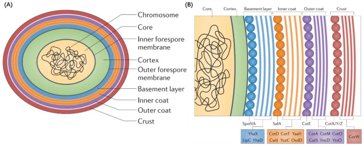
In B. subtilis, the spore coat is composed of three layers: a lamellar inner coat, a more coarsely layered outer coat and a crust layer (Fig. 9A). In recent years, coat proteins were identified through reverse genetics and immunogold-electron microscopy. Proteins playing a main role in coat morphogenesis include: SpoIVA, SpoVID and SpoVM, which play a role in anchoring the coat to the spore surface; SafA, which is necessary for inner coat assembly; and CotE, which is required for outer coat assembly (McKenney, Driks, and Eichenberger 2013). Another group of proteins including CotX, CotY and CotZ is essential to the assembly of the
31 crust layer (Fig. 9B) (McKenney, Driks, and Eichenberger 2013).
The spore coat is only permeable to molecules of 2 to 8 kDa and protects the spore from enzymatic aggressions, including peptidoglycan hydrolases.
Figure 9. Protective layers found in the mature B. subtilis spore. (A) Schematic diagram of the
mature B. subtilis spore. The chromosome is located in the dehydrated central core. The core is protected by multiple layers including the cortex (in green), the basement layer (in blue), the inner coat (in orange), the outer coat (in purple) and the crust (in red). (B) Composition of the different protein layers of the spore coat. The assembly of each layer may be driven by multimerization of the coat proteins. The interaction between each layer is still to be demonstrated. This figure is from McKenney et al., 2013.
d. Preparation for dormancy and mother cell lysis
To prepare for dormancy, the spore produces small DNA-binding proteins that compact the chromosome and protect it from irradiation and genotoxic stress.
In parallel, the mother cell produces the SpoVFA and SpoVFB enzymes that convert dihydroxydipicolinic acid into dipicolinic acid (DPA) (Daniel and Errington 1993). DPA is transported across the outer forespore membrane by the SpoVV transporter and then imported into the forespore by the SpoVA proteins (SpoVAC, SpoVAD, SpoVAEb and SpoVAF) (Ramirez-Guadiana et al. 2017). Accumulation of DPA in complex with Ca2+ (CaDPA) into the