Computational Tools for Enabling Longitudinal Skin
Image Analysis
by
Kang Qi Ian Lee
BEng, Electrical and Electronic Engineering
Imperial College London (2014)
MASSACHI TS NST ITUTE OFTECHNQLOGY
SEP 152016
LIBRARIES
ARCH\ES
Submitted to the Center for Computational Engineering
in partial fulfillment of the requirements for the degree of
Master of Science in Computation for Design and Optimization
at the
MASSACHUSETTS INSTITUTE OF TECHNOLOGY
September 2016
Massachusetts Institute of Technology 2016. All rights reserved.
Author ...
Certified by
Principal
Accepted by.
Signature redacted
Center for Computational Engineering
3ignature redacted
August 5, 2016
Brian W. Anthony
Research Scientist, Department of Mechanical Engineering
Thesis Supervisor
Signature redacted
(
Youssef M. Marzouk
Associate Professor, Department of Aeronautics and Astronautics
Co-Director, Computation for Design and Optimization
.
MiTLibraries
77 Massachusetts Avenue Cambridge, MA 02139 http://Iibraries.mit.edu/ask
DISCLAIMER NOTICE
Due to the condition of the original material, there are unavoidable
flaws in this reproduction. We have made every effort possible to
provide you with the best copy available.
Thank you.
The images contained in this document are of the
Computational Tools for Enabling Longitudinal Skin Image
Analysis
by
Kang Qi Ian Lee
Submitted to the Center for Computational Engineering on August 5, 2016, in partial fulfillment of the
requirements for the degree of
Master of Science in Computation for Design and Optimization
Abstract
We present a set of computational tools that enable quantitative analysis of longi-tudinally acquired skin images: the assessment and characterization of the evolution of skin features over time. A framework for time-lapsed skin imaging is proposed.
A nonrigid registration algorithm based on multiple plane detection for landmark
identification accurately aligns pairs of longitudinal skin images. If dense and thick hairs are present, then nonrigid registration is used to reconstruct the skin texture of occluded regions by recording multiple images from the same area. Realistic re-construction of occluded skin texture is aided by an automatic hair segmentation algorithm and guided painting method based on image blending. We demonstrate that constituent algorithms in this framework are accurate and robust in a multitude of scenarios. In addition, a methodology for rigorous longitudinal analysis of skin microrelief structure is introduced. Following rigid registration, a microrelief junction point matching algorithm based on point pattern matching is shown to accurately match two sets of junction points. Immediate applications for these computational tools are change detection for pigmented skin lesions and deformation field computa-tion of the skin surface under stress using only visual features of the skin. Prospective applications include new insights in skin physiology and diseases from the capability to precisely track movements of the microrelief structure over time and localization of skin images on the body.
Thesis Supervisor: Brian W. Anthony
Acknowledgments
After 2 years at MIT, I am quite reluctant to leave the community and the memorable experiences behind. The journey has been nothing short of amazing. And I have many people to thank for.
The first person I must thank is my advisor, Dr. Brian Anthony. I would always come out of meetings with him with more ideas. So lie has been a great help towards helping me find my research direction in skin research. There was a lot of uncertainty in exploring this field of study and with his guidance, we found interesting areas to work on. From him I learned to: try out things, and avoid thinking too much; always be ready to present; appreciate his sarcasm when he jokes with fellow lab mates. So working with him has been immensely pleasurable.
Next, I would like to thank my colleague Dr. Xian Du. He knows everything; name any field of engineering and lie has done something on it. So I am thankful to have him talk about algorithms, paper writing and his current projects at the interface of so many fields. Most importantly, I am grateful for our collaboration in skin research that was made into part of this thesis. Also, as a friend and colleague in Team Skin, I thank Ina Kundu for her hard work in designing the imaging system used in this thesis and for meticulously acquiring the skin images I needed.
Over the past year, the lab doubled in size and I really enjoyed all the friendships and fun activities we had. A special mention goes to Rishon and Alex Benjamin, who let me stay in their Tang apartment while I finish writing this thesis. It is great to have fellow CDO coursemates in the lab. We are a rare breed.
Being part of CDO brought me into unfamiliar territory when its came to classes. As such, I am sincerely grateful for my friends from our program who supported me through the toughest psets and projects in the core CDO classes, hung out for countless hours in the old CCE office and went for hikes in and around Boston. Special thanks go to Ming Qing Foo, Lutao Xie, Park Sinchaisri, Larson Hogstrom and Ken Wang.
team-mates I ever worked with in class. I am truly glad that our 6.869 project kickstarted Lezhi's journey into discovering the visual identity of cities.
I thank the Singaporeans in MIT for all the fun and good times, meals together,
board games and travels. Special mention goes to Goh Siongthye, Kong Jian Feng, Dax Koh, Gladia Hotan, Chen Hanrong, Low Guang Hao, Alvin Tan, Tan Zheng Jie, Liu Yun and Yeong Li Qian.
My heartfelt thanks go to my small group, who has been a great source of love
and support throughout my time here: Ryan Chang, Alice Kao, Wu Mengfei, David
Kwabi, Lyndon Zhang, Tim Yee and Kim MinJung.
My sincere gratitude go to my good friends in Graduate Christian Fellowship:
Yong Sze Zheng, Nigel Chou, Grace Goon, Sam Elder and Kevin Meng.
Finally, I would like to thank my parents and brother for their love and support, whom I have spent 5 years away from due to my university studies.
Contents
1 Introduction 17
1.1 Contributions . . . . 19
1.2 Thesis Outline. . . . . 20
2 Background 21 2.1 Visible Light Optical Skin Imaging System . . . . 21
2.2 Skin Features . . . . 22
2.2.1 Skin Pigmentation . . . .. . . . 23
2.2.2 H air . . . . 23
2.2.3 Microrelief Structure . . . . 25
2.3 Skin Registration . . . . 25
2.3.1 Skin Appearance and Correspondence . . . . 28
2.3.2 Related Work on Skin Registration . . . . 31
2.4 Digital Hair Removal . . . . 33
2.4.1 Related Work on Digital Hair Removal . . . . 34
2.5 Microrelief Junction Point Matching . . . . 39
2.5.1 Related Work on Microrelief Extraction . . . . 40
2.5.2 Related Work on Non-Rigid Point Pattern Matching . . . . . 40
3 Skin Registration 43 3.1 Algorithm Design . . . . 43
3.1.1 Overview . . . . 43
3.1.3 Keypoint Matching and Global Registration . . . .
3.1.4 Fast Initial Keypoint Matching . . . .
3.1.5 Keypoint Matching by Multiple Plane Detection . .
3.1.6 Local Motion Model . . . .
3.2 Experimental Results . . . .
3.2.1 System and Parameters . . . . 3.2.2 Real Images with Differing Viewpoints . . . .
3.2.3 Comparing Multiple Plane Detection Methods . . . 3.2.4 Real Images of Stretched Skin . . . .
3.2.5 Real Time-Lapsed Skin Images . . . . 3.2.6 Simulated Images . . . .
3.2.7 Accuracy Evaluation Criteria . . . .
3.2.8 Varying the Number of Levels in Multilevel B-Spline
3.2.9 Varying Rotation and Distortion . . . .
3.2.10 Varying Noise Levels . . . .
3.2.11 Computation Time for Keypoint Matching . . . . . 3.3 Final Design and Discussion . . . .
4 Microrelief Junction Point Matching
4.1 Algorithm Design . . . .
4.1.1 Overview . . . .
4.1.2 Homography and Keypoint Matching 4.1.3 Convex Hull Registration . . . . 4.1.4 Microrelief Structure Extraction . . .
4.1.5 Junction Point Detection . . . . 4.1.6 Point Pattern Matching . . . .
4.2 Experimental Results . . . . 4.2.1 Implementation and Parameters . . .
4.2.2 Rigid Skin Registration . . . . 4.2.3 Microrelief Matching . . . . 8 . . . . 46 . . . . 51 . . . . 52 . . . . 58 . . . . 61 . . . . 61 . . . . 62 . . . . 65 ... 69 . . . . 74 . . . . 78 . . . . 80 . . . . 81 . . . . 83 . . . . 85 . . . . 86 88 91 . . . . 91 . . . . 91 . . . . 92 . . . . 94 . . . . 95 . . . . 95 . . . . 96 . . . . 99 . . . . 99 . . . . 99 . . . . 107
4.3 Discussion . . . . . 110
5 Hair Segmentation 5.1 Overall Algorithm Design and Implementation 111 ... . ... .. ... .. 111
5.1.1 Overall Design . . . . 5.1.2 Implementation . . . . 5.1.3 D atasets . . . . 5.2 Curvilinear Structure Detection . . . . 5.2.1 Exploring Color Spaces . . . . 5.2.2 Top-Hat Transform . . . . 5.2.3 Multiscale Matched Filter . . . . 5.2.4 Increasing Color Sensitivity . . . . 5.2.5 Comparison with Other Detection Methods . . . . 5.3 Adaptive Thresholding . . . . 5.3.1 Observations . . . . 5.3.2 Threshold Searching . . . . 5.4 k-Nearest Neighbor Classification . . . . 5.5 Morphological Processing . . . . 5.6 Experimental Results . . . . 5.6.1 Comparison with Other Hair Segmentation Methods 5.6.2 Computation Time . . . . 5.6.3 Dermnoscope Images . . . . 5.6.4 Light Hair Detection on Dark Hairs . 5.6.5 Low-Contrast Hairs . . . . 5.7 Discussion . . . . 6 Guided Inpainting of Skin Images 6.1 Algorithm Design . . . . 6.1.1 Overall Design . . . . 6.1.2 Guided Inpainting . . . . 6.2 Experimental Results . . . . . . . . 111 . . . . 112 . . . . 113 . . . . 114 . . . . 114 . . . . 116 . . . . 119 . . . . 123 . . . . 124 . . . . 126 . . . . 127 . . . . 130 . . . . 131 . . . . 133 . . . . . 134 . . . . 135 . . . . 135 . . . . 137 . . . .. ... 139 . . . . 1 3 9 143 . . . . 143 . . . . 1 4 3 . . . . 144 . . . . 1 4 9 9
.
6.2.1 Implementation ... 149
6.2.2 D ataset . . . . 150
6.2.3 Algorithm Demonstration . . . . 150
6.2.4. Visual Comparison with Other Methods . . . . 153
6.2.5 Visual Comparison Using Microrelief Extraction . . . . 154
6.3 D iscussion . . . . 158
7 Conclusion 161
List of Figures
Visible Light Optical Skin Imaging Device . . . .
Skin Layers, Hair, Melanocytes and Blood Vessels . . . .
Images of Various Hair and Skin Types . . . . Imaging System Artifacts in Hair-Occluded Skin Images Microrelief Structure . . . .
Assumed Behavior of Light in BRDF . . . .
BRDF of Human Skin at Normal Illumination . . . . Intensity Profile of Hair . . . .
2-1 2-2 2-3 2-4 2-5 2-6 2-7 2-8 3-1 3-2 3-3 3-4 3-5 3-6 3-7 3-8 3-9 3-10 3-11 3-12 3-13 . . . . . 22 . . . . . 24 . . . . . 26 . . . . . 26 . . . . . 27 . . . . . 30 . . . . . 30 . . . . . 37
Flowchart for Skin Registration . . . .
RANSAC Algorithm . . . .
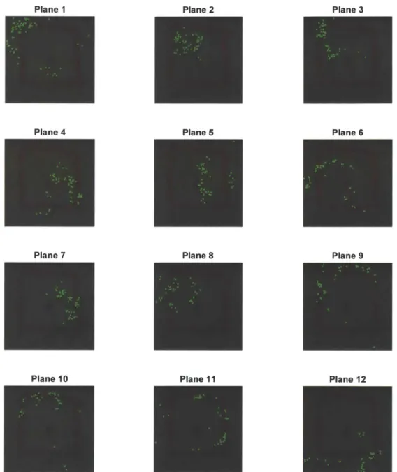
Minimum Number of Models Against Different Inlier Probabilities . . Tiles in Skin Im age . . . . Initial Matched Keypoints After Removal of Matched Keypoints in First P lane . . . .
Sequential RANSAC Algorithm . . . . UpdateCS Routine . . . . MultiRANSAC Algorithm . . . . J-Linkage Algorithm . . . . Images for Registration and Global Registration Results . . . . Comparison of Initial Keypoint Matching Results . . . . Failure of Iconic Deformable Registration Methods . . . . Number of Keypoints in Each Cluster for J-Linkage . . . .
44 48 49 52 53 55 56 57 59 64 64 65 67
3-14 Registration Result for Differing Viewpoints . . . . 70
3-15 Matched Keypoints in Planes Identified Using Sequential RANSAC . 71 3-16 Matched Keypoints in Planes Identified Using MultiRANSAC . . . . 72
3-17 Matched Keypoints in Planes Identified Using J-Linkage . . . . 73
3-18 Registration Result for Stretched Skin . . . . 75
3-19 Number of keypoints in Each Plane for Stretched Skin . . . . 76
3-20 Registration Result for Time-Lapse Skin Images . . . . 77
3-21 Number of keypoints in Each Plane for Time-Lapse Skin Images . . . 78
3-22 Matched Keypoints in Corresponding Planes for Simulated Image S3 80 3-23 Matched Keypoint Set with 10% Outliers and Junction Points Within the R O I . . . . 82
3-24 RMSE and Mean Distance Error for Varying B-Spline Maximum Grid Levels and Outlier Percentages . . . .. . 83
3-25 RMSE Against Warping Factors and In-Plane Rotation for Skin Reg-istration . . . . 84
3-26 Rigidly Registered Simulated Image S3 Corrupted by Noise . . . . 85
3-27 RMSE Against Gaussian Noise Level and RMSE Against Initial Matched K eypoints . . . . 87
3-28 Runtime Against Number of Initial Matched Keypoints . . . . 87
4-1 Flowchart for Microrelief Junction Point Matching . . . . 93
4-2 Rutovitz Crossing Number . . . .96
4-3 Time-Lapsed Image Pair . . . . 100
4-4 Matched Keypoints and Rigidly Registered Overlapping Images . . . 101
4-5 Simulated Images for Global Registration and Global Registration Re-sults . . . . 105
4-6 RMSE and Number of Correspondences as a Function of Warp Factor and Yaw . . . . 106
4-7 Microrelief Matching Process . . . . 108
4-8 Matching Error for Microrelief Matching . . . . 109
5-1 Flowchart for Hair Segmentation . . . . 112
5-2 Hair-Occluded Skin Image Dataset Used for Algorithm Comparison 113 5-3 Skin-Only Image Dataset . . . . 114
5-4 Dermoscope Images Dataset . . . . 114
5-5 Test Images for Hair Segmentation Demonstration . . . . 116
5-6 Single-Channel Images for Dark Hair With Microrelief . . . . 117
5-7 Single-Channel Images for Mixture of Dark and Light Hair . . . . 118
5-8 Top-Hat Transform on Image with Dark and Light Hairs . . . . 119
5-9 Convolutional Peaks for Second Derivative of Gaussian . . . . 121
5-10 Multiscale Matched Filter on Image with Dark and Light Hairs . . . 123
5-11 Average ROC Curves for Various Curvilinear Structure Detectors . . 125
5-12 Response Images from Various Curvilinear Structure Detectors . . . . 127
5-13 ED and MBL Against Threshold and Corresponding Masks for Hair-Occluded Skin Image . . . . 129
5-14 Hair Masks After Thresholding at MBL = 10 . . . . 130
5-15 ED and MBL Against Threshold for Skin-Only Image . . . . 130
5-16 Input Masks for k-NN Classifier for Test Image . . . . 132
5-17 Hair Mask After k-NN Classification and Final Hair Mask . . . . 133
5-18 Hair Masks Overlaid on Skin Images for Various Hair Segmentation A lgorithm s . . . . 136
5-19 Hair Masks Overlaid on Dermoscope Images . . . . 138
5-20 Hair Mask Found Using Light Hair Detection on Images with Only D ark H airs . . . . 139
5-21 ED and MBL Against Threshold for Image with Mixture of Dark and Light H airs . . . . 140
5-22 Hair Orientations at Each Pixel from Multiscale Matched Filter . . . 142
6-1 Flowchart for Overall Design of Guided Inpainting of Skin Images 145 6-2 Algorithm Flowchart for Compositing . . . . 148
6-4 Hair Segmentation Masks . . . . 151
6-5 DHR-Processed Images After Initial Inpainting and Image Registration 151 6-6 Sets of Final Matched Keypoints Found During Registration and After Keypoint Removal . . . . ... . . . . 152
6-7 Region of Validity of Different Image Pairs . . . . 152
6-8 Assignment of Pixels in Mask Mh . . . . 153
6-9 Comparison of Final Images for Various Inpainting Methods . . . . . 155
6-10 Zoomed-In Comparison of Final Images for Various Inpainting Methods 156 6-11 Extracted Microrelief in Images with Dense Hair . . . . 157
List of Tables
3.1 Comparison between Initial Keypoint Matching Results . . . . 65 3.2 Mean and Median RMSE Across All Rotations and Distortion scales . 84 4.1 Correspondences Found For Different Image Pairs . . . . 103
4.2 Parameters for Simulated Images for Testing Rigid Registration . . . 104 4.3 RMSE and Number of Correspondences for Simulated Images . . . . 104
5.1 AUC of Average ROC for Various Curvilinear Structure Detectors 125 5.2 Mean Sensitivity, Specificity and Accuracy for Hair Segmentation for
Hair D ataset . . . . 134
Chapter 1
Introduction
A visual inspection of human skin reveals many salient visual features. Variations in
color pigmentation, hair shafts and hair follicle openings are immediately noticeable. There are numerous intersecting fine ridges appear like irregular geometric patterns; these are the microrelief or glyphic patterns of the skin. Wrinkles appear alongside the microrelief patterns or deepen their appearance. Many of these skin features correspond to visual features that are quite uniformly distributed in visible light optical images of the skin.
The field of computational skin analysis is traditionally concerned with problems such as computer-aided diagnosis of skin cancer from skin images [471 and extract-ing and matchextract-ing dermatoglyphic patterns on fextract-ingerprints for biometric identification
11051. To the best of our knowledge, no work has quantitatively assessed and
char-acterized the evolution of skin features over time. A nonrigid skin image registration would be required to address this gap in research. In this thesis, we describe an accurate and robust skin image registration algorithm that first identifies and match visual features of the skin between image pairs. Then, a local motion model uses these corresponding landmarks to register the skin images. An accurate non-rigid registra-tion method certainly has some novel applicaregistra-tions in computaregistra-tional skin analysis. For example, change detection of a pigmented skin lesion (PSL) [47] could be accu-rately determined using a freehand imaging device. Also, the capability to track the movements of the microrelief structure over time can potentially lead to new insights
in skin physiology, pathological analysis and disease diagnosis. Another possible ap-plication for the algorithm is in finding the dense deformation field of the skin surface under stress without using physical markers. Currently, this measurement is obtained using markers on the skin [64]. Moreover, there are potential uses for registration in localizing of skin images on the body; to elaborate, the skin surface could be utilized as a map for determining 3D ultrasound probe pose [921.
Accurate segmentation of pathological skin features is a major research area in computational skin analysis. For instance, segmentation of a PSL allow algorithms to ascertain whether melanoma is present using the ABCDE criteria [47]: The shape of tile segmentation is used to assess Asymmetry, Border, Color, Diameter and Evolving characteristics of a mole. But oftentimes hair occlude these skin images and negatively affect the accuracy of PSL segmentation. Consequently, digital hair removal (DHR) is used as a preprocessing step to PSL segmentation [45]. In our case, we employ DHR as a preprocessing step to general skin feature extraction, including skin mi-crorelief structure extraction. However, existing DHR techniques in the literature are optimized for removing dark hairs that often share similar colors with PSLs in dermo-scope images [53, 3, 104]. Additionally, dermodermo-scope images are recorded by pressing a piece of glass onto the skin [65], so the shallow, net-like microrelief structures are not very visible in these images. Therefore, existing DHR methods are prone to falsely identifying microrelief structure, when visible, as hairs. In this thesis, we propose a framework for realistic reconstruction of skin texture and microrelief from multiple images of the same patch of skin. First, our automatic hair segmentation algorithm segments hairs that occlude the mnicrorelief structure of the skin images. Second, the images are registered using our nonrigid skin registration algorithm. Third, areas of the first image with missing information due to removed hairs are filled in with what we know about the skin texture in other images by guided inpainting.
A potential use for skin registration is in the assessment of the stability and
evolution of microrelief structure over time. Although some studies have attempted to extract microrelief structure in order to create measurement indices for skin aging assessment [111, 43, 151, they did not attempt to track shifts in the structure over
time. Skin registration enables rigorous scientific inquiry into the long-term stability of microrelief structure. In this thesis, we describe a method that initially uses feature-based rigid skin registration to identify and register a region of interest (ROI) from a pair of time-lapsed skin images. After extracting junctions points from the net-like microrelief, a microrelief junction point matching (MJPM) algorithm employs nonrigid point pattern matching (PPM) to match both point sets. This is a first step towards prospective applications such as novel methods for tracking skin diseases over time and skin-based biometric identification.
1.1
Contributions
A key contribution of this thesis is the development and evaluation of enabling
tech-nologies for quantitative time-lapsed dermatology. Nonrigid skin image registration accurately aligns pairs of skin images recorded with a freehand visible light optical skin imaging system at regular intervals of time over a time scale of months. If dense and thick hairs are present in the time-lapsed images, then nonrigid skill image registration is used to reconstruct the skin texture of occluded regions by record-ing multiple images from the same area. Next, hairs are accurately extracted with our automatic hair segmentation algorithm and subsequently removed with guided inpainting to produce a realistic reconstruction of skin texture. Moreover, skin regis-tration can be applied towards tracking deformations of the skin surface using only visual features of the skin.
Another major contribution is in the introduction of novel methodology for rigor-ous time-lapsed analysis of microrelief structure. A preliminary step we have taken is the proof-of-concept validation of MJPM for accurate mnatching of two sets of rigidly registered microrelief structure.
1.2
Thesis Outline
The thesis is structured as follows. Chapter 2 gives the reader background knowledge about the freehand optical imaging system and skin features. This is followed by a literature review on skin registration, DHR, microrelief structure extraction and non-rigid PPM. Then, Chapter 3 describes methodology for our skin registration algorithm in two parts: A feature-based homography registration algorithm gives an initial guess, and then a (MPD) registration technique finds the deformation transformation. This is followed by an evaluation of the MPD-based nonrigid registration method. In Chapter 4, a description of MJPM consisting of two parts is given. A enhanced feature-based rigid homography registration method for the initial guess and PPM algorithm for matching are detailed. Next, the performance of both rigid registration and PPM are assessed. Chapter 5 presents the automatic hair segmentation algorithm and then evaluates its performance. Afterwards, Chapter 6 reports the methodology and proof-of-concept evaluation for guided inpainting. Finally, Chapter 7 concludes this thesis with summary of results, limitations and future work.
Chapter 2
Background
In this chapter, key features of the visible light skin optical imaging system used to
capture skin images are highlighted. Second, skin features relevant to design choices in this thesis are described. Third, the registration problem is defined and prior work in skin registration is discussed. Fourth, related work on digital hair removal (DHR) in terms of image segmentation and image inpainting is examined. Finally, a review of literature related to microrelief junction point matching (MJPM) is given: inicrorelief extraction and nonrigid point pattern matching (PPM).
2.1
Visible Light Optical Skin Imaging System
The freehand visible light optical imaging system is designed by Kundu [48j. We summarize the key design features of the system in this section.
The imaging device consists of both custom-made and off-the-shelf parts that provide high-resolution images of the skin surface so that skin features can be resolved in detail. A LabView-controlled Basler ac2040-90uc color camera provides a resolution of 26.6 pm per pixel at a working distance of 6.8 cm. Tests conducted in [481 concluded that the camera provides sufficient resolution to resolve skin features. The focal length of the lens is 16 mm. Ai Edmund Optics DC LED light source with a light diffuser is used to illuminate the skin surface. The lens hood is lined with a enihanced-reflectivity metallic polymer substrate on the outside to reduce the effects of ambient lighting,
vhile the ilside (4 f the hood is fined with white paper to ensure uniform illumination of
the skill from the light so1rce. These conponents are coiiibiiied into a (:Iistomn-cmde, ham-Iheld probe illistrated in Figure 2-1.
To acquire images. the human operator places the imaging device with optical
axis perpendicular to the skin surface at the region of interest (B-01). An RGB image
of resolution 2040 x 2040 is acquired and is saved in the lossless inig format. As the
device is handheld, the operator can only approximately align the region of interest (Ro1) oi1 successive images of the sane region in serial examinations.
Figure 2-1: Imaging dv-ice design: a high-resolution Basler camera and LED light ring built into custom-made handheld probe.
2.2
Skin Features
The imaging system resolves skin features that are at least 26.6 pim in length, and
outputs images that are approximately 5. 4 cm x 5. 1 cm in area. In this thesis, several skin features are relevant to the design choices of our algorithms: (1) skin
pigmnenta-tion, (2) hair, and (3) microrelief structure. These three skin features are distributed all across the human body, but their density and appearance are variable in different parts of the body. In the rest of this section, we briefly describe the skin features and
their relation to our work.
2.2.1
Skin Pigmentation
The color of the skin arises froii pigments that absorb light called chromophores. The dominant chronophore in the epidermis, the outermost layer of the skin, is melanin, which ranges in colors from black to brown (eurmelanin) and red to yellow (pheome-lanin). Melanin is produced by ielanocytes, which are cells found in the basal layer of the epidermis. When exposed to sunlight, melanocytes produce melanin, and this biological reaction eventually causes skin to appear tanned. Another chromophiore is hemoglobin, a red pigment carried in erythrocytes (red blood cells). These cells are found in the capillaries and veins of the dermis, the layer beneath the epidermis. The amount of blood flow determines the amount of reddish hue in skin [551. The aboveirentioned skin layers and structures are displayed in Figure 2-2. More details on the physiology of skin pigmentation is found in [351.
Clusters of these chromophores in the skin layers give rise to salient skin features such as moles and freckles. These skin features are visible to the naked eye and occur at a scale of up to approximately 2 cm [35]. Due to variability of these features over time, they are not desirable visual features for visual landmarks in skin registration.
Furthermore, some of these features are also the subject of image segmentation for computer-assisted diagnosis by change detection, and thus should not be used to register images for longitudinal analysis.
2.2.2
Hair
Hair is composed of cylindrical structures made of compacted cells that grow from hair follicles in the dermal layer, emerging from the surface of the skin. This is illustrated in 2-2. Brown and black hairs are pigmented by eumielamnin, red and yellow hairs are pigmented by phomelanin and white hairs 'are unpigmnented. In humnanis, the diameter of each hair shaft ranges from 15 pm to 160 pm. Human hair can be classified into 3 kinds: (1) terminal hair, (2) vellus hair and (3) intermediate
Hair Epidermis Melanocyte Dermis Melanin -- Vein Subcutis - - Capillaries Hair Follicle
Figure 2-2: Layers of the skin and hair structure on left, and enlarged view of inelanocytes, melanin and blood vessels on right. Images courtesy of A.D.A.M. [1].
hair. Terminal hairs are long, thick ( >60 1mi) and pigmented, and are found iii areas
such as the scalp, beard and armpit. Vellus hair are short (approximately 1 mum), thin
(<.30 pmi) and unpigmnented; they are found in areas such as the forehead and bald
scalp. Intermediate hairs have intermediate lengths and diameters and are found on the legs and arms. [11, 30, 351
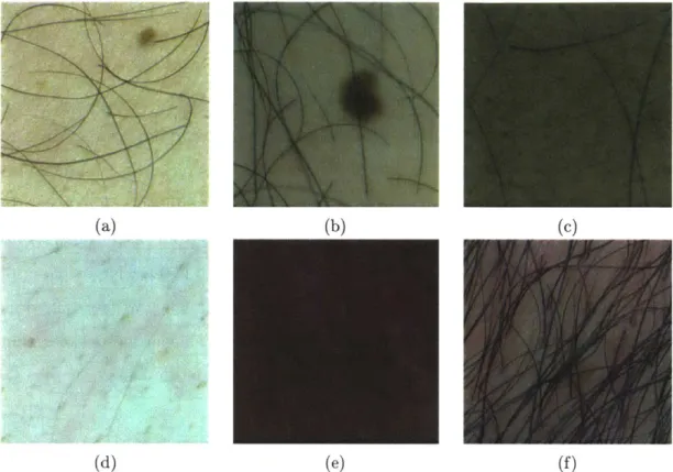
When dense amounts of terminal and intermediate hairs partially obstruct the view of skin pigmentation and microrelief structure, complications occur in accu-rately extracting the underlying skin features. For instance, hairs cause occlusion in images of pigmented skin lesions (PSL) and without first removing the hairs, PSL seg-inentation algorithms often give erroneous results because hair colors may be similar to those of the lesion. [45] Consequently, to address this problem, a number of DHR algorithms were developed. A summary of these algorithis is described in Chapter 2.4. These techniques mainly exploit the property that hair is curvilinear; local op-erators that detect lines in images are used in order to enhance these ridge or valley structures. These algorithms performi very well on skin images with thick, dark hairs with few intersections between hairs and high contrast between hair and skin (Figure 2-3a). However, it remains challenging for algorithms to distinguish between hair and microrelief with similar contrast iii intensity (Figure 2-3c), differentiate thin, faintly-colored intermediate hairs and skin (Figure 2-3d), and to discriminate dark and light hair structures (Figure 2-3e). Also, difficulty arises when hairs clump together as
they do not appear like curvilinear structures (Figure 2-3f). Moreover, the imaging
system introduces several artifacts in hair-occluded skin images. At certain viewing
angles, severe amounts of specular reflections are observed on hair (Figure 2-4a); this
occassionally causes gaps in detected hair strands. In addition, hairs may be out of
focus as the depth of field of the camera is small at a short working distance (Figure
2-4a). Compared to hairs in focus, out of focus hairs have larger widths and exhibit
lower contrast with respect to skin, thus making them more difficult to extract. In
contrast to dermoscope images
[651,
the imaging system does not flatten hairs onto
the skin with a transparent glass plate or lens as it aims to cover a larger patch of
skin. The Bayer filter RGB camera sensor also causes demosaicing artifacts [581
dur-ing image conversion (Figure 2-4b) that is made worse by high contrast between skin
and hair. This makes it difficult to ascertain the true edges of the hairs.
2.2.3
Microrelief Structure
The net-like lines forming triangles and quadrilaterals on the skin surface is the
mi-crorelief structure, seen in Figure 2-5. Two types of skin lines are present: (1) primary
lines, which are wide and uniformly directed, with depth 20 pm to 100 Pm; (2)
sec-ondary lines, which are thin and proceed in all other directions, with depth 5 1m to
40 llm. [851 The appearance of the microrelief is affected by aging and environmental
factors such as sun exposure and humidity.
[791
We postulate that the microrelief structure give rise to stable skin features in a
time scale of months to years. Furthermore, the microrelief structure contributes
most to the appearance of detailed visual features on the skin surface. Therefore,
landmarks from the skin could be used to register images taken over some period of
time or images taken from different viewpoints in one session.
2.3
Skin Registration
Given a target image T and source image S in the image domain Q, and a
transfor-mation W, the aim of registration is to estimate an optimal transfortransfor-mation W* that
(a)(b) (C)
(d) (e) (1)
Figure 2-3: a) Thick, dark, individual hair strands with few intersections are easy to detect with existing DHR algorithms. (b-f) Hair and skin types that are difficult to detect. (b) Hairs obstructing the view of moles and microrelief structure. (c)
Microrelief on skin exhibits similar contrast to that of hair on skin. (d) Thin and faint intermediate hairs. (e) Mixture of dark and light hairs. (f) Hair clumping
together do not appear like curvilinear structures.
jK
(a) (b)
Figure 2-4: Imaging system artifacts in hair-occluded skin images. (a) Specular reflections of hair and hairs out of focus. (b) Demosaicing artifacts from Bayer filter camera sensor.
Figure 2-5: Manually annotated net-like structure of the microrelief on skin.
minimizes the energy of the form
1891:
M(T.
S o W) + R(W) (2.1)where A quantifies the agreement between T and S, and R is the regularization term that favors user-defined specific properties in the transformation and helps to assuage difficulties arising from the ill-posedness of the registration problem. Transformation 14 is a mapping function of the domain Q to itself; it maps locations in S to T. Specifically, for every point x E Q, 1' defines a displacement field u such that 1W(x) x
+
u(x).There are three components in an image registration algorithm: deformation model, objective function and optimization method. [89J The deformation model
defines the parameter space and inherently incorporates regularization so as to in-form what transin-formations are acceptable and include prior knowledge, thus greatly helping to limit the solution. The optimization method navigates the parameter space of the often ill-posed problem with a non-convex objective function to find the local minimum. The ill-posed nature of the registration problem is due to the number of unknowns being greater than the number of constraints, as a displacement vector is estimated for the scalar information (i.e. image intensity) at every location.
The rest of this section discusses image correspondence from skin appearance and related work in skin registration and related fields.
2.3.1
Skin Appearance and Correspondence
Two general approaches exist for constructing the matching criteria M between
tar-get image T and source image S: (1) iconic method and (2) geometric method,
using Sotiras' terminology [89]. Iconic methods evaluate image alignment based on
intensity-based or information theoretic-based measures using correlation-like
meth-ods. According to [110], this approach has the advantage of better representing the
dense correspondence between images accurately and does not rely on the presence of
high-frequency details, thus it is widely used in medical imaging. On the other hand,
the skin surface lacks high-contrast boundaries and has low-contrast glyphic patterns
that appears to be repeated across the image. Thus, these attributes of the skin
surface lead to a highly nonconvex optimization problem such that gradient-based
optimization algorithms blindly converges to local minima far from the true solution
in most cases.
The geometric method assesses image alignment by creating a matching
cri-teria based on landmarks found in images. To find landmarks, first, a keypoint
detector [70] is applied to image T and image S to obtain 2 sets of landmarks
T = {0i | i E Q,i = 1,...,n} and A = {Nj | A E Q~j = 1, ...,m} respectively.
Un-known variables in the registration problem comprise correspondences between
land-marks and parameters of the deformation model. This approach is advantageous as it
is robust to various initial conditions and large deformations. After correspondences
between landmarks have been found, fitting a deformation model to the landmarks is
a relatively straightforward process. However, as only a sparse set of correspondences
is used, the accuracy of interpolation or approximation of the dense deformation
field decreases as the distance between landmarks increases. Therefore, accurate
ge-ometric method-based registration certainly requires identifying a uniform and dense
distribution of correctly matched landmarks.
Finding correspondence between keypoints is a crucial step in the geometric
method. In this thesis, we attempt to register a pair of images (P E R
2for 2D
images) of the skin taken in same area from different viewpoints. To find the
point matches, the assumption we make is that same locations in both images appear to have similar intensities at the scale of skin features. We shall investigate this assumption in the following paragraphs.
The complex optical properties of skin layers and surface microgeometry of skin features give rise to the skin taking on different appearances when it is viewed and illuminated from different directions [18]. We can easily observe this phenomenon from specular reflections in regions of the skin surface when viewing it from an ar-bitrary viewpoint. In computer graphics, one widely used model for rendering skin is the Bidirectional Reflectance Distribution Function (BRDF) [75]. Given a 2D sur-face, the BRDF gives the relation of the reflected radiance dL, at a point x in the direction 6, to the irradiance dE from a light source in direction 62:
BRDF(x,Or,,O) - dL '(x,60) (2.2)
dEj(x,6 )
A visual reference in Figure 2-6 shows that the BRDF assumes that incident light
hits the same point as reflected light, despite light travelling through the epidermis and dermis layers of the skin. Despite the fact that skin is a volume scatterer [661, the model gives an approximately correct representation of surface reflection of the skin [35]. To our advantage, Marschner et al. [661 found that the BRDF of human skin with respect to reflected angle is close to constant for a light source near normal incidence, as seen in the plot in Figure 2-7. Hence, in this case, skin approximates a Lambertian surface: a surface in which the reflected radiance of every point on the surface is the same, regardless of the observer's angle of view. Moreover, the imaging device described in Chapter 2.1 is designed to achieve constant illumination of the skin in the ROI. Due to constant illumination and a Lambertian scene, our skin images fulfill the conditions [991 needed to confidently establish local correspondences between both viewpoints of the scene.
dLr
dEj
/
x
Figure 2-6: BRDF assumes that incident and reflected light enter and exit at the same point. When light enters the skin, some subsurface scattering occur. Image adapted from [35. BRDF 0.2 0.1
0
-80
-40 0 40Reflected Angle
Or
(in
degrees)
80
Figure 2-7: ID BRDF at a single point of the human skin surface with angle of view when illumination normal to the surface is used. Image [661.
respect to the adapted from
30
2.3.2
Related Work on Skin Registration
Change detection of individual PSLs can be used to diagnose diseases like melanoma
[671. Due to camera motion relative to the skin surface, accurate registration is desired for this application. However, in tile commnericial market, many computer-assisted diagnosis (CAD) systems for PSLs only offer the functionality of side-by-side or blink comparison of skin dermoscope images taken over periods of time [471. In the literature, a few rigid registration methods have been developed specifically for change detection of PSLs. These methods assume that the correspondence between images is related by a global transformation. This assumption may hold for PSLs in dermoscope images because the skin surface is flattened into a plane by the dermoscope's glass plate or lens
[271.
For instance, [861 registered two skin images with a PSL in the translation and rotation space using stochastic gradient descent to optimize mutual information. Also,[771
and [62] devised similar methods for estimating scaling and rotation parameters by a fast Fourier transform-based cross-correlation algorithm and translation parameters by hill-climbing optimization using non-parametric statistical similarity measures. These abovementioned iconic approach-based methods share the share similarity measures as some of the common techniques used in nonrigid registration of medical images [441. But for skin surfaces without PSLs, the iconic approach may be unreliable, as explained earlier.Another group of methods for rigid skin registration involves identifying a sparse set of landmarks in both images and aligning the images by a homography or affine transformation (i.e. the geometric method). These algorithms follow the well-known method proposed by Lowe
159].
The workflow for Lowe's method is as follows: First, keypoints are detected from each image. Such keypoints may be detected using for example, Harris corners [68], information theory [40] or Laplacian-of-Gaussian [591. Second, a local descriptor around each keypoint is computed. For instance, some affine-invariant local descriptors [69] are based on image gradients [591, Haar wavelets[101, moment invariance [981 or shape contexts [6]. Third, correspondences between
variants of random sample consensus (RANSAC) [221. These methods have been applied to constructing panoramas
191,
multiview geometry matching [82] and fun-damental matrix es.timation [871. One of the earliest use of the geometric method for registering skin images can be traced to Zambanini et al.[1061,
who used it to register skin images with cutaneous hemangioma, which are a benign red PSL on the skin developed in infancy due newly-formed blood vessels1191.
They employed Lowe's Scale Invariant Feature Transform (SIFT)[591
and RANSAC to calculate the optimal homography transformation. However, keypoints found were constrained within the region of the lesion after segmentation. Later, [4] used a similar method to register images of melanocytic nevus using keypoints found in the entire image domain. Next, Dong [541 demonstrated that keypoint matching via homography warping could be applied to matching skin images on faces by modifying SIFT to be more receptive to the shape of facial pores. This result was used to match human faces from multiple viewpoints using skin features. Lastly, Madan et al. [61, 601 showed that the same ap-proach (using traditional SIFT) could be used to register multimodal images of facial skin by homography transformation. In this case, five imaging modalities of the skin were registered: optical images of the skin illuminated with unpolarized, parallel-polarized and cross-parallel-polarized light, and florescence images of the skin illuminated with ultraviolet and blue light.Nonetheless, rigid registration does not allow us to accurately register the free skin surface. For example, homography transformation assumes that the scene is planar, hence the transformation does not model situations in which the skin is lo-cally deformed or different viewpoints of a slightly curved surface like the forearm. Literature we have reviewed above shows that rigid registration is generally success-ful for a small, locally planar ROI or a fixed viewpoint shared between images. In contrast, for a freehand imaging system, a registration algorithm that can handle a larger viewpoint changes and a larger field of view is desirable. Thus, we may not assume a planar scene.
Nonrigid or deformable registration has been previously explored in skin-related literature to assess the mechanical properties of skin in vivo. Several studies have
proposed methods to find the full-field 2D displacement of the skin surface in response to stress. For example, in [641, an array of reflective markers was placed on the forearm and the displacement of the marker set was tracked with an optical imaging system, and the deformation field was interpolated. Using confocal laser scanning microscopy
(CLSM) to reveal layers within the skin,
171]
compared the results of a cubic B-spline free-form deformation (FFD) registration algorithm [80] with the mean squared error metric to that of the the Demon registration algorithm1941
for registering pairs stress-deformed and fixed 2D CLSM images. In comparison to existing works, our skin registration algorithm described in Chapter 3 is able to compute the deformation field of the skin from visible light images without physical markers. Furthermore, it can accurately register deformed skin images resulting from realistic viewpoint changes or small stretches in a 1 inch by 1 inch sized ROI.2.4
Digital Hair Removal
In the literature, the primary use of DHR methods is to disocclude hair in dermoscopic images for automated analysis of PSLs. In these methods, the first step is to identify pixels in the image that contain hair and the second step is to replace these pixels with plausible skin colors based on neighboring skin colors and structure by image impainting. This implies that statistical estimates of missing data in hair-occluded regions are used instead of true, observed data. The major challenges lie in finding and replacing hairs that are thin, overlapping, or of similar contrast or color to underlying skin and nmicrorelief structure. [461
The key focus of our work in Chapter 5 is to address the first step of DHR: the image segmentation problem. Image segmentation is defined as the optimal parti-tioning of an image I into non-overlapping regions that are similar in characteristics such as texture or image intensity. Given image domain Q, we wish to determine sets
,5
C
Q such that [78]:K
= (2.3)
k=1
where Sk n S = 0 and K is the number of regions. In DHR, we have Q
C
R2 forpixels in 2D images, and K = 2 to represent the set of hair pixels and set of skin pixels. As convention, we assign binary labels of 0 to skin pixels and 1 to hair pixels in segmentation masks.
The rest of this section describes prior work on DHR in terms of hair segmentation and image inpainting methods.
2.4.1
Related Work on Digital Hair Removal
Hair Detection and SegmentationIn DHR, hair detection based on mathematical morphology [291 is widely used. Many of these methods are based on the closing-based top-hat transform. This morpholog-ical operation returns objects that are smaller than the structuring element and are darker than their surroundings. In 1997, Lee et al. [531 released DullRazor, the first and most well-known method for DHR, as a readily available software on Windows or Unix platforms [2]. The software uses generalized morphological closing operations with three structuring elements that model line orientations in horizontal, vertical and diagonal directions and then takes the maximum response from each pixel in the RGB image. Next, the difference between the intensity and response at each pixel is taken, and a predetermined threshold is applied to obtain a hair segmentation mask as the union of hair masks from each channel. Furthermore, Schmid-Saugeon et al.
[83] used similar method to that of Lee et al. by applying generalized morphological
closing operations with a disk structuring element in the luminance component of the CIELUV color space to obtain a hair mask. Moreover, Xie et al.
[1031
and Fiorese et al.121]
both used variants of the top-hat transform and hard thresholding to obtain a hair mask. The top-hat transform used in these methods is reportedly generallyeffective at enhancing dark hairs with sufficient contrast on light skin, including both thin and thick hairs. In addition, to find light hairs on dark skill, the opening-based top-hat operator can be used.
Koehoorn et al. [461 presented a DHR approach that decompose the skill image into its luminance threshold set (256 binary image layers) and applies a multiscale morphological gap detection technique to find thin structures in each threshold layer. The layers are then merged to create a hair mask. The authors evaluated their algorithm to be effective at detecting many types of hair oil different skin types. Due to the need to apply morphological operations on up to 512 binary threshold layers to detect dark and light hairs, the algorithm requires fast parallel processing techniques in order to process the skin image in a comparable time frame to other methods.
The next group of hair detection methods are edge detection-based. The authors of E-shaver [421 used the Radon transformn to detect the predominant orientation of hairs and then used the Prewitt edge filter and thresholding to find light and/or thin hair edges in the predominant orientation. Tossi et al. [971 proposed a hair detection method using the adaptive canny edge detector. Lastly, Maglogiannis [631 compared edge detection results from combinations of Laplacian, Laplacian of Gaussian and Sobel methods. In these hair detection methods, the hair mask is obtained from the edge image by dilation with a fixed structuring element to fill holes in the interior regions of hair edges. However, we observed that this group of methods is prone to identifying non-curvilinear objects on the skin as hair because they assume that all high-contrast edges in skin images are belong to those of hair objects.
Matched filtering methods make up another group of methods used for hair de-tection. A 2D kernel, designed to model a the cross-sectional intensity profile of hair in the image at some unknown orientation and position, is convolved with the skin image. The resulting matched filter response indicates confidence in the prescence of a feature at each pixel. These methods make the assumptions [741 that the cross-section intensity profile of hair is approximated by a Gaussian (see Figure 2-8), and that hair has a small curvature and can be approximated by piecewise linear seg-ments. In addition, the Gaussian profile may be inverted to extract dark or light
hairs. Abbas et al. [3] presented a matched filter with a 1D Gaussian profile and adaptive threshold based on the local mean of the ID first derivative of Gaussian that helps to de-emphasize step edges that arise from a PSL in the image. Nguyen [741 used the normalized correlation of a 1D Gaussian profile, thresholding and skele-tonization to find the centerlines of the hairs. Then, the hair mask is reconstructed based on the Gaussian cross-sectional profile of hairs. The normalized correlation (as opposed to convolution) helps to extract both light and dark hairs at the same time, but we found that the author introduced many parameters and steps in order distinguish both structures in a method that may not be robust enough to extract hairs from images with dense amounts of hair. Lastly, Huang et al. [33] found the maximum normalized correlation response using a multiscale ID Gaussian profile and used hysteresis thresholding to obtain a hair mask. Thereafter, local linear discrimi-nant analysis using the partial information of hair colors in the Lab color space and region growing recovered hair pixels at intersections. However, Koehoorn et al. [461 reported that this method is susceptible to producing dark halos where hairs were identified and removed. In general, matched filtering methods are able to produce smooth hair outlines and recover small gaps in the hair structure due to noise in hair pixels. But the skin image needs to be convolved with a large set of filters, which increases its computational cost. However, in practice, this process can be imple-mented in parallel. In our hair segmentation algorithm in Chapter 5, we introduce a multiscale second deriative of Gaussian matched filter and show that its response to hair objects is superior to many hair detection methods discussed so far.
Other methods to extract hair have also been used. Zhou et al. [109] used a curve fitting approach. They first enhanced dark hairs using generalized morpholog-ical closing operations with a disk structuring element and then used a differential geometric approach [901 to detect lines. The line segments are refined by Bezier curve fitting using RANSAC and a line intersection refinement procedure. Finally, the hair mask is created by dilating the skeleton according to the estimated width of each hair from the average normal intensity profile of the lines. Although the authors claim that the results are a clear improvement compared to the work of Schmid-Saugeon
Dark Hair 180 170 160 15 0 140 130 120 Light Hair 220 215 -210 -205 200
Intensity Profile of Dark Hair
./
105 110 115 120 Index
Intensity Profile of Light Hair
20 25 30 35
Index
Figure 2-8: Intensity profile of hair showing the intensity profiles taken along the red lines in grayscale images of dark and light hair. In matched filtering, the intensity profile is assumed to be Gaussian.
.4
4'
-4 <K
et al. on 460 dermoscopy images, the results for this work are not well-reported and
not reproducible due to lack of details in the algorithm description.
Xie et al. [104] proposed using an isotropic nonlinear filtering [57] to detect hairs
as wide lines. They optimized their hair detection parameters to segment dense
dark hairs on light skin that exhibit clumping in dermoscope images. To acquire the
preliminary hair mask, an adaptive threshold based on linear regression of a linear
combination of several global parameters is used. Then, pixels that are similar to the
median of the skeleton image of the preliminary hair mask are marked as additional
hair pixels. The drawback of this hair detector is that the isotropic nonlinear filter,
however, also functions as a small object detector. Thus, the response appears noisy
as it does not only detect curvilinear objects.
Typically, after obtaining a hair mask, many DHR methods use mathematical
mor-phology to identify long and thin hair objects and remove non-hair objects. This step
removes many false positives from the hair mask; the hair mask is usually peppered
with short, disconnected, hair-like fragments of multiple orientations arising from skin
features. Hence, removing these false positives help to preserve skin pigmentation,
microrelief structure and texture. For example, Fiorese et al. [21J removes non-hair
connected objects in the hair mask according to density, sphericity and convex hull
sphericity metrics. Abbas et al.
[31
filters out connected objects using morphological
area opening and Haralick circularity [28] conditions. Xie et al. [104] uses circularity
defined by the ratio between area and the minimum circumscribed circle radius of the
connected regions to remove those regions. Lastly, Koehoorn et al. [461 discarded and
refined objects from the hair skeleton mask: First, they discarded connected regions
in the hair skeleton mask with small areas. Second, they pruned sours from the hair
skeleton. Third, they removed objects according to junction-based metrics. Fourth,
they constructed the hair mask by dilation using structuring elements of various radii.
Image Inpainting of Hair
Inpainting methods in existing DHR algorithms include simple bilinear interpolation
[53, 74], replacement of pixels with pixels from the morphologically closed image
38
[831 and median filtering [33]. The above-mentioned methods do not attempt to preserve image structure and may lead to noticeable blurred appearance and color bleeding [1091. Other inpainting methods used include partial differential equation-based [21, 103j, coherence transport
[31,
fast marching [451 and exemplar-based[1091
methods. These methods reportedly model the texture of surrounding skin. However, we show in Chapter 6 that they do not preserve the true underlying skin texture. To overcome this shortcoming, our inpainting method records multiple skin images of the same area and combines information on underlying skin texture from the images in order to reconstruct a hair-free skin image with true skin texture.2.5
Microrelief Junction Point Matching
Junction points of fingerprint patterns, or fingerprint minutiae, form the basis for many fingerprint matching methods [39, 37, 95, 1081 due to the stability of these features over time. Similarly, we postulate that junction points of the microrelief structure of skin in general form a set of good candidate features that do not change over a period of a few years. In order to assess the stability of junction points, we need to be able to find matches between two sets of them. The problem of matching junc-tion points is defined as finding the optimal matches between a reference point pattern set and target point pattern set in R2
. To solve this problem, several challenges exist: First, different image acquisition parameters and the microrelief extraction method introduce many missing or spurious patterns to the point pattern sets. Second, the transformation is nonrigid due to the non-planar and stretchable nature of the skin surface. Third, there are as many as 7000 points in each set within a 1 inch by 1 inch patch of skin on the forearm. Thus, a well-known non-rigid PPM algorithm, thin-plate spline-robust point matching (TPS-RPM), with complexity O(N3) in the
worst case, where N is the number of points in the set, would take a prohibitively long time to find matches. Hence, developing an efficient and robust non-rigid PPM algo-rithm to solve this problem is desirable. In Chapter 4, we present our nonrigid PPM algorithm and show- that in preliminary experiments that it achieves low matching