Computational and theoretical modeling of intermediate
filament networks: Structure, mechanics and disease
The MIT Faculty has made this article openly available.
Please share
how this access benefits you. Your story matters.
Citation
Qin, Zhao, and Markus J. Buehler. “Computational and Theoretical
Modeling of Intermediate Filament Networks: Structure, Mechanics
and Disease.” Acta Mechanica Sinica 28, no. 4 (August 2012): 941–
950.
As Published
http://dx.doi.org/10.1007/s10409-012-0124-5
Publisher
The Chinese Society of Theoretical and Applied Mechanics; Institute
of Mechanics, Chinese Academy of Sciences
Version
Author's final manuscript
Citable link
http://hdl.handle.net/1721.1/105536
Terms of Use
Creative Commons Attribution-Noncommercial-Share Alike
Acta Mechanica Sinica (2012) 28(4):941–950 DOI 10.1007/s10409-012-0124-5
REVIEW ARTICLE
Computational and theoretical modeling of intermediate filament networks:
Structure, mechanics and disease
Zhao Qin··· Markus J. Buehler
Received: 8 June 2012/ Revised: 2 July 2012 / Accepted: 2 July 2012
©The Chinese Society of Theoretical and Applied Mechanics and Springer-Verlag Berlin Heidelberg 2012
Abstract Intermediate filaments, in addition to micro-tubules and actin microfilaments, are one of the three major components of the cytoskeleton in eukaryotic cells. It was discovered during the recent decades that in most cells, in-termediate filament proteins play key roles to reinforce cells subjected to large-deformation, and that they participate in signal transduction, and it was proposed that their nanome-chanical properties are critical to perform those functions. However, it is still poorly understood how the nanoscopic structure, as well as the combination of chemical composi-tion, molecular structure and interfacial properties of these protein molecules contribute to the biomechanical proper-ties of filaments and filament networks. Here we review recent progress in computational and theoretical studies of the intermediate filaments network at various levels in the protein’s structure. A multiple scale method is dis-cussed, used to couple molecular modeling with atomistic detail to larger-scale material properties of the networked material. It is shown that a finer-trains-coarser method-ology as discussed here provides a useful tool in under-standing the biomechanical property and disease mechanism of intermediate filaments, coupling experiment and simu-lation. It further allows us to improve the understanding
Z. Qin · M. J. Buehler
Laboratory for Atomistic and Molecular Mechanics (LAMM), Department of Civil and Environmental Engineering,
77 Massachusetts Ave., Massachusetts Institute of Technology, Room 1-235 A&B, Cambridge 02139, MA, USA
Center for Computational Engineering, Massachusetts Institute of Technology,
77 Massachusetts Ave., Cambridge, MA 02139, USA e-mail: qinzhao@MIT.EDU
M. J. Buehler
Center for Materials Science and Engineering, Massachusetts Institute of Technology,
77 Massachusetts Ave., Cambridge, MA 02139, USA e-mail: mbuehler@MIT.EDU
of associated disease mechanisms and lays the foundation for engineering the mechanical properties of biomaterials.
Keywords Intermediate filament network· Multiple scale method· Nanoscopic structure · Mechanics · Disease mech-anism· Molecular mechanics
1 Introduction
Intermediate filaments exist in nearly all eukaryotic cells and many biological materials including wool, hair and hooves [1–4]. As many other fibrous proteins such as col-lagen or fibrin, they linearly and laterally self-assemble into filaments with a diameter of 8–12 nm [1, 5] which appear to be intermediate in size between those of the other cytoskele-ton components, microtubules (25 nm) and microfilaments (7–9 nm). These three components work together to enhance the structural integrity, and motility of cells. Besides the diameter differences, intermediate filaments are more flex-ible to adopt multiple conformations than microtubules, and they are more extensible when subjected to the tensile stress than those two components [5]. Its most fundamental subunit is called a dimer and has a common tri-partite organization characterized by a central alpha-helical coiled-coil domain and amorphous “head” and “tail” domains of variable length and sequence [2, 6].
Compared to microtubules and microfilaments, the in-termediate filament family has more members and each of them features specialized functions. For instance, the in-termediate filament protein family in humans contains more than 70 proteins [2, 3], which are classified into five distinct types based on their similarities in sequence and the tissue distribution [6, 7]. Type I and II intermediate filaments are acidic keratin and basic keratin produced by different epithe-lial cells, respectively. Keratins play the key role to maintain the cell’s stability in its morphology and contribute to the stability of mechanical connection to adjacent cells [8]. Type III intermediate filaments include vimentin, desmin, glial fib-rillary acidic protein, syncoilin and peripherin. Vimentin is
found in the cytoplasm of fibroblasts, leukocytes, and blood vessel endothelial cells. It plays a significant role in main-taining cell integrity by supporting and anchoring the posi-tion of the organelles in the cytoplasm [2]. Type IV interme-diate filaments are mostly inside the nervous system includ-ing neurofilament, internexin and nestin. Neurofilaments are found specifically in neurons and it provides structural sup-port for axonal radial growth, leading to an axon diameter in-crease by a factor of five [9]. Type V intermediate filaments include lamin which forms a filamentous support inside the inner nuclear membrane. It contributes largely to the struc-tural integrity of the nucleus and plays a vital biological role in the process of the re-formation of the nuclear envelope after cell division [10, 11].
Due to their important mechanical roles in cells and tis-sues, intermediate filaments have been referred to as the “safety belts of cells”. There is evidence to show that the mechanical function of intermediate filaments plays a central role among their physiological functions [12] but conven-tional material or biological models have failed to explain the mechanism behind [13]. For example, the complete mech-anism of Hutchinson–Gilford progeria syndrome, which is also known as rapid aging disease, is still not clear. Children with this syndrome experience severe hardening of the arter-ies and this condition significantly increases the chances hav-ing a heart attack at an early age and die prematurely. By sys-tematically comparing the patients’ DNA sequences to nor-mal ones, a point mutation in DNA has been identified that causes a cryptic splice site that leads to a 50 amino acid dele-tion in the lamin tail domain [14]. In vivo experimental work has provided evidence that the mutated lamin forms more or-dered and rigid networks than the wild type [15]. However,
there is relatively little theoretical or modeling work to ex-plain the mechanism by which the peptide deletion causes the nuclear lamina to become unstable, leaving the disease mechanism and process largely elusive.
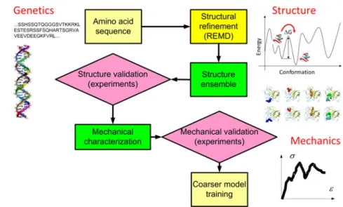
It is now clear that the nanostructure and nanomechan-ical properties of intermediate filaments are the key to un-derstanding the mechanisms of intermediate-filament-related diseases, since most known genetic diseases stem from sin-gle point mutations [3, 13]. However, to fully understand the mutation effects one needs to develop a systematic per-spective by considering the tissue as an integrated system. Some of the molecular interactions are strong and appear to be critical during physiological processes, and thus can not be removed or altered without changing the tissue func-tion. For example, the head domain at the N-terminal and the tail domain at the C-terminal end of intermediate ments are essential in the association of intermediate fila-ment dimers into higher order assemblies such as head-to-tail polymers [6, 16]. Those domains are highly conserved in se-quence and mutations there usually relate to diseases such as various muscular dystrophy conditions for vimentin and lamin. Moreover, molecular modeling has shown that a sin-gle point mutation is hardly sufficient to affect the mechani-cal features of a lamin dimer as a standalone system, but its influence appears in the hierarchical context [17]. These evi-dences make us believe in that any study restricted to a single scale level is far from enough to model and understand an in-termediate filament system. Thus researchers are challenged intensify the efforts in studying the hierarchical structures and material functions at multiple scales as summarized in Fig. 1.
mm
ms
10-1nm
Fig. 1 A schematic figure to summarize the basic approach of the step-by-step finer-trains-coarser procedure of studying the structure and
mechanical properties of intermediate filament networks by invoking multiple scale computational and theoretical modeling methods. For each subunit level (such as amino acid, monomer, dimer and so on), the corresponding time and length scales are as noted by the y and x axis and sample modeling methods are noted in the gray shaded ellipses
Computational and theoretical modeling of intermediate filament networks: Structure, mechanics and disease 943
There are persistent experimental challenges in identi-fying the entire whole structure of intermediate filaments. Some intermediate filaments regions, including the head, tail domains and linkers, are composed of largely intrinsically disordered structures that take many possible conformations. They are typically difficult to crystallize, making traditional structural analyses such as X-ray crystallography and NMR challenging. X-ray diffraction method on naturally occur-ring or recombinantly produced intermediate filament bun-dles has been applied to investigate the structure of the rod-like domain of intermediate filament dimers and has par-tially solved their coiled-coil structures [18]. However, it does not provide sufficient data to model the disordered re-gions. Solid state nuclear magnetic resonance can be used to derive the 3D structure of molecules in solution but its successful usage limits to small particles like amyloid pep-tides [19]. Cryo-electron tomography offers the promise of molecular-level imaging of single intermediate filaments, but the method is limited by the low temperature requirement and the best tomograms are currently limited to a resolution around 5 nm [20]. Far-field fluorescence microscopy tech-niques combined with recently developed super-resolution techniques manage to actively control the fluorophore emis-sion in any imaging frame and thus the resulting image can reach a resolution of 20 nm [21]. It can be used to observe the 3D profile of intermediate filaments and their dynamic behavior inside cells, but the resolution is even further from being sufficient for molecular modeling.
The intrinsic limitations in experimental methods call for the development of complementary computational meth-ods that can provide a more controlled condition to assess the relation between the nanostructure and nanomechanics of this class of materials. Atomic interactions, which are difficult to be directly measured from experiments, can be computed by first-principles based calculations such as den-sity functional theory (DFT) and relevant numerical imple-mentations. Several massive sampling techniques have been developed during recent years, making protein structure pre-diction and refinement increasingly feasible [22, 23]. How-ever, even the most powerful computer face severe length and time scale limits simulating an entire material (or cell) with full electronic structures and atomic details. A finer-trains-coarser computational modeling approach can be ap-plied as shown in Fig. 1 to investigate the mechanical proper-ties of a complex system like intermediate filament networks. This methodology holds great advantages in probing the ma-terial behavior at various scale levels without losing the un-derline atomic mechanism to explain coarser scale phenom-ena [24].
In this review article, we focus on recent progress in the application of computational and theoretical modeling of the structure and mechanics of intermediate filament networks. The structure of the entire network is exceedingly complex (in a hierarchical structure) that its complete description is clearly beyond the ability of single scale modeling. There-fore, in the current study we explore the topic with a
mul-tiple scale approach. By invoking a “divide-and-conquer” strategy we discuss four central issues: (1) How to use com-putational simulation to investigate the atomic structure of the most amorphous region of intermediate filaments? (2) How does a single intermediate filament respond to external forces and what is the mechanism of this highly nonlinear response? (3) How do single filaments associate with one another by various types of crosslinks, and how can physio-logical conditions play an important role in affecting the me-chanical response of crosslinks? (4) How does an interme-diate filament network respond to extreme stress conditions and what is the mechanism of its failure? Multiple examples are provided to showcase the application of computational simulations at different scales.
2 Multiple scale modeling of intermediate filament net-works
2.1 Atomic structure of lamin tail and molecular mecha-nism of progeria syndrome
The tail domain of lamin A is composed of 261 amino acids with mostly disordered structure, and demonstrates the characteristic qualities of intrinsically disordered pro-teins including a promiscuity in protein binding, tendency to aggregate in a high proline and glycine content [25, 26]. It is difficult to predict how the removal of 50 amino acids in this disordered region lacking secondary structure will affect the protein function because of no specific conforma-tion. The structural characteristic of this domain may be only described as an ensemble of local energy minimum conformations which statistically represent conformations allowing protein bindings and lead to statistical mechanical functions. Other intrinsically disordered biological materials include spider silk, huntingtin (an unstructured molecule that relates to Huntington’s disease) and talin rods share similar characteristics [27–29]. Classic molecular dynamics method is difficult to investigate such a structural property because the molecular conformation is easy to be trapped by its local energy minimum state. Instead, the replica exchange molec-ular dynamics (REMD) has been shown to be successfully utilized as a computational method used to improve the dy-namic properties of conventional molecular dydy-namics and Monte Carlo methods, aiming at obtaining an ensemble of global minimum free energy states by overcoming kinetic trappings as shown in Fig. 2 [28, 30, 31]. Compared to con-ventional molecular dynamics that runs under a constant temperature, REMD utilizes many replicas to run in paral-lel under various temperature conditions. The replica under higher temperature enhances barrier crossing ability in the purpose of larger conformation samplings while the replica under lower temperature converges to a more accurate pre-diction of local minimum. This design is based on the fact thatτ = τ0exp(ΔG/kBT ) is the expected time for conforma-tion change by crossing the energy barrier, whereτ0 = 1 ps is the natural vibration period,ΔG the energy barrier to cross,
Fig. 2 A schematic of the overall procedure to investigate the molecular structure and mechanical property of intermediate filaments by
computational modeling and simulations and validation by comparison against experimental tests.
kBthe Boltzmann constant and T is the ensemble tempera-ture. The metropolis criterion is used to judge whether or not the configurations exchange between the higher and lower temperature replicas.
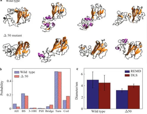
REMD is efficient under the condition of massively par-allelized computational power. For example, here we take the sequence of human lamin A/C (SwissProt/TrEMBL ac-cession number P02545) and study the conformation of the
wild type lamin (AA428-646) and theΔ50 mutant
(AA428-614). Here, 128 replicas with 128 CPUs are applied for par-allel computing and CHARMM force field is used to model the atomic interactions [32]. 20 000 energy exchange cy-cles (10 ns for each constant temperature replica) are per-formed to ensure the convergence of the conformation en-ergy. We classify the conformations in the lowest tempera-ture replica (corresponding to conformations with local min-imum energy) according to the mutual similarity of atomic coordinates, and K-means algorithm is used to identify con-formation clusters with root mean square deviation less than 0.2 nm. For each structure cluster, we select the represen-tative conformation with lowest energy and as shown in Fig. 3a. The Ig-fold is observed as conservedβ-sheet sec-ondary structure while the rest are mainly composed of disor-dered structures. We statistically study all the lowest temper-ature conformations to obtain the expected secondary struc-ture distribution as shown in Fig. 3b. It is shown that most of the secondary structure in this tail domain is composed of β-sheet structure (21.9% for the wild type and 18.6% for the Δ50) and random coil (turn + coil as 66% for the wild type and 72% for theΔ50), which agree to the circular dichro-ism measurement as it gives structural composition of main β-sheet (27% ± 5% for the wild type and 23% ± 7% for theΔ50) and random coil (66% ± 5% for the wild type and 70%± 7% for the Δ50) with little α-helix [30]. The simula-tion result statistically shows that the overall diameter of the
Δ50 mutant (3.2 ± 0.3) nm smaller than the wild type lamin (5.0 ± 1.4) nm, which agrees with the dynamic light scatter-ing (DLS) measurements shown in Fig. 3c. The result sug-gests that theΔ50 mutant has a more compact structure than the wild type lamin. Combining with further investigation to show that theΔ50 mutant is of a higher thermal stability (by approximately 155–293 kcal/mol [30]), the result, which is obtained both in computational calculations and experimen-tal tests, provides the molecular mechanism of the progeria syndrome.
2.2 Mechanical properties of single intermediate filaments The mechanical behavior of single intermediate filaments under extreme condition is highly nonlinear, and is mostly defined by the coiled-coil secondary structure of the rod-like domain of the dimer [33]. It has received considerable atten-tion because of the variability cross different types of inter-mediate filaments lies on the length and sequence of the head and tail domains but the rod-like domain is conserved. Its tensile properties have been investigated in atomistic simula-tions by steered molecular dynamics (SMD) as well as exper-iments by atomic force microscopy (AFM) for stretching [5]. For the SMD technique, one end of a deformable spring is tethered to the protein end and the increasing pulling force is applied to the other end of the spring as shown in Fig. 4a. This method is similar as the tapping mode of AFM to avoid any direct ambitious force that instantly breaks the structure, and thus it is promising to find the relation between mechan-ical and microstructural properties of protein structures [34]. The typical S-shape of the force-extension curve of interme-diate filaments before rupture, which is obtained by stretch-ing the vimentin both by SMD and AFM, gives a signature of intermediate filaments as shown in Fig. 4d. We find the mechanical properties of intermediate filaments are closely related to their secondary structures during the stretching
Computational and theoretical modeling of intermediate filament networks: Structure, mechanics and disease 945
process. The first linear region at the small strain (< 50%) corresponds to the intact coiled-coil structure under homoge-nous deformation. The secondary plateau region (up to 90% strain) corresponds to the material plasticity comes from the unfolding of alpha helices. Hydrogen bonds in different seg-ments break sequentially in this region [35]. The third stiff-ening region (up to 180% stain) corresponds to the alpha-beta transition caused by reforming of the hydrogen bonds between the unfolded adjacent polypeptides [35–37]. This phenomenon is explained by that the unfolded polypeptides lose their alpha-helix conformation but gain more degrees of freedom (can be explained by the significant decrease in persistence length, from several tens of nanometers for an alpha-helix to approximately 0.4 nm for a random coil), and the hydrophobic interactions squeeze the unfolded chains to-gether to form beta-sheet structures as shown in Fig. 4b. This
process is validated by experimental observation in scanning electron microscope (SEM) (as shown in Fig. 4c) as the fil-ament diameter significantly shrinks under stretching and its secondary structural character changes from α-helix to β-sheet under wide angle x-ray scattering (WAXS) and infrared spectroscopy (IS) [35, 38]. It is observed in experiments that the secondary structure characteristic after transition keeps stable for 24 hours, which agrees to the simulation result as theβ-sheet structure content keeps on a constant level after force relaxation as shown in Fig. 4e. The last plateau re-gion before rupture corresponds to the sticky sliding of the beta-strands with respect to each other. Hydrogen bonds de-form cooperatively and rupture in clusters with a unide-form size (∼ 3.5 hydrogen bonds), leading to a constant average force. D 50 mutant Wild type Wild type D 50 REMD
AH BS 3-10H PiH Bridge Turn Coil Wild type D50 0.4 0.2 0 6 4 2 0 Probability Diameter/nm a b c DLS
Fig. 3 Quasi-equilibrated conformations of the tail domain of wild type lamin A and itsΔ50 mutant. a Conformations with high statistical significance factors of the wild type lamin tail domain (AA428-646) andΔ50 lamin tail domain (AA428-614); b We study the secondary structure character for each amino acid by looking into its dihedral angle and hydrogen bonding patterns within the wild type tail (AA428-646) and progerin tail domain (AA428-614) and the probability for each secondary structure is thereby calculated from its percentage in content; c The statistical result of the diameter of the wild type lamin andΔ50 mutant, and it is shown that the Δ50 mutant gives a more compact structure.
2.3 Mechanical and dynamic properties of filament
crosslinks
Although the mechanical property of the single intermedi-ate filament under extreme conditions is greatly controlled by a conserved rod-like domain, different types of inter-mediate filaments have different strategies to make the sin-gle filaments connected and form the integrated network. For example, the individual filaments can be crosslinked by forming covalent bonds caused by chemical reactions
among amino acids [39, 40], or it can be crosslinked by di-valent cations, such as Ca2+and Mg2+, which are widely ob-served crosslinking agents in the vimentin network [16], or they can be connected by the non-bonded adhesion of small molecules like filament tail domain or plectin, which is a protein with coiled-coil structure, plays a role as linker to connect cytoskeletal filaments and is important for network organization [41].
a b c d e 0 % 90 % 60 % i ii iii ii iii 100 nm Alpha-helixBeta-sheet Alpha-helix Beta-sheet 0 5 10 15 t20 Times/ns 60 50 40 30 20 10 Number of amino-acids 0 1 2 3 90 60 30 0 Engineering strain Stress/MPa Simulation Experiment i
Fig. 4 Simulation snapshots and structural/mechanical analysis during pulling of the tetramer. a Displays snapshots as the applied tensile strain is increased. Theα–β transition leads to significant thinning of the filament diameter; b the schematic figure for the molecular mechanism of structural transition; c Electron microscopy picture of an intermediate filament (desmin DesA360P filament) before and after shearing (reproduced from Ref. [38] with permission, copyright c 2009 Elsevier). Significant thinning of the filament diameter of < 55% seen in these experimental images suggests that α–β transitions and interfilament sliding occur, in agreement with simulation results; d Stress-strain behavior of intermediate filaments, quantitative comparison between simulation and experiment. The black curve depicts experimental results of stretching the single intermediate filament [60] (figures in panels a and d are reproduced from Ref. [35] with permission); e the amount of amino acids with secondary structure asα-helix and β-sheet in equilibrium after releasing the stretching force for the 2B coiled-coil segment of vimentin filament.
Keratin proteins, as Type I and II intermediate filaments, are important structural stabilizers of epithelial cells. This intermediate filament is rich of cysteine amino acids and a disulfide bond can form between two of them in an oxidizing environment according to the formula [40, 42]
R—SH+ R—SH−−−−−−→ R—S—S—R+2HOxidation ++ 2e−, (1)
The bond strength is greatly controlled by the concentration of a reducing agent, which can induce a reversed reaction as
R—S—S—R+2H++ 2e−−−−−−−−−−−→ R—SH + R—SH.Reducing agent (2)
Many reducing agent including dithiothreitol, thioredoxin enzymes and hydrogen molecule can cause this reaction. Thereby the disul-fide bond in keratin can create a reversible crosslink that is gov-erned by the microscopic chemical environment, and this crosslink property controls the structure and mechanical properties of the ker-atin filaments and its networks. The quantitative understand of the mechanism of this process will provide insight into the tunable me-chanics of protein crosslinks. Experiments have shown the reac-tions in experiments [40] but have never created an ideal environ-ment for quantitative measureenviron-ment of the bond strength and provide the molecular mechanism. Quantum mechanics methods have been applied to study the single bond breaking pathway but their com-putational complexity limits their applications to study the solvent
effect by modeling the water and other agents explicitly. In a recent study, the investigators apply the first-principles derived ReaxFF re-active force field to study the disulfide bond strength [42, 43]. This force field is capable of modeling chemical reactions (including transition states during reactions, charge equilibration) while retain-ing computational efficiency, which has been previously demon-strated for a variety of reactive systems including oxidization of hydrocarbons, silicon fracture and rupture of collagen [44–46]. It is found that the mechanical property of disulfide bond in protein structures is strongly influenced by the reduction potential of the chemical microenvironment, where the concentration of reducing agents can drastically changes the strength of a protein disulfide bond, and controls the rupture pathway by reducing the energy bar-rier of bond breaking [42].
The Ig-fold within the lamin tail domain plays an important role in intermediate filament binding and aggregation [47]. The Ig-fold as shown in Fig. 5a is basically composed of theβ-sheet structure in two halves inside the surrounding disordered regions as shown in Fig. 3a [30, 48]. The molecule conformation of the Ig-fold affects the adhesion property for filament binding and aggre-gation [47]. By using the block normal mode (BNM) method [49], we analyze the dynamic properties of this Ig-fold. This method has the purpose in obtaining the protein structures’ dynamics property by analyzing the structure’s intrinsic vibration mode and frequency (as has been used to computationally study the elastic property of
Computational and theoretical modeling of intermediate filament networks: Structure, mechanics and disease 947 amyloid structures [50]) as show in Fig. 5b. Each component of
the mass-weighted Hessian matrix is generated by the second finite differences of the potential energy between any of the two amino acids, each of which is taken as a single block, within the equili-brated structure. The eigenvalue of the Hessian matrix gives the frequency of a certain mode while the corresponding eigenvector represents the mode shapes. Among all the normal modes, the low frequency modes (excepted mode 1–6 as they represent rigid body motion) are collected and they correspond to large-amplitude move-ments and conformational changes of the structure. It is shown that for this Ig-fold, the first three modes capture the opening behavior into two halves in different directions. This analysis provides a di-rect evidence that the structure of the Ig-fold facilitate its
conforma-tion change to alter its binding property. This observaconforma-tion agrees to the result that is obtained in experiments to show that the calcium ions effectively alter the molecular binding property by changing the dihedral angle of this Ig-fold (unpublished data). By using the well-tempered metadynamics method [51], we are able to obtain the free energy landscape of this Ig-fold structure as a function of the dihedral angle. We find the most energy favored dihedral angle is 66◦and the energy height for structure transition is 22.59 kJ/mol
as shown in Fig. 5c. This energy is the same order of a single hydro-gen bond energy between two water molecules and such a shallow energy barrier provides the basis for the hypothesis that the chem-ical environment plays an important role to alter the conformation and binding property of this protein structure.
a b c 429 SER 478 LEU 437 SER 497 ILE θ 4 6 8 40 60 80 0 -- 2 -- 4 -- 6 0.12 0.06 0.04 0 Ener gy/(kcal . mol -1) G/ (kcal . mol -1) Wave number/cm-1 θ /(Ο)
Fig. 5 The structure and dynamic properties of the Ig-fold. a The Ig-fold is mainly composed of aβ-sheet structure, which forms a dihedral angle between the two halves. The angle is defined and can be measured by the position of the mass center of the 4 amino acid as shown. The first three vibration modes are as shown by the cones pointing from the alpha carbon of each amino acid involved in vibrations; b Frequencies of the first few vibration modes as functions of the deformation energy, the low energy level for the low-frequency modes indicates that the corresponding deformations are more dominant; c Free energy landscape of the Ig-fold as a function of the dihedral angle as obtained from well-tempered metadynamics calculations.
2.4 Mechanical properties of intermediate filament networks Intermediate filament network is involved in propagating mechani-cal stresses from the cell surface to the nucleus. It has been shown recently that there are linkages between cytoskeletal intermediate filaments and the nuclear lamina, namely, nesprin and SUN pro-teins [52, 53]. Moreover the interactions between intermediate fila-ment networks and integrins have been identified to involve in cell migrations [54]. However most of the cell level studies on interme-diate filament mutants have only focused on their assembly proper-ties and their involvement in signal transduction pathways but few have looked into the fundamental property of the entire network structure. To model the network material, one needs to combine the network geometry, single filament property as well as the crosslink property. An elastic network model has shown its power in model-ing the structure and properties of gels and good agreements to ex-perimental tests have been found in the elastic regime [55]. More-over, this model has shown that the material property of vimentin
networks is effectively controlled by its microscopic topology and crosslink strength, as the concentration of divalent cations plays an important role in affecting the networks’ mechanical and dynam-ical property. They also identify that the last few amino acids in the vimentin tail domain account for forming those ionic crosslinks between filaments [16, 56].
One question about intermediate filament network is why it can effectively withstand extreme dilations and deformation (> 100%) without rupture even with the existence of defects [15, 57]. Con-ventional continuous material models have failed to explain this significant capacity to expand. This is because in many engineering materials such as metal and silicon, locations of structural imperfec-tions such as crack-like inclusions usually present singularities for stresses, which typically causes a severe deterioration of material properties [46]. However, intermediate filament networks are com-posed of highly nonlinear materials and may lead to different mate-rial behavior. This key hypotheses has been tested by building a
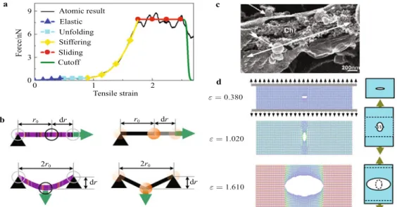
net-work model based on the bottom-up design as shown in Fig. 6. This blueprint illustrates the basis for setting up the mesoscale model of nuclear lamina networks basing on the constitutive behavior of in-dividual single filament building blocks as shown in Figs. 6a and 6b. The mesoscale network model integrates the tensile property, bending property and density of the single filament by designing the bond term, angular term and particle mass in the potential function, respectively. The model also includes the inter-filament crosslink by designing the inter-filament connecting strength between parti-cles in different filaments. This model captures the mechanism be-hind the deformation capacity (> 100% strain) of intermediate fil-ament networks despite the presence of structural defects as shown in Fig. 6d. This flaw-tolerant characteristic is greatly caused by the stiffening behavior during secondary structural transitions for a sin-gle filament. The behavior significantly increases the capacity of the meshwork to withstand loading by delocalizing the
deforma-tion energy around flaws, and preventing catastrophic crack prop-agation. A more severe condition causes the crack to propagate in two directions without brunching for energy dissipation as shown in Fig. 6c. This phenomenon is also captured by simulations as shown in Fig. 6d. The effect of crosslink strength has been studied basing on this model. It is found that the crosslink strength between filaments plays a critical role to affect the materials strength by tun-ing the failure mechanism of the network. The unpinntun-ing process between crosslinked filaments is the key mechanism to enable the tuning function. Comparing to the failure strength of the network with very strong crosslinks, it has been find that the networks with moderate crosslink are 23% stronger [58]. This result, combining with the fact that theΔ50 mutant makes the lamin tail as a crosslink become more compact and rigid for binding, provides the mecha-nism in explaining the progeria syndrome as the mutated cellular nuclear behaves less stable and more vulnerable to defects.
a b d c 0 1 2 Tensile strain 9 6 3 0 Force/nN Atomic result Elastic Unfolding Stiffering Sliding Cutoff r0 dr r0 dr 2r0 2r0 dr dr ε=0.380 ε=1.020 ε=1.610
Fig. 6 Mesoscopic modeling of intermediate filament network and its mechanical behavior under the extreme mechanical condition. a
tensile parameters within the mesoscopic model are fitted so that the force–strain curve of a stretching mesoscopic simulation is identical to the one obtained from an atomistic stretching simulation (reproduced from Ref. [61] with permission, Copyright (2011) American Chemical Society); b Illustration of the mechanical loading conditions used to fit tensile and bending potential parameters for intermediate filament network model; c Crack forms and propagates during the disassembling process of the nuclear envelop of Xenopus egg cell (reproduced from Ref. [62] with permission, Copyright (2007) Elsevier). The chromatin (chr) is exposed during this process and the white double arrow indicates the crack. Scale bar: 200 nm; d Snapshots of the intermediate filament network under the uniaxial tension from its initial state to extreme applied strain that exceeds the material limit. The schematic figures to the right show the transition of the crack shape in stretching.
3 Conclusion
By studying the mechanical and biological properties of intermedi-ate filament proteins from a bottom-up perspective, we have shown the power of computational modeling as a tool to fill in the gap be-tween genotype and phenotype of intermediate filaments by setting up bridges of the molecular structure, mechanism and the materials functions. Establishing such links is critical to reach a better un-derstanding of the complete gene expression and modification path, which is not limited to intermediate filaments but may be generally applicable to other proteins structures in studying their mechani-cal properties. With the multismechani-cale modeling scheme and computa-tional tools, we can racomputa-tionally design novel polymer materials with
greater reliability and tunable material properties by adjusting their compositions and microscopic structures.
Another strength of this methodology is that it enables us to simulate a system with tens of micrometers dimension while re-taining information about molecular details and mechanisms, which has been demonstrated recently in a study of spider web mechan-ics [59]. As single point mutations in protein materials represent typical sources that create flaws in biological systems, it is very important to understand the pathway of the disease because the de-signs of effective treatments of genetic diseases. Over 4 000 known human diseases are estimated to be caused by single gene defects that can be either inherent or accumulated. Since experimental methods are limited both in time and space resolution,
computa-Computational and theoretical modeling of intermediate filament networks: Structure, mechanics and disease 949 tional modeling has an advantage to investigate biological systems
in a bottom-up fashion. In the multiple scale modeling, quantum effects, thermal fluctuations, chemical reactions, structure stabili-ties, conformities and interaction networks can all be considered for quantitative understanding of material functions and processes, which are features rooted in the nanoscale realm and extended to the macro realm. By combining with the experimental and clinical effort, we hope that this methodology will facilitate breakthroughs in biomedical and biomaterial innovation in future applications.
Acknowledgement We acknowledge support from AFOSR, NSF
and ONR.
References
1 Ishikawa, H., Bischoff, R., Holtzer, H.: Mitosis and intermediate-sized filaments in developing skeletal muscle. Journal of Cell Biology 38, 538–555 (1968)
2 Herrmann, H., Bar, H., Kreplak, L., et al.: Intermediate fila-ments: from cell architecture to nanomechanics. Nature Re-views Molecular Cell Biology 8, 562–573 (2007)
3 Omary, M. B., Coulombe, P. A., McLean, W. H. I.: Mecha-nisms of disease: Intermediate filament proteins and their asso-ciated diseases. New England Journal of Medicine 351, 2087– 2100 (2004)
4 Hearle, J. W. S.: A critical review of the structural mechan-ics of wool and hair fibres. International Journal of Biological Macromolecules 27, 123–138 (2000)
5 Kreplak, L., Fudge, D.: Biomechanical properties of intermedi-ate filaments: from tissues to single filaments and back. Bioes-says 29, 26–35 (2007)
6 Chang, L., Goldman, R. D.: Intermediate filaments mediate cy-toskeletal crosstalk. Nature Reviews Molecular Cell Biology 5, 601–613 (2004)
7 Strelkov, S. V., Herrmann, H., Aebi, U.: Molecular architecture of intermediate filaments. Bioessays 25, 243–251 (2003) 8 Moll, R., Divo, M., Langbein, L.: The human keratins: biology
and pathology. Histochemistry and Cell Biology 129, 705–733 (2008)
9 Jafari, S. S., Maxwell, W. L., Neilson, M., et al.: Axonal cy-toskeletal changes after non-disruptive axonal injury. Journal of Neurocytology 26, 207–221 (1997)
10 Dahl, K. N., Kahn, S. M., Wilson, K. L., et al.: The nuclear envelope lamina network has elasticity and a compressibility limit suggestive of a molecular shock absorber. J. Cell. Sci.
117, 4779–4786 (2004)
11 Wilson, K. L., Zastrow, M. S., Lee, K. K.: Lamins and disease: Insights into nuclear infrastructure. Cell 104, 647–650 (2001) 12 Wang, N., Stamenovic, D.: Mechanics of vimentin
intermedi-ate filaments. J. Muscle Res. Cell. Motil. 23, 535–540 (2002) 13 Buehler, M. J., Yung, Y. C.: Deformation and failure of
pro-tein materials in physiologically extreme conditions and dis-ease. Nature Materials 8, 175–188 (2009)
14 Eriksson, M., Brown, W. T., Gordon, L. B., et al.: Recurrent de novo point mutations in lamin A cause Hutchinson-Gilford progeria syndrome. Nature 423, 293-298 (2003)
15 Dahl, K. N., Scaffidi, P., Islam, M. F., et al.: Distinct structural and mechanical properties of the nuclear lamina in
Hutchinson-Gilford progeria syndrome. Proceedings of the Na-tional Academy of Sciences of the United States of America
103, 10271–10276 (2006)
16 Lin, Y. C., Broedersz, C. P., Rowat, A. C., et al.: Divalent cations crosslink vimentin intermediate filament tail domains to regulate network mechanics. Journal of Molecular Biology
399, 637–644 (2010)
17 Zhang, H., Ackbarow, T., Buehler, M. J.: Muscle dystrophy single point mutation in the 2B segment of lamin A does not affect the mechanical properties at the dimer level. Journal of Biomechanics 41, 1295–1301 (2008)
18 Nicolet, S., Herrmann, H., Aebi, U., et al.: Atomic structure of vimentin coil 2. Journal of Structural Biology 170, 369–376 (2010)
19 Luca, S., Yau, W. M., Leapman, R., et al.: Peptide conforma-tion and supramolecular organizaconforma-tion in amylin fibrils: Con-straints from solid-state NMR. Biochemistry 46, 13505–13522 (2007)
20 Goldie, K. N., Wedig, T., Mitra, A. K., et al.: Dissecting the 3-D structure of vimentin intermediate filaments by cryo-electron tomography. Journal of Structural Biology 158, 378– 385 (2007)
21 Lee, H. L. D., Sahl, S. J., Lew, M. D., et al.: The double-helix microscope super-resolves extended biological structures by localizing single blinking molecules in three dimensions with nanoscale precision. Applied Physics Letters 100, 153701 (2012)
22 Zagrovic, B., Snow, C. D., Shirts, M. R., et al.: Simulation of folding of a small alpha-helical protein in atomistic detail using worldwide-distributed computing. Journal of Molecular Biol-ogy 323, 927–937 (2002)
23 Khatib, F., DiMaio, F., Cooper, S., et al.: Crystal structure of a monomeric retroviral protease solved by protein folding game players. Nature Structural & Molecular Biology 18, 1175–1177 (2011)
24 Buehler, M. J., Keten, S.: Colloquium: Failure of molecules, bones, and the Earth itself. Reviews of Modern Physics 82, 1459–1487 (2010)
25 Rauscher, S., Pomes, R.: Molecular simulations of protein dis-order. Biochemistry and Cell Biology-Biochimie Et Biologie Cellulaire 88, 269–290 (2010)
26 Zwanzig, R., Szabo, A., Bagchi, B.: Levinthal’s paradox. Proc Natl Acad Sci USA 89, 20–22 (1992)
27 del Rio, A., Perez-Jimenez, R., Liu, R. C., et al.: Stretching single talin rod molecules activates vinculin binding. Science
323, 638–641 (2009)
28 Keten, S., Buehler, M. J.: Nanostructure and molecular me-chanics of spider dragline silk protein assemblies. J. R. Soc. Interface 7, 1709–1721 (2010)
29 Bates, G.: Huntingtin aggregation and toxicity in Huntington’s disease. Lancet 361, 1642–1644 (2003)
30 Qin, Z., Kalinowski, A., Dahl, K. N., et al.: Structure and sta-bility of the lamin A tail domain and HGPS mutant. Journal of Structural Biology 175, 425–433 (2011)
31 Sugita, Y., Okamoto, Y.: Replica-exchange molecular dynam-ics method for protein folding. Chemical Physdynam-ics Letters 314, 141–151 (1999)
32 Brooks, B. R., Bruccoleri, R. E., Olafson, B. D., et al.: Charmm - a program for macromolecular energy, minimization, and dy-namics calculations. Journal of Computational Chemistry 4,
187–217 (1983)
33 Ackbarow, T., Buehler, M. J.: Superelasticity, energy dissipa-tion and strain hardening of vimentin coiled-coil intermediate filaments: atomistic and continuum studies. Journal of Materi-als Science 42, 8771–8787 (2007)
34 Sotomayor, M., Schulten, K.: Single-molecule experiments in vitro and in silico. Science 316, 1144–1148 (2007)
35 Qin, Z., Kreplak, L., Buehler, M. J.: Hierarchical structure con-trols nanomechanical properties of vimentin intermediate fila-ments. PLoS One 4, e7294 (2009)
36 Qin, Z., Buehler, M. J.: Molecular dynamics simulation of the alpha-helix to beta-sheet transition in coiled protein filaments: evidence for a critical filament length scale. Phys. Rev. Lett.
104, 198304 (2010)
37 Keten, S., Buehler, M. J.: Asymptotic strength limit of hydrogen-bond assemblies in proteins at vanishing pulling rates. Phys. Rev. Lett. 100, 198301 (2008)
38 Kreplak, L., Bar, H.: Severe myopathy mutations modify the nanomechanics of desmin intermediate filaments. J. Mol. Biol.
385, 1043–1051 (2009)
39 Parbhu, A. N., Bryson, W. G., Lal, R.: Disulfide bonds in the outer layer of keratin fibers confer higher mechanical rigidity: Correlative nano-indentation and elasticity measurement with an AFM. Biochemistry 38, 11755–11761 (1999)
40 Wiita, A. P., Ainavarapu, S. R. K., Huang, H. H., et al.: Force-dependent chemical kinetics of disulfide bond reduction ob-served with single-molecule techniques. Proceedings of the National Academy of Sciences of the United States of America
103, 7222–7227 (2006)
41 Wiche, G.: Role of plectin in cytoskeleton organization and dy-namics. Journal of Cell Science 111, 2477–2486 (1998) 42 Keten, S., Chou, C. C., van Duin, A. C. T., et al.: Tunable
nanomechanics of protein disulfide bonds in redox microenvi-ronments. Journal of the Mechanical Behavior of Biomedical Materials 5, 32–40 (2012)
43 Aryanpour, M., van Duin, A. C. T., Kubicki, J. D.: Develop-ment of a reactive force field for iron-oxyhydroxide systems. Journal of Physical Chemistry A 114, 6298–6307 (2010) 44 Chenoweth, K., van Duin, A. C. T, Goddard, W. A.: ReaxFF
re-active force field for molecular dynamics simulations of hydro-carbon oxidation. Journal of Physical Chemistry A 112, 1040– 1053 (2008)
45 Buehler, M. J.: Atomistic and continuum modeling of me-chanical properties of collagen: Elasticity, fracture, and self-assembly. Journal of Materials Research 21, 1947–1961 (2006) 46 Buehler, M. J., Tang, H., van Duin, A. C. T., et al.: Threshold crack speed controls dynamical fracture of silicon single crys-tals. Physical Review Letters 99, 165502 (2007)
47 Goldberg, M. W., Fiserova, J., Huttenlauch, I., et al.: A new model for nuclear lamina organization. Biochemical Society Transactions 36, 1339–1343 (2008)
48 Krimm, I., Ostlund, C., Gilquin, B., et al.: The Ig-like structure of the c-terminal domain of lamin A/C, mutated in muscular dystrophies, cardiomyopathy, and partial lipodystrophy. Struc-ture 10, 811–823 (2002)
49 Tama, F., Gadea, F. X., Marques, O., et al.: Building-block ap-proach for determining low-frequency normal modes of macro-molecules. Proteins-Structure Function and Genetics 41, 1–7 (2000)
50 Xu, Z. P., Paparcone, R., Buehler, M. J.: Alzheimer’s a beta(1-40) amyloid fibrils feature size-dependent mechanical proper-ties. Biophysical Journal 98, 2053–2062 (2010)
51 Barducci, A., Bussi, G., Parrinello, M.: Well-tempered meta-dynamics: A smoothly converging and tunable free-energy method. Physical Review Letters 100, 020603 (2008) 52 Wilhelmsen, K., Litjens, S. H. M., Kuikman, I., et al.:
Nesprin-3, a novel outer nuclear membrane protein, associates with the cytoskeletal linker protein plectin. Journal of Cell Biology 171, 799–810 (2005)
53 Ostlund, C., Folker, E. S., Choi, J. C., et al.: Dynamics and molecular interactions of linker of nucleoskeleton and cy-toskeleton (LINC) complex proteins. Journal of Cell Science
122, 4099–4108 (2009)
54 Kim, H., Nakamura, F., Lee, W., et al.: Filamin A is required for vimentin-mediated cell adhesion and spreading. American Journal of Physiology-Cell Physiology 298, C221-C236 (2010) 55 Lindstrom, S. B., Vader, D. A., Kulachenko, A., et al.: Biopoly-mer network geometries: Characterization, regeneration, and elastic properties. Physical Review E 82, 051905 (2010) 56 Lin, Y. C., Yao, N. Y., Broedersz, C. P., et al.: Origins of
elastic-ity in intermediate filament networks. Physical Review Letters
104, 058101 (2010)
57 Panorchan, P., Schafer, B. W., Wirtz, D., et al.: Nuclear enve-lope breakdown requires overcoming the mechanical integrity of the nuclear lamina. J. Biol. Chem. 279, 43462–43467 (2004)
58 Qin, Z., Buehler, M. J.: Mechanical properties of crosslinks controls failure mechanism of hierarchical intermediate fila-ment networks. Theoretical and Applied Mechanics Letters 2, 014005 (2012)
59 Cranford, S. W., Tarakanova, A., Pugno, N. M., et al.: Nonlin-ear material behaviour of spider silk yields robust webs. Nature
482, 72–76 (2012)
60 Kreplak, L., Herrmann, H., Aebi, U.: Tensile properties of single desmin intermediate filaments. Biophysical Journal 94, 2790–2799 (2008)
61 Qin, Z., Buehler, M. J.: Flaw tolerance of nuclear intermediate filament lamina under extreme mechanical deformation. ACS Nano 5, 3034–3042 (2011)
62 Cotter, L., Allen, T. D., Kiseleva, E., et al.: Nuclear membrane disassembly and rupture. Journal of Molecular Biology 369, 683–695 (2007)