HAL Id: inserm-02351977
https://www.hal.inserm.fr/inserm-02351977
Submitted on 6 Nov 2019HAL is a multi-disciplinary open access archive for the deposit and dissemination of sci-entific research documents, whether they are pub-lished or not. The documents may come from teaching and research institutions in France or abroad, or from public or private research centers.
L’archive ouverte pluridisciplinaire HAL, est destinée au dépôt et à la diffusion de documents scientifiques de niveau recherche, publiés ou non, émanant des établissements d’enseignement et de recherche français ou étrangers, des laboratoires publics ou privés.
Expansion of allogeneic NK cells with efficient
antibody-dependent cell cytotoxicity against multiple
tumors
Diego Sanchez, Nerea Allende-Vega, Stefania Orecchioni, Giovanna Talarico,
Amélie Cornillon, Dang-Nghiem Vo, Céline René, Zhao-Yang Lu, Ewelina
Krzywinska, Alberto Anel, et al.
To cite this version:
Diego Sanchez, Nerea Allende-Vega, Stefania Orecchioni, Giovanna Talarico, Amélie Cornillon, et al.. Expansion of allogeneic NK cells with efficient antibody-dependent cell cytotoxicity against multiple tumors. Theranostics, Ivyspring International Publisher, 2018, 8 (14), pp.3856-3869. �10.7150/thno.25149�. �inserm-02351977�
Expansion of allogeneic NK cells with efficient antibody-dependent cell cytotoxicity against multiple tumor cells
Diego Sanchez1, #,Nerea Allende-Vega1,2,#, Stefania Orecchioni3, Giovanna Talarico3, Amelie Cornillon1, Dang-Nghiem Vo1, Celine Rene1, Zhao-Yang Lu1, Ewelina Krzywinska1, Alberto Anel4, Eva M. Galvez4, Julian Pardo4, Bruno Robert5, Pierre Martineau5, Yosr Hicheri6, Francesco Bertolini3, Guillaume Cartron6 and Martin Villalba*,1,2
1 IRMB, Univ Montpellier, INSERM, CHU Montpellier, Montpellier, France. 2 IRMB, CHU Montpellier, France.
3 Laboratory of Hematology-Oncology, European Institute of Oncology, Milan, Italy 4 University of Zaragoza/Institute of Health Research of Aragón (IIS-Aragón), Spain. 5 IRCM, INSERM U1194, Univ Montpellier, France.
6 Département d'Hématologie Clinique, CHU Montpellier, Univ Montpellier, France
# These two authors have equally contributed to this manuscript * corresponding author
E-mail: martin.villalba@inserm.fr (MV)
Abstract
Monoclonal antibodies (mAbs) have significantly improved the treatment of certain cancers. However, in general mAbs alone have limited clinical activity. One of their main mechanisms of action is to induce antibody-dependent cell-mediated cytotoxicity (ADCC), which is mediated by natural killer (NK) cells. However, most cancer patients present severe immune dysfunctions affecting NK activity. This could be circumvented by the injection of allogeneic, expanded, NK cells, which is safe. Nevertheless, despite the strong cytolytic potential of expanded NK cells against different tumors, clinical results have been poor.
Methods: We thus propose the combination of allogeneic NK cells and mAbs to improve cancer treatment. We generated expanded NK cells (e-NK) with strong in vitro and in vivo ADCC response against different tumors and mediated by different therapeutic mAbs, i.e. rituximab, obinutuzumab, daratumumab, cetuximab and trastuzumab.
Results: Remarkably, e-NK cells can be stored frozen and after thawing armed with mAbs. They mediate ADCC through degranulation dependent and independent mechanisms. Furthermore, they overcome some of the anti-apoptotic mechanism found in leukemic cells. Conclusion: In summary, we have established a new protocol for activation/expansion of NK cells with high ADCC activity. The use of mAbs in combination with e-NK cells could potentially improve cancer treatment.
Introduction
Recent progresses in cancer treatment are mainly related to the development of new-targeted therapies [1]. These require the identification of suitable targets that are mainly expressed by the tumor cell population and/or playing a critical role for neoplastic cell development. In this context, monoclonal antibodies (mAbs) have improved the treatment of certain cancers. Indeed, rituximab (RTX), an IgG1 mAb directed against CD20 antigen, has now become the backbone for the treatment of most B-cells chronic lymphocytic leukemia (B-CLL) and B-cells non-Hodgkin’s lymphoma (B-NHL) patients. The combination of RTX with conventional chemotherapy has shown better efficacy in randomized clinical trials. This success has also been found with other cytotoxic mAbs such as trastuzumab in breast cancer or cetuximab in colorectal carcinoma and squamous cell carcinoma of the head and neck [2, 3]. However, it is remarkable to note that the mAbs themselves have modest clinical activity. For example, RTX or obinutuzumab (OBZ; previously GA101, Roche, Genentech) when used as monotherapy in patients with relapsed follicular lymphoma (FL) have demonstrated short progression-free survival (PFS) [4]. These data indicate that there is a need to optimize their use in co-therapy. RTX success is related to its capacity to induce Fc-antibody-dependent cell-mediated cytotoxicity (ADCC). One receptor for human IgG1 is FcγRIIIa (CD16a), which is expressed on natural killer (NK) cells and macrophages. The influence of FcγRIIIa-158VF polymorphism on RTX clinical response strongly suggests that ADCC is critical [5]. Based on these results, there has been an attempt to produce new anti-CD20 mAbs that exhibit higher affinity for FcγRIIIa either by Fc mutations or by glycoengeenering [4, 6]. This later strategy, leading to low fucose content of the N-glycan, is currently under clinical investigations in B-cell malignancies with the mAb OBZ, which shows stronger ADCC in vitro and in a lymphoma xenograft mouse model compared to RTX and improved clinical activity for treating B-CLL [4]. This clinical benefit has been observed in other B-cell malignancies [4]. OBZ is approved for first-line B-CLL in association with chlorambucil and in combination with bendamustine for the treatment of patients with FL who relapse or are refractory to RTX-containing regimen [4]. Initial results show that lenalidomide, which stimulates NK cell activity [7], activates NK cells in OBZ-treated patients [8].
Hematological cancer patients possess antitumor NK cells that are unable to control disease [9, 10]. Blood-borne tumor cells use different mechanisms for immune escape [11, 12], e.g. inducing NK cell dysfunction [7, 13]. This mechanism has also been observed in a variety of solid tumor patients [3]. In addition, NK cell differentiation may be inhibited by the presence of tumor cells e.g. acute myeloid leukemia (AML) cells infiltrating bone marrow [14, 15].
Therefore, the failure of mAb as monotherapy could be related to impaired NK cell function and hence, there is a clinical interest to reactivate or replace patient NK cells [16]. Clinical-grade production of allogeneic NK cells is efficient and NK cell–mediated therapy after hematopoietic stem cell transplantation (HSCT) seems safe [15, 17, 18]. However, despite the strong cytolytic potential of expanded NK cells against different tumors, clinical results have been very limited [15, 17, 18].
The combination of allogeneic NK cells with mAb could improve cancer treatment by replacing the defective effector immune cells. In addition, mAbs would effectively guide these effectors to their tumor targets. Several groups have tried this combination with miscellaneous results that could be due to deficient CD16 expression or lack of proper activation of expanded NK [19-22]. In addition, these works did not include a systematic study of the effect of these cells in combination with several mAbs on different tumors, and also they did not include primary tumor cells.
The aim of this work is to generate allogeneic NK cells with strong ADCC response against different tumors and mediated by different therapeutic mAbs. In addition, NK cell production should be easily scaled up and developed with good manufacturing practices (GMP). We have produced umbilical cord blood (UCB)-derived NK cells because UCB are rapidly available, present low risk of viral transmission and have less strict requirements for HLA matching and lower risk of graft-versus-host disease (GvHD) [17]. For NK cell expansion we used Epstein– Barr virus (EBV)-transformed lymphoblastoid B cell lines as accessory cells, which induce a unique NK cell genetic reprogramming [23], generating effectors that overcome the anti-apoptotic mechanism of leukemic cells [24] and that are able to eliminate tumor cells from patients with poor prognosis [25]. We show that NK cells obtained with our protocol are able to perform ADCC in vitro and in vivo. The ADCC response was induced by different therapeutic antibodies and against multiple target cells.
Materials and Methods Ethical statement
Experimental procedures were conducted according to the European guidelines for animal welfare (2010/63/EU). Protocols were approved by the Animal Care and Use Committee “Languedoc-Roussillon” (approval number: CEEA-LR-12163). The use of human specimens for scientific purposes was approved by the French National Ethics Committee. All methods were carried out in accordance with the approved guidelines and regulations of this committee. Written informed consent was obtained from each patient prior to surgery.
Chemicals
The D1D2 peptide has been previously described [26]. IL-2 and IL-15 were obtained from Miltenyi Biotec. To produce deglycosylated cetuximab, a commercial cetuximab solution was treated overnight with PNGAseF (Promega) at 37°C in 50 mM sodium bicarbonate buffer (pH 7.8) at 125 U/mg of cetuximab. Deglycosilated cetuximab was purified by gel filtration using a Sephadex 75 column in PBS, and sterilized by filtration. The AF 647 Goat F(AB')2 anti human IgG (H+L) min x (BOV,HRS,MS) was from Interchim.
B-CLL patients
Data and samples from patients were collected at the Clinical Hematology Department of the CHU Montpellier, France, after patient’s written consent and following French regulations. Patients were enrolled in two independent clinical programs approved by the “Comités de Protection des Personnes Sud Méditerranée I”: ref 1324 and HEMODIAG_2020 (ID-RCB: 2011-A00924-37). Samples were collected at diagnosis and kept by the CHU Montpellier [10, 27]. For analysis peripheral blood mononuclear cells (PBMCs) were obtained by ficoll gradient and stored frozen in liquid nitrogen until use. The percentage of CD19+CD5+ cells was always higher than 90%. Other samples from hematological cancer patients were obtained from the same collections.
Cell lines
The (EBV)-transformed lymphoblastoid B cell line PLH, the hematopoietic cell lines HL60 and MV4-11 (acute myeloid leukemia), Daudi and Raji (Burkitt's lymphoma derived, CD20+), K562 (erythroleukemia), MM.1S and U266 (multiple myeloma), MEC1 (B-chronic lymphocytic leukemia) and its variants MEC-1-BCL-XL and MEC-1-MCL-1 were cultured in 10% FBS RPMI medium. The adherent cell lines Calu-1 (lung cancer), A549 (lung
adenocarcinoma), SK-OV-3 (ovarian carcinoma) and SKBR3 (breast adenocarcinoma) were cultured in 10% FBS DMEM medium.
PBMC and UCBMC purification
UCBs were obtained from healthy donors from the CHU Montpellier. PBMC and UCB mononuclear cells (UCBMC) were respectively collected from peripheral blood samples and UCB units using Histopaque -1077 (Sigma). Briefly, 13 ml Histopaque were added to 50 ml centrifugation tubes and 30 ml of 1/2 diluted blood in RPMI, (Invitrogen) were slowly added at the top. Tubes were centrifuged at 1600 rpm for 30 minutes at 20 °C without break. Mononuclear cells were collected from the interlayer white ring, washed in RPMI and suspended in RPMI medium supplemented with 10% FBS (Invitrogen).
Isolation and activation of human NK cells
Frozen UCBMCs were depleted of T cells by using EasySepTM CD3 Positive Selection Kit (STEMCELL technologies). Cells were cultured for 10 to 20 days with γ-irradiated PLH cells at 1:1 NK cell:accessory cell ratio in presence of IL-2 (100 U/ml) and IL-15 (5 ng/ml), or with IL-2 alone (1000 U/ml). PLH cells were added every four days and fresh cytokines every two days. At the end of the process NK cell purity (CD56+/CD3-) was always higher than 90%.
FACS analysis
For phenotype analysis, cells were stained with 7AAD (Beckman) to identify viable cells and antibodies against the surface markers CD25-FITC, CD45RO-FITC, CD69-PE, CD62L-PE, CD19-PE, CD3-PE, CD19-ECD, CD56-PECy7, CD56-APC, CD3-APC, CD45-APCAlexaFluor750, CD45RA-CD45-APCAlexaFluor750, CD16-PacificBlue, CD57-PacificBlue, CD45-KromeOrange, CD16-KromeOrange (Beckman), CD158b-FITC, CD158a-PE, CD107a-HV500 (BD Biosciences), CD158e-Vioblue (Miltenyi). 1x10⁵–3x10⁵ cells were incubated for 20-30 minutes at 4 °C with different antibodies in PBS containing 2.5% FBS. Cells were then washed and suspended in 200-250 µl of the same media. Staining was analyzed on a Gallios flow cytometer (Beckman) using the Kaluza software. Alive lymphocytes were gated using FSC/SSC and 7AAD staining. B lymphocytes (CD19+), T lymphocytes (CD3+CD56-) and NK cells (CD56+CD3-) were distinguished using respectively CD19, CD3 and CD56 antibodies.
Fresh or frozen (stored in liquid nitrogen) NK cells were labeled with 3 µM of CellTracker™ Violet BMQC Dye (Life Technologies) and incubated overnight with target cells at different E:T ratios. Subsequently, phosphatidylserine (PS) translocation and membrane damage were analyzed in the violet fluorescence negative target cell population by flow cytometry using Annexin V-FITC (Immunostep) and 7AAD (BD Biosciencies) or propidium iodide (PI) as previously described [24, 28]. We consider all cells positive for annexin-V and/or PI (or 7-ADD) as death (or dying).
In ADCC experiments, we incubated target cells with the relevant antibodies (RTX and OBZ at 10 µg/ml; daratumumab, cetixumab and trastuzumab at 5 µg/ml) for 30 minutes at 37°C. When we armed NK cells, we incubated them at the same concentration of antibodies before washing and incubation with target cells. EGTA was used at 1 mM to block the granular exocytosis pathway and MgCl2 at 1.5 mM to maintain the osmotic pressure.
We used the tetrazolium dye MTT (3-(4,5-dimethylthiazol-2-yl)-2,5-diphenyltetrazomium bromide) to determinate cellular viability. We added 10 µL of MTT (5 mg/ml) to the adherent cells (100 µL of medium after 2 washes with PBS) and we incubated them for 1 h at 37°C, and added 100 µL of 0.05 M HCL in isopropanol to dissolve the crystals and quantified color at 550 nm in a spectrophotometer.
In all experiments we calculated the basal cell death in the absence and presence of the different mAbs. These values were subtracted from those obtained after NK cell or NK cell+mAb treatments to generate the specific natural cytotoxicity or specific ADCC, respectively. All mAbs gave very low levels (<3%) of cytotoxicity in presence of heat inactivated serum media.
Evaluation of RTX-armed e-NK
e-NK cells (2x10⁵) were incubated for 1 h with 10 µg/ml RTX, washed and incubated with 1:800 solution of a goat F(ab')2 anti human IgG (H+L) for 30 min at 4°C. After incubation, NK cells were washed with PBS and RTX binding was analyzed by FACs. As a control cells were only stained with the a goat F(ab')2 anti human IgG.
NK degranulation assay
Briefly, 50x103 target cells per well were placed in RPMI, 10% FBS, IL-2 100 U/ml with monensin (BD Biosciences) in a 96-well V-bottom plate. NK and target cells were incubated overnight at 37°C in 5% CO2 and living cells were counted using a Muse cytometer
without targets. CD107a+ NK cells were analyzed on a Gallios flow cytometer (Beckman Coulter) using 7AAD, CD45RO-FITC, CD19-PE, CD56-PECy7, CD3-APC, CD45RA-APCAlexaFluor750, CD16-KromeOrange and CD107a-HV500 (BD Biosciences). Results were analyzed using Kaluza software.
In vivo experiments
In vivo experiments were carried out using 6 to 8 weeks/old male NOD scid gamma (NSG)
mice. Mice were bred and housed in pathogen-free conditions in the animal facility of the European Institute of Oncology–Italian Foundation for Cancer Research (FIRC), Institute of Molecular Oncology (Milan, Italy). For engraftment of human cells, mice were subcutaneously engrafted with 5x10⁶ BCL-P2 or 10x106 LNH1 primary tumor cells derived
from a BCL (P2) patient or a diffuse large B-cell lymphoma (DLBCL) patient (LNH1). At day 4, we engrafted 15 (BCL-P2) or 10 (LNH1) million e-NK cells and at day 6, mice were treated i.p. with RTX (in saline medium) 3 mg/kg once a week x 3 weeks; or with a combination of both. Tumor growth was monitored at least once a week using a digital calliper, and tumor volume was calculated according to the formula: L × W2/2 = mm3, where W represents the width and L the length of the tumor mass.
Results
Costimulation with the EBV lymphoblastoid PLH cell line is more efficient to expand UCB NK cells for clinical use than high IL-2 stimulation
Cytokines and encounter with target cells induce dissimilar gene expression on NK cells [23]. We used umbilical cord blood (UCB) cells and compared two NK cell activation/expansion protocols: one using a high dose of the cytokine IL-2 (1000 U/ml) and the other one by cell costimulation. The costimulation protocol was performed with the EBV cell line PLH together with low concentrations of IL-2 (100 U/ml) and IL-15 (5 ng/ml) [29]. NK cell expansions are jeopardized by T cells, therefore we depleted them from UCB before expansion. We observed that NK cell cultures underwent massive cell death at day 12 in IL-2-drive expansion (data no shown). Hence, we compared at day 10 different parameters that reflect NK activity.
Proliferation. Costimulation-drive expansion was largely more efficient (Fig. 1A).
Cytotoxicity. IL-2-drive expansion induced NK cells with superior natural cytotoxicity against K562 (Fig. 1B), Daudi (supplementary. Fig. 1A) and primary CD20+ B cell lymphoma cells (BCL P2; Fig. 1C). Moreover, they also showed higher ADCC activity with RTX (Fig. 1C and supplementary Fig. S1A). However, natural cytotoxicity gradually increased with the costimulation protocol when cells were activated for longer periods of time (Fig. 1D). This correlated with a notable large-scale expansion of cells (Median+/-STDV, IL-2 10 d (16.8 +/- IL-2IL-2.IL-2), costimulation 10 d (140.5 +/- IL-235.8) and costimulation IL-20 d (IL-260.9 +/- 141.2), n=10).
Activation markers. Both protocols increased the expression of the activation marker CD69 (supplementary Fig. S2B) and decreased of CD45RA expression to originate CD45RAdim cells (supplementary. Fig. 2B), which was associated to increase in the activation marker CD45RO as previously published [9]. Costimulation kept higher CD16 expression (supplementary Fig. S2B).
Exhaustion markers. We investigated the expression of two markers that could suppress NK cell-mediated cytotoxicity: TIM-3 [30] and PD-1 [31]. Both protocols did not affected their expression PD-1 expression (supplementary Fig. S2B).
Maturation and homing markers. UCB NK cells showed low percentage of CD62L+ cells (18.1+/-6.7, average+/-STD, n=3) that increased 10 d after IL-2 treatment (68.2+/-13.5) and costimulation (56.7+/-11.9). However at day 20 post-costimulation, CD62L expression was lost (1.6+/-0.4). IL-2-stimulated cells did not survive so long; hence we could not observe a
possible decrease in CD62L. CD62L is a "homing receptor" facilitating naive lymphocytes to enter secondary lymphoid tissues. Mature NK cells express low CD62L, which favors their peripheral trafficking [32].
In agreement with others [33], few naïve UCB NK cells expressed CD57 (1.2 +/- 1.3, average+/-STD, n=3). IL-2 stimulation barely increased expression (7.3 +/- 3.9) and costimulation did not change it (0.6 +/- 0.6). Longer costimulation, i.e. 20 days, did not alter it (1.6 +/- 0.5). However, the lack of CD57 expression did not impair NK cell cytolytic activity (see below).
In summary, costimulation showed higher number of activated and functional NK cells with higher CD16 expression. This prompted us to exclusively perform costimulation for the next experiments. However IL-2 induced higher and faster cytotoxicity and could be the best option for autologous NK cell grafts.
Frozen/thawed NK cells keep their cytolytic activity
For clinical purposes, it will be advantageous to store and have a bank of cryopreserved expanded NK cells that will be “ready to use” [34]. Compared with fresh expanded NK cells, frozen/thawed NK cells roughly lost on average 50% of their cytolytic activity (Fig. 1E). As shown in Fig. 1D, 20 day-activation showed higher cytolytic activity than shorter expansions. For next experiments we used 20-21 days costimulation-induced expansion of UCB-derived NK cells containing more than 90% of NK cells and kept frozen until use. Hereafter, we called them e-NK.
e-NK cells mediate ADCC against target cells with diverse CD20 levels
We observed that e-NK performed ADCC similarly on Raji and Daudi cells, which express high CD20 levels (supplementary Fig. S2A), than on primary tumor cells (supplementary Fig. S2B). P2 cells probably express more CD20 than P148, however, they were slightly less sensitive to RTX-mediated ADCC. In fact, e-NK performed ADCC even if their natural cytotoxicity against some patient cells was low or absent (Fig. 2A supplementary Fig. S2B-E). Hence, e-NK show strong ADCC with RTX independently of their natural cytotoxicity and with lower variability (Fig. 2A-B). The glycoengeenering mAb OBZ [4, 35] induced higher ADCC than RTX (Fig.2C and supplementary Fig. S2B-C).
We next used e-NK cells coated with anti-CD20 mAb (“armed” e-NK cells) as an alternative to antibody-coated target cells. “CD20-armed” e-NK showed similar results than opsonizing tumor cells with anti-CD20 (supplementary Fig. S3A). The presence of RTX after e-NK “arming” was visualized by using a fluorescent anti-IgG (supplementary Fig. S3B). “OBZ-armed” e-NK were also efficient to generate ADCC (supplementary Fig. S4).
Statistical analysis of several e-NK productions on 5 CLL patients did not show any differences between opsonizing targets or “arming” e-NK (Fig. 3A). Moreover, the analysis of these 4 e-NK expansions on the CLL targets showed that all productions could be armed (Fig. 3B). Combining all these results statistically showed the significant ADCC mediated by e-NK (Fig. 3C).
Cytotoxicity requires degranulation and cell interaction by ICAM
Primary human NK cell cytotoxicity is largely independent of degranulation [36] and resides in death receptor binding on tumor cells by death receptors ligands expressed by NK cells. In agreement, e-NK natural cytotoxicity was only partially diminished by the degranulation inhibitor EGTA (Fig. 4A). In contrast, EGTA largely blocked ADCC.
The interaction of NK cell expressed LFA-1 with its target cell ligand ICAM modulates NK cell cytotoxicity [37]. Blocking this interaction with the D1D2 peptide [26] partly reduced natural cytotoxicity and almost completely abolished ADCC (Fig. 4B). Therefore, our e-NK keep similar mechanisms of eliminating target cells than primary human cells.
e-NK mediate ADCC with daratumumab
Next we tested if e-NK produced ADCC with the anti-CD38 daratumumab [38]. We used 3 target cells that expressed CD38 (MM.1S, MV4-11 and BCL-P2) and observed that three different e-NK preparations showed ADCC with daratumumab (supplementary Fig. S5A-C). In contrast daratumumab failed to induce ADCC against U266 that did not express CD38 (Supplementary Fig. 5D). Statistical analysis of several e-NK productions on MM.1S and P2 showed that e-NK efficiently performed ADCC with daratumumab (Fig. 4C).
e-NK mediate ADCC with cetuximab
Next we analyzed if e-NK cells mediated ADCC with other mAbs used to treat solid tumors. We used the cell lines Calu-1 and A549. Calu-1 cells express more epidermal growth factor receptor (EGFR) than A549 (data not shown). Both lines are targets of the anti-EGFR cetuximab, which has been proposed for using with adoptively transferred expanded
allogeneic NK cells in clinical trials for cervical cancer [39]. In fact, in vitro and clinical data suggest that cetuximab mediates ADCC through NK cells [22, 34]_ENREF_35. We observed a relatively large diversity of the natural cytotoxicity of the different e-NK donors versus these target cells. The decreased on cell viability, measured by MTT formation, was low (supplementary Fig. S6A). However, the increased in cell death, measured by annexin-V/7-ADD staining, was higher. This showed that e-NK had induced on targets the initial steps of apoptosis (annexin-V staining) but longer times were required to evaluate cell viability with MTT. Cetuximab increased early apoptosis and accelerated the process of cell death, decreasing viability. Statistical analysis of several e-NK productions on CALU-1 and A549 showed that e-NK efficiently performed ADCC with cetuximab (Fig. 5A).
EGTA diminished ADCC but not significantly (supplementary Fig. S6B-C). This suggested that the mechanism of action only partly involved degranulation, indicating a possible participation of death ligand-induced apoptosis.
e-NK mediate ADCC with trastuzumab
We next tested the anti-HER2 mAb trastuzumab on SK-BR-3, which express high HER2, and A549, which express low levels, cells. e-NK showed ADCC with trastuzumab in both cell lines by decreasing viability or increasing apoptosis (supplementary Fig. S7A). Natural cytotoxicity as well as ADCC heavily depends on degranulation because EGTA largely decreased both (supplementary Fig. S7A-B). Statistical analysis of several e-NK productions on SK-BR-3 and A549 showed that e-NK efficiently performed ADCC with trastuzumab (Fig. 5B). Finally, we extended this study to the ovarian cell line SK-OV-3 that was mainly resistant to both, natural cytotoxicity and ADCC, during short treatment (data not shown). However, 6-day treatment revealed that NK cells, mainly with trastuzumab, efficiently killed these cells (Fig. 5C).
e-NK mediated ADCC in vivo
We next evaluated e-NK activity in vivo by engrafting primary tumor cells from a BCL patient (P2) or from a DLBCL patient (LNH1) into NSG mice. Four days later mouse were engrafted with e-NK and 2 days later treated with RTX. e-NK, and mainly RTX, decreased tumor growth (Fig. 6A). But co-treatment was more effective, totally protecting mice from BCL cells and 4 out of 5 mice from DLBCL cells.
EBV-activated NK cells overcome anti-apoptotic mechanisms of chemoresistant cells [24, 25]. Overexpression of BCL-XL and MCL1 are common features of several hematological
cancers [40]. Jurkat cells over-expressing BCL-XL are resistant to doxorubicin and to soluble
TRAIL [41]. MCL1 over-expression protects from ibrutinib cytotoxicity (O. Gonzalo and I. Marzo, Doctoral Thesis, University of Zaragoza, 2017). e-NK killed the CD20+ MEC-1 B-CLL cell line overexpressing BCL-XL or MCL1, which gave them low protection against
ADCC compared to the wild type cell line (supplementary Fig. S8). Statistical analysis of several e-NK productions showed that they kill chemoresistant cell lines (Fig. 6B). However, BCL-XL overexpression significantly decreases ADCC at high E:T ratios. This suggests that
chemoresistance could partially protect tumor cells from e-NK-mediated ADCC, but not from e-NK natural cytotoxicity.
Discussion
High numbers of tumor-infiltrating NK correlates with a better prognosis in some tumors [15]. NK are the first lymphocytes to recover after HSCT including after UCBT. The speed of recovery correlates with the prognostic [17]. Despite these findings, NK cell adoptive immunotherapy has given low clinical benefit. Perhaps current expansion protocols fail to produce enough NK cells to support clinical success or generate cells with impaired activity [15]. We focused on developing a protocol to obtain NK cells in sufficient number and with high ADCC activity that could be mediated by different therapeutic mAbs. From UCB-derived NK we produce e-NK that only partially decrease ADCC function after cryopreservation (Fig. 1), preserving ADCC in vivo (Fig. 6). Interestingly, we have recently described that expression of the CD45 isoform CD45RO identifies anti-tumor NK cells in human patients [10]. e-NK decrease CD45RA and increase CD45RO expression suggesting that they would show elevated antitumor natural cytotoxicity as we have confirmed here. e-NK do not require relatively high Ag levels to perform ADCC because they were effective with cells expressing variables Ag levels in different cell lines.
Coupling mAbs with e-NK should improve the use of both treatments in several clinical contexts. First, most cancer patients possess impaired NK cells [3, 15, 17]. This should diminish the clinical activity of mAb. e-NKs should overpass this hitch by increasing mAb-induced ADCC in patients with immune defects. Second, the clinical activity of NK cells in solid cancers is uncertain [15, 17]. Probably these effectors scarcely recognize these targets in
vivo and/or fail to infiltrate these tumors e.g., in some tumors NK have been detected in the
tumor stroma but not within the tumor lesion [42-44]. Moreover, the adoptive transfer of autologous NK cells in patients as single therapy maintains high levels of circulating NK cells but did not mediate tumor regression [3, 17, 45]. mAbs should bring e-NK to the selected targets and can also facilitate target elimination by favoring the recognition of opsonized cells. In fact, haploidentical NK cells plus anti-GD2 mAb therapy has promising antitumor activity in pediatric recurrent/refractory neuroblastoma [46, 47]. Third, e-NKs overcome the anti-apoptotic mechanism of leukemic cells (Figure 7 and [24]) allowing elimination of tumor cells from poor prognosis patients [25].
An inconvenient of allogeneic expanded NK cell engraftment is that they survive few days in
vivo [48]. The persistence of ex vivo haploidentical IL-2-activated and -expanded NK-DLIs
reaches a maximum of 7 days in lymphoma patients [49]. This leaves grafted NK cells short time to eliminate their targets. The advantage is that NK will be less prompted to generate clinical problems as those found with CAR-T cells, which produce some chronic effects
related to their long-term persistence ([50]; http://www.medscape.com/viewarticle/876591). One of the main concerns for the use of allogeneic immune cells is the incidence of GVHD. Allogeneic NK cells infusion is well tolerated in cancer patients [3, 17] and the severity of aGVHD correlates with impaired reconstitution of the NK cell compartment [51]. To our knowledge, engraftment of NK cells have been linked to GVHD only when combined to HLA-matched, T-cell–depleted nonmyeloablative peripheral blood stem cell transplantation [52]. NK cells likely contributed to GVHD in this setting by augmenting underlying T-cell alloreactivity [52].
An interesting alternative to allogeneic NK are KIR/KIRL blocking antibodies that activate endogenous NK cells [53]. This approach has the inconvenient that cancer patient NK cells are hyporeactive [3, 15, 17], suggesting that they could be inefficient to totally eliminate tumors. Moreover, recent clinical data suggest that such antibodies modify the endogenous NK repertoire making KIR-expressing NK cells, which are those with higher cytolytic activity, hyporeactive [54]. This raises concerns to the clinical use of these antibodies. There are other ways to activate endogenous NK cells such as the use of lenalidomide (LEN) [7, 8]. Preliminary results from the Phase Ib/II clinical trial GALEN suggest that LEN could facilitate OBZ-mediated NK cell activation [8] as was observed with RTX [55].
Acknowledgements
FACs analysis was performed at MRI. The Région Languedoc Roussillon supports the clinical data and samples (HEMODIAG_2020).
Funding
This work was supported by an AOI from the CHU Montpellier (N°221826;GC/MV), La Ligue Regionale contre le Cancer (GC), Fondation de France (0057921;MV), the PRT-K program 2018 (MV/GC/BR) and “Investissements d’avenir” Grant LabEx MAbImprove: ANR-10-LABX-53.
References:
1. Neves H, Kwok HF. Recent advances in the field of anti-cancer immunotherapy. BBA clinical. 2015; 3: 280-8.
2. Scott AM, Wolchok JD, Old LJ. Antibody therapy of cancer. Nat Rev Cancer. 2012; 12: 278-87.
3. Hu z. Overcome the Impairment of NK Cells for Icon and Antibody Immunotherapy of Cancer Journal of Immune Based Therapies, Vaccines and Antimicrobials. 2013; 2: 1-8. 4. Cartron G, Watier H. Obinutuzumab: what is there to learn from clinical trials? Blood. 2017.
5. Cartron G, Dacheux L, Salles G, Solal-Celigny P, Bardos P, Colombat P, et al.
Therapeutic activity of humanized anti-CD20 monoclonal antibody and polymorphism in IgG Fc receptor FcgammaRIIIa gene. Blood. 2002; 99: 754-8.
6. Jefferis R. Glycosylation as a strategy to improve antibody-based therapeutics. Nat Rev Drug Discov. 2009; 8: 226-34.
7. Giuliani M, Janji B, Berchem G. Activation of NK cells and disruption of PD-L1/PD-1 axis: two different ways for lenalidomide to block myeloma progression. Oncotarget. 20PD-L1/PD-17; 8: 24031-44.
8. Vo DN, Alexia C, Allende-Vega N, Morschhauser F, Houot R, Menard C, et al. NK cell activation and recovery of NK cell subsets in lymphoma patients after obinutuzumab and lenalidomide treatment. Oncoimmunology. in press.
9. Krzywinska E, Allende-Vega N, Cornillon A, Vo D, Cayrefourcq L, Panabieres C, et al. Identification of anti tumor cells carrying natural killer (NK) cell antigens in patients with hematological cancers. EBioMedicine. 2015; 2: 1364-76.
10. Krzywinska E, Cornillon A, Allende-Vega N, Vo DN, Rene C, Lu ZY, et al. CD45 Isoform Profile Identifies Natural Killer (NK) Subsets with Differential Activity. PLoS One. 2016; 11: e0150434.
11. Villalba M, Rathore MG, Lopez-Royuela N, Krzywinska E, Garaude J, Allende-Vega N. From tumor cell metabolism to tumor immune escape. Int J Biochem Cell Biol. 2013; 45: 106-13.
12. Villalba M, Lopez-Royuela N, Krzywinska E, Rathore MG, Hipskind RA, Haouas H, et al. Chemical metabolic inhibitors for the treatment of blood-borne cancers. Anti-cancer agents in medicinal chemistry. 2014; 14: 223-32.
13. Baier C, Fino A, Sanchez C, Farnault L, Rihet P, Kahn-Perles B, et al. Natural Killer Cells Modulation in Hematological Malignancies. Frontiers in immunology. 2013; 4: 459. 14. Ambrosini P, Loiacono F, Conte R, Moretta L, Vitale C, Mingari MC. IL-1beta
inhibits ILC3 while favoring NK-cell maturation of umbilical cord blood CD34(+) precursors. Eur J Immunol. 2015; 45: 2061-71.
15. Moretta L, Pietra G, Vacca P, Pende D, Moretta F, Bertaina A, et al. Human NK cells: From surface receptors to clinical applications. Immunology letters. 2016; 178: 15-9.
16. Muntasell A, Ochoa MC, Cordeiro L, Berraondo P, Lopez-Diaz de Cerio A, Cabo M, et al. Targeting NK-cell checkpoints for cancer immunotherapy. Curr Opin Immunol. 2017; 45: 73-81.
17. Sarvaria A, Jawdat D, Madrigal JA, Saudemont A. Umbilical Cord Blood Natural Killer Cells, Their Characteristics, and Potential Clinical Applications. Frontiers in immunology. 2017; 8: 329.
18. Kottaridis PD, North J, Tsirogianni M, Marden C, Samuel ER, Jide-Banwo S, et al. Two-Stage Priming of Allogeneic Natural Killer Cells for the Treatment of Patients with Acute Myeloid Leukemia: A Phase I Trial. PLoS One. 2015; 10: e0123416.
19. Besser MJ, Shoham T, Harari-Steinberg O, Zabari N, Ortenberg R, Yakirevitch A, et al. Development of allogeneic NK cell adoptive transfer therapy in metastatic melanoma patients: in vitro preclinical optimization studies. PLoS One. 2013; 8: e57922.
20. Deng X, Terunuma H, Terunuma A, Takane T, Nieda M. Ex vivo-expanded natural killer cells kill cancer cells more effectively than ex vivo-expanded gammadelta T cells or alphabeta T cells. International immunopharmacology. 2014; 22: 486-91.
21. Voskens CJ, Watanabe R, Rollins S, Campana D, Hasumi K, Mann DL. Ex-vivo expanded human NK cells express activating receptors that mediate cytotoxicity of allogeneic and autologous cancer cell lines by direct recognition and antibody directed cellular
cytotoxicity. Journal of experimental & clinical cancer research : CR. 2010; 29: 134. 22. Wang W, Erbe AK, Hank JA, Morris ZS, Sondel PM. NK Cell-Mediated Antibody-Dependent Cellular Cytotoxicity in Cancer Immunotherapy. Frontiers in immunology. 2015; 6: 368.
23. Sanchez-Martinez D, Krzywinska E, Rathore MG, Saumet A, Cornillon A, Lopez-Royuela N, et al. All-trans retinoic acid (ATRA) induces miR-23a expression, decreases CTSC expression and granzyme B activity leading to impaired NK cell cytotoxicity. Int J Biochem Cell Biol. 2014; 49: 42-52.
24. Sanchez-Martinez D, Azaceta G, Muntasell A, Aguilo N, Nunez D, Galvez EM, et al. Human NK cells activated by EBV lymphoblastoid cells overcome anti-apoptotic
mechanisms of drug resistance in haematological cancer cells. Oncoimmunology. 2015; 4: e991613.
25. Sanchez-Martinez D, Lanuza PM, Gomez N, Muntasell A, Cisneros E, Moraru M, et al. Activated Allogeneic NK Cells Preferentially Kill Poor Prognosis B-Cell Chronic
Lymphocytic Leukemia Cells. Frontiers in immunology. 2016; 7: 454.
26. Nunez D, Domingo MP, Sánchez-Martínez D, Cebolla V, Chiou A, Velázquez-Campoy A, et al. Recombinant production of human ICAM-1 chimeras by single step on column refolding and purification. Process in Biochemistry. 2013; 48: 708-15.
27. Allende-Vega N, Krzywinska E, Orecchioni S, Lopez-Royuela N, Reggiani F, Talarico G, et al. The presence of wild type p53 in hematological cancers improves the efficacy of combinational therapy targeting metabolism. Oncotarget. 2015; 6: 19228-45. 28. Aguilo JI, Garaude J, Pardo J, Villalba M, Anel A. Protein kinase C-theta is required for NK cell activation and in vivo control of tumor progression. J Immunol. 2009; 182: 1972-81.
29. Anel A, Aguilo JI, Catalan E, Garaude J, Rathore MG, Pardo J, et al. Protein Kinase C-theta (PKC-theta) in Natural Killer Cell Function and Anti-Tumor Immunity. Frontiers in immunology. 2012; 3: 187.
30. Ndhlovu LC, Lopez-Verges S, Barbour JD, Jones RB, Jha AR, Long BR, et al. Tim-3 marks human natural killer cell maturation and suppresses cell-mediated cytotoxicity. Blood. 2012; 119: 3734-43.
31. Benson DM, Jr., Bakan CE, Mishra A, Hofmeister CC, Efebera Y, Becknell B, et al. The PD-1/PD-L1 axis modulates the natural killer cell versus multiple myeloma effect: a therapeutic target for CT-011, a novel monoclonal anti-PD-1 antibody. Blood. 2010; 116: 2286-94.
32. Luetke-Eversloh M, Killig M, Romagnani C. Signatures of human NK cell development and terminal differentiation. Frontiers in immunology. 2013; 4: 499.
33. Nielsen CM, White MJ, Goodier MR, Riley EM. Functional Significance of CD57 Expression on Human NK Cells and Relevance to Disease. Frontiers in immunology. 2013; 4: 422.
34. Trivedi S, Srivastava RM, Concha-Benavente F, Ferrone S, Garcia-Bates TM, Li J, et al. Anti-EGFR Targeted Monoclonal Antibody Isotype Influences Antitumor Cellular
Immunity in Head and Neck Cancer Patients. Clin Cancer Res. 2016; 22: 5229-37.
35. Evans SS, Clemmons AB. Obinutuzumab: A Novel Anti-CD20 Monoclonal Antibody for Chronic Lymphocytic Leukemia. Journal of the advanced practitioner in oncology. 2015; 6: 370-4.
36. Zhu Y, Huang B, Shi J. Fas ligand and lytic granule differentially control cytotoxic dynamics of Natural Killer cell against cancer target. Oncotarget. 2016.
37. Barber DF, Faure M, Long EO. LFA-1 contributes an early signal for NK cell cytotoxicity. J Immunol. 2004; 173: 3653-9.
38. Shallis RM, Terry CM, Lim SH. The multi-faceted potential of CD38 antibody targeting in multiple myeloma. Cancer Immunol Immunother. 2017.
39. Veluchamy JP, Heeren AM, Spanholtz J, van Eendenburg JD, Heideman DA, Kenter GG, et al. High-efficiency lysis of cervical cancer by allogeneic NK cells derived from umbilical cord progenitors is independent of HLA status. Cancer Immunol Immunother. 2017; 66: 51-61.
40. Beroukhim R, Mermel CH, Porter D, Wei G, Raychaudhuri S, Donovan J, et al. The landscape of somatic copy-number alteration across human cancers. Nature. 2010; 463: 899-905.
41. De Miguel D, Basanez G, Sanchez D, Malo PG, Marzo I, Larrad L, et al. Liposomes decorated with Apo2L/TRAIL overcome chemoresistance of human hematologic tumor cells. Molecular pharmaceutics. 2013; 10: 893-904.
42. Balsamo M, Vermi W, Parodi M, Pietra G, Manzini C, Queirolo P, et al. Melanoma cells become resistant to NK-cell-mediated killing when exposed to NK-cell numbers compatible with NK-cell infiltration in the tumor. Eur J Immunol. 2012; 42: 1833-42.
43. Carrega P, Morandi B, Costa R, Frumento G, Forte G, Altavilla G, et al. Natural killer cells infiltrating human nonsmall-cell lung cancer are enriched in CD56 bright CD16(-) cells and display an impaired capability to kill tumor cells. Cancer. 2008; 112: 863-75.
44. Halama N, Braun M, Kahlert C, Spille A, Quack C, Rahbari N, et al. Natural killer cells are scarce in colorectal carcinoma tissue despite high levels of chemokines and cytokines. Clin Cancer Res. 2011; 17: 678-89.
45. Parkhurst MR, Riley JP, Dudley ME, Rosenberg SA. Adoptive transfer of autologous natural killer cells leads to high levels of circulating natural killer cells but does not mediate tumor regression. Clin Cancer Res. 2011; 17: 6287-97.
46. Federico SM, McCarville MB, Shulkin BL, Sondel PM, Hank JA, Hutson P, et al. A pilot trial of humanized anti-GD2 monoclonal antibody (hu14.18K322A) with chemotherapy and natural killer cells in children with recurrent/refractory neuroblastoma. Clin Cancer Res. 2017.
47. Talleur AC, Triplett BM, Federico S, Mamcarz E, Janssen W, Wu J, et al. Consolidation Therapy for Newly Diagnosed Pediatric Patients with High-Risk
Neuroblastoma Using Busulfan/Melphalan, Autologous Hematopoietic Cell Transplantation, Anti-GD2 Antibody, Granulocyte-Macrophage Colony-Stimulating Factor, Interleukin-2, and Haploidentical Natural Killer Cells. Biol Blood Marrow Transplant. 2017.
48. Shevtsov M, Multhoff G. Immunological and Translational Aspects of NK Cell-Based Antitumor Immunotherapies. Frontiers in immunology. 2016; 7: 492.
49. Bachanova V, Burns LJ, McKenna DH, Curtsinger J, Panoskaltsis-Mortari A,
Lindgren BR, et al. Allogeneic natural killer cells for refractory lymphoma. Cancer Immunol Immunother. 2010; 59: 1739-44.
51. Ullrich E, Salzmann-Manrique E, Bakhtiar S, Bremm M, Gerstner S, Herrmann E, et al. Relation between Acute GVHD and NK Cell Subset Reconstitution Following Allogeneic Stem Cell Transplantation. Frontiers in immunology. 2016; 7: 595.
52. Shah NN, Baird K, Delbrook CP, Fleisher TA, Kohler ME, Rampertaap S, et al. Acute GVHD in patients receiving IL-15/4-1BBL activated NK cells following T-cell-depleted stem cell transplantation. Blood. 2015; 125: 784-92.
53. Vey N, Bourhis JH, Boissel N, Bordessoule D, Prebet T, Charbonnier A, et al. A phase 1 trial of the anti-inhibitory KIR mAb IPH2101 for AML in complete remission. Blood. 2012; 120: 4317-23.
54. Carlsten M, Korde N, Kotecha R, Reger R, Bor S, Kazandjian D, et al. Checkpoint Inhibition of KIR2D with the Monoclonal Antibody IPH2101 Induces Contraction and Hyporesponsiveness of NK Cells in Patients with Myeloma. Clin Cancer Res. 2016; 22: 5211-22.
55. Lagrue K, Carisey A, Morgan DJ, Chopra R, Davis DM. Lenalidomide augments actin remodeling and lowers NK-cell activation thresholds. Blood. 2015; 126: 50-60.
Figure legends.
Figure 1. Optimization of NK cell expansion protocol. A) Comparison of costimulation
(PLH accessory cells + IL-2 100 U/ml + IL-15 5 ng/ml) and IL-2 (1000 U/ml) expansion protocols using three UCB donors (2903, 3464, 2928). B) K562 cells were incubated overnight with costimulation- (circles) or IL-2-activated (triangles) NK cells from two different donors at different effector:target (E:T) ratios. Cell death was analyzed by 7-AAD staining. C) BCL Patient 2 cells were incubated overnight with costimulation- or IL-2-activated NK cells from three different donors, in presence (black symbols) or absence (white symbols) of RTX (10 µg/ml). D) NK cells were expanded by costimulation for different days from 2 donors. At these days NK cells were frozen. They were thawed at the same time and tested for cytotoxicity.against the cell lines. E) NK cells from 2 donors were expanded for 20 days and frozen or kept fresh before testing their cytotoxicity.
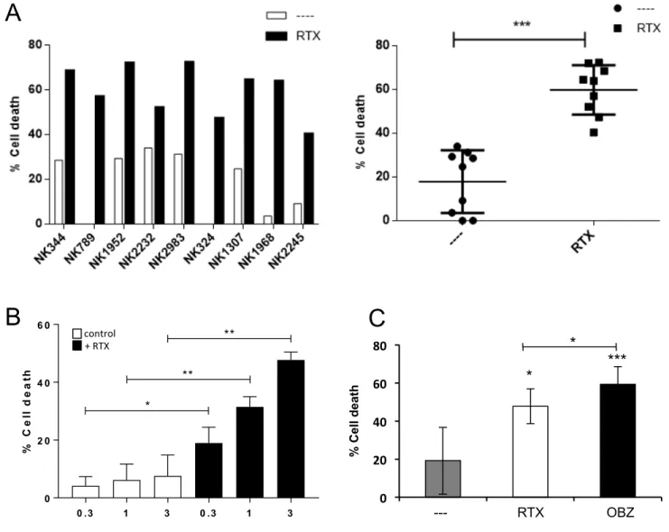
Figure 2. e-NK mediate ADCC with anti-CD20 mAbs. A) PBMCs from CLL patient 148
were incubated for 1h with 10 µg/ml of RTX and overnight with e-NK cells obtained from nine different donors at 1:1 E:T ratio. Cell death was analyzed by 7AAD staining. The right panel shows the statistical analysis. B) 7 e-NKs were tested against 13 CD20+ target samples at different E:T ratios as described in (A). C) 5 e-NKs were tested against 6 CD20+ target samples at different 1:1 E:T ratio as described in (A).
Figure 3. Anti-CD20-armed e-NK show ADCC activity. PBMCs from CLL samples were
incubated for 1h with 10 µg/ml of RTX and overnight with donor e-NK cells at 3:1 E:T ratio (antibody-coated target cells condition). Alternatively, e-NK cells were incubated for 1 h with 10 µg/ml of RTX before incubating them overnight with target cells (antibody-armed NK cell condition). B-CLL cell death was analyzed by 7-AAD.
Figure 4. ADCC requires degranulation and LFA-1/ICAM interaction. A-B) Daudi cells
were incubated overnight with e-NK cells from two different donors and/or RTX (10 µg/ml) as described in Fig. 2. Cytotoxic assays were performed also in the presence of 1 mM EGTA (A) or 15 µg/ml D1D2 protein (B). Cell death was analyzed by 7-AAD staining. C) e-NK produce ADCC with daratumumab. Three different e-NK cell productions were tested against the CD38+ cell line MM.1S and BCL-P2 cells that expressed CD38. Target cells were
pre-incubated for 1 h with 5 µg/ml daratumumab before overnight incubation at different E:T ratios with e-NK. Cell death was analyzed by 7-AAD staining.
Figure 5. e- NK perform ADCC with cetuximab and trastuzumab against EGFR and HER2 positive cell lines, respectively. A) Tumor cells were incubated with 5 µg/ml
cetuximab for 1 h and overnight with 4 e-NK preparations at 3:1 E:T ratio. Subsequently, we measured cell viability (MTT) and cell death (annexin-V/PI). B) Tumor cells were incubated with 5 µg/ml trastuzumab for 1 h and overnight with e-NK cells from 4 different donors at 3:1 E:T ratio. Subsequently, we measured cell viability (MTT). C) 1x10⁴ SK-OV-3 cells were plated and 24h later treated with trastuzumab and cultured with 5x10⁵ NK cells. After 6 days, medium was removed and cells were fixed and stained. The right graph shows the statistical analysis comparing cells incubated with e-NK alone or with trastuzumab. Two areas of 2 different experiments were counted and the average of cells/area depicted in the graphic.
Figure 6. e-NK show ADCC in vivo and overcome mechanisms of drug resistance. A) 5
NSG mice/group were subcutaneously engrafted with BCLP2 (left) or 10x106 (right) cells and treated with e-NK and/or RTX. B) e-NK cell-induced ADCC overcome anti-apoptotic mechanisms of drug resistance. CD20+ MEC-1 cells overexpressing BCL-X
L and MCL1 were
incubated with of RTX (10 µg/ml). After 1 hour, e-NK cells from 3 different donors were added overnight. Cell death was analyzed by 7AAD/annexin-V labeling.
Figure 1
D
E
A
B
E:TC
E:T E:T E:T
E:T E:T E:T
Figure 2
A
C
B
0 .3 1 3 0 .3 1 3 0 2 0 4 0 6 0 E :T % C e ll d e a th * ** ** C O N T R O L + R IT control + RTX 0 20 40 60 80 % Cell d ea th * * *** RTX OBZ ---Figure 3
0 5 1 0 1 5 2 0 2 5 % C e ll d e a th N K c e lls A n tib o d y -c o a te d ta rg e t c e lls A n tib o d y -a rm e d N K c e lls C L L P 4 2 1 C L L P 5 2 0 C L L P 4 2 4 C L L P 5 7 0 C L L P 5 7 6 * * * * * * * * * * * * * * * 0 5 1 0 1 5 2 0 2 5 % C e ll d e a th N K c e lls A n tib o d y -c o a te d ta rg e t c e lls A n tib o d y -a rm e d N K c e lls * * * * * * * *A
B
0 5 1 0 1 5 2 0 2 5 % C e ll d e a th N K c e lls A n tib o d y -c o a te d ta rg e t c e lls A n tib o d y -a rm e d N K c e lls N K 3 1 6 8 * * * * * * * * * * * * * * * N K 3 5 3 3 N K 2 9 0 3 N K 3 4 6 4C
Antibody-coated target cells Antibody-armed NK cells
A
B
2 0 4 0 6 0 8 0 B C L P 2 % C e ll d e a th * * * 2 0 4 0 6 0 M M .1 S % C e ll d e a th * * * * * 0 2 0 4 0 6 0 le g e n d % C e ll d e a th * * * * * -+ D A R AC
0 .3 1 3 0 .3 1 3 0 .3 1 3 0 .3 1 3 0 .3 1 3 0 .3 1 3 0 .3 1 3 0 .3 1 3 0 1 0 2 0 3 0 4 0 5 0 E : T % C e ll d e a th N K 2 9 2 8 N K 2 9 2 7 0 .3 1 3 0 .3 1 3 0 .3 1 3 0 .3 1 3 0 .3 1 3 0 .3 1 3 0 .3 1 3 0 .3 1 3 0 1 0 2 0 3 0 4 0 5 0 E : T % C e ll d e a th N K 2 9 2 8 N K 2 9 2 7EGTA
D1D2
Figure 4
N K 1 N K + R T X3 1 N K ( E G T A)N K N K 0 .3N K ( E G T A) + R T X0 .3 1 3 0 .3 1 3 0 .3 1 3 0 .3 1 3 0 1 0 2 0 3 0 4 0 5 0 E : T % C e ll d e a th 2 9 2 8 D A U D I N K s N K + R IT N K E G T A N K E G T A + R IT N K N K + R T X N K (E G T A ) N K (E G T A )+ R T X N K 1 N K + R T X3 1 N K ( D 1 D 2 )N K N K 0 .3N K ( D 1 D 2 ) + R T X0 .3 1 3 0 .3 1 3 0 .3 1 3 0 .3 1 3 0 1 0 2 0 3 0 4 0 5 0 E : T % C e ll d e a th 2 9 2 8 D A U D I N K s N K + R IT N K E G T A N K E G T A + R IT N K N K + R T X N K (D 1 D 2 ) N K (D 1 D 2 )+ R T XFigure 5
--- C E T --- C E T 0 2 0 4 0 6 0 8 0 1 0 0 % C e ll v ia b il it y C A L U -1 A 5 4 9 * --- T R A S T --- T R A S T 0 2 0 4 0 6 0 8 0 1 0 0 % C e ll v ia b il it y S K B R 3 A 5 4 9 *A
B
C
Control Trastuzumab NK cells --- C E T --- C E T 0 2 0 4 0 6 0 8 0 1 0 0 % C e ll v ia b il it y C A L U -1 A 5 4 9 * * --- + C E T --- + C E T 0 1 0 2 0 3 0 4 0 % C e ll d e a th * * * --- T R A S T --- T R A S T 0 2 0 4 0 6 0 8 0 1 0 0 % C e ll v ia b il it y S K B R 3 A 5 4 9 * * * * **Figure 6
A
B
0 .3 1 3 0 .3 1 3 0 .3 1 3 0 .3 1 3 0 .3 1 3 0 .3 1 3 0 2 0 4 0 6 0 E :T % C e ll d e a th C O N T R O L + R IT M E C -1 M E C - 1 B C L -X L M E C -1 M C L -1 * * * * * * * * * * * 0 .3 1 3 0 .3 1 3 0 .3 1 3 0 .3 1 3 0 .3 1 3 0 .3 1 3 0 2 0 4 0 6 0 E :T % C e ll d e a th C O N T R O L + R T X M E C -1 M E C - 1 B C L -X L M E C -1 M C L -1 * * * * * * * * * * * 1 5 2 0 2 5 3 0 3 5 0 2 0 0 4 0 0 6 0 0 8 0 0 D a y s fr o m tu m o r in je c tio n T u m o r v o lu m e ( m m 3) C O N T R O L R T X N K s N K s + R T X * * * * * * 1 0 1 5 0 2 0 0 4 0 0 6 0 0 8 0 0 1 0 0 0 D a y s fr o m tu m o r in je c tio n T u m o r v o lu m e ( m m 3) C O N T R O L R T X N K s N K s + R T X * * * * * 5 x 1 06 B C L P 2NK cell CD16 NK Expansion Antibody-armed NK Antibody-coated target cells Degranulation Degranulation Antigen mAb Target cell