HAL Id: hal-01656511
https://hal.archives-ouvertes.fr/hal-01656511
Submitted on 14 Mar 2019
HAL is a multi-disciplinary open access
archive for the deposit and dissemination of
sci-entific research documents, whether they are
pub-lished or not. The documents may come from
teaching and research institutions in France or
abroad, or from public or private research centers.
L’archive ouverte pluridisciplinaire HAL, est
destinée au dépôt et à la diffusion de documents
scientifiques de niveau recherche, publiés ou non,
émanant des établissements d’enseignement et de
recherche français ou étrangers, des laboratoires
publics ou privés.
Distributed under a Creative Commons Attribution - NonCommercial| 4.0 International
Antiplatelet Drug Regimen in Patients With Stent
Thrombosis – Insights From the PESTO French
Optical Coherence Tomography Registry –
Nicolas Amabile, Guillaume Cayla, Pascal Motreff, Charlotte Trouillet,
Grégoire Range, Olivier Dubreuil, Estelle Vautrin, François Derimay, Lionel
Mangin, Nicolas Meneveau, et al.
To cite this version:
Nicolas Amabile, Guillaume Cayla, Pascal Motreff, Charlotte Trouillet, Grégoire Range, et al..
An-tiplatelet Drug Regimen in Patients With Stent Thrombosis – Insights From the PESTO French
Optical Coherence Tomography Registry –. Circulation Journal, Japanese Circulation Society, 2017,
81 (10), pp.1469 - 1476. �10.1253/circj.CJ-17-0181�. �hal-01656511�
doi: 10.1253/circj.CJ-17-0181
Thrombosis) French multicenter prospective observational registry reported that a stent mechanical abnormality was present in 97% of subjects with ST explored by intracoro-nary optical coherence tomography (OCT) imaging.5
Premature discontinuation and incomplete individual response to APT have previously been reported as major risk factors for ST.6,7 However, the interactions between
APT regimen and potential underlying stent abnormalities remain largely unknown, even though different mechanical substrates could favor a rheological environment that might promote thrombosis. For example, uncovered/poorly healed struts behave as foreign bodies that induce platelet
A
lthough its frequency has decreased over the recent past years, stent thrombosis (ST) remains a major complication of percutaneous coronary interven-tions (PCI) and still carries a high mortality.1 ST is amul-tifaceted process that can be triggered by different factors such as the patient’s clinical characteristics, efficacy of anti-platelet therapy (APT) or underlying mechanical stent abnormalities.2 However, coronary angiography has
lim-ited sensitivity to correctly identify these latter mechanisms and recent studies suggest that intracoronary imaging is a valuable option to achieve this purpose.3,4 Therefore, the
PESTO (Morphological Parameters Explaining Stent
Received February 22, 2017; revised manuscript received April 19, 2017; accepted April 25, 2017; released online May 23, 2017 Time for primary review: 27 days
Cardiology Department, Institut Mutualiste Montsouris, Paris (N.A., C.C.); Cardiology Department, CHU Nimes, Nimes (G.C.); Cardiology Department, CHU Clermont-Ferrand, Clermont-Ferrand (P.M., G.S.); Cardio Vascular Interventional Therapy and Imaging, Image Science for Interventional Techniques, UMR CNRS 6284, Auvergne University, Clermont-Ferrand (P.M., G.S.); Cardiology Department, GH La Rochelle-Re-Aunis, La Rochelle (C.T.); Cardiology Department, CH Albert Schweitzer, Chartres (G.R.); Cardiology Department, Clinique St Joseph-St Luc, Lyon (O.D.); Cardiology Department, CHU Grenoble, Grenoble (E.V.); Cardiology Department, Hospices Civils de Lyon, Bron (F.D.); Cardiology Department, CH Annecy, Annecy (L.M.); and Cardiology Department, CHU Jean Minjoz, Besançon (N.M.), France
Mailing address: Nicolas Amabile, MD, PhD, Department of Cardiology, Institut Mutualiste Montsouris, 42 Bd Jourdan, 75014 Paris, France. E-mail: nicolas.amabile@imm.fr
ISSN-1346-9843 All rights are reserved to the Japanese Circulation Society. For permissions, please e-mail: cj@j-circ.or.jp
Antiplatelet Drug Regimen in Patients With
Stent Thrombosis
― Insights From the PESTO French Optical
Coherence Tomography Registry ―
Nicolas Amabile, MD, PhD; Guillaume Cayla, MD, PhD; Pascal Motreff, MD, PhD; Charlotte Trouillet, MD; Grégoire Range, MD; Olivier Dubreuil, MD; Estelle Vautrin, MD;
François Derimay, MD; Lionel Mangin, MD; Nicolas Meneveau, MD, PhD; Christophe Caussin, MD; Géraud Souteyrand, MD
Background: Stent thrombosis (ST) may be triggered by different phenomena, including underlying device abnormalities and
modification of the antiplatelet therapy (APT) regimen. This work investigated the characteristics of APT regimens and their relation-ships with ST mechanisms among a large cohort of patients evaluated by optical coherence tomography (OCT).
Methods and Results: A prospective multicenter registry was screened for patients with confirmed ST. OCT was performed after
the initial intervention to the culprit lesion. ST was classified as acute (AST), subacute (SAST), late (LST) and very late (VLST). OCT records were analyzed in a central core laboratory. A total of 120 patients (median age 62 years, 89% male) were included in the study. VLST was the clinical presentation in 75%, LST in 6% and SAST+AST in 19% of the patients. Single APT (SAPT) was given in 61%, double APT (DAPT) in 27% and no APT in 12% of the cases at the time of the ST. A recent (≤15 days) APT modification was reported in 22% of the patients. An underlying mechanical abnormality was identified by OCT in 96.7% of the cases. Ruptured neoatherosclerotic lesions were significantly more frequent in patients without APT compared with the others.
Conclusions: ST mostly occurs in patients receiving DAPT or SAPT. Any underlying mechanical abnormality of ST can be involved,
irrespective of the APT regimen.
Key Words: Antiplatelet drugs; Optical coherence tomography; Stent thrombosis
1470 AMABILE N et al.
Inc./St. Jude Medical, Westford, MA, USA) in all facilities as previously described.14
Offline analysis was performed with proprietary software (LightLab Imaging Inc./St. Jude Medical) after confirming the calibration settings of the Z-offset. All images were recorded digitally, stored and read in a centralized core laboratory by 3 independent investigators blinded to the patients’ clinical and angiographic baseline characteristics, and ST type.5 Discordance around the leading cause of ST
was resolved by consensus.
The methods used for stent analysis have been extensively described elsewhere.5,15 The region of interest, including the
stented as well as the surrounding normal coronary artery segments, was analyzed systematically at 1-mm intervals. The outlines of the stent and lumen were drawn for area measurements. If thrombus with low attenuation was pres-ent, the visible lumen contour could still be drawn behind the thrombus. If thrombus with high attenuation was pres-ent, the lumen contour was allowed to be extrapolated behind the thrombus when the lumen contour was visible in >3 quadrants. The stent area (SA) was measured by join-ing the middle points of the endoluminal signal-rich strut surface of the stent. The intra-stent lumen area and SA were measured for each interval within the stent. Proximal and distal reference lumen areas were also determined. The most “normal-appearing” segments 5 mm proximal and distal to the lesion shoulders identified by OCT were used as the references.
Thrombi were defined as masses attached to the vessel wall or stent and protruding into the vessel lumen, and char-acterized according to consensus documents.16 The
throm-bus score was graded according to the method proposed by the ESC OCT expert review document,17 based on
semi-quantitative assessment of thrombus (number of involved quadrants in the cross-sectional OCT images) and the lon-gitudinal extension of the thrombus itself. Neointimal thick-ness (per strut) and area (per cross-sectional area) were calculated as previously reported.14 An uncovered strut was
defined as a strut of measured neointimal thickness equal to 0 µm.17 In the presence of a highly attenuating layer of
thrombus, struts were categorized as not analyzable for coverage.
The etiologies of mechanical ST were classified as major stent malapposition, severe stent underexpansion, in-stent ruptured neoatherosclerosis, coronary evaginations, isolated uncovered struts without associated abnormality, edge dis-section, edge-related disease progression and intra-stent neointimal hyperplasia with adherent thrombus (Figure).5
A malapposed strut was defined as a strut with a measured distance between its surface and the adjacent vessel surface greater than the strut thickness for bare-metal stents (BMS) or greater than the sum of the thickness of the strut plus polymer for drug-eluting stent (DES).17 The
malap-position was considered significant if the stent lumen dis-tance was ≥200 μm and severe if the disdis-tance was >300 μm.18
Severe stent underexpansion was defined as intra-stent min-imal area ≤70% of the average reference lumen area or ≤80% of the lumen area of the reference segment with the lowest lumen area.19 Neo-atheroma was defined as the
combina-tion of neointimal diffuse thickening with atherosclerotic plaque architectural features including lipid-laden intima, presence of a fibrous cap, neovessels and potential neointi-mal rupture.14 Neointimal rupture referred to a break in
the fibrous cap connecting the lumen with the underlying lipid pool.20 A coronary evagination was defined as the
adhesion and activation of the coagulation cascade.8
Coro-nary evaginations and strut malapposition can affect blood flow within the stent, decrease shear stress and potentially enhance local stasis.9 Neoatherosclerotic lesion formation
results from coverage of struts by an incompetent or poorly maturated endothelium,10 which could also affect in-stent
thrombogenicity.11 However, most of these hypotheses
have yet to be confirmed in vivo.
Our nested OCT study within the PESTO registry aimed to investigate the characteristics of APT regimens in a large cohort of patients with ST explored by OCT.
Methods
PatientsThe PESTO multicenter prospective observational registry included patients treated in 29 French catheterization facil-ities from January 2013 to October 2014. Patients who were referred with acute coronary syndromes (ACS) were prospectively screened for the presence of confirmed ST. The definite diagnosis of ST was made based on the angio-gram and classified as acute ST (AST: 0–24 h after stent implantation), subacute (SAST: >24 h to 30 days after stent implantation), late (LST: >30 days to 1 year after stent implantation) and very late (VLST: >1 year after stent implantation), in accordance with the Academic Research Consortium definitions.12
Patients were included if the following criteria were ful-filled: (1) TIMI 3 flow in the target vessel after initial treat-ment, (2) OCT deemed feasible by the operator, and (3) patient gave written informed consent. Exclusion criteria included: age <18 years, duration of symptoms >12 h, and OCT judged unfeasible by the operator.
The clinical characteristics that were collected at base-line included medical history, and clinical, biological and angiographic parameters. The data were prospectively entered into a predefined standardized case report form.
The study complied with the Declaration of Helsinki. The Ethics Committee of The Gabriel Montpied Univer-sity Hospital approved the research protocol.
ACS Management
All patients included in the study were routinely treated with oral APT including aspirin and clopidogrel or prasug-rel, or ticagrelor. Parenteral antithrombotic therapy included glycoprotein IIb/IIIa inhibitors, and unfractionated or low-weight heparin in accordance with the European Society of Cardiology (ESC) guidelines for management of patients with ST- and non-ST-elevation myocardial infarction (STEMI, NSTEMI).13
PCI was performed with a 6Fr guiding catheter in all patients. The decision whether to use a thromboaspiration device and the options for ST management (culprit lesion deocclusion, balloon dilation alone, redo stenting or med-ical treatment with anticoagulant therapy) were left to the discretion of the local operators. The timing of OCT imaging (immediately following restoration of TIMI 3 flow, or dur-ing subsequent control angiography) was not prespecified by the study protocol and selected by the local investiga-tors according to individual experience and their patients’ clinical and angiographic characteristics.
OCT Image Acquisition and Analysis
Frequency-domain OCT images were acquired using a com-mercially available system (C7 System; LightLab Imaging
in the cohort.
Statistical Analysis
The statistical analysis was performed with SPSS 21.0 soft-ware (SPSS, Chicago, IL, USA). Continuous variables are expressed as median and interquartile ranges, and the nor-mality of their distributions was assessed by the Kolmogorov-Smirnov test. The differences between clinical and OCT parameters among the DAPT, SAPT and no APT groups were compared using χ2 or Fisher exact tests (for
categori-cal variables) and Kruskal-Wallis test (for continuous vari-ables), as appropriate. Incidence of MACE during follow-up was evaluated according to the Kaplan-Meier method in the different groups and compared with the log-rank test. P<0.05 was considered statistically significant.
Results
Baseline CharacteristicsA total of 229 patients in 17 active centers were treated for ST during the inclusion period; 134 patients were screened, and of these 123 provided informed consent. Only 3 patients (2.4%) were excluded from the analysis because of inade-quate OCT image quality.
The population study included 89% of men and the median age was 62 [51–70] years. The treated vessels were large caliber and stent/lesion length was relatively short. The delay between initial PCI and ST was 31.5 [11.6–86.5] months: AST occurred in 4%, SAST in 15%, LST in 6% and VLST in 75% of the cases. A DES or BMS stent was used in 71 (59%) and 47(39%) patients, respectively. A bio-resorbable vascular scaffold (BVS) thrombosis was used in presence of an outward bulge in the luminal vessel contour
between apposed struts with a maximum depth of the bulge exceeding that of the actual strut thickness.21 Edge
dissection was defined as a disruption or discontinuity of the endoluminal vessel surface connecting to the proximal or distal stent edge.22 Edge-related disease progression was
defined as the presence of a ruptured necrotic core plaque at the proximal edge of the stent in an incompletely cov-ered lesion.5
Antiplatelet Therapy Regimen Definitions
The APT regimen at the time of ST was prospectively col-lected in the predefined standardized case report form for each patient and identified as double (DAPT), single (SAPT) or no APT. Modifications of APT within the 15 days prior to ST were identified as recent modifications and their underlying causes were defined according to the classifica-tion established by Mehran et al:23 discontinuation
corre-sponded to premature drug cessation based on physician recommendation; disruption corresponded to cessation of APT because of bleeding or non-compliance; and interrup-tion was defined as temporary cessainterrup-tion of APT because of surgical or invasive intervention with reinstitution of APT within 14 days.
Clinical Follow-up
Clinical follow-up was obtained by clinic visits and/or by telephone contact. The incidence of major adverse cardio-vascular events (MACE: cardiocardio-vascular death, non-fatal stroke, non-fatal myocardial infarction and the need for urgent target vessel revascularization) in the 180 days after hospital discharge following ST was prospectively assessed
Figure. Examples of different types of
stent thrombosis (ST) illustrating the diversity of underlying mechanical abnor-malities and antiplatelet therapy (APT) regimens in the study. (A1–A3) Severe stent underexpansion complicated with subacute ST in a 62-year-old man under DAPT. The minimal stent area (3.22 mm2:
A3) is 45% of the proximal reference stent area (7.15 mm2: A1). (B1–B3) Major malapposition complicated with late ST in a 65-year-old man under SAPT. (C1–
C3) Ruptured neoatherosclerotic lesion with thrombus occurring 14 years after BMS implantation in a 61-year-old man with no APT. BMS, bare-metal stent; DAPT, dual APT; SAPT, single APT.
1472 AMABILE N et al.
characteristics, including stent type. Interestingly, there were comparable proportions of patients with BMS and DES in the no APT group. We observed that patients with AST+SAST were under DAPT in 78.4%, SAPT in 17.4% and no APT in 4.3% of the cases. On the other hand, LST+VLST occurred in patients with SAPT in 72.2%, DAPT in 12.4% and no APT in 15.4% of the cases.
APT Regimen Modification and ST
A recent (≤15 days) change in APT regimen was reported in 22% of the patients prior to ST, including switching from DAPT to SAPT in 65% of the cases and from SAPT/ DAPT to no APT in 35% of the subjects. Recent APT change was present in 23.7% of the patients with LST+VLST and 13% of the patients with AST+SAST. The median delay between APT modification and index ST was 4.0 [2–10] days. The main reason for recent APT change was disrup-tion (42% of the cases, all related to non-compliance), fol-lowed by discontinuation (31%) and interruption (27%). Moreover, APT disruption was significantly more frequently 2 (2%) patients.
The initial intervention to the culprit lesion was manual thrombectomy in 101 patients and balloon predilation in 19 subjects. Intravenous glycoprotein IIb–IIIa inhibitor therapy was administered to 89 patients. OCT images were acquired during a deferred procedure in 85 subjects (69%) (median delay between deocclusion and OCT: 4.0 [2–7] days).
ST mostly occurred in patients treated by either SAPT (60.8%) or DAPT (26.7%), leaving 12.5% of patients expe-riencing ST while not taking any antiplatelet drug. The baseline characteristics of the different groups, as well as the anticoagulation and antiplatelet agents used in these patients, are given in Table 1.
The delay between index PCI and ST and the incidence of VLST were significantly longer and higher in patients without APT compared with the other groups. On the other hand, patients with DAPT experienced acute and subacute ST (AST+SAST) more often. There was no other differ-ence between groups regarding the clinical or angiographic
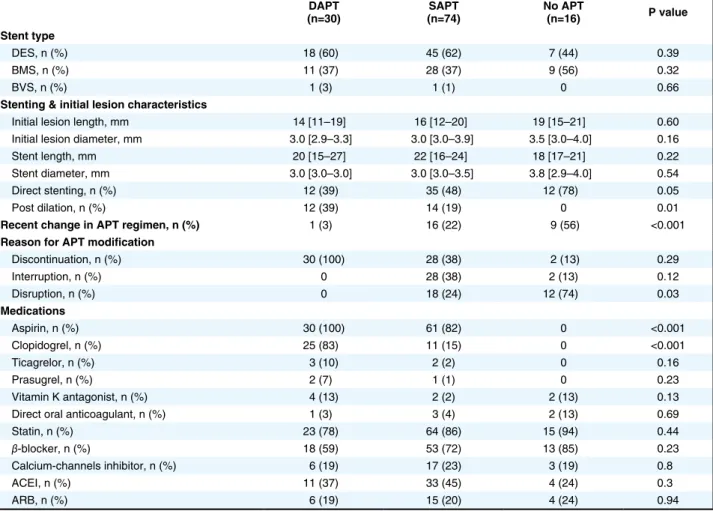
Table 1. Baseline Characteristics of the Different Groups of Patients With Stent Thrombosis DAPT
(n=30) (n=74)SAPT No APT (n=16) P value
Age (years) 63 [51–73] 61 [52–69] 61 [49–67] 0.59
Male sex, n (%) 24 (80) 67 (91) 16 (100) 0.1
Presentation at event
STEMI, n (%) 24 (80) 61 (82) 14 (87) 0.86
NSTEMI, n (%) 6 (20) 13 (18) 2 (13) 0.83
Cardiovascular risk factors
Active smoking, n (%) 11 (37) 24 (33) 8 (50) 0.43 Dyslipidemia, n (%) 21 (70) 61 (82) 15 (93) 0.14 Hypertension, n (%) 18 (60) 38 (52) 11 (67) 0.49 Diabetes, n (%) 11 (37) 18 (24) 4 (25) 0.43 Overweight, n (%) 14 (47) 49 (66) 10 (63) 0.24 Atrial fibrillation, n (%) 4 (13) 4 (5) 1 (6) 0.37
Peripheral artery disease, n (%) 4 (13) 5 (7) 0 0.24
Previous CABG, n (%) 1 (3) 1 (1) 0 0.66
Prior myocardial infarction, n (%) 21 (70) 52 (70) 9 (57) 0.40
Clinical presentation at index PCI
STEMI, n (%) 11 (37) 40 (54) 7 (44) 0.40
NSTEMI or unstable angina, n (%) 10 (33) 13 (18) 5 (31) 0.29
Stable angina, n (%) 9 (30) 21 (28) 4 (25) 0.99
Delay between index PCI and ST (months) 0.33 [0.13–11.6] 42.6 [21.105.5] 79.0 [31.8–99.2] <0.001
AST, n (%) 4 (13) 0 0 0.002 SAST, n (%) 14 (47) 4 (5) 1 (6) <0.001 LST, n (%) 4 (13) 2 (3) 1 (6) 0.11 VLST, n (%) 8 (27) 68 (92) 15 (88) <0.001 Culprit vessel LAD, n (%) 19 (63) 31 (42) 6 (37) 0.10 LCX, n (%) 6 (20) 15 (20) 3 (19) 0.99 RCA, n (%) 5 (17) 28 (38) 7 (44) 0.07 Bifurcation lesion, n (%) 10 (32) 11 (15) 2 (13) 0.13
Initial TIMI flow
Grade 0, n (%) 23 (76) 50 (68) 11 (69) 0.96
Grade 1, n (%) 2 (7) 8 (11) 1 (6)
Grade 2, n (%) 2 (7) 4 (5) 2 (13)
Grade 3, n (%) 3 (10) 12 (16) 2 (13)
OCT Findings According to APT Regimen
An underlying mechanical abnormality was identified by OCT in 116 (96.7%) patients (Table 2). Ruptured neoath-erosclerotic lesions were significantly more frequently observed in patients with no APT compared with the oth-ers, whereas the edge dissection-related ST incidence was higher in patients under DAPT. However, these results observed in patients withdrawing from any APT compared
with those switching to SAPT. We did not observe any significant different baseline characteristics in patients with recent APT change compared with the other subjects (data not shown).
DAPT
(n=30) (n=74)SAPT No APT (n=16) P value
Stent type
DES, n (%) 18 (60) 45 (62) 7 (44) 0.39
BMS, n (%) 11 (37) 28 (37) 9 (56) 0.32
BVS, n (%) 1 (3) 1 (1) 0 0.66
Stenting & initial lesion characteristics
Initial lesion length, mm 14 [11–19] 16 [12–20] 19 [15–21] 0.60
Initial lesion diameter, mm 3.0 [2.9–3.3] 3.0 [3.0–3.9] 3.5 [3.0–4.0] 0.16
Stent length, mm 20 [15–27] 22 [16–24] 18 [17–21] 0.22
Stent diameter, mm 3.0 [3.0–3.0] 3.0 [3.0–3.5] 3.8 [2.9–4.0] 0.54
Direct stenting, n (%) 12 (39) 35 (48) 12 (78) 0.05
Post dilation, n (%) 12 (39) 14 (19) 0 0.01
Recent change in APT regimen, n (%) 1 (3) 16 (22) 9 (56) <0.001
Reason for APT modification
Discontinuation, n (%) 30 (100) 28 (38) 2 (13) 0.29 Interruption, n (%) 0 28 (38) 2 (13) 0.12 Disruption, n (%) 0 18 (24) 12 (74) 0.03 Medications Aspirin, n (%) 30 (100) 61 (82) 0 <0.001 Clopidogrel, n (%) 25 (83) 11 (15) 0 <0.001 Ticagrelor, n (%) 3 (10) 2 (2) 0 0.16 Prasugrel, n (%) 2 (7) 1 (1) 0 0.23 Vitamin K antagonist, n (%) 4 (13) 2 (2) 2 (13) 0.13
Direct oral anticoagulant, n (%) 1 (3) 3 (4) 2 (13) 0.69
Statin, n (%) 23 (78) 64 (86) 15 (94) 0.44
β-blocker, n (%) 18 (59) 53 (72) 13 (85) 0.23
Calcium-channels inhibitor, n (%) 6 (19) 17 (23) 3 (19) 0.8
ACEI, n (%) 11 (37) 33 (45) 4 (24) 0.3
ARB, n (%) 6 (19) 15 (20) 4 (24) 0.94
ACEI, angiotensin-converting enzyme inhibitor; APT, antiplatelet therapy; ARB, angiotensin-receptor blocker; AST, acute stent thrombosis; BMS, bare metal stent; BVS, bioresorbable vascular scaffold; CABG, coronary artery bypass graft; DAPT, double antiplatelet therapy; DES, drug-eluting stent; LAD, left anterior descending artery; LCX, left circumflex artery; LST, late stent thrombosis; NSTEMI, non-ST elevation myocardial infarction; PCI, percutaneous coronary intervention; RCA, right coronary artery; SAPT, single antiplatelet therapy; SAST, subacute stent thrombosis; STEMI, ST elevation myocardial infarction; VLST, very late stent thrombosis.
Table 2. Underlying Mechanical Abnormalities Analyzed by OCT According to Baseline APT Regimen in Patients With Stent Thrombosis
DAPT
(n=30) (n=74)SAPT No APT (n=16) P value
Underlying mechanical abnormality
Malapposition, n (%) 11 (36) 25 (34) 3 (18) 0.43
Ruptured neoatherosclerosis, n (%) 3 (10) 18 (23) 7 (44) 0.03
Underexpansion, n (%) 6 (20) 5 (8) 1 (6) 0.17
Coronary evagination, n (%) 1 (3) 8 (11) 1 (6) 0.43
Edge-related disease progression, n (%) 2 (7) 5 (8) 2 (13) 0.72
Isolated uncovered struts, n (%) 0 8 (11) 2 (13) 0.16
Neointimal hyperplasia, n (%) 2 (7) 4 (5) 0 0.59
Edge dissection, n (%) 2 (7) 0 0 0.05
No cause identified, n (%) 3 (10) 1 (1) 0 0.06
1474 AMABILE N et al.
The thrombogenic potential of a stent-related problem could enhanced by platelet thrombogenicity associated with sys-temic health problems (e.g., inflammation, diabetes) or inad-equate APT therapy (e.g., poor compliance, premature discontinuation or blunted individual response to P2Y12 inhibitors or aspirin).2 Other hypotheses have been proposed
to explain the relationships between stent mechanical fail-ure and local thrombotic factors. Stent malapposition can increase local shear stress.25 which could in turn delay strut
coverage25 and promote platelet activation.26 Coronary
evaginations are suspected to affect flow distribution within and around the stent and favor local blood stasis, a well-identified rheological factor promoting thrombosis.9
Neo-atherosclerotic lesions result from interaction between the device and an incompetent endothelium:10 the necrotic
core contains highly prothrombotic material that could create in situ thrombus formation following in-stent plaque rupture.10 Thus, it could be speculated that the absence of
APT would favor ST occurrence in patients with such stent structural abnormalities. However, our results showed that the vast majority (>80%) of patients in this cohort were receiving APT at the time of the thrombotic event and a morphological abnormality was identified by OCT in almost all patients.5,18
SAPT was the most frequent regimen in this cohort (61.7%) and the vast majority (>90%) of these patients experienced VLST. In this group, malapposition (which can result from an initial fault in the stent implantation or appears over time in relation to vessel remodelling5) was
the most frequent mechanical abnormality, as reported by others.27 The high prevalence of ruptured
neoatheroscle-rotic lesions is also not surprising in this group, because the incidence of this process increases over time10 and the
prob-ability of receiving APT decreases after 1 year.28
Further-more, 26.7% of our subjects suffered ST while on DAPT. In the latter group, complications mostly occurred during the first month (60%) and were essentially related to abnor-malities (acute stent malapposition, underexpansion and edge dissection) that probably resulted from potential pit-falls of the initial PCI. Surprisingly, we also observed that AST+SAST occurred in subjects receiving DAPT in 78% of cases, yet premature discontinuation of APT or clopido-grel withdrawal are reported to be major risk factors for early ST.7,29 Finally, stent thrombosis associated with no
might just reflect the differences in delays between index PCI and ST, as ruptured neoatherosclerotic lesions are more frequently observed in VLST cases (in which patients were more likely to receive SAPT or no APT). Further-more, we observed that patients treated by DAPT had a higher intra-stent thrombotic score, a smaller minimal SA and a higher percentage of uncovered struts compared with subjects with SAPT or no APT. There was no significant differences between red and white thrombus frequencies among the different groups (Table 3) Finally, we did not observe any significant differences in mechanical abnor-malities (Table S1) or thrombus characteristics between patients with recent APT change and the others (respective white thrombus incidence: 85% vs. 92%, P=NS).
Clinical Follow-up
Follow-up data were obtained for 117 (99%) patients. The actuarial freedoms from MACE at 6 months in patients with DAPT, SAPT and no APT did not differ and were 89.9% [95% confidence interval (CI): 84.4–95.4%], 92.8% [95% CI: 89.7–95.9%] and 87.5% [95% CI: 79.2–95.8%], respectively (P=0.74 log-rank test). Moreover, we did not observe sig-nificant differences in outcome among patients with recent APT modification vs. the others (survival from MACE: 87.8% [95% CI: 81.2–94.4%] vs. 92.3% [95% CI: 89.5–95.1%]; P=0.48 log-rank test). Furthermore, in patients with recent APT modification, there was no difference in outcome in cases of disruption vs. discontinuation+interruption (sur-vival from MACE: 86.7% [95% CI: 77.9–95.5%] vs. 86.7% [95% CI: 79.6–93.8%]; P=0.96 log-rank test).
Discussion
The main findings of this subanalysis of the PESTO national multicenter registry were: (1) ST mostly occurred in patients receiving APT (87.5% of the study population) and very late thrombosis was the predominant presentation; (2) A recent modification of APT regimen was reported in 22% of the subjects prior to the thrombotic event; and (3) dis-ruption related to poor compliance was the main reason for APT modification.
ST is a complex process resulting from the interaction of different factors including patient’s characteristics, platelet adhesiveness and mechanical problems with the stent(s).4,24
Table 3. Thrombus and Quantitative OCT Parameters According to Baseline APT Regimen of Patients With Stent Thrombosis DAPT
(n=30) (n=74)SAPT No APT (n=16) P value
Thrombotic score 22 [11–37] 13 [5–27] 8 [5–26] 0.04
Red thrombus, n (%) 2 (7) 8 (11) 2 (13) 0.76
White thrombus, n (%) 28 (93) 66 (89) 14 (87) 0.99
No. of struts analyzed per lesion 180 [149–262] 204 [139–267] 171 [122–233] 0.58
Minimal luminal intra-stent area (mm2) 4.6 [2.4–5.8] 3.3 [2.4–5.2] 3.6 [1.7–5.0] 0.38
Mean luminal intra-stent area (mm2) 5.1 [3.8–6.9] 5.7 [4.1–7.0] 4.7 [3.4–7.5] 0.71
Minimal stent area (mm2) 4.3 [3.6–6.4] 5.5 [4.2–6.8] 6.8 [5.4–8.0] 0.02
Mean stent area (mm2) 6.4 [5.1–7.9] 6.7 [5.7–8.2] 7.6 [6.3–9.4] 0.10
Mean neointimal thickness (μm) 70 [15–234] 225 [142–282] 333 [132–550] 0.01
Malapposed struts (%) 4 [0–11] 3.8 [0–13.6] 0.6 [0–6.7] 0.28
Maximal malapposition (μm) 240 [0–668] 360 [0–805] 0 [0–443] 0.21
Uncovered struts (%) 36.7 [8.2–74.6] 16.4 [2.3–37.1] 6.9 [0–31.2] 0.007
in the study because of an inadequate clinical presentation or impossibility to restore TIMI 3 flow without PCI before OCT image acquisition. Thus, our results might be biased by patient selection and may not reflect the true incidence of the different ST mechanisms in the general population (229 cases of ST during this period in the 17 active centers). Furthermore, most of the OCT data in the present study were acquired during a deferred procedure following the initial deocclusion and potential anti GPIIb–IIIa infusion, according to the operators’ discretion. This strategy might have positively influenced the image quality, as the throm-bus load decreases over time under these conditions.37 This
could explain the low rate of OCT acquisitions that were rejected for inadequate quality by the core laboratory. Hence, our data might differ from series in which the intra-coronary imaging analysis was not deferred and thus make generalization of our conclusions more difficult. Finally, the PESTO registry was designed to analyze the incidence of ST characteristics in the French population. There was no control group of patients with non-thrombotic stents analyzed with OCT that would have allowed us to specifi-cally investigate the predictive factors for ST. However, the study was not designed to achieve this specific aim.
Conclusions
In conclusion, our results showed that the majority of ST cases occurred under DAPT or SAPT. All types of stents and mechanisms of ST can be involved, irrespective of the APT regimen. A recent modification of ATP regimen could favor ST but might have different effects according to the mode of cessation and clinical factors that remain to be identified by future studies. In the light of these data, the decision to modify APT in patients with previous stents should be made cautiously to prevent adverse events.
Disclosures
G.S., P.M., C.C., N.M. and N.A. have received consulting fees from St. Jude Medical. G.S. and P.M. have received consulting fees from Terumo.
Source of Funding
This work was funded by an educational grant from Medtronic and St. Jude Medical.
References
1. Yeo KK, Armstrong EJ, Soni K, Waldo SW, Patel M, Reeves R, et al. Long-term outcomes of angiographically confirmed coronary stent thrombosis: Results from a multicentre California registry.
EuroIntervention 2015; 11: 188 – 195.
2. Byrne RA, Joner M, Kastrati A. Stent thrombosis and restenosis: What have we learned and where are we going?: The Andreas Grüntzig Lecture ESC 2014. Eur Heart J 2015; 36: 3320 – 3331. 3. Alfonso F, Dutary J, Paulo M, Gonzalo N, Perez-Vizcayno MJ,
Jimenez-Quevedo P, et al. Combined use of optical coherence tomography and intravascular ultrasound imaging in patients undergoing coronary interventions for stent thrombosis. Heart 2012; 98: 1213 – 1220.
4. Claessen BE, Henriques JPS, Jaffer FA, Mehran R, Piek JJ, Dangas GD. Stent thrombosis: A clinical perspective. J Am Coll
Cardiol Intv 2014; 7: 1081 – 1092.
5. Souteyrand G, Amabile N, Mangin L, Chabin X, Meneveau N, Cayla G, et al. Mechanisms of stent thrombosis analysed by opti-cal coherence tomography: Insights from the national PESTO French registry. Eur Heart J 2016; 37: 1208 – 1216.
6. Cayla G, Hulot JS, O’Connor SA, Pathak A, Scott SA, Gruel Y, et al. Clinical, angiographic, and genetic factors associated with early coronary stent thrombosis. JAMA 2011; 306: 1765 – 1774. 7. Iakovou I, Schmidt T, Bonizzoni E, Ge L, Sangiorgi GM,
APT was essentially VLST, with a significantly longer delay and associated with various stents abnormalities. In this latter group, the proportions of BMS and DES were com-parable. We observed that ruptured neoatherosclerotic lesions were more frequently identified in patients with no APT compared with patients with SAPT or DAPT. These results might be, once again, explained by the longer delay between initial PCI and ST in patients without aspirin and P2Y12 inhibitors compared with the others (see above). Thus, we cannot draw firm conclusions between ST, APT regimen and any particular stent structural problem from the current data. The incidence of morphological abnor-mality was high, irrespective of APT regimen, and might thus represent a major risk factor for ST, as previously suggested by others.30
We identified a recent modification of the APT regimen prior to ST in 22% of patients (essentially patients under SAPT or no APT at the time of the ST), which was related to treatment disruption in 42% of these cases. In the PARIS registry, DAPT cessation was observed in 54.2% of patients within the first 2 years following PCI and was mainly driven by physician-guided discontinuation. However, Mehran et al23 reported that the effect of DAPT cessation on cardiac
risk after PCI is not uniform but varies substantially based on the underlying mode; that is, DAPT disruption was associated with an increased risk for ST, whereas discon-tinuation and interruption were not.23 Although the designs
and objectives of the 2 studies could not be compared, our data are in line with the results from the PARIS registry, where a recent APT modification (mostly disruption) was only found in a minority of subjects with ST (14/71 cases, i.e. 20%).23 The present results could not, however, confirm
the deleterious effect of APT disruption on ST onset or investigate the potential mechanisms supporting this hypoth-esis. Interestingly, the median delay between APT change and ST was 4 days in our series, irrespective of ST type (acute, subacute, late, very late), which is in line with some previous reports.23,30 The biological reasons underlying this
observation remain unidentified. Although “rebound” plate-let adhesiveness in patients under long-term APT, mainly related to clopidogrel withdrawal and subsequent conse-quences (loss of platelet inhibition, enhanced platelet acti-vation and increased inflammation) has been suspected,31–34
other data suggest no significant biological effect of APT cessation in the general population.35,36 Moreover, the
pres-ent analysis did not idpres-entify a relationship between any stent structural abnormality and ST in these patients: there was no significant difference in the underlying mechanisms in subjects with recent APT modification compared with the others. Whether the effect of APT changes is different in patients with underlying stent mechanical abnormalities compared with those without also remains, in the light of these data, unknown.
Study Limitations
Several limitations of this study deserve consideration. First, ST is a multifactorial process that potentially involves a stent architecture abnormality and clinical factors, includ-ing the degree of platelet inhibition under therapy and com-pliance with treatment. Although we evaluated declared compliance with APT, we do not have data regarding platelet function or genetic testing for all our population, because the different tests were not performed routinely in the different centers. Moreover, a substantial number of patients presenting with ST were not screened for inclusion
1476 AMABILE N et al.
events after percutaneous coronary intervention (PARIS): 2 year results from a prospective observational study. Lancet 2013; 382: 1714 – 1722.
24. Nakagawa Y. Speculative mechanisms for very late stent throm-bosis after drug-eluting stent implantation. Circ J 2011; 75: 779 – 780.
25. Foin N, Gutiérrez-Chico JL, Nakatani S, Torii R, Bourantas CV, Sen S, et al. Incomplete stent apposition causes high shear flow disturbances and delay in neointimal coverage as a function of strut to wall detachment distance: Implications for the manage-ment of incomplete stent apposition. Circ Cardiovasc Interv 2014;
7: 180 – 189.
26. Miyazaki Y, Nomura S, Miyake T, Kagawa H, Kitada C, Taniguchi H, et al. High shear stress can initiate both platelet aggregation and shedding of procoagulant containing micropar-ticles. Blood 1996; 88: 3456 – 3464.
27. Taniwaki M, Radu MD, Zaugg S, Amabile N, Garcia-Garcia HM, Yamaji K, et al. Mechanisms of very late drug-eluting stent thrombosis assessed by optical coherence tomography.
Circula-tion 2016; 133: 650 – 660.
28. Yano M, Natsuaki M, Morimoto T, Nakagawa Y, Kawai K, Miyazaki S, et al. Antiplatelet therapy discontinuation and stent thrombosis after sirolimus-eluting stent implantation: Five-year outcome of the j-Cypher Registry. Int J Cardiol 2015; 199: 296 – 301.
29. van Werkum JW, Heestermans AA, Zomer AC, Kelder JC, Suttorp MJ, Rensing BJ, et al. Predictors of coronary stent thrombosis: The Dutch stent thrombosis registry. J Am Coll Cardiol 2009; 53: 1399 – 1409.
30. Waksman R, Kirtane AJ, Torguson R, Cohen DJ, Ryan T, Räber L, et al. Correlates and outcomes of late and very late drug-eluting stent thrombosis: Results from DESERT (International Drug-Eluting Stent Event Registry of Thrombosis). J Am Coll
Cardiol Intv 2014; 7: 1093 – 1102.
31. Sambu N, Dent H, Englyst N, Warner TD, Leadbeater P, Roderick P, et al. Effect of clopidogrel withdrawal on platelet reactivity and vascular inflammatory biomarkers 1 year after drug-eluting stent implantation: Results of the prospective, single-centre CESSATION study. Heart 2011; 97: 1661 – 1667.
32. Sambu N, Warner T, Curzen N. Clopidogrel withdrawal: Is there a “rebound” phenomenon? Thromb Haemost 2011; 105: 211 – 220. 33. Mylotte D, Peace AJ, Tedesco AT, Mangiacapra F, Dicker P,
Kenny D, et al. Clopidogrel discontinuation and platelet reactivity following coronary stenting. J Thromb Haemost 2011; 9: 24 – 32. 34. Aarnoudse W, Fearon WF, Manoharan G, Geven M, van de
Vosse F, Rutten M, et al. Epicardial stenosis severity does not affect minimal microcirculatory resistance. Circulation 2004; 110: 2137 – 2142.
35. Sibbing D, Stegherr J, Braun S, Mehilli J, Schulz S, Seyfarth M, et al. A double-blind, randomized study on prevention and exis-tence of a rebound phenomenon of platelets after cessation of clopidogrel treatment. J Am Coll Cardiol 2010; 55: 558 – 565. 36. Ford I, Scott NW, Herd V, Mitchell LR, Williams DJP, Brittenden
J. A randomized controlled trial of platelet activity before and after cessation of clopidogrel therapy in patients with stable cardiovascular disease. J Am Coll Cardiol 2014; 63: 233 – 239. 37. Amabile N, Hammas S, Fradi S, Souteyrand G, Veugeois A,
Belle L, et al. Intra-coronary thrombus evolution during acute coronary syndrome: Regression assessment by serial optical coherence tomography analyses. Eur Heart J Cardiovasc Imaging 2015; 16: 433 – 440.
Supplementary Files Supplementary File 1
Table S1. Underlying mechanical abnormalities analyzed by OCT
according to recent change in APT regimen in patients with stent thrombosis
Please find supplementary file(s); http://dx.doi.org/10.1253/circj.CJ-17-0181 Stankovic G, et al. Incidence, predictors, and outcome of
throm-bosis after successful implantation of drug-eluting stents. JAMA 2005; 293: 2126 – 2130.
8. Lüscher TF, Steffel J, Eberli FR, Joner M, Nakazawa G, Tanner FC, et al. Drug-eluting stent and coronary thrombosis: Biological mechanisms and clinical implications. Circulation 2007; 115: 1051 – 1058.
9. Radu MD, Pfenniger A, Räber L, De Marchi SF, Obrist D, Kelbak H, et al. Flow disturbances in stent-related coronary evaginations: A computational fluid-dynamic simulation study.
EuroIntervention 2014; 10: 113 – 123.
10. Otsuka F, Byrne RA, Yahagi K, Mori H, Ladich E, Fowler DR, et al. Neoatherosclerosis: Overview of histopathologic findings and implications for intravascular imaging assessment. Eur Heart
J 2015; 36: 2147 – 2159.
11. Otsuka F, Finn AV, Yazdani SK, Nakano M, Kolodgie FD, Virmani R. The importance of the endothelium in atherothrom-bosis and coronary stenting. Nat Rev Cardiol 2012; 9: 439 – 453. 12. Cutlip DE, Windecker S, Mehran R, Boam A, Cohen DJ, van Es
GA, et al. Clinical end points in coronary stent trials. Circulation 2007; 115: 2344 – 2351.
13. Steg PG, James SK, Atar D, Badano LP, Blomstrom-Lundqvist C, Borger MA, et al. ESC Guidelines for the management of acute myocardial infarction in patients presenting with ST-segment elevation. Eur Heart J 2012; 33: 2569 – 2619.
14. Amabile N, Souteyrand G, Ghostine S, Combaret N, Slama MS, Barber-Chamoux N, et al. Very late stent thrombosis related to incomplete neointimal coverage or neoatherosclerotic plaque rupture identified by optical coherence tomography imaging. Eur
Heart J Cardiovasc Imaging 2014; 15: 24 – 31.
15. Amabile N, Trouillet C, Meneveau N, Tissot CM, Belle L, Combaret N, et al. Mechanical abnormalities associated with first- and second-generation drug-eluting stent thrombosis analyzed by optical coherence tomography in the national PESTO French registry. Int J Cardiol 2017; 227: 161 – 165.
16. Tearney GJ, Regar E, Akasaka T, Adriaenssens T, Barlis P, Bezerra HG, et al. Consensus standards for acquisition, measure-ment, and reporting of intravascular optical coherence tomogra-phy studies: A report from the International Working Group for Intravascular Optical Coherence Tomography Standardization and Validation. J Am Coll Cardiol 2012; 59: 1058 – 1072. 17. Prati F, Guagliumi G, Mintz GS, Costa M, Regar E, Akasaka
T, et al. Expert review document part 2: Methodology, terminology and clinical applications of optical coherence tomography for the assessment of interventional procedures. Eur Heart J 2012; 33: 2513 – 2520.
18. Prati F, Kodama T, Romagnoli E, Gatto L, Di Vito L, Ramazzotti V, et al. Suboptimal stent deployment is associated with subacute stent thrombosis: Optical coherence tomography insights from a multicenter matched study. From the CLI Foundation investiga-tors: The CLI-THRO study. Am Heart J 2015; 169: 249 – 256. 19. Meneveau N, Ecarnot F, Souteyrand G, Motreff P, Caussin C,
Van Belle E, et al. Does optical coherence tomography optimize results of stenting?: Rationale and study design. Am Heart J 2014;
168: 175 – 181, e171 – e172.
20. Kang SJ, Mintz GS, Akasaka T, Park DW, Lee JY, Kim WJ, et al. Optical coherence tomographic analysis of in-stent neoathero-sclerosis after drug-eluting stent implantation. Circulation 2011;
123: 2954 – 2963.
21. Radu MD, Raber L, Kalesan B, Muramatsu T, Kelbaek H, Heo J, et al. Coronary evaginations are associated with positive vessel remodelling and are nearly absent following implantation of newer-generation drug-eluting stents: An optical coherence tomography and intravascular ultrasound study. Eur Heart J 2014; 35: 795 – 807.
22. Muramatsu T, Garcia-Garcia HM, Onuma Y, Zhang YJ, Bourantas CV, Diletti R, et al. Intimal flaps detected by optical frequency domain imaging in the proximal segments of native coronary arteries. Circ J 2013; 77: 2327 – 2333.
23. Mehran R, Baber U, Steg PG, Ariti C, Weisz G, Witzenbichler B, et al. Cessation of dual antiplatelet treatment and cardiac
![Table 1. Baseline Characteristics of the Different Groups of Patients With Stent Thrombosis DAPT (n=30) SAPT (n=74) No APT (n=16) P value Age (years) 63 [51–73] 61 [52–69] 61 [49–67] 0.59 Male sex, n (%) 24 (80) 67 (91) 16 (100) 0.1](https://thumb-eu.123doks.com/thumbv2/123doknet/14641471.549380/5.879.83.805.444.1076/table-baseline-characteristics-different-groups-patients-stent-thrombosis.webp)
![Table 3. Thrombus and Quantitative OCT Parameters According to Baseline APT Regimen of Patients With Stent Thrombosis DAPT (n=30) SAPT (n=74) No APT (n=16) P value Thrombotic score 22 [11–37] 13 [5–27] 8 [5–26] 0.04 Red thrombus, n (%) 2 (](https://thumb-eu.123doks.com/thumbv2/123doknet/14641471.549380/7.879.83.807.96.337/thrombus-quantitative-parameters-according-baseline-patients-thrombosis-thrombotic.webp)
