HAL Id: inserm-02398221
https://www.hal.inserm.fr/inserm-02398221
Submitted on 7 Dec 2019
HAL is a multi-disciplinary open access
archive for the deposit and dissemination of
sci-entific research documents, whether they are
pub-lished or not. The documents may come from
teaching and research institutions in France or
abroad, or from public or private research centers.
L’archive ouverte pluridisciplinaire HAL, est
destinée au dépôt et à la diffusion de documents
scientifiques de niveau recherche, publiés ou non,
émanant des établissements d’enseignement et de
recherche français ou étrangers, des laboratoires
publics ou privés.
an abnormal capillary remodelling in the skeletal muscle
of COPD patients
Léo Blervaque, Emilie Passerieux, Pascal Pomiès, Matthias Catteau, Nelly
Héraud, Marine Blaquière, François Bughin, Bronia Ayoub, Nicolas Molinari,
Jean-Paul Cristol, et al.
To cite this version:
Léo Blervaque, Emilie Passerieux, Pascal Pomiès, Matthias Catteau, Nelly Héraud, et al.. Impaired
training-induced angiogenesis process with loss of pericyte-endothelium interactions is associated with
an abnormal capillary remodelling in the skeletal muscle of COPD patients. Respiratory Research,
BioMed Central, 2019, 20 (1), pp.278. �10.1186/s12931-019-1240-6�. �inserm-02398221�
R E S E A R C H
Open Access
Impaired training-induced angiogenesis
process with loss of pericyte-endothelium
interactions is associated with an abnormal
capillary remodelling in the skeletal muscle
of COPD patients
Léo Blervaque
1*, Emilie Passerieux
1, Pascal Pomiès
1, Matthias Catteau
1, Nelly Héraud
2, Marine Blaquière
3,
François Bughin
3, Bronia Ayoub
3, Nicolas Molinari
4, Jean-Paul Cristol
3, Antonia Perez-Martin
5, Jacques Mercier
3,
Maurice Hayot
3and Fares Gouzi
3Abstract
Chronic obstructive pulmonary disease (COPD) is associated with exercise intolerance and limits the functional gains in response to exercise training in patients compared to sedentary healthy subjects (SHS). The blunted skeletal muscle angiogenesis previously observed in COPD patients has been linked to these limited functional improvements, but its underlying mechanisms, as well as the potential role of oxidative stress, remain poorly understood. Therefore, we compared ultrastructural indexes of angiogenic process and capillary remodelling by transmission electron microscopy in 9 COPD patients and 7 SHS after 6 weeks of individualized moderate-intensity endurance training. We also assessed oxidative stress by plasma-free and esterified isoprostane (F2-IsoP) levels in
both groups. We observed a capillary basement membrane thickening in COPD patients only (p = 0.008) and abnormal variations of endothelial nucleus density in response to exercise training in these patients when compared to SHS (p = 0.042). COPD patients had significantly fewer occurrences of pericyte/endothelium
interdigitations, a morphologic marker of capillary maturation, than SHS (p = 0.014), and significantly higher levels of F2-IsoP (p = 0.048). Last, the changes in pericyte/endothelium interdigitations and F2-IsoP levels in response to
exercise training were negatively correlated (r =− 0.62, p = 0.025). This study is the first to show abnormal capillary remodelling and to reveal impairments during the whole process of angiogenesis (capillary creation and
maturation) in COPD patients.
Trial registration:NCT01183039&NCT01183052, both registered 7 August 2010 (retrospectively registered). Keywords: COPD, Capillaries, Angiogenesis, Skeletal muscle, Exercise training
© The Author(s). 2019 Open Access This article is distributed under the terms of the Creative Commons Attribution 4.0 International License (http://creativecommons.org/licenses/by/4.0/), which permits unrestricted use, distribution, and reproduction in any medium, provided you give appropriate credit to the original author(s) and the source, provide a link to the Creative Commons license, and indicate if changes were made. The Creative Commons Public Domain Dedication waiver (http://creativecommons.org/publicdomain/zero/1.0/) applies to the data made available in this article, unless otherwise stated.
* Correspondence:leo.blervaque@umontpellier.fr 1
Physiologie & médecine expérimentale du Cœur et des Muscles (PhyMedExp), INSERM - CNRS - Montpellier University. CHU Arnaud De Villeneuve, 371 avenue du Doyen Gaston Giraud, 34295 Montpellier cedex 5, France
Background
Chronic obstructive pulmonary disease (COPD) is characterized by a persistent airflow limitation due to airway and/or alveolar abnormalities, and it is system-atically associated with other concomitant chronic diseases [1]. Limb skeletal muscle dysfunction, for ex-ample, affects the morbi-mortality of these patients [2–4]. Yet, skeletal muscle capillarization, despite its key role in this dysfunction in COPD patients [5], is an under-considered explanation of this phenomenon. A previous study of our group nevertheless demon-strated a significantly lower increase in skeletal muscle capillarization in response to exercise training in COPD patients compared with sedentary healthy subjects (SHS) [6]. This blunted angiogenesis was linked to the limited functional gains from exercise training observed in the patients [6]. Thus, while im-paired angiogenesis appears to be relevant to the muscle misadaptations to exercise training in COPD patients, it remains insufficiently understood.
Physiologically, the angiogenic process in response to exercise training is highly dynamic, resulting from a bal-ance between pro- and anti-angiogenic factors. However, two main phases of angiogenesis have been described: an early phase of new capillary creation and a later phase of neo-capillary maturation [7]. The morphologic markers of capillary creation [abluminal endothelial (EC) sprouts or intraluminal EC folding] and maturation [pericyte/endothelium (Pc/EC) interdigitations] in skel-etal muscle were initially described in studies using transmission electron microscopy (TEM) [8–11], which is the reference method for their identification [12, 13]. Capillary remodelling in response to exercise training also appears to be linked to angiogenesis [12], and it too can be assessed by TEM through a morphometric ana-lysis of the main capillary components (lumen, endothe-lium, basement membrane and pericytes) [8].
In chronic diseases like diabetes, both impaired angio-genesis [14] and pathological capillary remodelling [15] have been described in response to exercise training. Oxidative stress is a potential factor of these impair-ments, causing basement membrane protein accumula-tion [16] and pericyte apoptosis [17]. Moreover, oxidative stress promotes isoprostane formation, [18] which is a direct inhibitor of angiogenesis [19] and a promotor of pericyte loss [20]. In COPD patients, in-creased levels of plasma-free and esterified isoprostanes (F2-IsoP) and muscle oxidative stress have been reported
[5, 21, 22]. We thus hypothesized that the blunted angiogenesis in COPD patients in response to exercise training [6] would be associated with abnormal capillary remodelling, an impaired angiogenic process (i.e. capil-lary creation and maturation), and increased plasmatic oxidative stress levels.
Therefore, the aim of this study was to compare capil-lary remodelling and the morphologic markers of the an-giogenic process in skeletal muscle after a 6-week exercise training programme in COPD patients and SHS. In addition, we sought to determine whether the changes in the capillary ultrastructure and the changes in plasmatic levels of F2-IsoP were correlated in both
groups.
Materials and methods
Study design
This study was conducted on skeletal muscle samples from subgroups of both COPD patients and SHS re-cruited in two previous studies (registration numbers for
clinicaltrial.gov: NCT01183039 and NCT01183052). The
subjects were recruited at PhyMedExp (INSERM-CNRS-UM), Montpellier University Hospital, Montpellier, France, or the “La Solane” and “La Vallonie” Pulmonary Rehabilitation Centres in Osseja and Lodève, France. These studies were the subject of a previous publication [Gouzi et al., 2013] [6]. The present study used TEM to more closely investigate the underlying mechanisms of the blunted angiogenesis in response to exercise training found in the main study. The inclusion criteria are avail-able in the previous study [6] and in clinicaltrials.gov/. Briefly, COPD patients were defined by dyspnoea, chronic cough or sputum production and/or a history of exposure to risk factors for the disease, and post-bronchodilator forced expiratory volume in 1 s/forced ventilator capacity ratio < 70% [23]. The exclusion cri-teria were other respiratory diagnoses, decompensated comorbidity, exacerbation within the last 3 months, and previous participation in an exercise training programme. Study protocols followed the European guidelines for good clinical practice and the Declaration of Helsinki.
Pulmonary function assessment
Pulmonary function was assessed using whole-body plethysmography (Transmural Bodybox 2800; Sensome-dics, Yorba Linda, CA, USA). PaO2was measured in
ra-dial artery blood samples from seated COPD patients under room air condition, with a blood gas analyser (Roche OMNIS, Roche Diagnostics, Mannheim, Germany).
Assessment of skeletal muscle function and exercise
The 6-min walking test was conducted in accordance with the ATS statement [24]. COPD patients and SHS also performed an incremental cycloergometric test on a calibrated, electrically-braked cycloergometer (Ergoselect 200P, Ergolyne, Bitz, Germany) in accordance with the ATS/ACCP statement [25]. The mean V̇O2 value over
the last 20 s of the test was used as symptom-limited oxygen uptake (V̇Osl). The endurance time of the quad-riceps (Tlimquad) was assessed at a frequency of 10
movements per minute at 30% of the maximal voluntary contraction, as previously described [6]. The level of physical activity was assessed using the Voorrips ques-tionnaire (modified Baecke’s quesques-tionnaire) [26].
Exercise training programme
The exercise training programme was conducted for 6 weeks with 3 or 4 sessions per week (20 sessions in total) and was supervised by an experienced clinician. The protocol was consistent with the ATS/ERS statements [27]. The sessions lasted 1.5 h, with 45 min of endurance training (10 min of work at the intensity of the ventila-tory threshold followed by 5 min of active recovery, re-peated 3 times) completed by 30 min of strength-building exercise (8–10 exercises, with sets of 10–15 repetitions). The exercise intensity for the endurance ex-ercise was set individually to the heart rate at the venti-latory threshold [28] and continuously monitored during the session with a cardiofrequency meter. The cycloerg-ometer load was increased over the course of the ses-sions to maintain the targeted heart rate. The load for the resistance exercise was set at 40% of the isotonic one-repetition maximum (1-RM) of each muscle (del-toid, biceps, triceps and quadriceps) at the first session and progressively increased, targeting 5–6 out of 10 on an exertion scale [29]. This exercise training programme was part of a multi-component and comprehensive pul-monary rehabilitation programme, and it was completed by six small group education sessions (1–1.5 h each) conducted by an experienced therapist specialized in im-proving health-related behaviours.
Muscle biopsies
Biopsies from the vastus lateralis muscle of the COPD patients and SHS were performed before and after exer-cise training, using the Bergström technique as previ-ously described [6, 30]. Each biopsy was separated into two samples: one part was immediately frozen in isopen-tane, precooled in liquid nitrogen, and stored at − 80 °C for histochemical analysis of muscle capillarization (C/F ratio and fibre type characterization; for more details, see Gouzi et al., 2013 [6]); and one part was fixated with a solution of 2.5% glutaraldehyde in PHEM buffer (1X, pH 7.4) and stored at 4 °C for TEM analysis of capillary ultrastructure. The TEM samples were recut into smaller samples (about 12 mm3) and immersed in a sec-ond fixative solution (0.5% osmic acid) for 2 h in the dark at room temperature. After dehydration (graded series of ethanol, 30–100%), these blocks were embed-ded in EmBed 812 resin using an Automated Microwave
Tissue Processor for Electronic Microscopy (Leica EM AMW) and cut into transversal sections 70 nm thick (Leica-Reichert Ultracut E). These sections were stained with a solution of uranyl acetate and lead citrate and ob-served with a Tecnai F20 transmission electron micro-scope at 120 kV (CoMET MRI facilities, INM France) to analyse the capillary ultrastructure.
TEM image analysis
Quantitative and semi-quantitative analysis of TEM im-ages was performed using ImageJ software. The software was systematically scaled for each image. A mean 12.3 ± 3.2 capillaries/subject for each time (pre- and post-training) was analysed by three independent blinded op-erators. The reproducibility of the measurement of capil-lary morphometry between the three operators was assessed. The mean value of the Lin concordance coeffi-cient [31] was 0.98 ± 0.02. The capillary remodelling was assessed using morphometric analysis of the principal capillary components, as previously described [32]. The area (A) and circumference (C) of the capillary struc-tures (Lum, EC, BM, Nucl and Pc) were obtained with the free-hand surrounding tool, as previously described [32]. The thickness (T) of Lum, EC and BM and Lum and the relative volume (RV) of EC, BM and Pc were calculated using formulae proposed by Bigler et al. (2016) [32]: LumT= 2*ALum/CLum; ECT= 2*[(AEC– ALum
– ANucl)/(CEC+ CLum+ CNucl)]; BMT= 2*[(ABM – AEC –
APc)/(CBM+ CEC+ CPc)]; LumRV= ALum/ABM; ECRV= [(
AEC– ALum)/ABM]; BMRV= [(ABM– AEC)/ABM]; PcRV=
APc/ABM. Endothelial nucleus density (ECNuclD) was
calculated in the same manner: ECNuclD = [ECNuclA/
(AEC – Alum)]. Morphologic markers of the angiogenic
process were assessed according to previous studies [12,
32]. The pericyte coverage (PcCov), considered as the ra-tio of the lines crossing the BM/EC interface and at least one pericyte to the total lines crossing the BM/EC inter-face, was obtained using grid-based analysis with test points equal to 0.56μm2. A semi-quantitative analysis was performed to identify the percentage of capillaries associated with Pc/EC interdigitations, abluminal EC sprouts and intraluminal EC folding [9,12,32].
Markers of oxidative stress
The levels of plasma-free and esterified isoprostanes (F2
-IsoP) were assessed to obtain an index of oxidative stress, as previously reported by Kadiiska et al. [33] and Roberts and Milne (2009). Venous blood samples were collected before and after exercise training and centri-fuged (2500 rpm, 4 °C, 10 min) and plasma was stored at − 80 °C until analysis. Free and esterified plasmatic F2
-IsoP levels were determined using gas chromatography-negative ion chemical ionization mass spectrometry, as previously described [22].
Statistical analysis
Baseline characteristics of the COPD and SHS groups were compared with t-tests or the Mann-Whitney U de-pending on the normality and homoscedasticity of the data. Normality was assessed with the Kolmogorov-Smirnov (K-S) test and homoscedasticity with the Brown and Forsythe test [34]. The data are presented as MEAN
(SD) or MEDIAN (Q1-Q3) according to the K-S test result.
Correlations are described using the Pearson coefficient. As the two groups (SHS and COPD) were evaluated twice (before and after exercise training), we analysed the data with a linear mixed-effect model, taking into ac-count the repeated measures and using the subject effect as a random effect. For this model, the fixed effects were the Time and Group effects and the interaction between these factors (Group x Time). For variables for which a single parametric value was obtained for each capillary, we performed a multilevel linear mixed-effect model, clustering each capillary value according to its subject of origin to avoid the inflation of Type I error [35]. Linear mixed models were fit using the lme function from the nlme R package [36]. To further detail our results, we completed the analysis with Fisher’s LSD post-hoc test when the Group x Time interaction term was significant. The normality of the residual distribution was assessed by a Q-Q plot after each lme test [37]. Data were ana-lysed with Statistica 7.1 (StatSoft, Inc.) and R 3.5.0
soft-ware (www.r-project.org). Data were plotted using
GraphPad Prism 5 (GraphPad Software). A p-value< 0.05 was considered significant.
Results
Baseline values of clinical, functional and
histomorphologic parameters and changes in response to exercise training
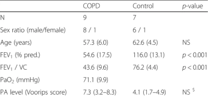
Among 24 COPD patients and 23 SHS included in the main study [Gouzi et al., 2013] [6], we selected all pa-tients for whom we had at least one muscle sample con-ditioned for TEM analysis, i.e. 9 COPD patients and 7 SHS. The baseline clinical characteristics of the COPD patients and SHS are presented in Table1.
As shown in Table2, exercise training induced signifi-cant improvement in 6-min walking distance (%pred.) and in endurance time of the quadriceps (Tlimquad) in
both groups (Time effect: p < 0.001 and p = 0.009, re-spectively). As expected, these two functional parameters were significantly lower in COPD patients than in SHS, regardless of the training effect (Group effect: p = 0.006 and p = 0.024, respectively). A differential effect of exer-cise training on symptom-limited oxygen uptake (V̇O2slÞ was found in COPD patients compared with SHS (Group x Time interaction: p = 0.020) with a significant increase in the SHS group only (SHS: 13.8 ± 10.4%, p =
0.003 vs COPD: 5.1 ± 8.1%, p = 0.28). Regarding the histochemical data, COPD patients showed lower base-line values for the capillary-to-fibre (C/F) ratio compared with SHS (p = 0.002 and p = 0.024, respectively; Table2). In response to exercise training, we observed lower im-provement in the C/F ratio of COPD patients than SHS (+ 15% versus + 30%; Group x Time interaction: p = 0.003), leading to persistent lower post-training values in COPD patients compared with SHS (p < 0.001). Simi-larly, COPD patients presented a reduction of type I fibre proportion in response to exercise training leading to lower post-training values compared to SHS (Group x Time interaction: p = 0.01; Additional file1: Figure. S1).
Angiogenesis-related capillary remodelling in response to exercise training.
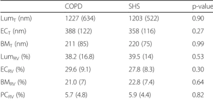
Capillaries from the vastus lateralis muscle biopsies, obtained before and after exercise training in the 9 COPD patients and 7 SHS, were analysed by TEM. The analyses were performed on 392 capillary profiles, which were always composed of at least Lum, EC and BM. Nei-ther total capillary area nor the number of analysed ca-pillaries differed between groups or training times (p > 0.05; data not shown). As shown in Table3, the baseline values of lumen (Lum), endothelium (EC), basement membrane (BM) and pericyte (Pc) relative volume (RV) and thickness (T) did not differ between COPD patients and SHS.
Standard transmission electron micrographs of the skeletal muscle capillaries in COPD patients and SHS, before and after exercise training, are shown on Fig. 1a. Variations of relative volume and thickness of each ca-pillary compartment in response to exercise training are presented in Fig.1b. In response to exercise training, we observed a significant decrease in LumRV (COPD: −
16.4 ± 21.5%; SHS: − 23.3 ± 22.1%; Fig. 1b) and LumT
(COPD: − 7.1 ± 36.8%; SHS: − 17.2 ± 22.4%; Fig. 1b) in both groups (Time effect: p < 0.001 and p = 0.002, re-spectively). Conversely, a significant training-induced in-crease was found in both ECRV (COPD: + 17.0 ± 24.1%;
Table 1 Baseline clinical and functional characteristics of COPD patients and SHS
COPD Control p-value
N 9 7
Sex ratio (male/female) 8 / 1 6 / 1
Age (years) 57.3 (6.0) 62.6 (4.5) NS FEV1(% pred.) 54.6 (17.5) 116.0 (13.1) p < 0.001
FEV1/ VC 43.6 (9.6) 76.2 (4.4) p < 0.001
PaO2(mmHg) 71.1 (9.9)
PA level (Voorips score) 7.3 (3.2–8.3) 4.1 (1.7–4.9) NS$
Data are presented as mean (SD) or median (Q1-Q3). FEV1Forced expiratory
volume in 1 s, VC slow vital capacity, % pred: % predicted, PaO2 arterial oxygen tension, PA Physical activity.§
SHS: + 22.5 ± 18.5%; Fig. 1b) and ECT (COPD: + 27.1 ±
24.9%; SHS: + 24.7 ± 31.0%; Fig. 1b), regardless of the group (Time effect: p < 0.001 for both). Similarly, we ob-served a significant increase in PcRV in both the COPD
and SHS groups (COPD: + 68.4 ± 60.2%; SHS: + 40.3 ± 65.7%; Time effect: p < 0.001; Fig. 1b). Last, a signifi-cantly different response to exercise training was ob-served in COPD patients compared with SHS for BMRV
(Group x Time interaction: p = 0.008; Fig.1b) and BMT
(Group x Time interaction: p = 0.050; Fig.1b), with a sig-nificant increase only in the COPD group for both BMRV
(p = 0.005; + 14.1 ± 22.6%; SHS: − 2.2 ± 17.3%; Fig. 1b) and BMT (p = 0.043; + 9.7 ± 13.8%; SHS: − 0.8 ± 20.2%;
Fig.1b).
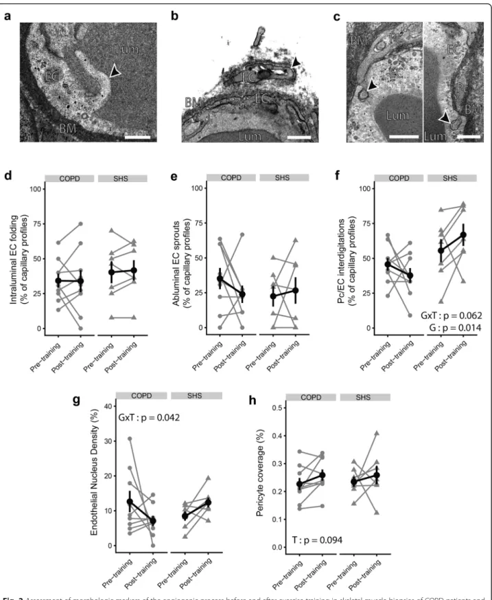
Changes in morphologic markers of the angiogenic process in response to exercise training.
We quantified the ultrastructural morphologic markers of angiogenesis, which are presented on the transmission electron micrographs in Fig. 2a-c. As shown in Fig. 2d and e, the occurrence of intralum-inal EC folding or ablumintralum-inal EC sprouts did not in-crease significantly with exercise training (p > 0.05). COPD patients and SHS did not show different pre-or post-training values fpre-or these indexes either (p > 0.05). Conversely, Pc/EC interdigitation occurrence was significantly lower in the COPD group than in SHS (Fig. 2f) (pre-training: 0.46 ± 0.14% vs 0.56 ± 0.22%; post-training: 0.38 ± 0.16% vs 0.67 ± 0.21%; Group effect: p = 0.014). Moreover, taking into ac-count the baseline difference between COPD patients and SHS, we noted a tendency towards a differential effect of exercise training on this parameter between the two groups (Group x Time interaction: p = 0.062). Similarly, we observed significantly different variations of endothelial nucleus density (ECNuclD) in the capil-laries of COPD patients compared with SHS in re-sponse to exercise training (Group x Time interaction: p = 0.042; Fig. 2g). ECNuclD tended to de-crease in response to exercise training only in the COPD patients (− 7.3 ± 67.1%, p = 0.053; Fig. 2g), lead-ing to a tendency towards lower post-training ECNuclD values in the patients compared with SHS (p = 0.080; Fig. 2g). The pericyte coverage (PcCov) tended to increase significantly with exercise training in both groups (Time effect: p = 0.094; Fig. 2h), with no difference between the groups (COPD: + 17.6 ± 26.1%; SHS: + 16.9 ± 50.5%).
Table 2 Functional and histomorphologic changes in response to exercise training in COPD patients and SHS
Pre-training Post-training p-value
COPD SHS COPD SHS Functional parameters V̇O2sl(% pred.) 69.3 (17.3)‡ 113.7 (12.7) 72.44 (17.2)‡ 125.6 (9.6)###$ T: p = 0.004 G: p < 0.001 GxT: p = 0.020 V̇O2sl(ml/min/kg) 19.8 (4.7)‡ 30.1 (4.4) 21.6 (4.1)‡ 33.1 (7.9)##$ T: p = 0.009 G: p < 0.001 GxT: p = 0.046 6MWD (%pred.) 77.2 (10.7) 94.0 (7.3) 84.2 (11.5) 98.7 (7.7) T: p < 0.001 G: p = 0.006 GxT: NS 6MWD (m) 539.6 (60.9) 643.3 (60.9) 591.2 (72.4) 684.6 (60.5) T: p < 0.001 G: p = 0.010 GxT: NS Tlimquad(sec) 299.6 (123.4) 588.6 (382.2) 388.6 (201.2)£ 830.3 (467) T: p = 0.009
G: p = 0.024 GxT: NS Histomorphologic parameters C/F ratio 1.29 (0.3) * 1.67 (0.3) 1.49 (0.3)## ‡ 2.18 (0.4)### T: p < 0.001 G: p = 0.004 GxT: p = 0.003 Data are presented as mean (SD). V̇O2sl: symptom-limited oxygen uptake, 6MWD 6-min walking distance, Tlimquad: endurance time of quadriceps, C/F ratio
Capillary-to-fibre ratio.$:n = 5,£: n = 8. Linear mixed-effect model, G Group effect, T Time effect, GxT Group x Time interaction. Post-hoc analysis for GxT: different
from SHS group at same time: *p < 0.05; † p < 0.01; ‡ p < 0.001. Different from pre-training:##
p < 0.01;###
p < 0.001
Table 3 Baseline morphometric characteristics of capillary components in COPD patients and SHS
COPD SHS p-value LumT(nm) 1227 (634) 1203 (522) 0.90 ECT(nm) 388 (122) 358 (116) 0.27 BMT(nm) 211 (85) 220 (75) 0.99 LumRV(%) 38.2 (16.8) 39.5 (14) 0.53 ECRV(%) 29.6 (9.1) 27.8 (8.3) 0.30 BMRV(%) 21.0 (7) 22.8 (7.4) 0.64 PCRV(%) 5.7 (4.8) 5.9 (4.4) 0.82
RV Relative volume, T Thickness, Lum Lumen, EC, Endothelium, BM Basement membrane, Pc Pericyte. Data are presented as Mean (SD)
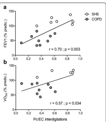
Correlations between the morphologic markers of the angiogenic process and the clinical, functional and histo-chemical parameters of COPD patients and SHS are pre-sented in Fig. 3. After the 6-week exercise training programme, we found significant and positive correla-tions between the occurrence of Pc/EC interdigitacorrela-tions
and FEV1 (%pred.; r = 0.70, p = 0.003; Fig. 3a), V̇O2sl (%pred.; r = 0.57, p = 0.034; Fig.3b).
Training-induced changes in plasmatic F2-IsoP levels
As shown in Fig. 4a, plasmatic levels of F2-IsoP
de-creased significantly with exercise training in both
Fig. 1 Angiogenesis-related capillary remodelling in response to exercise training in skeletal muscle biopsies of COPD patients and SHS. a Transmission electron micrographs of capillaries from vastus lateralis biopsies of COPD patients and SHS. b Quantification of changes in relative volume and thickness of each capillary component following exercise training. RV: relative volume; T: thickness; Lum: lumen; EC: endothelium; BM: basement membrane; Pc: pericyte; Nucl: nucleus. Data are presented as means and individual values. Linear mixed-effect model: T: Time effect; GxT: Group x Time interaction. Post-hoc: Post-training different from Pre-training: *p < 0.05; ** p < 0.01. Scale bar: 1μm
Fig. 2 Assessment of morphologic markers of the angiogenic process before and after exercise training in skeletal muscle biopsies of COPD patients and SHS. a-c Transmission electron micrographs focused on morphologic markers of angiogenesis, shown with black arrows: a intraluminal endothelial (EC) folding, b abluminal EC sprout, and c pericyte/endothelium (Pc/EC) interdigitations. d-f) Semi-quantitative analysis of the occurrence of intraluminal EC folding (d), abluminal EC sprouts (e), and Pc/EC interdigitations (f) on transmission electron micrographs. g, h Quantitative analysis of endothelial nucleus density (g) and pericyte coverage (h) on transmission electron micrographs. Lum: lumen; EC: endothelium; BM: basement membrane; Pc: pericyte. Data are presented as mean ± SE. Linear mixed-effect model: T: Time effect; G: Group effect; GxT: Group x Time interaction. Scale bar: 500 nm
COPD patients and SHS (− 24.5 ± 19.1% and − 31.1 ± 17.2%, respectively; Time effect: p < 0.001). However, regardless of exercise training, plasma F2-IsoP was
sig-nificantly higher in COPD patients than in SHS (pre-training: 335.7 ± 96.1 vs 280.0 ± 71.4; post-training: 235.6 ± 42.2 vs 183.5 ± 16.9; Group effect: p = 0.048; Fig. 4a). Moreover, as shown in Fig. 4b, we found a significant and negative correlation between the changes in Pc/EC interdigitations and the changes in plasmatic levels of F2-IsoP in response to exercise
training (r =− 0.62, p = 0.025). Discussion
This study is the first to assess skeletal muscle capillary ultrastructure after exercise training-related angiogenesis in COPD patients. The main result is that the blunted training-induced angiogenesis previously found in COPD patients [6] is associated with abnormal capillary remodelling and morphologic features of an impaired angiogenic process.
Consistent with the previous study in this trial, COPD patients showed significant but blunted improvements in exercise capacity and muscle angiogenesis (Table2) and an impaired adaptation of the muscle typology (Additional
file1: Figure S1) in response to exercise training in com-parison with sedentary healthy subjects. Together, these observations highlight an impairment of the oxidative me-tabolism of COPD patients and a misadaptation of this last to exercise training. However, the changes in fibre type under exercise training and the training-induced angiogenesis were poorly correlated (Additional file1: Fig-ure S1). Moreover, another study on COPD patients found differential angiogenic responses to exercise
Fig. 3 Correlations between morphologic markers of the angiogenic process and clinical and functional parameters of COPD patients and SHS. Positive significant correlations between the proportion of capillary profiles associated with Pc/EC interdigitations in post-training vastus lateralis biopsies and a forced expiratory volume in 1 s (FEV1) (% predic.), b post-training values of symptom-limited
oxygen uptake (V̇O2sl), in both COPD patients (grey circles) and SHS
(white circles)
Fig. 4 Changes in plasma-free and esterified isoprostane (F2-IsoP) levels
in response to exercise training in COPD patients and SHS and the relation to the angiogenic process. a Effect of exercise training on plasmatic levels of F2-IsoP in COPD and SHS. Linear mixed-effect
model: T: Time effect, G: Group effect. Pre-training: COPD: n = 9, SHS: n = 5; Post-training: COPD: n = 8, SHS: n = 6. Data are presented as Mean ± SE. b Correlation between the pre-to-post training variations of plasmatic F2-IsoP levels and the pre-to-post training variations in
Pc/EC interdigitation occurrence in COPD patients (grey circles) and SHS (white circles)
training between two groups of COPD patients, although the groups presented similar adaptations of skeletal muscle typology to the training [38]. Muscle typology alone was thus insufficient to explain the blunted angio-genesis found in these COPD patients, which led us to suspect that other cellular mechanisms might be involved. We therefore conducted this study to investigate capillary remodelling and the morphologic markers of the angio-genic process associated with the blunted training-induced angiogenesis found in these patients. The capillary remod-elling and the morphologic markers of the angiogenic process were assessed blindly following cross-validated and previously published methods [12,32]. The inter-observer reliability of the measurements of capillary remodelling was assessed with three observers and was“excellent” (ac-cording to Lin’s concordance coefficient) [39].
Our study brings the first evidence of an exercise training effect on capillary ultrastructure in both COPD patients and healthy subjects between 50 and 70 years old and identified as strictly sedentary using a validated method. Both exercise training and ageing are known to alter the capillary ultrastructure in skeletal muscles [12,
32]. To isolate the impact of COPD on capillary ultra-structure and its response to exercise training, we se-lected COPD patients and SHS who did not differ in age or physical activity level for this study. In SHS, we ob-served a significant increase in both ECRV and ECT and
no changes in BMRV and BMT in response to the
train-ing (Fig. 1b,c), as previous described in younger and more active healthy subjects [12, 40]. In contrast, we found a decrease in both LumRVand LumTin these SHS
following the training programme (Fig. 1b,c), and this decrease has been described as part of the angiogenic process in skeletal muscle [9,10]. Physiologically, angio-genesis occurs in two phases, an early phase of capillary creation (by sprouting and/or intussusception) in the first 5 weeks of exercise training and then a late phase of capillary maturation [7, 41]. Consistent with this time-course, we observed later angiogenesis-related changes (increased PcCov and PCRV) in SHS at 6 weeks of
exer-cise training (Fig. 1b and Fig. 2f,h) but not the early angiogenesis hallmarks (abluminal EC sprouts or intra-luminal EC folding; Fig.2d,e), in line with the findings of an earlier study in younger healthy subjects [12].
Conversely, the COPD patients presented an abnormal remodelling of capillaries in response to exercise train-ing. While their adaptation of lumen (LumRVand LumT)
and endothelium (ECRVand ECT) in response to training
was physiological, they showed an unexpected increase in BMRV and BMT (Fig. 1b,c). The BM thickening after
training was a non-physiological response, and this ori-ginal result contrasts with what has been shown in other chronic diseases like hypertension, in which training in-duced BM thinning [40]. Exercise-induced oxidative
stress [42], which is a hallmark of COPD, may be a mechanism in this modification, because oxidative stress disturbs the BM turnover in favour of an accumulation of the BM component [16]. Last, the loss of Pc/EC inter-digitations (Fig.2f) may also have contributed to the BM thickening. Indeed, these interdigitations act as a mech-anical link between the endothelium and pericytes, help-ing the capillaries to resist the dramatic increase in haemodynamic forces during exercise [43,44]. Thus, the loss of the pericyte-endothelium link in COPD patients could be the cause of compensatory BM thickening.
In addition, our study showed an impaired angiogenic process in response to exercise training in the COPD pa-tients. The skeletal muscle angiogenic process of COPD patients and SHS in response to exercise training was assessed through TEM analysis of the morphologic markers of specific capillary alterations [8–10,12]. These markers inform about angiogenesis-related mechanisms and possible impairments [9, 12]. As described above, our measures were performed too late to assess the mor-phologic markers of the early phase of angiogenesis [7] (i.e. abluminal EC sprouts and intraluminal EC folding), and we thus found no changes in these markers in the COPD patients (Fig.2d,e). However, it is possible to as-sess the final result of capillary proliferation by measur-ing the nucleus density in endothelial cells [8,12]. Here, we found a significantly different response to exercise training for the ECNuclD in the COPD patients com-pared with that of healthy subjects (Fig. 2g), suggesting impaired capillary proliferation during the angiogenic process. This finding is consistent with the blunted angiogenesis found at the histomorphologic level in these patients (Table2). After 6 weeks of exercise train-ing, we assessed the later phase of the angiogenic process, capillary maturation [7]. The Pc/EC interdigita-tions, as observed by TEM, are defined as the morpho-logic expression of capillary maturation [11], which increases during skeletal muscle stimulation [9]. In our study, we found a tendency towards a differential vari-ation in the occurrence of the Pc/EC interdigitvari-ations in the COPD patients compared with SHS (Fig. 2f). More-over, independently of exercise training, the patients pre-sented a significantly lower occurrence of Pc/EC interdigitations. Surprisingly, the PcCov was not im-paired in the COPD patients. However, while PcCov is described as a marker of capillary maturation, the mat-uration effect of pericyte is mainly driven by the ex-change of multiple angiogenic factors. Among these, Ang1 plays a crucial role [45] and its exchange mostly occurs at the level of Pc/EC interdigitations [46]. Thus, while the PcCov in COPD patients was not impaired, the loss of Pc/EC interdigitations could lead to a dys-functional pericyte coverage. However, both Ang1/Tie2 axis and pericyte function remains to be experimentally
assessed in skeletal muscle capillaries of COPD patients. With these results, we bring the first evidence of im-paired capillary maturation during exercise training-related angiogenesis in COPD patients. Regardless of ex-ercise training, the COPD patients in our study pre-sented higher levels of plasmatic F2-IsoP, a marker of
oxidative stress-related lipid peroxidation [18], than SHS (Fig. 4a). In addition, the changes in the occurrence of Pc/EC interdigitations in response to the training were negatively correlated with the changes in the plasmatic level of F2-IsoP (Fig. 4b). In the literature, oxidative
stress has been experimentally related to pericyte loss [17, 47]. Indeed, it can increase angiopoeitin-2 levels [48], which antagonize the interaction between endothe-lial cells and pericytes [49, 50]. Moreover, partial sup-pression of F2-IsoP by antioxidant supplementation
prevents pericyte loss [20]. Based on the literature and the F2-IsoP results of the present study, we hypothesize
that oxidative stress can contribute to the impairment of capillary maturation found in COPD patients. Further experimental studies with oxidative stress intervention (pro- and anti-oxidant treatment in combination with training) in an animal model and COPD patients, associ-ated with TEM analysis, are required to confirm this hypothesis.
The present study shows impairments in capillary re-modelling and the angiogenic process in response to ex-ercise training in COPD patients. As discussed in the previous paragraphs, our results constitute an original contribution to knowledge on the muscle cellular adap-tations to cardiopulmonary rehabilitation. This study may be of interest to researchers working in the field of skeletal muscle dysfunction and angiogenesis in chronic respiratory diseases because it provides evidence that can guide their research questions and it specifically tar-gets the angiogenesis mechanisms, i.e. the Pc/EC inter-actions. Our study constitutes a step forward in the search for complementary therapeutic interventions to restore physiological muscle adaptations to exercise training in COPD patients. While it was not the aim of our study, our results raise the question of their clinical relevance. Indeed, BM thickening increases the width of the capillary wall and thus the distance between erythro-cytes and mitochondria. Because this distance is crucial in the process of oxygen diffusion and supply to the exercised muscle [51], this BM thickening might limit the improvements in muscle oxygen consumption and V̇O2sl in patients, as in peripheral artery disease patients [13]. Also, contractile pericytes [43] can directly regulate capillary blood flow [52]. Thus, the loss of the Pc/EC interdigitations in COPD patients might also impact the oxygen supply [53] and V̇O2sl. Consistently, we found a positive correlation between post-training Pc/EC
interdigitation occurrence and V̇O2sl (Fig.3b). Last, given that capillary maturation prevents the regression of neo-capillaries [54, 55], the impaired capillary maturation might constitute a mechanism for the blunted training-related angiogenesis in COPD patients. Specifically de-signed studies with larger samples are required to generalize these results to the whole COPD population and to establish the clinical implications of the defective capillary remodelling and angiogenic process in these pa-tients, as reported in our exploratory study.
Conclusion
Exercise training improves muscle function in COPD pa-tients but to a lesser extent than in healthy subjects, es-pecially regarding the increase in muscle capillarization (i.e. angiogenesis). Here, we provide the first evidence that this blunted angiogenesis is characterized by abnor-mal capillary remodelling and an impairment in both phases of the angiogenic process, i.e. capillary creation and maturation, in response to exercise training. The loss of pericyte-endothelium interaction appears to be a key factor for the capillary maturation. This study con-tributes to our understanding of the defective angiogenic process in COPD patients and constitutes a significant step towards isolating the blunted cellular mechanisms in endothelial cells and pericytes. Our observations sug-gest a hypothetic role for oxidative stress in the angio-genesis defect and open the way for further studies investigating this relationship in COPD patients.
Supplementary information
Supplementary information accompanies this paper athttps://doi.org/10. 1186/s12931-019-1240-6.
Additional file 1 Figure S1. Changes in skeletal muscle typology in response to exercise training in COPD patients and SHS and the relation to the angiogenic process. a) Effect of exercise training on the proportion of type I fibre in vastus lateralis cryosections of COPD patients (grey bars) and SHS (white bars). Linear mixed-effect model: GxT: Group x Time inter-action. Post-hoc: *p < 0.05; *** p < 0.001. Data are presented as mean ± SE. b) Correlation between the post-to-pre training variations of type I pro-portion and the post-to-pre training variations of capillary-to-fibre ratio in COPD patients (grey circles) and SHS (white circles).
Abbreviations
A:Area; BM: Basement Membrane; C: Circumference; C/F: Capillary-to-fibre; COPD: Chronic obstructive pulmonary disease; EC: Endothelial;
ECNuclD: Endothelial nucleus density; F2-IsoP: Plasma-free and esterified isoprostane; FEV1: Forced expiratory volume in one second; Lum: Lumen; Pc: Pericyte; PcCov: Pericyte coverage; RV: Relative volume; SHS: Sedentary healthy subjects; T: Thickness; TEM: Transmission electron microscopy; Tlimquad: Endurance time of the quadriceps; V̇O2sl: Symptom-limited oxygen uptake
Acknowledgements
The authors would like to thank Chantal Cazevieille from the CoMET MRI facilities (INM, France) for her valuable help in tissue preparation for the transmission electron microscopy and micrograph acquisitions. We also would like to thank C. Stott for her critical reading of the manuscript.
Authors’ contributions
LB, EP, MB, PP, FG and JPC performed the experiments. JM, PP, LB, FG, EP, MH and MC conceived and designed the experiments and discussed the data and the manuscript. JM, FG, PP and MH supervised the work. BA, EP, LB and NM collected and analysed the data. APM, FB and NH provided expertise and resources and discussed the data and the manuscript. LB wrote the manuscript. All authors reviewed the manuscript. All authors read and approved the final manuscript
Funding
This study was supported by joint grants from the Centre Hospitalier Régional Universitaire Montpellier, patient associations and the Association Pour l’Assistance et la Réhabilitation à domicile (APARD). FG was supported by a Conventions Industrielles de Formation par la Recherche (CIFRE) grant from the 5 Santé group, France, and the French Ministère Délégué à la Recherche et aux Nouvelles Technologies.
Availability of data and materials
The datasets used and/or analysed during the current study are available from the corresponding author on reasonable request.
Ethics approval and consent to participate
The two studies were approved by the institutional ethics committee of the Montpellier University Hospitals (2008-EESSS-V2 and 2009–04-BPCO-V2). All subjects had given informed written consent before starting the studies and agreed to the utilization of their muscle samples in future studies. Consent for publication
Not applicable. Competing interests
FG received financial support for congress participation from SOS Oxygène, LVL Médical, Eole Santé, APARD, Agir à Dom’ and Bastide Médical. FB received financial support for congress participation from SOS Oxygène, LVL Médical, Orkyn, Eole Santé, APARD and Agir à Dom’. All other authors declare that they have no competing interest.
Author details
1Physiologie & médecine expérimentale du Cœur et des Muscles
(PhyMedExp), INSERM - CNRS - Montpellier University. CHU Arnaud De Villeneuve, 371 avenue du Doyen Gaston Giraud, 34295 Montpellier cedex 5, France.2Les Cliniques du Souffle®, Groupe 5 Santé, Lodève, France. 3PhyMedExp, INSERM– CNRS, Montpellier University, CHU Montpellier,
Montpellier, France.4IMAG, CNRS, Montpellier University, CHU Montpellier,
Montpellier, France.5Vascular Medicine Department and Laboratory, CHU Nîmes and EA2992 Research Unit, Montpellier University, Nimes, France. Received: 9 August 2019 Accepted: 13 November 2019
References
1. Vanfleteren LEGW, Spruit MA, Groenen M, Gaffron S, van Empel VPM, Bruijnzeel PLB, et al. Clusters of Comorbidities Based on Validated Objective Measurements and Systemic Inflammation in Patients with Chronic Obstructive Pulmonary Disease. Am J Respir Crit Care Med. 2013;187(7): 728–35.
2. Marquis K, Debigaré R, Lacasse Y, LeBlanc P, Jobin J, Carrier G, et al. Midthigh muscle cross-sectional area is a better predictor of mortality than body mass index in patients with chronic obstructive pulmonary disease. Am J Respir Crit Care Med. 2002;166(6):809–13.
3. Oga T, Nishimura K, Tsukino M, Sato S, Hajiro T. Analysis of the factors related to mortality in chronic obstructive pulmonary disease: role of exercise capacity and health status. Am J Respir Crit Care Med. 2003;167(4): 544–9.
4. Swallow EB, Reyes D, Hopkinson NS, Man WD-C, Porcher R, Cetti EJ, et al. Quadriceps strength predicts mortality in patients with moderate to severe chronic obstructive pulmonary disease. Thorax. 2007;62(2):115–20. 5. Maltais F, Decramer M, Casaburi R, Barreiro E, Burelle Y, Debigaré R, et al. An
official American Thoracic Society/European Respiratory Society statement: update on limb muscle dysfunction in chronic obstructive pulmonary disease. Am J Respir Crit Care Med. 2014;189(9):e15–62.
6. Gouzi F, Préfaut C, Abdellaoui A, Roudier E, de Rigal P, Molinari N, et al. Blunted muscle angiogenic training-response in COPD patients versus sedentary controls. Eur Respir J avr. 2013;41(4):806–14.
7. Haas TL, Nwadozi E. Regulation of skeletal muscle capillary growth in exercise and disease. Appl Physiol Nutr Metab déc. 2015;40(12):1221–32. 8. Egginton S, Zhou A-L, Brown MD, Hudlická O. Unorthodox angiogenesis in
skeletal muscle. Cardiovasc Res. 2001;49(3):634–46.
9. Egginton S, Hudlicka O, Brown MD, Graciotti L, Granata AL. In VivoPericyte– endothelial cell interaction during angiogenesis in adult cardiac and skeletal muscle. Microvasc Res mars. 1996;51(2):213–28.
10. Zhou A-L, Egginton S, Brown MD, Hudlicka O. Capillary growth in overloaded, hypertrophic adult rat skeletal muscle: an ultrastructural study. Anat Rec Off Publ Am Assoc Anat. 1998;252(1):49–63.
11. Caruso RA, Fedele F, Finocchiaro G, Pizzi G, Nunnari M, Gitto G, et al. Ultrastructural descriptions of pericyte/endothelium peg-socket interdigitations in the microvasculature of human gastric carcinomas. Anticancer Res janv. 2009;29(1):449–53.
12. Baum O, Gübeli J, Frese S, Torchetti E, Malik C, Odriozola A, et al. Angiogenesis-related ultrastructural changes of capillaries in human skeletal muscle in response to endurance exercise. J Appl Physiol. 2015;119:1118-1126. jap.00594.2015.
13. Baum O, Bigler M. Pericapillary basement membrane thickening in human skeletal muscles. Am J Physiol Heart Circ Physiol. 2016;311(3): H654–66.
14. Wallberg-Henriksson H, Gunnarsson R, Henriksson J, Östman J, Wahren J. Influence of Physical Training on Formation of Muscle Capillaries in Type I Diabetes. Diabetes. 1984;33(9):851–7.
15. Raskin P, Pietri AO, Unger R, Shannon WA. The Effect of Diabetic Control on the Width of Skeletal-Muscle Capillary Basement Membrane in Patients with Type I Diabetes Mellitus. N Engl J Med. 1983;309(25):1546–50.
16. Mason RM, Wahab NA. Extracellular Matrix Metabolism in Diabetic Nephropathy. J Am Soc Nephrol. 2003;14(5):1358–73.
17. Hayden MR, Yang Y, Habibi J, Bagree SV, Sowers JR. Pericytopathy: oxidative stress and impaired cellular longevity in the pancreas and skeletal muscle in metabolic syndrome and type 2 diabetes. Oxidative Med Cell Longev. 2010; 3(5):290–303.
18. Roberts LJ, Milne GL. Isoprostanes. J Lipid Res avr. 2009;50(Suppl):S219–23. 19. Benndorf RA, Schwedhelm E, Gnann A, Taheri R, Kom G, Didié M, et al.
Isoprostanes inhibit vascular endothelial growth factor-induced endothelial cell migration, tube formation, and cardiac vessel sprouting in vitro, as well as angiogenesis in vivo via activation of the thromboxane A(2) receptor: a potential link between oxidative stress and impaired angiogenesis. Circ Res. 2008;103(9):1037–46.
20. Tsai GY, Cui JZ, Syed H, Xia Z, Ozerdem U, McNeill JH, et al. Effect of N-acetylcysteine on the early expression of inflammatory markers in the retina and plasma of diabetic rats. Clin Experiment Ophthalmol mars. 2009;37(2): 223–31.
21. Couillard A, Prefaut C. From muscle disuse to myopathy in COPD: potential contribution of oxidative stress. Eur Respir J oct. 2005;26(4):703–19. 22. Delample D, Durand F, Severac A, Belghith M, Mas E, Michel F, et al.
Implication of xanthine oxidase in muscle oxidative stress in COPD patients. Free Radic Res janv. 2008;42(9):807–14.
23. Vogelmeier CF, Criner GJ, Martinez FJ, Anzueto A, Barnes PJ, Bourbeau J, et al. Global strategy for the diagnosis, management, and prevention of chronic obstructive lung disease 2017 report. GOLD Executive Summary Am J Respir Crit Care Med mars. 2017;195(5):557–82.
24. Statement ATS. ATS statement: guidelines for the six-minute walk test. Am J Respir Crit Care Med. 2002;166(1):111–7.
25. ATS/ACCP Statement. ATS/ACCP Statement on Cardiopulmonary Exercise Testing. Am J Respir Crit Care Med. 2003;167(2):211–77.
26. Voorrips LE, Ravelli AC, Dongelmans PC, Deurenberg P, Van Staveren WA. A physical activity questionnaire for the elderly. Med Sci Sports Exerc août. 1991;23(8):974–9.
27. Spruit MA, Singh SJ, Garvey C, ZuWallack R, Nici L, Rochester C, et al. An official American Thoracic Society/European Respiratory Society statement: key concepts and advances in pulmonary rehabilitation. Am J Respir Crit Care Med. 2013;188(8):e13–64.
28. Vallet G, Ahmaïdi S, Serres I, Fabre C, Bourgouin D, Desplan J, et al. Comparison of two training programmes in chronic airway limitation patients: standardized <I>versus</I> individualized protocols. Eur Respir J. 1997;10(1):114–22.
29. Nelson ME, Rejeski WJ, Blair SN, Duncan PW, Judge JO, King AC, et al. Physical activity and public health in older adults: recommendation from the American College of Sports Medicine and the American Heart Association. Med Sci Sports Exerc août. 2007;39(8):1435–45.
30. Hayot M, Michaud A, Koechlin C, Caron M-A, LeBlanc P, Préfaut C, et al. Skeletal muscle microbiopsy: a validation study of a minimally invasive technique. Eur Respir J. 2005;25(3):431–40.
31. Lawrence I, Lin K. A concordance correlation coefficient to evaluate reproducibility. Biometrics. 1989;45:255–68.
32. Bigler M, Koutsantonis D, Odriozola A, Halm S, Tschanz SA, Zakrzewicz A, et al. Morphometry of skeletal muscle capillaries: the relationship between capillary ultrastructure and ageing in humans. Acta Physiol oct. 2016;218(2): 98–111.
33. Kadiiska MB, Gladen BC, Baird DD, Germolec D, Graham LB, Parker CE, et al. Biomarkers of oxidative stress study II: are oxidation products of lipids, proteins, and DNA markers of CCl4 poisoning? Free Radic Biol Med mars. 2005;38(6):698–710.
34. Brown MB, Forsythe AB. Robust tests for the equality of variances. J Am Stat Assoc juin. 1974;69(346):364–7.
35. Aarts E, Verhage M, Veenvliet JV, Dolan CV, van der Sluis S. A solution to dependency: using multilevel analysis to accommodate nested data. Nat Neurosci avr. 2014;17(4):491–6.
36. Pinheiro J, Bates D, DebRoy S, Sarkar D. R Core Team (2014) nlme: linear and nonlinear mixed effects models. R package version 3; 2014. p. 1–117. Available H TtpCRAN R-Proj Orgpackage Nlme
37. Faraway JJ. Extending the linear model with R: generalized linear, mixed effects and nonparametric regression models, vol. 124; 2016. CRC press 38. Costes F, Gosker H, Feasson L, Desgeorges M, Kelders M, Castells J, et al.
Impaired exercise training-induced muscle fiber hypertrophy and Akt/mTOR pathway activation in hypoxemic patients with COPD. J Appl Physiol. 2015; 118(8):1040–9.
39. Partik BL, Stadler A, Schamp S, Koller A, Voracek M, Heinz G, et al. 3D versus 2D ultrasound: accuracy of volume measurement in human cadaver kidneys. Investig Radiol. 2002;37(9):489–95.
40. Gliemann L, Buess R, Nyberg M, Hoppeler H, Odriozola A, Thaning P, et al. Capillary growth, ultrastructure remodelling and exercise training in skeletal muscle of essential hypertensive patients. Acta Physiol juin. 2015;214(2):210–20. 41. Jain RK. Molecular regulation of vessel maturation. Nat Med juin. 2003;9(6):
685–93.
42. Couillard A, Maltais F, Saey D, Debigaré R, Michaud A, Koechlin C, et al. Exercise-induced Quadriceps Oxidative Stress and Peripheral Muscle Dysfunction in Patients with Chronic Obstructive Pulmonary Disease. Am J Respir Crit Care Med. 2003;167(12):1664–9.
43. Tilton RG, Kilo C, Williamson JR. Pericyte-endothelial relationships in cardiac and skeletal muscle capillaries. Microvasc Res. 1979;18(3):325–35. 44. Andersen P, Saltin B. Maximal perfusion of skeletal muscle in man. J Physiol
sept. 1985;366:233–49.
45. Suri C, McClain J, Thurston G, McDonald DM, Zhou H, Oldmixon EH, et al. Increased Vascularization in Mice Overexpressing Angiopoietin-1. Science. 1998;282(5388):468–71.
46. Wakui S, Yokoo K, Muto T, Suzuki Y, Takahashi H, Furusato M, et al. Localization of Ang-1,-2, Tie-2, and VEGF expression at endothelial-pericyte interdigitation in rat angiogenesis. Lab Investig. 2006;86(11):1172. 47. Cacicedo JM, Benjachareowong S, Chou E, Ruderman NB, Ido Y.
Palmitate-Induced Apoptosis in Cultured Bovine Retinal Pericytes: Roles of NAD(P)H Oxidase, Oxidant Stress, and Ceramide. Diabetes. 2005;54(6):1838–45. 48. Mofarrahi M, Sigala I, Vassilokopoulos T, Harel S, Guo Y, Debigare R, et al.
Angiogenesis-related factors in skeletal muscles of COPD patients: roles of angiopoietin-2. J Appl Physiol. 2013;114(9):1309–18.
49. Armulik A. Endothelial/Pericyte Interactions. Circ Res. 2005;97(6):512–23. 50. Zeng H, He X, Tuo Q, Liao D, Zhang G, Chen J. LPS causes pericyte loss and
microvascular dysfunction via disruption of Sirt3/angiopoietins/Tie-2 and HIF-2α/Notch3 pathways. Sci Rep. [cité 12 déc 2018]; 2016;6:20931. Disponible sur:https://www.ncbi.nlm.nih.gov/pmc/articles/PMC4751495/. 51. Weibel ER. The Structural Conditions for Oxygen Supply to Muscle Cells: The
Krogh Cylinder Model. J Exp Biol. 2013;216(22):4135–7.
52. Hall CN, Reynell C, Gesslein B, Hamilton NB, Mishra A, Sutherland BA, et al. Capillary pericytes regulate cerebral blood flow in health and disease. Nature avr. 2014;508(7494):55–60.
53. Kisler K, Nelson AR, Rege SV, Ramanathan A, Wang Y, Ahuja A, et al. Pericyte degeneration leads to neurovascular uncoupling and limits oxygen supply to brain. Nat Neurosci mars. 2017;20(3):406–16.
54. Gianni-Barrera R, Burger M, Wolff T, Heberer M, Schaefer DJ, Gürke L, et al. Long-term safety and stability of angiogenesis induced by balanced single-vector co-expression of PDGF-BB and VEGF164 in skeletal muscle. Sci Rep. 2016; [cité 12 déc 2018];6. Disponible sur:https://www.ncbi.nlm.nih.gov/ pmc/articles/PMC4756385/.
55. Erber R, Thurnher A, Katsen AD, Groth G, Kerger H, Hammes H-P, et al. Combined inhibition of VEGF and PDGF signaling enforces tumor vessel regression by interfering with pericyte-mediated endothelial cell survival mechanisms. FASEB J. 2004;18(2):338–40.
Publisher’s Note
Springer Nature remains neutral with regard to jurisdictional claims in published maps and institutional affiliations.