HAL Id: hal-01243558
https://hal-amu.archives-ouvertes.fr/hal-01243558
Submitted on 15 Dec 2015
HAL is a multi-disciplinary open access
archive for the deposit and dissemination of
sci-entific research documents, whether they are
pub-lished or not. The documents may come from
teaching and research institutions in France or
abroad, or from public or private research centers.
L’archive ouverte pluridisciplinaire HAL, est
destinée au dépôt et à la diffusion de documents
scientifiques de niveau recherche, publiés ou non,
émanant des établissements d’enseignement et de
recherche français ou étrangers, des laboratoires
publics ou privés.
deleterious IFN-I effects to design innovative
immunotherapies targeting cytokine activity to specific
cell types
Elena Tomasello, Emeline Pollet, Thien-Phong Vu Manh, Gilles Uzé, Marc
Dalod
To cite this version:
Elena Tomasello, Emeline Pollet, Thien-Phong Vu Manh, Gilles Uzé, Marc Dalod. Harnessing
mech-anistic knowledge on beneficial versus deleterious IFN-I effects to design innovative immunotherapies
targeting cytokine activity to specific cell types. Frontiers in Immunology, Frontiers, 2014, 5, pp.1-27
�10.3389/fimmu.2014.00526�. �hal-01243558�
Harnessing mechanistic knowledge on beneficial versus
deleterious IFN-I effects to design innovative
immunotherapies targeting cytokine activity to specific
cell types
Elena Tomasello1,2,3†, Emeline Pollet1,2,3†, Thien-Phong Vu Manh1,2,3†, Gilles Uzé4and Marc Dalod1,2,3*
1UM2, Centre d’Immunologie de Marseille-Luminy (CIML), Aix-Marseille University, Marseille, France 2
U1104, Institut National de la Santé et de la Recherche Médicale (INSERM), Marseille, France 3
UMR7280, Centre National de la Recherche Scientifique (CNRS), Marseille, France 4
UMR 5235, Centre National de la Recherche Scientifique (CNRS), University Montpellier II, Montpellier, France
Edited by:
Yoichi Furuya, Albany Medical College, USA
Reviewed by:
Christine Anne Biron, Brown University, USA
Alan Chen-Yu Hsu, The University of Newcastle, Australia
Danushka Kumara Wijesundara, The Basil Hetzel Institute, Australia *Correspondence:
Marc Dalod , Centre d’Immunologie de Marseille-Luminy, Parc scientifique et technologique de Luminy, case 906, F-13288 Marseille Cedex 09, France
e-mail: dalod@ciml.univ-mrs.fr †Elena Tomasello, Emeline Pollet and Thien-Phong Vu Manh have contributed equally to this work.
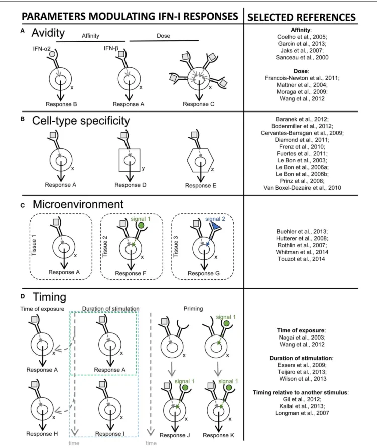
Type I interferons (IFN-I) were identified over 50 years ago as cytokines critical for host defense against viral infections. IFN-I promote anti-viral defense through two main mecha-nisms. First, IFN-I directly reinforce or induce de novo in potentially all cells the expression of effector molecules of intrinsic anti-viral immunity. Second, IFN-I orchestrate innate and adaptive anti-viral immunity. However, IFN-I responses can be deleterious for the host in a number of circumstances, including secondary bacterial or fungal infections, several autoimmune diseases, and, paradoxically, certain chronic viral infections. We will review the proposed nature of protective versus deleterious IFN-I responses in selected diseases. Emphasis will be put on the potentially deleterious functions of IFN-I in human immunod-eficiency virus type 1 (HIV-1) infection, and on the respective roles of IFN-I and IFN-III in promoting resolution of hepatitis C virus (HCV) infection. We will then discuss how the balance between beneficial versus deleterious IFN-I responses is modulated by several key parameters including (i) the subtypes and dose of IFN-I produced, (ii) the cell types affected by IFN-I, and (iii) the source and timing of IFN-I production. Finally, we will specu-late how integration of this knowledge combined with advanced biochemical manipulation of the activity of the cytokines should allow designing innovative immunotherapeutic treat-ments in patients. Specifically, we will discuss how induction or blockade of specific IFN-I responses in targeted cell types could promote the beneficial functions of IFN-I and/or dampen their deleterious effects, in a manner adapted to each disease.
Keywords: type I interferons, dendritic cells, chronic viral infections, immunotherapy, bioengineering
INTRODUCTION
Type I interferons (IFN-I) were the first cytokines discovered, over 50 years ago, based on their potent anti-viral effects (1,2). IFN-I play a crucial, non-redundant role in vertebrate anti-viral defenses (3–5). IFN-I also mediate protective effects in other phys-iopathological contexts, including cancer (6) and multiple sclerosis (MS) (7). On the contrary, IFN-I responses can be deleterious in a number of circumstances, including bacterial or fungal infec-tions (8–10), many autoimmune diseases (11), and, paradoxically, certain chronic viral infections (12–14). It is only recently that an integrated picture has emerged of the cellular and molecular mechanisms regulating the production of IFN-I and underlying their functions. Much knowledge was gained recently on another class of potent innate anti-viral interferons, the lambda, or type III IFNs (IFN-III). We will review knowledge on IFN-I/III (IFNs) and discuss how it could be harnessed to develop innovative ther-apeutic strategies aimed at surgically tuning IFN activity toward protective responses in a manner adapted to each disease. We will focus on IFN-α/β/λ because they are the best characterized
IFNs and already used therapeutically. Recent reviews are cover-ing information on other IFN-I subsets includcover-ing IFN-ε, which is produced at mucosal sites and promotes local anti-viral defenses (15,16).
Dendritic cells (DCs) are rare heterogeneous mononuclear phagocytes functionally characterized by their unique efficacy for antigen-specific activation of naïve T lymphocytes. DCs are sentinel cells of the immune system, able to sense and inte-grate a variety of danger input signals for delivery of output signals instructing the activation and functional polarization of effector immune cells. In mammals, five major DC sub-sets exist, which differ in their expression of innate immune recognition receptors (I2R2s) and in their functional
special-ization: monocyte-derived DCs (MoDCs), Langerhans cells, CD11b+
DCs, XCR1+
DCs, and plasmacytoid DCs (pDCs) (17). A recurrent theme of this review will be the intricate rela-tionships between IFNs and DCs, since these cells can be a major source and/or target of these cytokines under various conditions.
The first section will synthesize current knowledge on IFN production and protective anti-viral functions. The I2R2s and
downstream signaling pathways responsible for IFN-I production during viral infection will be listed. The roles of different cell types for this function will be discussed. The two main mechanisms through which IFN-I promote anti-viral defense will be reviewed, succinctly for direct anti-viral effects and in greater details for immunoregulatory functions.
The second section will focus on the detrimental functions of IFN-I. Selected diseases will be discussed to illustrate how differ-ent, and sometimes opposite, processes underlie deleterious IFN-I responses depending on the physiopathological contexts. IFN-I induction of unbridled inflammatory responses causing lethal tis-sue damage will be discussed as a major pathological mechanism during bacterial encounters secondary to influenza infection or in some autoimmune diseases. Inappropriate functional polarization of immune responses by IFN-I will be discussed as one potential cause for enhanced susceptibility to bacterial or fungal infections. The complex and disputed role of IFN-I in chronic viral infec-tions will be reviewed, with emphasis on the physiopathology of the infections by human immunodeficiency virus type 1 (HIV-1) and human hepatitis C virus (HCV), with an outlook for the development of novel immunotherapeutic strategies to combine with anti-viral drugs.
The third section will recapitulate how the balance between beneficial versus deleterious IFN-I responses is modulated by sev-eral key parameters including (i) the source and timing of IFN-I production, (ii) the cell types affected by IFN-I, and (iii) the signaling pathways activated by IFN-I.
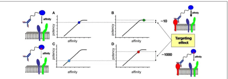
In the last section, we will speculate how integration of all the knowledge discussed before combined with advanced biochem-ical manipulation of the activity of the cytokines should allow designing innovative immunotherapeutic treatments, based on induction or blockade of specific IFN-I responses in targeted cell types. This“activity-by-targeting”concept is based on the design of novel “immuno-IFNs” consisting in covalent association between a mutated IFN-I with decreased affinity for its receptor and an antibody with high avidity for a molecule specifically expressed on target cell types (18). This design ensures lack of activity of the immuno-IFNs on all cell types but those targeted, contrary to previous strategies using IFNs with close to maximal potency that were still able to mediate strong off-target effects despite their coupling to cell-type specific antibodies and/or their local delivery.
GENERAL CONCEPTS ON IFN PRODUCTION AND FUNCTIONS
HOW IS THE PRODUCTION OF IFN CONTROLLED?
Type I interferons expression is not detectable under steady state conditions in vivo using classical methods such as gene expression analysis by RT-PCR or protein titration by ELISA or bioassays. However, mice deficient for the expression of the alpha chain of the IFN-I receptor (IFNAR1) harbor alteration in the ontogeny or functions of various cell types (19–26). Hence, extremely small or localized but functionally relevant quantities of IFN-I must be produced under steady state conditions (27). Indeed, the exis-tence of steady state responses to IFN-I in various organs in vivo was demonstrated by using reporter mice expressing the firefly luciferase under the control of the promoter of Ifnb1 (28) or
of Mx2 (29), a canonical IFN-I-stimulated gene (ISG). Steady state IFN-I responses are promoted by gut commensals (30). Early and transiently after many viral infections, large amounts of IFNs can be detected, in blood and spleen in the case of sys-temic infections or locally in the case of confined infections. IFN induction during viral infections results from the detection of specific danger signals by specialized I2R2s. This includes the
detection of pathogen-associated molecular patterns as well as the sensing of stress signals or damage-associated molecular pat-terns (31,32). Based on the nature and intracellular location of the danger signals that induce the production of the cytokines, the cellular sources of IFNs during viral infection can be classi-fied in two main groups. Infected cells often contribute to IFN production as a response to their sensing of endogenous viral replication, or consecutive to the metabolic stress induced during massive translation of viral structural proteins, or as a result of plasma membrane perturbations upon viral entry. Specific sub-sets of uninfected cells can also significantly contribute to IFN production upon engulfment of material containing viral-derived nucleotide sequences and sensing of these molecules in endosomes by specific I2R2s. All sensing pathways leading to IFN induction
converge on the activation of interferon response factors 3 or 7 (IRF3/7), which are the master transcription factors inducing IFN genes. Most cell types constitutively express IRF3 but not IRF7 or only at low levels. IRF7 expression requires IFN-I stimula-tion. IFN-β can directly be induced by IRF3. All but one of the IFN-α subtypes require IRF7 for their induction. Hence, IFN-β secretion promotes its own production and that of IFN-α in an autocrine manner (33,34). This positive feedback loop strongly amplifies IFN production during viral infections, promoting fast and widespread induction of cell-intrinsic anti-viral defenses in uninfected cells to prevent virus dissemination. Other feedback loops tightly regulate IFN-I production positively or negatively. This section reviews different mechanisms controlling IFN pro-duction and how they could play different roles in host/virus interactions.
IFN production in infected cells is initiated by sensing of endogenous viral replication
Plasma membrane modifications occur upon virus entry which can induce IFN-I production and ISGs through a STING-dependent signaling. Infected cells can sense abnormal changes in the physical or biochemical properties of their plasma mem-brane upon virus entry, which can trigger their production of IFN-I (35,36). This event depends on signaling by the endoplas-mic reticulum (ER) – resident transmembrane protein stimulator of interferon genes (STING). Upon virus entry, STING translo-cates to the cytosol where it is activated by phosphatidylinositol 3-kinase (PI3K) and calcium-dependent pathways to initiate a sig-naling cascade leading to IRF3-dependent induction of IFN-I and ISGs (Figure 1) (31,37).
Viral nucleotide sequences are sensed by dedicated I2R2s in the cytosol of infected cells, which induces IFN-I production. Some I2R2s are located in the cytosol and bind viral nucleotide sequences
to induce IFN-I production in infected cells. These I2R2s are
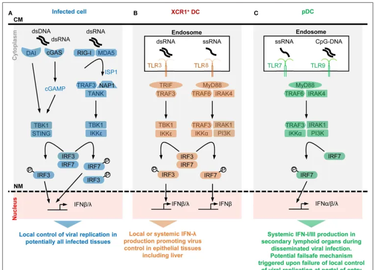
FIGURE 1 | A simplified model of the potential contributions of selective sensors and cell types to IFN production during viral infections. Different
innate immune recognition receptors are involved in sensing various types of viral nucleic acids in distinct categories of cells during viral infections, which may promote different types of anti-viral defenses. For each selected sensor shown, the types of viral nucleic acids recognized and the downstream signaling cascade induced are represented in a simplified, schematic manner. The potential specific role of each cell type in anti-viral defenses is also indicated at the bottom of each panel.(A) Potentially all types of infected cells
can detect endogenous viral replication through cytosolic sensors triggering their local production of IFN-β/λ to control viral replication in an autocrine and paracrine fashion in infected tissues.(B) Uninfected XCR1+DCs selectively
produce high levels of IFN-λ and IFN-β upon engulfment of materials containing dsRNA and the consecutive triggering of TLR3 in endosomes. The receptor of IFN-λ is mostly expressed by epithelial cells. Hence, XCR1+
DCs might be involved in inducing local IFN responses in virally infected epithelial
tissues. Since XCR1+DCs are especially efficient at producing IFN-III upon
HCV stimulation, they might contribute to local or systemic IFN production during infection with this virus, to promote IFN-λ-mediated protection of hepatocytes. Uninfected XCR1+DCs and other uninfected cells may produce
some IFN-β upon engulfment of materials containing ssRNA and the consecutive triggering of TLR8 in endosomes. The contribution of this pathway to anti-viral defense is not well understood yet, in part because mouse TLR8 is deficient for this function.(C) Uninfected pDCs selectively
produce high levels of all subsets of IFNs upon engulfment of materials containing ssRNA or CpG DNA and the consecutive triggering of TLR7/9 in endosomes. However, pDCs seem to be activated for this function only in lymphoid tissues. Hence, pDC might contribute to systemic IFN production during blood-borne viral infections or as a failsafe mechanism activated upon abnormal widespread dissemination of a viral infection once it has escaped local confinement at its portal of entry. CM, cell membrane; NM, nuclear membrane.
particular nucleotide sequences or tertiary structures, their sig-naling pathways and their physiological significance have been recently reviewed (31, 32). Cytosolic RNA sensors encompass DExD/H helicases among which the retinoic-acid-inducible gene (RIG)-I-like receptors (RLRs) have been the most studied, namely RIG-I and melanoma differentiation associated gene 5 (MDA5). RIG-I recognizes RNA with a 50
-PPP or 50
-PP (38) (uncapped) moiety, or double-stranded RNA (dsRNA), both structures being
present in viral, but not in cytosolic eukaryotic, RNA molecules. MDA5 might specifically recognize long dsRNA fragments. Both RIG-I and MDA5 contain a DexD/H box-containing RNA heli-case domain, and 2 caspase recruitment domains (CARD1/2), which bind to mitochondrial anti-viral signaling protein (MAVS). RNA/RLR/STING molecular complexes initiate a signaling cas-cade leading to IRF3/7-dependent induction of IFNs (Figure 1). Other DExD/H helicases can promote IFN-I production in DCs,
although their physiological roles for in vivo immune defenses against viral infections remain to be established (32). Cytosolic DNA sensors able to induce I (mostly β) and IFN-III encompass molecules belonging to different protein fami-lies, including DExD/H helicases, the inflammasome component IFN-γ-inducible protein 16 (IFI16), the Z-DNA binding pro-tein 1 (ZBP1), and the cyclic GMP-AMP (cGAMP) synthase (cGAS) (31, 32). Most of the cytosolic DNA sensors activate STING and lead to IRF3/7- and NFκB-dependent induction of IFN-β and IFN-III. Many cell types express ZBP1 and are able to produce IFN-I upon triggering of this molecule, including macrophages, DCs, and fibroblasts following an HSV-1 infection (39,40). Upon DNA binding, cGAS catalyzes the production of cGAMP. cGAS is critical for the detection of lentiviruses including HIV-1/2 (41,42) and can contribute to sensing of, and protec-tion against, other RNA viruses, including in vivo in mice (43). cGAMP also acts as a secreted second messenger signal alerting uninfected cells to directly induce their expression of intrinsic immune anti-viral defenses. The cGAS/STING/IRF3 signaling cas-cade and the IRF1 transcription factor are “master” inducers of cell-intrinsic immunity able to control the replication of most DNA and some RNA viruses at least in part independently of IFNs (43).
Viral hijacking of the protein synthesis apparatus of the host cell triggers ER overload, a stress, which synergizes with cytosolic sensing to promote IFN-I production. Infected cells become a factory for production of viral particles. Hijacking of the trans-lation apparatus of the host cell for massive production of viral structural proteins leads to an overload of the capacity of the ER for correct folding of newly synthesized proteins. ER overload induces a homeostatic response of the cell, the unfolded protein response (UPR). UPR aims at restoring normal ER functions by inhibiting translation. UPR activation in infected cells contributes to prevent viral replication, including through inhibition of the production of viral proteins, promotion of IFN-I production, and induction of cell suicide (44).
IFN-I production in uninfected cells is initiated by endosomal sensing of viral nucleotide sequences derived from engulfed virions or infected cells
Toll-like receptors (TLRs) are among the first and best character-ized I2R2s. TLRs are transmembrane proteins with a leucine-rich
repeat extracellular domain involved in ligand recognition and an intracellular toll/interleukin-1 receptor domain essential for signaling (45). Among the nine TLRs conserved between mouse and human, TLR3, TLR7, TLR8, and TLR9 are located in endo-somes where they can detect the abnormal presence of nucleic acids such as occurs upon endocytosis of virions or of virally infected cell material. TLR3 recognizes dsRNA, TLR7/8 ssRNA, and TLR9 DNA sequences containing unmethylated cytidine-phosphate-guanosine (CpG) motifs. TLR fine specificity and sig-naling pathways have been reviewed recently (32) and are sum-marized in Figure 1. We will discuss the expression patterns and functions of endosomal TLRs with regards to IFN pro-duction in uninfected specialized immune cell types, pDCs and XCR1+
DCs.
Selective expression of TLR7, TLR9, and IRF7 in pDCs endows them with a unique ability to produce very high amounts of all subtypes of IFNs upon virus stimulation irrespective of their own infection. Plasmacytoid DCs uniquely produce very large amounts of IFNs in response to in vitro stimulation with many viruses, without being infected (46). IFN-I mRNAs represent up to 40% of all mRNAs in pDCs at the peak of their activation (47).
In vitro, upon exposure to influenza virus, herpes virus type 1,
cytomegaloviruses, or vesicular stomatitis virus, individual pDCs produce 100–1000 times more IFNs than total PBMCs, monocytes, MoDCs, cDCs, neutrophils, and fibroblasts (47–52). However,
in vitro, high molarity infection of cDCs with certain viruses
unable to inhibit IFN-I production in their target cells can also induce massive IFN-β secretion (53). pDCs produce high levels of all subtypes of IFNs, contrary to many other cell types including infected cells, which often preferentially produce IFN-β (46, 47).
In vivo, pDC depletion during systemic viral infections leads to
over 95% decrease of IFN-I production, while the total number of pDCs producing IFN-I (<100,000 in one mouse) is much lower than the total number of infected cells (54–59). This shows that
in vivo also individual activated pDCs produce much more
IFN-I/III than most other cell types, including virus-infected cells. The professional IFN-producing function of pDCs largely results from their high constitutive and selective expression of IRF7, TLR7, and TLR9 (Figure 1). These molecules are pre-associated in ready-to-signal complexes located in specialized endosomes specific to pDCs (60,61). pDCs must also be equipped for efficient sensing and up-take of virions and virus-infected cells. The corresponding cell surface I2R2s remain to be identified.
Selective expression of TLR3 in XCR1+
DC endows them with a unique ability to produce very high amounts of IFN-β and IFN-III upon stimulation with dsRNA or HCV irrespective of their own infection. XCR1+
DCs are very potent for antigen-specific activation of CD8+
T cells, in particular through cross-presentation of exogenous antigens that they have captured from other cells and processed for association with class I major com-plex histocompatibility (MHC-I) molecules (62). In mice, XCR1+
DCs are crucial for the initiation of protective adaptive immune responses against tumors and a variety of viruses (63). Mouse and human XCR1+
DCs constitutively and selectively express high levels of TLR3 (Figure 1). They produce large amounts of IFN-III and IFN-β upon stimulation with a synthetic mimetic of dsRNA, Polyinosinic:polycytidylic acid (PolyI:C) (64,65). Human XCR1+ DCs uniquely respond to stimulation with HCV by producing large amounts of IFN-III in a TLR3-dependent manner (66,67), irrespective of their own infection.
Positive and negative feedback loops regulating IFN-I production
Positive feedback loops. In addition to IRF7 induction, other positive feedback mechanisms exist to amplify the production of IFNs rapidly after initiation of a viral infection as illustrated by the following selected examples. IFNs induce the expression of many cytosolic RNA/DNA sensors and of TLR7. This broad-ens the spectrum of host’s cell types able to detect endogenous viral replication for IFN induction. Induction of OASL by IFNs in human cells enforces RIG-I signaling, counteracting viral immune
evasion genes interfering with this sensing pathway (68). The IFN-inducible ribonuclease L (RNaseL) generates viral and cellular RNA degradation products, which engage RLRs for amplification of IFN production (69,70). The IFN-inducible Protein kinase R (PKR) stabilizes IFN-I mRNA (71).
Negative feedback loops. To prevent unbridled responses delete-rious for the host, IFN activity must be tightly controlled including during viral infections. Several negative feedback loops exist to terminate IFN production, after anti-viral defenses have been acti-vated. The ISG ubiquitin specific peptidase 18 (USP18) binds to IFNAR2, preventing it from recruiting signal transducer and activator of transcription 1 (STAT1). IFNs induce the expres-sion of TAM receptor tyrosine kinases in DCs, monocytes, and macrophages. TAM receptors associate and signal in part through IFNAR1. They activate the suppressors of cytokine signaling-1/3 (SOCS-1/3). SOCS inhibit TLR and RLR signaling, thereby ter-minating IFN production (72). TAM receptor ligands, Gas6 and ProS, bind phosphatidylserine on dying cells and are produced by activated DCs and monocytes/macrophages. Thus, IFN induction of TAM inhibitory receptors on uninfected phagocytic immune cells could limit their propensity to produce the cytokines upon engulfment of dying virally infected cells. IFNs induce Tetherin on most cell types. pDCs express a receptor for Tetherin, leukocyte immunoglobulin-like receptor, subfamily A (with TM domain), member 4 (LILRA4). LILRA4 triggering on pDCs inhibits their production of IFN-I. Hence, through LILRA4 engagement by Tetherin, pDCs can monitor their efficacy at inducing an anti-viral gene expression program in neighboring cells through IFNs, and timely terminate their IFN production.
How positive and negative feedback loops integrate in time and space to promote optimal kinetics and intensity of IFN produc-tion in order to efficiently control viral infecproduc-tion without causing severe immunopathology is not completely understood. Positive feedback loops may occur very rapidly after initiation of viral infection to allow rapid secretion of high levels of the cytokines for fast and strong induction of anti-viral cell-intrinsic immu-nity. Negative feedback loops occur likely later to terminate the response and thus avoid chronicity of cytokine production and its ensuing deleterious effects.
What are the respective roles of infected versus uninfected cells in IFN production during viral infections?
IFN production by infected cells serves as first line of defense to block viral replication at his portal of entry in the body, while IFN production by uninfected pDCs might constitute a failsafe mechanism activated only when viral infection gets systemic. pDCs do not constitute the major source of IFN production upon local infections by several viruses in the lung or in the female reproductive tract. pDCs are dispensable for resistance against these infections (56,73,74). During pulmonary infection by New-castle disease virus (NDV), IFN-I are produced locally in the lungs mainly by infected alveolar macrophages. Lung pDCs do not express the cytokines (73). Selective depletion of lung alveo-lar macrophages leads to systemic dissemination of NDV, and to a strong activation of pDCs for IFN-I production specifically in the spleen. Even in the case of systemic viral infections such as
caused by intravenous injection of NDV or intraperitoneal injec-tion of mouse cytomegalovirus (MCMV), pDC IFN producinjec-tion is confined to the spleen. It is not detected in other organs even those with high viral replication (59,73). Hence, splenic pDCs are especially prone to high level IFN production upon systemic acute viral infections. pDCs located in non-lymphoid organs, in particular mucosal barrier tissues, appear to be inhibited for IFN production. Thus, IFN production by infected cells serves as first line of defense to block virus replication at its portal of entry in the body. IFN production by uninfected pDCs might constitute a failsafe mechanism mainly activated in the spleen when viral infection gets systemic (75). Under these conditions, to promote health over disease, the benefits for the host of producing high cir-culating levels of IFNs in order to induce widespread cell-intrinsic anti-viral defenses might prevail over the deleterious effects that this could cause on certain cell types or tissues. Indeed, pDCs are required for protection against HSV-2 and HSV-1 in mice only in systemic but not local infections (56). This observation is consis-tent with the crucial role of pDCs for protection of mice against systemic infection by mouse hepatitis virus (MHV), a fast repli-cating coronavirus (55). Conflicting results have been obtained on the role of pDCs during intranasal influenza infection (74,76–78). A possible explanation is that pDC IFN production contributes to resistance to highly pathogenic influenza strains that might sys-temically spread from the lung early after infection, even if at low levels. Another intriguing observation is that IFNs are critical for host resistance to MCMV and that pDCs are the major source of IFNs in this infection model but are dispensable for virus con-trol (54). Studies are ongoing to understand this apparent paradox. Patients bearing genetic mutations disrupting endosomal TLR sig-naling do not appear to suffer from life-threatening viral infections (79,80), contrary to patients impaired in IFNAR signaling (4,81). A notable exception is the specific susceptibility to severe herpes virus encephalitis in individuals’ deficient for TLR3 signaling (82,
83). However, contrary to extracellular TLR, endosomal TLR have evolved under strong purifying selection in human beings (84). Hence, while pDCs and endosomal TLR might have been required for protection of our species against viral infections in the past, this appears not to be the case anymore perhaps due to improved social, hygiene, and health care in modern society (75).
IFN production by uninfected pDCs or XCR1+
DCs might pro-mote protection against viruses able to interfere with the sig-naling pathways inducing cytokine production in infected cells. Attesting to the importance of IFNs for anti-viral defense in vertebrates, many mammalian viruses encode immune evasion genes specifically inhibiting the production of IFNs in infected cells (39,85). pDCs or XCR1+
DCs might be essential for IFN-dependent host protection against these viruses, because they are spared from the intracellular functions of viral immune evasion genes (75). To the best of our knowledge, MCMV does not encode for immune evasion genes inhibiting IFN production. However, MCMV manipulates IFN-I responses through specific inhibition of STAT1 functions in infected cells. Thus, pDCs might be dis-pensable for resistance against systemic MCMV infection due to sufficient levels of IFN production by infected cells locally in all infected tissues. Hepatocyte responses to IFN-III appear to play a
critical role in human resistance to HCV. In infected hepatocytes, HCV induces the expression of cellular microRNAs binding to IFN-III mRNA and leading to its degradation. Uninfected XCR1+
DCs produce high levels of IFN-III in vitro upon HCV stimula-tion (66,67). Hence, during acute HCV infection in vivo, XCR1+
DC may be a strong and early source of IFN-III not subjected to virus immune evasion strategies, therefore, contributing to protect naturally resistant individuals.
Altruistic suicide of subcapsular sinus macrophages in secondary lymphoid organs promotes strong IFN responses to control viral dissemination. In secondary lymphoid organs, a subset of macrophages is critical for the clearance of viruses from the lymph (86). These macrophages are located on viral entry routes, near to subcapsular sinuses where the afferent lymph drained from non-lymphoid tissues flows. Contrary to other subsets of macrophages, subcapsular sinus macrophages are highly suscepti-ble to viral infection, because they constitutively express only low levels of effector molecules of cell-intrinsic anti-viral immunity and because their responses to IFNs are inhibited by their high constitutive expression of USP18. Subcapsular sinus macrophages rapidly become infected by viruses incoming from the lymph and produce large amounts of IFNs. This altruistic suicide prevents virus dissemination to other adjacent cell types and promotes the induction of innate and adaptive anti-viral immunity (87). HOW ARE IFNs PROMOTING ANTI-VIRAL IMMUNITY?
IFN direct anti-viral effector functions: induction of effector molecules of cell-intrinsic anti-viral immunity
Upon instruction by IFNs, cells express a wide variety of viral restriction factors, whose combined action blocks pathogen invasion by interfering with the different stages of viral life cycle (Figure 2A). This has been extensively reviewed recently (88) and will only be described succinctly here. Virus fusion with host cell membrane can be blocked by Cholesterol-25-hydrolase (CH25H) that inhibits sterol biosynthesis. Some viruses enter cells by escaping from endosomes/lysosomes, which can be blocked by interferon inducible transmembrane (IFITM) proteins. Virus uncoating can be blocked by tripartite motif (TRIM) proteins, such as TRIM5α, which bind to HIV-1 cap-sid thus promoting its degradation, and by Myxoma resistance GTPases, MX1, and MX2, which efficiently trap viral structural proteins at an early stage following virus entry into the cell. MX1 inhibits a number of viruses, including influenza virus through sequestration of its nucleocapsid. MX2 associates with host cyclophilin A and HIV-1 capsid protein. Virion assem-bly can be blocked at transcriptional, translational, and post-translational levels. The adenosine deaminase acting on RNA 1 (ADAR1) and the apolipoprotein B mRNA editing enzyme, catalytic polypeptide-like (APOBEC) deaminases induce viral RNA destabilization and hypermutation (89, 90). The sterile alpha motif and histidine-aspartic domain (HD) containing pro-tein 1 (SAMHD1) blocks reverse transcription by hydrolyz-ing dNTPs (91). ADAR1, APOBEC, and SAMHD1 functions have been mainly studied in infections by HIV-1 and other retroviruses. The 20
,50
-oligoadenylate synthase (OAS) proteins, the IFN-induced proteins with tetratricopeptide repeats (IFIT),
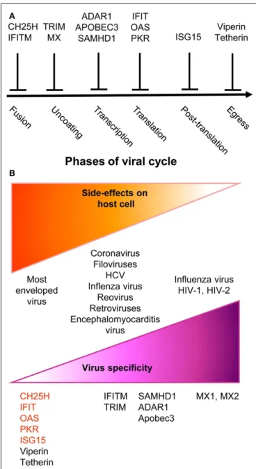
FIGURE 2 | A simplified and schematic view of the modes of action and specificities of ISG acting as direct anti-viral restriction factors. (A)
Different IFN-inducible restriction factors can block viral replication in infected cells in a cell-intrinsic manner at different stages of the viral life cycle.(B) Viral specificity (purple) might be inversely correlated to the
breadth of side effects on host cells (orange).
and PKR inhibit viral and host protein translation by using complementary mechanisms (88). The major post-translation modification induced by IFNs is the binding of the ubiquitin-like modifier ISG15 to several viral and host proteins, a process called ISGylation. Most of the known ISGylated proteins are tar-geted for degradation, with few exceptions that are on the contrary stabilized like IRF3 (88). Finally, the egress and budding of viri-ons of many enveloped viruses can be inhibited by Tetherin or by Viperin (88).
Many anti-viral ISGs have been functionally characterized only recently, largely thanks to large-scale screening approaches. They display a variable degree of viral specificity (43,92) that might inversely relate to the extent of their side effects on host cells (Figure 2B). Anti-viral effectors acting on a broad spectrum of viruses often target key metabolic pathways that are also crucial for host cell functions. This is the case for the control of cho-lesterol metabolism by CH25H (93) or of protein translation by PKR, OAS, or IFITs (88). Other anti-viral restriction factors such as MX2 may specifically target one molecule of a very restricted set of viruses with no apparent side effects on host cells. Some anti-viral ISGs target specific functions critical for only a restricted array of viruses and might similarly exert side effects only on a subset of host cell types. For example, SAMHD1 inhibits retrovirus replica-tion through dNTP deplereplica-tion, which might more specifically affect proliferating host cells. Hence, the infected host must balance the intensity, breadth, and location of ISG induction to circum-vent viral replication while precircum-venting life-threatening damages to vital cell types or tissues. One of the mechanisms contributing to this balance is translational control of the expression of ISGs, especially those with pro-apoptotic or anti-proliferative functions (94). While many anti-viral ISGs are transcriptionally activated in most IFN-stimulated cells, their translation can be specifically blocked in uninfected cells by cellular microRNA. This inhibi-tion is relieved upon cell infecinhibi-tion through negative regulainhibi-tion of the function of the RNA-induced silencing complex. Hence, IFN stimulation of uninfected cells prepares them for rapid and strong induction of cell-intrinsic anti-viral defenses upon viral infection while avoiding their unnecessary exposure to the toxic effects of certain ISGs.
Further knowledge on the functions and the dynamic regula-tion of ISGs is essential to develop novel therapeutic strategies against viral infections aiming at modulating IFN responses to promote their protective anti-viral cell-intrinsic functions over their deleterious toxic effects. A better understanding of the immunoregulatory effects of IFNs will also help.
IFN orchestration of anti-viral responses of both innate and adaptive immune cells
Type I interferon can modulate the functions of a broad spec-trum of immune cells (Figure 3A). We will review this knowledge, focusing on the functions of DCs, NK cells, T cells, and B cells, since they are involved in the control of most viral infections. We will discuss the hypothesis that DCs play a central role in IFN-I orchestration of innate and adaptive immunity for the induction of optimal anti-viral defenses (Figure 3).
During viral infections and cancer immunosurveillance, IFN-I constitute one of the most important input signal acting on DCs to promote their delivery of appropriate output signals to T cells, B cells, and NK cells for protective immunity (Figure 3A). DCs deliver three types of signals to activate and functionally polarize T cells. Signal 1 is the triggering of the T cell receptor by viral peptide-MHC complexes. Signal 2 is the triggering of activating T cell co-stimulation receptors such as CD28 or CD27 by the CD80/86 and CD70 co-stimulation molecules expressed on DCs. Signal 3 corresponds to cytokines, which can promote T cell proliferation and acquisition of specific effector functions. Under steady state conditions, most DCs are in an immature state characterized by low level expression of MHC-II (signal 1) and co-stimulation mol-ecules (signal 2) and by the lack of production of T cell-activating
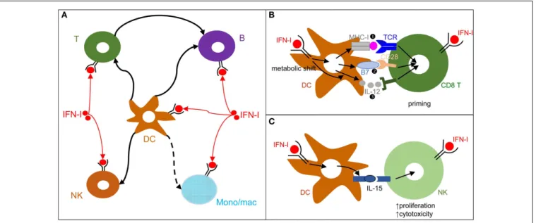
FIGURE 3 | DCs play a central role in IFN-I orchestration of innate and adaptive immune responses. (A) IFN-I exert cell-intrinsic as well as indirect
effects on a variety of immune cell populations. DC responses to IFN-I play a major role in promoting protective activation and functional polarization of other innate and adaptive immune cells, not only during viral infections but also in other physiopathological situations including cancer.(B) DC
cell-intrinsic responses to IFN-I endow them to deliver appropriate signals for T
cell priming and functional polarization. IFN-I can modulate all three types of signals delivered by DC to T cells: MHC-I/antigenic peptide complexes ( ), co-stimulation ( ), and cytokines ( ). This depends both on IFN-I-dependent transcriptional induction in DC of some of the corresponding genes and on IFN-I-dependent metabolic reprogramming of DC.(C) DC cell-intrinsic
responses to IFN-I endow them to deliver appropriate signals, in particular IL-15 trans-presentation, for NK cell activation. See main text for further details.
cytokines (signal 3). Upon activation, including early after viral infections in vivo, DCs up-regulate their expression of signal 1 and activating signal 2 and secrete T cell-activating cytokines. This process is called DC maturation. Gene expression profiling of DCs stimulated by microbial stimuli identified a core set of genes up-regulated in mature DCs irrespective of the stimulus they receive, irrespective of the subset they belong to, and conserved across evo-lution (95). Most of these genes are induced during DC maturation in part through cell-intrinsic IFN-I signaling (95). Consistently, cell-intrinsic IFNAR signaling in DCs is required in many cir-cumstances for the induction of protective immunity, including efficient CD8 T cell responses during viral infection or tumor development (96–98), Th1 responses upon PolyI:C injection inde-pendently of IL-12 or IFN-γ effects (99,100), as well as follicular helper T cell and humoral responses (101,102). Mechanistically, IFN-I promote DC immunogenicity for efficient T cell activation through a variety of effects (Figure 3B). It drives DC up-regulation of signal 2 in vivo during viral infections (103) and boosts their capacity to cross-present antigens for increased delivery of signal 1 to CD8 T cells (96–98). It shapes their delivery of activating sig-nal 3, in particular inducing IL-15 and promoting or inhibiting IL-12 depending on experimental conditions (58,104). Finally, it is necessary to induce their metabolic shift from mitochondrial oxidative phosphorylation to aerobic glycolysis, which fuels the increased needs in energy and the expansion of the intracellu-lar organelles required for the production and proper intracelluintracellu-lar routing of the signal 1 and 2 proteins (100,105). Selective inactiva-tion of IFNAR on cDCs compromises mouse resistance to MCMV and MHV infections (103,106). In contrast, IFNAR expression is not required on NK cells for protection against MCMV and on pDCs, T cells, and B cells for early control of MHV replica-tion (103,106). Although cell-intrinsic IFN-I signaling in NK cells can promote their activation (107) (Figure 3A), IFN-I-induced IL-15 trans-presentation by DCs plays a more prominent role for this function in many conditions including in vivo during MCMV infection (103,108) (Figure 3C).
Cell-intrinsic IFN-I signaling in CD4 T cells (109), CD8 T cells (110,111), and B cells (112) can also contribute to their efficient activation and functional polarization (Figure 3). This depends on experimental settings. CD8 T cell-intrinsic IFN-I responses are crucial for mounting efficient cytotoxic CD8 T cell responses against LCMV but are less critical against Vaccinia virus and vesic-ular stomatitis virus (110,113,114). Mechanistically, cell-intrinsic IFN-I signaling in CD8 T cells can promote their survival during their antigen-induced proliferation (110). Cell-intrinsic signaling in DCs and CD8 T cells may act in a synergistic manner. Indeed, conditional inactivation of IFN-I responsiveness was required to occur simultaneously in both of these two cell types to dramati-cally affect CD8 T cell expansion upon vaccination with a modified Ankara vaccinia virus (115).
In summary, IFN-I generally play a crucial, beneficial, role in immune defenses against viral infections, both through the induc-tion of cell-intrinsic anti-viral defenses and through the orchestra-tion of innate and adaptive immunity. However, if these responses are not properly regulated, they can contribute to diseases as we will now discuss.
DIFFERENT, AND SOMETIMES OPPOSITE, PROCESSES UNDERLIE DELETERIOUS IFN-I RESPONSES DEPENDING ON THE PHYSIOPATHOLOGICAL CONTEXTS
DELETERIOUS EFFECTS RESULTING FROM THE INDUCTION OF UNBRIDLED INFLAMMATORY RESPONSES CAUSING SEVERE TISSUE DAMAGE, AS EXEMPLIFIED IN AUTOIMMUNE DISEASES
A frequent side effect of IFN-I treatment against cancer or chronic viral infections is the induction of autoimmune reactions. Consis-tently, ISG expression is a hallmark of many spontaneous systemic or tissue-specific autoimmune diseases, including systemic lupus erythematosous (SLE), Sjogren’s syndrome, psoriasis, and other skin disorders (11). The dysregulation of IFN-I responses observed in patients with these autoimmune diseases likely results from both genetic and environmental factors. Genome-wide associa-tion studies show that polymorphisms in genes involved in IFN-I responses strongly correlate with increased susceptibility to many autoimmune diseases (11). Diverse environmental factors can also contribute to the onset of autoimmune diseases. Microbial infections often precede first clinical manifestations of autoim-mune diseases. Whether infections (116) and/or alterations in the commensal microbiota of the affected barrier tissues (117,118) are the cause or rather the consequence of autoimmunity is still matter of debate. Infection- or dysbiosis-induced tissue damages and unbridled IFN-I responses can contribute to initiate autoim-mune reactions. Gender is another prominent factor affecting susceptibility to autoimmune diseases. Women are more prone to autoimmunity, which may result from endocrine regulation of IFN-I responses. pDC IFN-I production is enhanced in human and mouse females, due at least in part to cell-intrinsic enhance-ment of TLR7/9 responses by the female hormone estradiol (119). In autoimmune diseases, different mechanisms could operate to initiate the dysregulation of immune responses leading to a vicious circle of reciprocal activation between innate IFN-I responses and adaptive self-reactive lymphocyte responses (Figure 4). Adaptive immune cells are educated to spare “self.” This occurs through negative selection of potentially autoimmune B and T cells during their development in the bone marrow or thymus, respectively, a process called central tolerance. Self-reactive B or T cells that have escaped this pruning can be either deleted or functionally inactivated once they have egressed in secondary lymphoid organs or non-lymphoid tissues, a process called peripheral tolerance. In some individuals, polymorphisms in genes involved in the pro-motion of central or peripheral tolerance lead to a higher number, diversity, and/or responsiveness of self-reactive lymphocytes in the periphery, in particular of B cells secreting DNA or anti-RNP antibodies (120,121). Mammalian DNA or RNA are poor inducers of pDC IFN-I induction under normal conditions. How-ever, pre-existing anti-DNA or anti-RNP autoantibodies can break this innate tolerance of pDC. Indeed, antibodies binding to self nucleic acids can protect them from degradation and compact them into nanoparticles that are very effective for the induction of IFN-I in pDC (Figure 4). DNA-containing immune com-plexes (ICs) are frequently found in the serum of SLE patients (SLE-ICs) and can activate pDC IFN-I production (122). In turn, pDC IFN-I activate cDCs, monocytes (123), and B cells, lead-ing to a vicious circle of reciprocal activation between DCs and
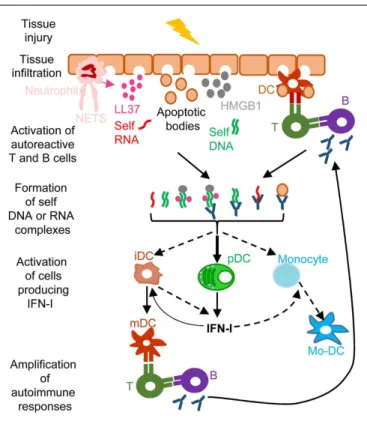
FIGURE 4 | A simplified model of the deleterious role of IFN-I in several autoimmune diseases. When exposed to different kinds of injuries
(microbial infection, commensal microbiota dysbiosis, chemical or physical insults), healthy tissues can undergo cell damage and death. These events induce the release of apoptotic bodies encompassing self RNA or DNA. Neutrophil recruitment and activation in inflamed tissues can also constitute a potent source of self nucleic acids, through the release of neutrophil extracellular traps (NET). Self RNA or DNA can associate with cationic peptides (e.g., LL37) as shown in psoriatic patients or with inflammatory molecules (e.g., high mobility group box 1, HMGB1) to generate nanoparticles that are extremely efficient for IFN-I production by pDC and eventually other cell types. pDC can also be efficiently activated for IFN-I production by immune complexes (ICs) generated by the association between self nucleic acids and auto-antibodies as frequently found in the serum of systemic lupus erythematosus patients. IFN-I promote the differentiation and/or the maturation of antigen-presenting cells, in particular different subsets of DC. Activated DC can then present self-antigens for activation of auto-reactive T CD4+
cells, including follicular helper lymphocytes, which in turn activate auto-reactive B cells for auto-antibody secretion, leading to a vicious circle of reciprocal activation between innate and auto-reactive adaptive immune cells. iDC, immature DC; mDC, mature DC; Mo-DC, monocyte-derived DC. See main text for further details.
self-reactive lymphocytes and to the exacerbation of autoimmune responses (Figure 4). Certain infections or dysbiosis of the com-mensal microbiota of the affected barrier tissues could promote chronic production of host amphiphatic peptides able to combine with eukaryotic DNA or RNA, likely released from dying cells, thus forming pDC-activating nanoparticles. Indeed, in psoriatic skin, both a high expression of LL37 and a massive infiltration of pDCs is observed (124) (Figure 4). Hence, to treat many autoim-mune diseases, novel therapeutic strategies could be designed to target dysregulated pDC IFN-I production or B cell activation by IFN-I.
DELETERIOUS RESPONSES RESULTING FROM INAPPROPRIATE FUNCTIONAL POLARIZATION OF IMMUNE RESPONSES, AS EXEMPLIFIED IN FAILURE TO CONTROL SECONDARY BACTERIAL OR FUNGAL INFECTIONS
One of the most common complications of primary infections by many respiratory viruses, in particular influenza virus, is a life-threatening pneumonia due to secondary pulmonary infections by bacteria, such as Streptococcus pneumoniae, Staphylococcus aureus, or Haemophilus influenza (125, 126). These pathologies affect especially infants, elderly, and immunocompromised patients. Retrospective studies indicate that secondary bacterial pneumonia was highly recurrent in lung tissues isolated from patients who died during last century influenza pandemics, independently of antibi-otic availability (127, 128). Influenza virus induces high IFN-I responses in human beings and mice. In both hosts, secondary bac-terial infections are lethal only when they occur in a limited time window following primary viral infection (3–7 days), around the peak of IFN-I responses, before complete virus clearance. Mouse models of viral/bacterial coinfections are being used to dissect disease mechanisms (129). IFNAR1-deficient mice appear more resistant to secondary pulmonary bacterial infections, showing that IFN-I responsiveness contributes to disease (130). Similarly, after lymphochoriomeningitis virus (LCMV) infection, wild-type but not IFNAR1-deficient mice are more susceptible to LPS-induced septic shock (131). Several mechanisms may contribute to the detrimental role of IFN-I in secondary bacterial infections (Figure 5). Early during viral infection, IFN-I decrease the host ability to control bacterial replication, by dominantly polarizing immune responses toward anti-viral functions, simultaneously inhibiting the development of the types of immune responses required for protection against most bacterial infections. IFN-I can inhibit the production of chemokines required for the recruitment to the respiratory tract of antibacterial effector innate immune cells, in particular neutrophils or monocytes/macrophages (132,
133) (Figure 5). Depending on the experimental models used, IFN-I can on the contrary induce a CCR2-dependant recruit-ment of classical monocytes (134). In infected tissue, IFN-I might skew the functional polarization of resident or infiltrating mono-cytic cells toward immunosuppression, because it does limit their antibacterial functions by inhibiting their IL-1 production (135–
137) while it might promote their production of IL-10 and nitric oxygen intermediates. The exact nature of infiltrating mono-cytic cells is not clear and could correspond to activated classical monocytes, MoDCs, monocyte-derived macrophages, or myeloid-derived suppressor cells (MDSCs). The boundaries between these putatively different cell types are currently ill-defined (138). These cells could fuel local replication of monocyte/macrophage-tropic bacteria (134), be immunosuppressive (139) or contribute to local immunopathology (140). The role of IFN-I on mono-cytes/macrophages is complex and will require further investiga-tions to determine when it is protective versus deleterious and what the underlying mechanisms are. Depending on the con-text, IFN-I can either promote or inhibit the induction of Th1 cytokines such as IL-12 and IFN-γ, and myeloid cell responses to IFN-γ (10, 141-143). IFN-I can also polarize CD4 T cell responses toward Th1 at the expense of Th17, while the Th17-type cytokines IL-17A and IL-22 are required for host defense against pulmonary
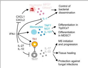
FIGURE 5 | A simplified model of the deleterious role of IFN-I in secondary pulmonary bacterial infections or in fungal infections and of their protective role in multiple sclerosis (MS). IFN-I-dependent signals
can block CXCL1 and CXCL2 production, thus inhibiting the recruitment and activation of neutrophils in inflamed tissues, hampering their protective functions against secondary pulmonary bacterial infections or fungal infections. IFN-I-dependent signals can enhance CCL2 production, promoting the recruitment in inflamed tissues of CCR2+monocytes that
can potentially differentiate into TipDC or into MDSC. This can contribute to disease either through enforcing T cell activation leading to
immunopathology or on the contrary through suppressing anti-microbial immune defenses. IFN-I can either inhibit or promote Th1 responses, which in the latter case occurs at the expense of Th17 responses thus
compromising the production of IL-17 and IL-22, which are respectively required for control of microbial replication and for tissue healing. IFN-I-induced IL-10 and IL-27 can directly inhibit Th17. In MS, inhibition of Th17 functions may contribute to the protective effects of IFN-β therapy. MPO, myeloperoxidase; TNF, tumor necrosis factor alpha; NO, nitric oxide. See main text for other abbreviations and further details.
bacteria by inducing the production of anti-microbial peptides and of tissue repair molecules (Figure 5) (141–143). IFN-I may not only affect host resistance to bacterial infection, but also host tolerance, i.e., the ability of the host to tolerate a given burden of pathogen without undergoing excessive tissue damages (143,
144). Hence, to counter IFN-I deleterious effects during secondary bacterial infections, it will be important to better delineate the respective contribution of lung tissue tolerance modulation and of immune-mediated resistance weakening.
Another well documented example of deleterious effects of IFN-I due to their inappropriate functional polarization of immune responses is the enhanced susceptibility to fungal infec-tions of patients with genetically determined hyperactive IFN-I responses, as exemplified in the hereditary disease Chronic Muco-cutaneous Candidiadis (CMC) (Figure 5) (145). Patients with CMC have a significant deficit in Th17 CD4 T cells, at least in part as a consequence of altered responsiveness to IL-6 or IL-21. Several STAT1 mutations were identified in patients with auto-somal dominant CMC. Gain-of-function STAT1 mutations were found to hard wire CD4 T cell responses to cytokines toward STAT1 signaling, compromising their STAT3-dependent ability to produce IL-17 upon IL-6 or IL-21 stimulation. This was associated
to induction of a global IFN-I transcriptomic signature in blood (145). Deleterious IFN-I effects on immunity to Candida might not only occur in CMC patients but also in other types of indi-viduals upon secondary fungal infections occurring shortly after a primary viral infection, likewise to the situation discussed above for secondary bacterial infections. Indeed, PolyIC induced IFN-I abrogate innate immunity to systemic candidiasis in mice (146), and IFNAR-deficient mice can be more resistant to Candida infec-tion under certain experimental settings (147). However, the role of IFN-I in the modulation of the ability of immunocompetent hosts to control fungal infection is disputed (148,149).
The inhibition of Th17 responses by IFN-I could be protec-tive in at least one important human pathology, MS (Figure 5). MS represents a striking exception to the previously discussed detrimental role of IFN-I in autoimmune diseases. Indeed, a large proportion of MS patients have low serum IFN-I activity and low ISG levels. These MS patients present a significant reduction of MS relapse upon IFN-β administration (150). The underlying mechanisms are not yet completely unraveled. However, in the experimental autoimmune encephalitis mouse model of MS, Th17 responses bear a major contribution to nervous system damages and are inhibited by the IL-10 and IL-27 induced upon IFN-I administration (151).
In summary, IFN-I responses can be deleterious in autoim-munity by promoting a vicious circle of reciprocal activation between innate immune cells and auto-reactive CD4 T or B lym-phocytes. IFN-I responses can also be deleterious upon secondary bacterial or fungal infections in the lung or the kidneys occur-ring shortly after a primary viral infection, by compromising the recruitment of anti-microbial innate effector cells and/or by pre-venting the proper functional polarization of immune responses. We will now discuss how IFN-I responses can also compromise host immune defenses against certain viruses and promote chronic infections.
DELETERIOUS RESPONSES RESULTING FROM THE INDUCTION OF IMMUNOSUPPRESSION, AS EXEMPLIFIED IN CHRONIC LCMV INFECTION
Different LCMV strains such as Armstrong and clone-13 (Cl13), respectively, lead to acute versus chronic infections in mice. A hallmark of chronic LCMV infection is the loss of the prolifer-ative potential and effector functions of anti-viral CD8 T cells, a process called exhaustion. Exhausted CD8 T cells are characterized by a high expression of the inhibitory receptors PD-1, CTLA4, and LAG-3 (152). In vivo blockade of these inhibitory receptors can reverse T cell exhaustion and allow resolution of the chronic infec-tion (152). IFN-I and ISGs are induced early after infection with all strains of LCMV, albeit to lower levels with those leading to chronic infection. This early IFN-I production is critical to limit viral repli-cation (3). In models of acute infection, IFN-I responses rapidly return to normal, undetectable, levels, before viral replication is completely controlled. In contrast, ISG induction is maintained in chronic infection, including the expression of PD-1 ligands on APCs and of the immunosuppressive IL-10 cytokine, consistent with a prolonged expression of IFN-I albeit at low levels (13,14).
In vivo neutralization of IFN-I by antibody administration
restoration of functional anti-viral CD8 T cell responses at least in part through CD4 T cell- and IFN-γ-dependent mechanisms (13, 14). During persistent LCMV Cl13 infection, chronic low level IFN-I production polarizes CD4 T cell responses toward T follicular helper (Tfh) rather than Th1 functions. Thus, chronic IFN-I responses promote enhanced anti-viral B cell responses but facilitate CD8 T cell exhaustion due to deficient CD4 T cell help, therefore contributing to host failure to prevent chronic infec-tion (153). Strikingly, establishment of chronic infection by LCMV Cl13 could also be prevented by early administration of two shots of a high dose of exogenous IFN-I, at days 2 and 5 post-LCMV inoculation. This treatment allowed viral clearance by rescuing anti-viral CD8 T cell from exhaustion (154). Altogether, these studies show that the timing and duration of IFN-I production during viral infections is critical in determining how this response will impact the balance between the virus and the host. An early and robust but transient production of IFN-I promotes strong induction of cell-intrinsic viral restriction mechanisms as well as adequate polarization of adaptive anti-viral immune responses, which combined effects lead to viral clearance. In contrast, if the production of IFN-I is too low and/or too late, both viral repli-cation and low IFN-I responses become chronic, their combined action leading to induction of immunosuppressive effects and to inadequate functional polarization of CD4 T cells. This results in CD8 T cell exhaustion and maintenance of chronic infection. Chronic viral replication and CD8 T cell exhaustion is also a hall-mark of HIV-1 infection. We will now discuss the complex and disputed role of IFN-I in this disease.
THE COMPLEX AND DISPUTED ROLE OF IFN-I IN HIV-1 INFECTION Both in HIV-1 infection and in its most relevant animal model, infection of non-human primates with simian immunodeficiency virus (SIV), disease progression after the acute phase of the infec-tion is associated with high and chronic expression of ISGs while IFN-I production is inconsistently detected (155–157). In con-trast, the individuals that do not progress toward disease despite persistent high viral loads show much lower immune activation, in particular low ISG expression, after the acute phase of the infec-tion (158–161). Hence, chronic low levels of IFN-I are associated to disease progression independently of the level of viral replication. Therefore, an outstanding question still open for a better under-standing of the physiopathology of HIV-1 infection is whether chronic IFN-I responses are merely a marker of progression, or whether they are implicated in driving disease development. In addition to mechanisms similar to those uncovered in the mouse model of chronic LCMV infection, during HIV-1 infection other effects of IFN-I could promote a vicious circle of reciprocal activa-tion between chronic viral replicaactiva-tion and sustained, deleterious immune responses (Figure 6). Very early after HIV-1 infection, in most individuals, IFN-I production might be too weak or too late to induce a combination of cell-intrinsic defense mechanisms and of immune responses efficient enough to prevent later establish-ment of chronic infection. On the contrary, as demonstrated in the case of the mouse model of LCMV infection, IFN-I responses could favor CD8 T cell exhaustion, either by direct cell-intrinsic effects on CD8 T cells (Figure 6, ) or by contributing to deprive them from CD4 T cell help (Figure 6, ). Several effects of IFN-I
might compromise anti-HIV-1 Th1 responses or more generally contribute to the global depletion of CD4 T cells. These mecha-nisms include functional polarization of anti-HIV-1 CD4 T cells toward Tfh rather than Th1 responses, CXCL10 production lead-ing to enhance recruitment of memory CD4 T cells to the sites of viral replication where they fuel chronic viral replication with new HIV-1 target cells (Figure 6, to ), direct pro-apoptotic and anti-proliferative effects on CD4 T cells (Figure 6, ), as well as TRAIL induction on pDCs licensing them for killing CD4 T cells irrespective of their infection (Figure 6, ) (162,
163). Altogether, these mechanisms entertain chronic viral repli-cation and continuous depletion of CD4 T cells, leading to the dramatically enhanced susceptibility to opportunistic infections (Figure 6, ) characteristic of the acquired immunodeficiency syndrome (AIDS) (Figure 6, ). Other lines of evidences have been reported to support a deleterious role of pDC activation during HIV-1 infection. Women undergo faster HIV-1 disease progression than men with similar viral loads, which may result in part from the highest IFN-I production of women’s pDCs includ-ing in response to HIV-1 stimulation (164). pDC recruitment and activation in the vaginal mucosa of female macaques early after local SIV inoculation contribute to attract and activate CD4 T cells, which can then be infected and promote virus dissemina-tion from its portal of entry (165). However, in vivo blockade of pDC IFN-α production by administration of TLR7/9-antagonistic oligonucleotides early after SIV infection of macaques did not decrease T lymphocyte activation, which suggests that additional sources of IFN-I likely contribute to the immune dysfunction observed in SIV/HIV-1 infections. Targeting dysregulated IFN-I responses during HIV-1 infection might represent an interest-ing adjuvant therapeutic strategy to highly active antiretroviral treatments. Administration of IFN-I in the non-pathogenic SIV infection model of sooty mangabeys was not sufficient to switch it into a pathogenic model. No CD4 T cell depletion ensued, no hyperactivation of immune responses were observed. Viral loads were even significantly decreased. However, this could be con-sistent with the positive impact of early and high dose IFN-I administration in chronic LCMV infection (154). Indeed, during the review process of this manuscript, it was reported that, early during primary SIV infection in the pathogenic rhesus macaque model, a high dose injection of IFN-I was protective while neutral-ization of endogenous IFN-I was deleterious. In contrast, in the same animal model, prolonged IFN-I administration accelerated disease development in the chronic stage of the infection (166). In mice with a humanized immune system, pDC depletion strongly decreased ISG induction and enhanced viral replication both in the acute and chronic phases of HIV-1 infection. However, pDC depletion during chronic infection decreased infection-induced T cell apoptosis and increased T cell numbers in lymphoid organs (167). These results further emphasize the dual role of IFN-I and pDCs in the physiopathology of HIV-1 infection. A strong and transient production of IFN-I early after infection benefits the host by lowering the set-point of viral replication during chronic infec-tion. Sustained production of low levels of IFN-I during chronic infection contributes to immune dysregulation and CD4 T cell depletion. Further studies will be necessary to examine whether complementing standard-of-use antiretroviral drugs with pDC
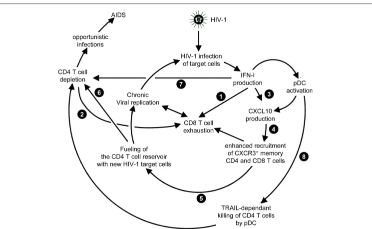
FIGURE 6 | Potential mechanisms through which chronic, low level IFN-I production might promote disease progression in HIV-1 infection. High
and sustained expression of ISG in blood and lymphoid organs is a hallmark of progressive infection with immunodeficiency viruses both in human beings and in non-human primates, irrespective of the levels of viral replication. Several mechanisms summarized here have been proposed to explain how chronic, low level IFN-I production might promote disease progression in HIV-1 infection. These mechanisms include direct ( ) and indirect ( )
promotion of the exhaustion of anti-viral CD8 T cell responses, as well as direct ( ) and indirect ( -to- , and ) promotion of CD4 T cell depletion with a proposed central role of pDC in this deleterious process. Altogether, these mechanisms may sustain a vicious circle of reciprocal activation between chronic viral replication and deleterious immune responses, driving the progressive depletion of all CD4 T cells ultimately causing the enhanced susceptibility to opportunistic infections characteristic of the acquired immunodeficiency syndrome (AIDS). See main text for further details.
depletion, IFN-I neutralization, or selective inhibition of T cell responses to IFN-I could yield additional benefits to chemotherapy in non-human primates during chronic SIV infection.
IFN-I administration has been used for many years to treat another human chronic viral disease, HCV infection. Roughly, half of the patients do not show sustained virological responses (SVR). The treatment causes severe side effects in many individ-uals. New chemotherapeutic drugs very potent at blocking HCV replication in vivo have recently become available. Hence, whether IFN-I administration still constitutes a viable treatment against chronic HCV infection is being questioned (168,169). We will now discuss this issue.
IFN-I TREATMENT OF CHRONIC HCV INFECTION: BALANCING BENEFITS FOR VIRUS CONTROL WITH SIDE EFFECTS STRONGLY AFFECTING PATIENT’S QUALITY OF LIFE
Chronic HCV infection is the main cause of liver cirrhosis and hepatocellular carcinoma. There is currently no vaccine against HCV. The most common therapy for chronic HCV patients is the administration of recombinant pegylated IFN-α (Peg-IFN-α) combined with the anti-viral drug ribavirin. However, because
of IFNAR pleiotropic expression, IFN-α administration induces severe side effects including flu-like syndrome, fever, fatigue, myalgia, and nervous depression (Figure 7A) (170). Moreover, only about half of treated patients harbor SVR (171). Prior-to-treatment high hepatic ISG expression is a negative predictor of SVR upon Peg-IFN-α therapy. High ISG expression in untreated patients likely results from chronic but low IFN-I production trig-gered by persistent HCV replication. Indeed, hepatocytes from non-responder patients were found to be infected at a greater fre-quency and to exhibit dampened antiviral and cell death responses (172). What the cellular sources of IFN-I production are and why they persist only in non-responder patients still remain to be estab-lished. In chronic HCV infection, cytotoxic effector lymphocytes may contribute to the development of hepatocarcinoma by causing low level but sustained hepatocyte death and renewal. In contrast, local production of IFN-γ in the liver by NK and T lymphocytes could promote resistance to disease through non-cytolytic control of viral replication. As discussed previously for LCMV and HIV-1, low chronic production of endogenous IFN-I in HCV patients could compromise both innate and adaptive anti-viral immune responses. Chronic exposure to IFN-I could dampen the ability of
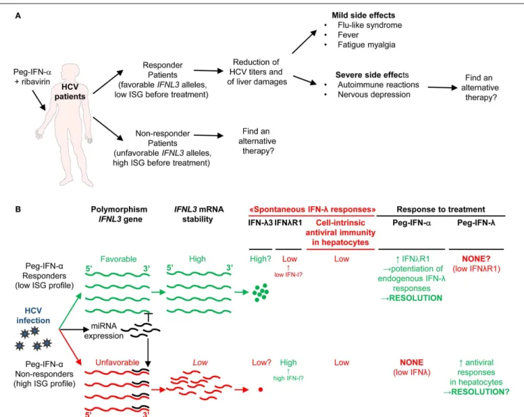
FIGURE 7 | A novel hypothetical model attempting to explain the respective roles of IFN-I and IFN-III in HCV infection. (A) Classification of
patients suffering from chronic HCV infection and treated with PEG-IFN-α in non-responders and responders, and identification of IFNL3 (IL28B) gene polymorphisms as the best predictors for treatment response. The side-effects induced by PEG-IFN-α treatment are also listed since they can severely affect the patient’s quality of life and lead to treatment failure due lack of compliance or suicide. Hence, new approaches are needed to promote beneficial over deleterious effects of IFN-I administration in chronic HCV infection.(B) Proposal of a new hypothesis explaining the relationships
between endogenous ISG levels in patients prior to treatment, IFNL3 gene polymorphism, endogenous expression of IFN-I, IFN-λ, and IFNλR1, and responsiveness to IFN-I administration. Efficient control of HCV infection may depend on hepatocyte response to IFN-λ rather than IFN-α. Upon HCV infection, the virus induces the expression of host miRNA able to bind the 30
UTR of IFNL3 mRNA to promote their degradation. The favorable IFNL3 allele associated with responsiveness to PEG-IFN-α treatment may allow endogenous expression of sufficient levels of IFNL3 for efficient induction of cell-intrinsic anti-viral defenses in hepatocytes. This process is, however, hampered by the limited expression of the receptor for this cytokine (IFNλR1) in these patients. PEG-IFN-α treatment might promote resolution of the infection by inducing IFNλR1 in these patients, potentiating their response to their endogenous production of IFNL3. In the patients that do not respond to PEG-IFN-α treatment, endogenous levels of IFNL3 are insufficient for efficient induction of cell-intrinsic anti-viral defenses in hepatocytes, due to the degradation of the corresponding mRNA in infected hepatocytes. In these patient’s hepatocytes, however, IFNλR1 is already expressed to high levels prior to treatment due to their high endogenous IFN-I responses.
Administration of exogenous IFN-λ might cure these patients. See main text for further details.
NK and CD8 T cells to produce IFN-γ (173,174) and promote CD8 T cell exhaustion (175). It could also induce an antagonist form of CXCL10, a chemokine required for recruitment to the liver of anti-viral NK and CD8 T cell effectors (176). It may also polarize monocytes toward immunosuppressive functions (177). Therefore, better understanding IFN-I effects in HCV infection is
critical to improve care of both responders and non-responder patients to Peg-IFN-α. For responder patients, the issue is to modify the treatment to favor beneficial antiviral and immunoac-tivating effects over side effects strongly affecting patient’s quality of life (Figure 7A). This might be achieved by specific delivery of IFN-I to targeted cell types as discussed later. For non-responder