Arginase-II Promotes Tumor Necrosis Factor-
a Release
From Pancreatic Acinar Cells Causing
b-Cell Apoptosis
in Aging
Yuyan Xiong,
1,2Gautham Yepuri,
1Sevil Necetin,
1Jean-Pierre Montani,
1,2Xiu-Fen Ming,
1,2and
Zhihong Yang
1,2Aging is associated with glucose intolerance. Arginase-II
(Arg-II), the type-IIL-arginine-ureahydrolase, is highly
ex-pressed in pancreas. However, its role in regulation of
pancreaticb-cell function is not known. Here we show
that female (not male) mice deficient in Arg-II (Arg-II2/2)
are protected from age-associated glucose intolerance and reveal greater glucose induced-insulin release, larger
islet size andb-cell mass, and more proliferative and less
apoptoticb-cells compared with the age-matched
wild-type (WT) controls. Moreover, Arg-II is mainly expressed in acinar cells and is upregulated with aging, which en-hances p38 mitogen-activated protein kinase (p38 MAPK)
activation and release of tumor necrosis factor-a (TNF-a).
Accordingly, conditioned medium of isolated acinar cells
from old WT (not Arg-II2/2) mice contains higher TNF-a
levels than the young mice and stimulatesb-cell
apopto-sis and dysfunction, which are prevented by a neutralizing
anti–TNF-a antibody. In acinar cells, our study
demon-strates an age-associated Arg-II upregulation, which pro-motes TNF-a release through p38 MAPK leading to b-cell
apoptosis, insufficient insulin secretion, and glucose
in-tolerance in female rather than male mice.
Aging is highly associated with insulin resistance and pancreatic islet b-cell dysfunction, leading to glucose intol-erance and type 2 diabetes (1–5). The pancreas is composed of the exocrine acini excreting digestive juices and the en-docrine islets to control blood glucose homeostasis (6). Yet, the underlying molecular mechanisms of age-associated glucose intolerance and b-cell dysfunction are not fully
understood. Recent studies (7,8) show that b-cells remain functional in advanced age and can increase their pro-liferative capacity under particular circumstances. The decrease in regenerative capacity, enhanced sensitivity to apoptosis, and defective insulin secretion capacity of b-cells in advanced age are largely due to deteriorating systemic or local environmental factors such as inflamma-tion and vascular dysfuncinflamma-tion in the pancreas and insulin resistance (9–12).
Recent research (13–21) has demonstrated an important role of arginase in age-associated vascular dysfunction, cel-lular senescence, apoptosis, and chronic inflammatory dis-eases such as atherosclerosis and obesity-associated insulin resistance. There are two arginase isoforms encoded by different genes (i.e., the cytoplasmic hepatic protein arginase-I) that mainly functions in the urea cycle for the detoxification of ammonia and the mitochondrial extra-hepatic protein arginase-II (Arg-II) (22). Our previous studies (19,20,23,24) have shown that Arg-II is upregulated in aging endothelial cells and promotes endothelial senes-cence associated with inflammatory responses and dys-function, causes smooth muscle cell apoptosis, and stimulates macrophage proinflammatory cytokine release including tumor necrosis factor-a (TNF-a) and interleukin (IL)-6. Mice with Arg-II deficiency (Arg-II2/2), which is attributable to the suppression of macrophage-mediated proinflammatory responses (23), are protected from vas-cular diseases, glucose intolerance, and insulin resis-tance while eating a high-fat diet (19,20,23,24). Similar to obesity, aging is also associated with chronic low-grade
1Cardiovascular and Aging Research, Department of Medicine, Division of
Phys-iology, University of Fribourg, Fribourg, Switzerland
2Kidney Control of Homeostasis, Swiss National Centre of Competence in
Re-search, Zurich, Switzerland
Corresponding authors: Xiu-Fen Ming, [email protected], and Zhihong Yang, [email protected].
This article contains Supplementary Data.
http://doc.rero.ch
Published in "Diabetes 66(6): 1636–1649, 2017"
which should be cited to refer to this work.
inflammation (25). It has been reported that Arg-II is ex-pressed in the pancreata of rodents and humans (26,27). However, the functions of Arg-II in the regulation of pan-creatic b-cells and in age-associated glucose intolerance are not known.
In this study, we demonstrate an age-associated Arg-II upregulation in pancreatic acinar cells that promotes TNF-a release through p38 mitogen-activated protein kinase (p38 MAPK), leading to b-cell apoptosis, insufficient compensa-tory insulin secretion, and glucose intolerance in mice in a sex-specific manner.
RESEARCH DESIGN AND METHODS
Materials
See Supplementary Data.
Animals
Arg-II2/2mice were provided by Dr. William O’Brien (28) and were backcrossed to C57BL/6J mice for more than eight generations. Wild-type (WT) and Arg-II2/2 off-spring (F2) from hetero/hetero (F1) cross were interbred to obtain WT and Arg-II2/2mice (F3), respectively, for our experiments. The mice were maintained in conventional conditions, as follows: temperature 23°C, on a 12-h light/ dark cycle, and fed a normal chow diet with free access to tap water. Young mice (3–4 months of age) and old mice (18–24 months of age) mice were sacrificed as previously described (18). The pancreas was isolated and subjected to immunofluorescence staining or snap-frozen in liquid nitro-gen and kept at280°C until processed. Animal work was approved by the Ethical Committee of the Veterinary Office of Fribourg (No. 2011_13_FR), Switzerland, and was per-formed in compliance with guidelines on animal experimen-tation at our institution.
Glucose Tolerance Test and Insulin Tolerance Test
A glucose tolerance test (GTT) (2 g/kg glucose i.p.) and an insulin tolerance test (ITT) (0.5 units/kg human insulin i.p.) were performed in young mice (3–4 months of age) and old mice (18–19 months of age) at 1-week intervals as previ-ously described (23).
Glucose-Induced Insulin Release In Vivo
After 6 h of fasting, mice received glucose (2 g/kg i.p.). Blood was collected from tail veins at 0, 15, and 30 min after injection. Plasma insulin was measured by the Metabolic Platform at Metabolic Evaluation Facility, Faculty of Biology and Medicine, University of Lausanne.
Ex Vivo Glucose-Stimulated Insulin Secretion From Isolated Islets
Mouse pancreatic islets were isolated as previously de-scribed (29). Approximately 50 islets with similar sizes were handpicked from each mouse and maintained in RPMI 1640 media supplemented with 10% FBS and 1% penicillin-streptomycin at 37°C under 5% CO2/95% air atmosphere
overnight. Prior to glucose-stimulated insulin secretion (GSIS), islets were collected and incubated in modified Krebs-Ringer bicarbonate buffer (in mmol/L: 118 NaCl,
4.7 KCl, 2.5 CaCl2, 1.2 MgSO4, 1.2 KH2PO4, 25 NaHCO3,
and 0.026 EDTA) containing 20 mmol/L HEPES and 0.25% BSA and 1.7 mmol/L glucose for 90 min followed by stim-ulation with glucose (25 mmol/L). The buffer was then collected at 15, 30, and 60 min. Insulin secreted into the buffer was measured by ELISA according to the manufac-turer instructions.
Pancreatic Islet Size andb-Cell Mass Quantification
Twenty to 25 slides per mouse pancreas were prepared and used for analysis of islet size and b-cell mass. The average values from 14 mice per group were calculated. The pancre-atic islet size was evaluated by hematoxylin-eosin staining of paraffin-embedded sections of the pancreas and quantified with NIH ImageJ version 1.49. b-Cell mass was quantified by multiplying the pancreas weight with the percentage of insulin-positive area, as described previously (30).
Tissue Immunofluorescence Staining and TUNEL Assay
Tissue immunofluorescence staining of paraffin-embedded sections (7 mm) was performed as previously described (23). For details, see the Supplementary Data.
Immunoblotting
Cell or tissue lysate preparation, SDS-PAGE, immunoblot-ting, antibody incubation, and signal detection were performed as previously described (23). Quantification of the signals was performed using NIH ImageJ version 1.49 software.
Quantitative RT-PCR Analyses
mRNA expression of cytokines was determined by two-step quantitative RT-PCR (qRT-PCR) as previously described (23). For details, see Supplementary Data.
Culture of Primary Pancreatic Acinar Cells and Depletion of Macrophages
Isolation and culture of mouse primary pancreatic acinar cells were performed as described by Gout et al. (31). For details, see Supplementary Data.
Culture of a Mouse Pancreaticb-Cell Line
A mouse pancreatic b-cell line (b-TC-6 cells) were pur-chased from the American Type Culture Collection (cata-log #CRL-11506) and cultured in high-glucose DMEM (catalog #D6429; Sigma-Aldrich) containing 15% heat-inactivated FBS (catalog #10500–064; Gibco), and 1% penicillin-streptomycin.
Statistics
Data are presented as the mean6 SEM. In all experiments, n represents the number of experiments or animals. Each subset of experiments was performed on mice derived from two to six different litters (two to six different cohorts). Some different subsets of experiments (e.g., GTT/ITT, ex vivo GSIS from isolated islets, macrophage depletion experiments, and experiments using neutralizing antibody) were further performed on mice from different cohorts. Statistical analysis was performed with a Student unpairedt test or ANOVA with Bonferroni post hoc test. Differences in
mean values were considered significant at two-tailed P value of #0.05.
RESULTS
Female Arg-II2/2Mice Are Protected From
Age-Associated Glucose Intolerance
The role of Arg-II in age-associated glucose intolerance was examined in WT and Arg-II2/2 mice at a young age (3– 4 months) and an old age (18–19 months) in both sexes. Genotype, aging, or sex did not affect fasting plasma glu-cose levels (Fig. 1A and B). No difference in body weight was observed between age- and sex-matched WT and Arg-II2/2 mice (Supplementary Fig. 1). However, in the female group, glucose intolerance was observed with aging in WT mice as shown by GTT (Fig. 1A), which was absent in Arg-II2/2
mice (Fig. 1A). ITT revealed an age-associated insulin resistance in WT mice, which was, however, not affected by Arg-II deficiency (Fig. 1C). In contrast to females, the age-associated glucose intolerance in males was not signif-icantly reversed by Arg-II deficiency (Fig. 1B). More pro-nounced age-associated glucose intolerance and insulin resistance were observed in the WT males mice compared with the WT female mice (Fig. 1C and D), which was not prevented by Arg-II deficiency (Fig. 1C and D). The results
demonstrate a sex difference in the role of Arg-II in age-associated glucose homeostasis and indicate that the im-proved glucose tolerance in old female Arg-II2/2 mice is not attributable to the improved insulin sensitivity.
Arg-II Disruption Improves Insulin Release in Old Female Mice
To examine whether the improved glucose tolerance in the old female Arg-II2/2mice is due to enhanced b-cell func-tion, in vivo glucose-induced insulin release was deter-mined. In response to intraperitoneal glucose injection (2 g/kg), the old female Arg-II2/2 mice revealed higher plasma concentrations of insulin compared with the age-matched WT control mice (Fig. 2A). This difference could not be observed in male mice (Fig. 2A). The glucose-induced plasma insulin elevation was stronger in the old female Arg-II2/2 mice than in the age-matched male Arg-II2/2 animals, although this was not statistically significant (Fig. 2A). The results indicate an improved insulin release from pancreatic islets of female Arg-II2/2mice during ag-ing. To confirm this observation, islets were isolated from old female and male WT and Arg-II2/2 mice to perform an analysis of in vitro GSIS. A significant enhancement of GSIS was observed in the old female Arg-II2/2mice com-pared with the aged-matched WT controls (Fig. 2B). This
Figure 1—Arg-II deficiency protects old female mice from glucose intolerance in aging. After 6 h of starvation, mice were subjected to GTT (2 g/kg glucose i.p.) (A and B) and ITT (0.5 units/kg human insulin i.p.) (C and D). A and C: Females. B and D: Males. AUC, area under the curve; O-KO, old Arg-II2/2; O-WT, old WT; Y-KO, young Arg-II2/2; Y-WT, young WT. GTT and ITT were performed at 1-week intervals.n indicates the number of animals in each experimental group. *P < 0.05, **P < 0.01, ***P < 0.001.
improvement is, however, not significant in the old male animals (Fig. 2B). Intriguingly, a higher pancreatic Arg-II level was observed in the old WT females than in the age-matched WT males (Fig. 2C), demonstrating a sex difference in pancreatic Arg-II expression.
Ablation of Arg-II Protects Against Age-Associated
Pancreatic Cell Apoptosis and Impairedb-Cell
Proliferation in Females
Since a significant age-associated metabolic phenotype was observed in females, the following experiments were
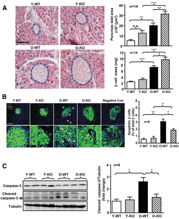
performed only in female mice. Figure 3A shows larger pancreatic islet size and bigger b-cell mass in the old com-pared with the young mice in both WT and Arg-II2/2mice. Moreover, within the old age groups, significantly larger islet size and b-cell mass were observed in the Arg-II2/2 mice compared with the WT mice (Fig. 3A). In the young age groups, Arg-II2/2mice tended to have larger islet size and b-cell mass than WT mice (Fig. 3A). The TUNEL assay demonstrated more apoptotic cells inside and outside the islets with aging in WT mice (Fig. 3B). More apoptotic Figure 2—More pronounced insulin release in old female Arg-II2/2mice as compared with WT counterparts.A: Plasma insulin levels were measured with plasma collected at 0, 15, and 30 min after intraperitoneal glucose injection (2 g/kg glucose i.p.) in female or male mice as indicated. O-KO, old Arg-II2/2; O-WT, old WT.B: Ex vivo GSIS (25 mmol/L) from isolated islets of the above-mentioned four groups of mice over 60 min. Secreted insulin in the incubation buffer was measured by ELISA.C: Immunoblotting analysis of Arg-II expression levels in pancreatic lysates of old WT female and male mice. Fold change of Arg-II/tubulin ratio to male O-WT group is shown in the bar graph at right.n indicates the number of animals of each experimental group. AUC, area under the curve. *P < 0.05.
b-cells (reflected by the percentage of insulin and TUNEL double-positive cells/insulin-positive cells) were observed in the old WT mice compared with the young animals (Fig. 3B). This age-associated b-cell apoptosis was signifi-cantly reduced in the old Arg-II2/2mice (Fig. 3B).
Immuno-blotting analysis also showed an age-associated increase in cleaved caspase-3 levels in WT mice, which was reduced in the Arg-II2/2animals (Fig. 3C). Moreover, coimmunostain-ing of a pancreatic section for proliferatcoimmunostain-ing cell nuclear antigen and insulin revealed an age-associated decrease in b-cell Figure 3—Ablation of Arg-II protects old female mice from age-associated apoptosis in the pancreas. Female young and old mice were sacrificed for collection of pancreata. A: Representative images of hematoxylin-eosin staining of the paraffin-embedded pancreas section (left panels). The border of the pancreatic islets was marked with blue dotted line. The size of pancreatic Langerhans islets was measured and expressed as the pancreatic islet area in square micrometers. Quantification of islet areas and the b-cell mass were presented in the bar graphs in the right panels. Scale bar = 100 mm.B: TUNEL assay of apoptotic cells (red) followed by immunofluorescence staining of insulin (green) in paraffin section of the pancreas. The image in the bottom panel is the enlargement of the selected area in the top panels. The white arrowheads indicate apoptotic cells outside of islets, and white arrows indicate apoptotic cells within islets. This slide of pancreatic tissue stained with label solution (without terminal transferase instead of TUNEL reaction mixture) from an old WT mouse served as the negative control. Scale bar = 100 mm. Quantification of the percentage of apoptotic b-cells is presented in the right bar graph. C: Immunoblotting analysis of total caspase-3 and cleaved caspase-3 in pancreas lysates and tubulin served as the loading control (left panel). Fold change of cleaved caspase-3/tubulin ratio to the Y-WT group is shown in the bar graph at right. n.s., not significant; O-KO, old Arg-II2/2; O-WT, old WT; Y-KO, young Arg-II2/2; Y-WT, young WT. *P < 0.05, **P < 0.01, ***P < 0.001.
proliferation in female WT mice, which was significantly prevented in Arg-II2/2mice (Supplementary Fig. 2A). These data suggest that reduced cell apoptosis and preserved b-cell proliferation could maintain b-cell function in the old female Arg-II2/2 mice. In the male animals, however, Arg-II deficiency did not statistically significantly affect b-cell proliferation (Supplementary Fig. 2B), islet size, b-cell mass, apoptotic cells, and cleaved caspase-3 levels (Supplementary Fig. 3).
Female Arg-II2/2Mice Show Reduced Age-Associated
Increases in TNF-a and p38 MAPK Signaling in Pancreas
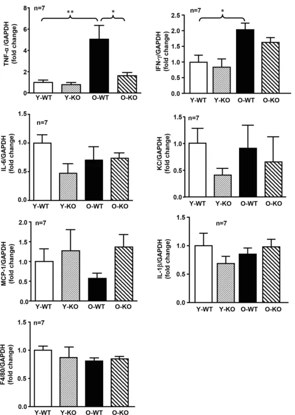
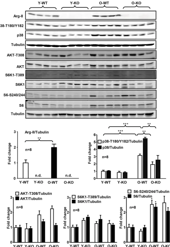
Given the role of inflammation in cell apoptosis, we assessed pancreatic inflammation. In WT females, a signif-icant increase in TNF-a and interferon-g (IFN-g) expres-sion was found with aging (Fig. 4). This age-associated increase in TNF-a (but not in IFN-g) expression was inhibited in the Arg-II2/2mice (Fig. 4). No consistent sig-nificant differences in IL-6, keratinocyte-derived che-mokine (the murine homolog of human IL-8), MCP-1, IL-1b, and F4/80 were found when comparing young and old groups (Fig. 4). We thus focused on TNF-a in further experiments. In the WT groups, a statistically significant age-associated elevation of Arg-II, p38 MAPK-(T180/ Y182) phosphorylation, total p38 MAPK, and S6-(S240/ 244) phosphorylation reflecting S6K1 activity, but not Akt-T308 or S6K1-T389 phosphorylation reflecting activa-tion of Akt or S6K1, respectively, were found (Fig. 5). Only an age-associated increase in p38 MAPK signaling, but not of S6K1-S6 signaling, was reduced in the Arg-II2/2 mice (Fig. 5). These data suggest that Arg-II in the pancreas may promote TNF-a production through p38 MAPK signaling in aging.
Aging Enhances Arg-II and TNF-a Expression
Predominantly in Pancreatic Acinar Cells
The origin of enhanced Arg-II and TNF-a in the aging pancreas was examined. Immunofluorescence staining dem-onstrated that Arg-II was mainly colocalized with carboxy-peptidase A1 (CPA1), the acinar cell marker (Supplementary Fig. 4A), and not with glucagon, the a-cell marker (Supple-mentary Fig. 4B). Arg-II was not detectable within the islet area (Supplementary Fig. 4A and B). Although quantitative PCR of Arg-II mRNA in isolated islets showed that Arg-II mRNA could be detected in islets, the Arg-II/GAPDH ratio was much lower than that in the whole pancreas (;20-fold lower) (Supplementary Fig. 5). Moreover, there was no age-associated change in Arg-II gene expression in islets (Sup-plementary Fig. 5). In contrast, Arg-I was mainly expressed in a-cells, as demonstrated by costaining with glucagon (Supplementary Fig. 4C). Arg-II was also found in F4/80-positive cells (macrophages) that are sporadically distrib-uted in the pancreas (Supplementary Fig. 6). In agreement with the results from immunoblotting, Arg-II expression in acinar cells was enhanced with aging in WT mice (Sup-plementary Fig. 4A). Furthermore, coimmunostaining of TNF-a with macrophage marker F4/80 (Fig. 6A) or with acinar cell marker CPA1 (Fig. 6B) revealed that enhanced
TNF-a production with aging is mainly in Arg-II–expressing acinar cells.
Acinar Cells Causeb-Cell Apoptosis Through Paracrine
Secretion of TNF-a in Aging
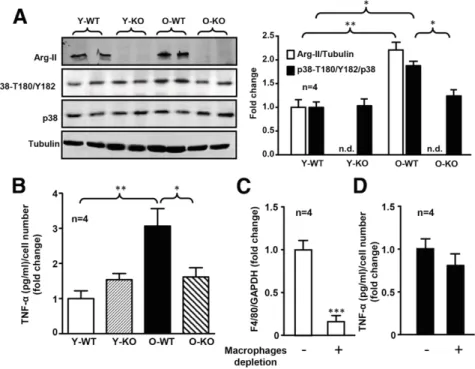
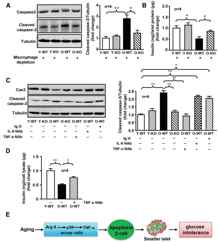
To demonstrate the role of Arg-II in cross talk between acinar cells and b-cells, acinar cells were isolated. Isolated acinar cells from old female WT mice exhibited enhanced Arg-II expression and p38 MAPK activation (Fig. 7A) and elevated TNF-a production (Fig. 7B) compared with the young animals. Arg-II deficiency prevented the age-associated increase in p38 MAPK activation and TNF-a production in acinar cells (Fig. 7A and B). In order to assess the extent to which TNF-a secreted by acinar cells is responsible for b-cell apoptosis, macrophages in exocrine cell cultures were efficiently depleted by magnetic cell sorting with CD11b microbeads (Fig. 7C). Under the condition of mac-rophage depletion, ;80% of TNF-a production remained (Fig. 7D), demonstrating that TNF-a is mainly derived from exocrine cells. Treatment of isolated islets from young WT mice (as bioassay) with conditioned medium (depleted of macrophages) from old WT female mice enhanced cleaved caspase-3 levels (Fig. 8A) and reduced insulin secretion (Fig. 8B). This effect was not seen with the acinar cell–derived conditioned medium of age-matched Arg-II2/2 mice (Fig. 8A and B) and was also abolished by a neutralizing anti– TNF-a antibody when compared with the control condition without antibody or with irrelevant antibodies (IgG and anti–IL-6 antibody) (Fig. 8C and D). The results demon-strate a paracrine role of acinar cell–derived TNF-a in age-associated islet b-cell dysfunction.
DISCUSSION
Recent research demonstrates an important role of Arg-II in chronic inflammatory diseases such as atherosclerosis, obesity-associated insulin resistance, and age-associated vascular dysfunction (19,20,23,32,33). Under these condi-tions, Arg-II, which promotes oxidative stress, proinflam-matory cytokine release, atherogenesis, insulin resistance, and glucose intolerance in mouse models (19,20,23,32,33), is upregulated in cardiovascular cells and macrophages. Here, we explored the role of Arg-II in age-associated pan-creatic b-cell dysfunction and glucose intolerance. We found that Arg-II deficiency protects mice from age-associated glu-cose intolerance resulting from preserved insulin release and larger islet size and b-cell mass in a sex-specific manner (in females but not in males), without affecting body weight. The underlying mechanisms are attributable to en-hanced Arg-II expression in acinar cells in aging, which causes b-cell apoptosis through paracrine release of TNF-a. Aging is a major risk factor for type 2 diabetes due to insulin resistance and alteration or insufficient compensa-tion of b-cell funccompensa-tions (2–5,34–36). In agreement with previous reports (30,37), we observe an age-associated in-sulin resistance and glucose intolerance in WT mice in both sexes. Our results are in line with the findings that age-associated insulin resistance and glucose intolerance are
accompanied with larger islet size and b-cell mass. This is considered to be an adaptive compensatory mechanism of b-cells to maintain glucose homeostasis in aging (35). How-ever, this adaptation in old WT mice is not sufficient to
compensate for insulin resistance, since glucose intolerance is observed in the old WT animals. Remarkably, Arg-II de-ficiency protects female mice from age-associated glucose intolerance. This can be explained by the larger islet size Figure 4—Age-associated increase in TNF-a production in the pancreas was prevented in female Arg-II2/2mice. mRNA expression levels of TNF-a, IFN-g, IL-6, keratinocyte-derived chemokine (the murine IL-8 homolog), MCP-1, IL-1b, and F4/80 were analyzed by qRT-PCR. GAPDH served as the reference. Data are expressed as the fold change of cytokines to the Y-WT group. O-KO, old Arg-II2/2; O-WT, old WT; Y-KO, young Arg-II2/2; Y-WT, young WT. *P < 0.05, **P < 0.01.
and b-cell mass and more pronounced insulin secretion in response to glucose in old female Arg-II2/2mice than in old WT mice, as evidenced by in vivo glucose-induced plasma insulin elevation and ex vivo GSIS. This protective effect of
Arg-II deficiency seems incomplete in males during aging, since there is no significant improvement of glucose toler-ance or significant difference in islet size and b-cell mass between the old WT and Arg-II2/2mice in males.
Figure 5—Age-associated increase in Arg-II expression and p38 MAPK activation in pancreas was prevented in old female Arg-II2/2mice. Immunoblotting analyses of Arg-II, p38-T180/Y182, total p38 MAPK, Akt-T308, total Akt, S6K1-T389, total S6K1, S6-S240/244, and total S6 levels in pancreas lysates. Tubulin served as loading control. n.d., not detectable; O-KO, old Arg-II2/2; O-WT, old WT; Y-KO, young Arg-II2/2; Y-WT, young WT. Data are expressed as the fold change compared with the Y-WT group. *P < 0.05, **P < 0.01, ***P < 0.001.
The mechanisms of the sex difference in the effect of Arg-II deficiency on b-cell functions and glucose tolerance in aging remain unknown. It should be noted that there is a
more pronounced age-associated glucose intolerance in male mice than in female mice, which could be explained by more pronounced insulin resistance in males than in Figure 6—Age-associated increase in TNF-a in pancreata of female mice mainly originates from acinar cells and is reduced by Arg-II deficiency. Confocal microscopy showing coimmunostaining of pancreatic section for F4/80 (green, macrophage marker) and TNF-a (red) (A) and CPA1 (red, acinar cell marker) and TNF-a (green) (B) followed by counterstaining of the nuclei with DAPI (blue). The right most images are the enlargements of the selected area in the corresponding pictures immediately to the left. The arrows indicate the costained cells. Scale bars = 100 mm. O-KO, old Arg-II2/2; O-WT, old WT; Y-KO, young Arg-II2/2; Y-WT, young WT.
females, as shown by ITT results. More stress in male mice during GTT/ITT than in female mice may also contribute to the more severe glucose intolerance in the former group. We wish to point out that higher levels of Arg-II in the pancreata of old females than in old males were observed. This may explain weak or no significant effects of Arg-II deficiency on b-cell function in the old male mice. Similar to our finding in the pancreas, higher Arg-II expression in the female mouse kidney than in male mouse kidney has been reported (38,39). The underlying mechanisms of the sex difference in Arg-II expression in the aging pancreas are not known. It is unlikely that it is attributable to sex hormones per se, since it has been reported that testosterone enhances Arg-II ex-pression in kidneys of both male and female mice (39) and supplementation of estradiol reduces Arg-II levels in rabbits (40). In this regard, the mechanism of higher Arg-II expres-sion in pancreata of female mice with aging requires further investigation.
The most importantfinding of this study is the role of Arg-II in age-associated b-cell dysfunction in female mice. The maintenance and adaptive changes in b-cell mass de-pend on replication/neogeneration and loss of the cells. In aging, b-cells reveal decreased proliferative capacity and are more vulnerable to apoptosis (10). In support of this no-tion, our data show an age-associated decrease in b-cell proliferation and an increase in cell apoptosis in female
WT mice, which are prevented by Arg-II deficiency. These results suggest that Arg-II plays an important role in age-associated b-cell dysfunction in females involving both b-cell proliferation and apoptosis. Insulin resistance is known to play a role in b-cell apoptosis (41). As shown in our study, age-associated glucose intolerance is accompanied by insulin resistance with increased islet area and b-cell mass. This suggests that age-associated glucose intolerance is primarily caused by insulin resistance, whereas b-cell dysfunction/apoptosis is secondary to insulin resistance and other factors such as the inflammatory microenviron-ment, as discussed below. It should be noted that Arg-II2/2 mice reveal an incomplete prevention of age-associated b-cell apoptosis, which could be attributed to incomplete inhibition of pancreatic inflammation and/or persistent in-sulin resistance in these mice.
Importantly, we demonstrate that Arg-II is mainly lo-calized in acinar cells and is upregulated in old animals. The enhanced expression of Arg-II in aging promotes acinar cell inflammation, leading to b-cell apoptosis and ultimately to reduced insulin secretion. The conclusion is supported by the following in vivo and in vitro evidence. First, b-cell apoptosis is increased with aging in female mice and re-duced by Arg-II deficiency. Second, enhanced production of the inflammatory cytokines TNF-a and IFN-g is detected in the pancreata of old mice. A previous study (42) showed Figure 7—Age-associated increase in TNF-a production mainly originated from acinar cells. A: Immunoblotting analysis showing expression levels of Arg-II, p38-T180/Y182, and total p38 in acinar cells isolated from young and old female WT and Arg-II2/2mice. Tubulin served as the loading control. Quantification of the signals is presented in the bar graph at right. Data are expressed as the fold change compare with the Y-WT group.B: Measurement of TNF-a in the conditioned medium of acinar cells by ELISA. Data are expressed as the fold change compared with the Y-WT group.C: qRT-PCR determination of mRNA levels of F4/80 in acinar cells without (2) or with (+) macrophage depletion. Data are expressed as the fold change compared with samples without macrophage depletion.D: TNF-a release in conditioned medium of acinar cells without (2) or with (+) macrophage depletion using CD11b microbeads. Data are expressed as the fold change compared with samples without macrophage depletion. n.d., not detectable; O-KO, old Arg-II2/2; O-WT, old WT; Y-KO, young Arg-II2/2; Y-WT, young WT. *P < 0.05, **P < 0.01, ***P < 0.001.
Figure 8—TNF-a released from acinar cells induces b-cell apoptosis. A: Approximately 100 isolated islets of similar size were handpicked from young WT mice (as a bioassay) and then were treated with conditioned medium prepared from macrophage-depleted acinar cells of the four groups of mice (Y-WT, Y-KO, O-WT, and O-KO) for 16 h. The islet lysates were then prepared and subjected to immunoblotting analysis of caspase-3 and cleaved caspase-3. Quantifications of the signals were presented in the bar graph at right. Tubulin served as the loading control. B: Conditioned media from islets as described in A were collected for insulin measurement. C: b-Cells (b-TC-6 cell line) were serum starved overnight in 0.2% BSA-high-glucose DMEM medium containing 1% penicillin-streptomycin then were incubated further for 72 h with condi-tioned medium of acinar cells isolated from different experimental groups of animals. The condicondi-tioned medium was either untreated (2) or pretreated (+) with control rat IgG (10 mg/mL) or neutralizing antibody against IL-6 (IL-6 NAb) (18 mg/mL) or neutralizing antibody against TNF-a (TNF-a NAb) (0.4 mg/mL) for 2 h prior to addition to the cells. The cell lysates were then prepared and subjected to immunoblotting analysis of caspase-3 and cleaved-caspase-3. Quantifications of the signals were presented in the bar graph at right. Tubulin served as the loading control. D: Experiments were carried out as described in C, and after 72 h of incubation the conditioned media were collected and subjected to insulin measurement by ELISA. O-KO, old Arg-II2/2; O-WT, old WT; Y-KO, young Arg-II2/2; Y-WT, young WT. *P < 0.05, **P < 0.01. E: Schematic summary of thefindings of this study.
that apoptosis in response to TNF-a is observed in insuli-noma cells, whereas apoptosis of primary b-cells requires both TNF-a and IFN-g. Moreover, TNF-a and IFN-g syn-ergistically induce apoptosis of b-cells through reactive ox-ygen species (43). It could not be excluded that both TNF-a and IFN-g are required for b-cell apoptosis through reac-tive oxygen species. Further experiments will be designed to confirm this hypothesis. Our current study provides ev-idence that the Arg-II–mediated b-cell apoptosis is attribut-able to enhanced TNF-a but not IFN-g from acinar cells, since b-cell apoptosis is prevented in Arg-II2/2 mice, in which decreased levels of TNF-a but not IFN-g were ob-served in the pancreas. Moreover, neutralizing anti–TNF-a antibody is able to reduce b-cell apoptosis evoked by con-ditioned medium derived from acinar cells of old WT mice. A role of Arg-II and of TNF-a in age-associated b-cell apo-ptosis and dysfunction shall be further confirmed by an in vivo pharmacological treatment study.
Immunofluorescence staining reveals acinar cells to be the major source of pancreatic TNF-a in aging. The capa-bility of acinar cells to produce a large amount of TNF-a has been shown under pathological conditions such as acute pancreatitis (44). We also found a few macrophages in the pancreas that also express TNF-a. The numbers of mac-rophages are, however, much lower than the numbers of acinar cells. Indeed, 80% of TNF-a is still present in the conditioned medium from isolated pancreatic exocrine cells after macrophages are depleted. The results confirmed acinar cells to be the major source of TNF-a in aging, although the contribution of macrophage-derived TNF-a to age-associated b-cell apoptosis could not be fully excluded. Taking into account that an age-associated increase in cell apoptosis is also observed outside of islets in WT mice and a reduction is observed in Arg-II2/2 mice, an increase in acinar cell apoptosis may also be promoted by increased Arg-II levels in aging. It is thus presumed that enhanced TNF-a in the conditioned medium from aged acinar cells is from both the true release of TNF-a and dying cells. Im-portantly, macrophage-depleted, acinar cell–derived condi-tioned medium from old WT mice but not Arg-II2/2mice is still able to cause cell apoptosis of isolated islets and b-cells and to reduce insulin secretion, which are prevented by a neutralizing antibody against TNF-a. Thus, our current study not only uncovers a novel function of Arg-II in acinar cell inflammation but also provides the first evidence for a role of acinar cells in regulating b-cell apoptosis and insulin secretion. This mechanism may explain the observation that the adaptive increase in pancreatic islet size and b-cell mass in old female mice is further enhanced when Arg-II is deficient.
Studies have demonstrated an important role for p38 MAPK in cytokine production in pancreatic acinar cells (45). Our previous studies (19–21,24,46) have shown cross talk between Arg-II and various signaling pathways, including p38 MAPK, in vascular cells. In accordance with the pre-vious report (45), we show here an age-associated increase in p38 MAPK signaling in both pancreata and isolated
acinar cells of WT mice. The results demonstrate the role of elevated Arg-II levels in the activation of p38 MAPK signaling in acinar cells, leading to TNF-a production, caus-ing b-cell apoptosis and dysfunction in agcaus-ing. The activation of p38 MAPK by Arg-II in endothelial cells was also ob-served in diet-induced obese mice (21). The underlying mechanisms remain to be investigated.
Age-associated elevation of Arg-II, which is associated with hyperactive S6K1 signaling, has been observed in various organs (14,20,47–49). mTORC1-S6K1 signaling is a key modulator of aging and age-related diseases (50). Our previous studies (20,24) demonstrated that Arg-II and S6K1 form a positive regulatory circuit, which maintains hyper-active S6K1 and elevated Arg-II levels to promote vascular aging. Our current study shows an enhanced S6K1 activity in the aging pancreas. It is tempting to speculate that hyperactive S6K1 is involved in the upregulation of Arg-II in the aging pancreas. This hypothesis requires further investigation.
In summarizing our findings in Fig. 8E, we conclude that Arg-II is upregulated in pancreatic acinar cells in aging and activates p38 MAPK, leading to paracrine release of TNF-a, which then causes b-cell apoptosis. This mechanism contributes to the incomplete adaptive compensation of islet b-cell mass extension with aging and results in age-associated glucose intolerance. Targeted disruption of Arg-II prevents this cross talk between acinar cells and b-cells in the aging pancreas and preserves glucose homeostasis dur-ing agdur-ing in a sex-specific manner. Since Arg-II deficiency reduces age-associated glucose intolerance through increas-ing insulin secretion without improvincreas-ing insulin sensitivity, targeting Arg-II together with insulin sensitizers can be a novel therapeutic approach for treating type 2 diabetes. We are aware that global Arg-II knockout mice are used in our study. Nevertheless, the ex vivo experiments using isolated acinar cells and islets demonstrate an acinar cell–auton-omous effect on b-cell function, although the possibility that Arg-II in other organs or even in the islet itself may also affect pancreatic function in aging could not be ex-cluded. Given that Arg-II expression is very low in islets and does not change with age, it is most unlikely that Arg-II in islets exerts a direct effect on age-associated b-cell dys-function. Future studies will confirm whether conditioned Arg-II knockout in acinar cells could achieve the same ef-fects on age-associated glucose homeostasis.
Acknowledgments.The authors thank the Metabolic Platform at Metabolic
Evaluation Facility, Faculty of Biology and Medicine, at the University of Lausanne for the measurement of plasma insulin concentration.
Funding.This work was supported by the Swiss National Science Foundation
(31003A_159582/1), the Swiss Heart Foundation, and the Swiss National Centre of Competence in Research Program (NCCR-Kidney.CH).
Duality of Interest.No potential conflicts of interest relevant to this article
were reported.
Author Contributions.Y.X. discussed the scientific concept, carried out the
project design, performed all experiments except the GTT and ITT, analyzed and interpreted the data, prepared thefigures, and contributed to the writing and editing of
the manuscript. G.Y. and S.N. performed the GTT and ITT and analyzed the data. J.-P.M. discussed the scientific concept, interpreted the data, and contributed to the editing of the manuscript. X.-F.M. and Z.Y. discussed the scientific concept, carried out the project design, interpreted the data, and contributed to the writing and editing of the manuscript. All authors reviewed the manuscript. Y.X., X.-F.M., and Z.Y. are the guarantors of this work and, as such, had full access to all the data in the study and take responsibility for the integrity of the data and the accuracy of the data analysis.
References
1. Stout MB, Justice JN, Nicklas BJ, Kirkland JL. Physiological aging: links among adipose tissue dysfunction, diabetes, and frailty. Physiology (Bethesda) 2017;32:9–19 2. Chang AM, Halter JB. Aging and insulin secretion. Am J Physiol Endocrinol Metab 2003;284:E7–E12
3. Szoke E, Shrayyef MZ, Messing S, et al. Effect of aging on glucose homeo-stasis: accelerated deterioration of beta-cell function in individuals with impaired glucose tolerance. Diabetes Care 2008;31:539–543
4. Evans JL, Goldfine ID. Aging and insulin resistance: just say iNOS. Diabetes 2013;62:346–348
5. Ryan AS. Insulin resistance with aging: effects of diet and exercise. Sports Med 2000;30:327–346
6. Hindley CJ, Cordero-Espinoza L, Huch M. Organoids from adult liver and pancreas: stem cell biology and biomedical utility. Dev Biol 2016;420:251–261
7. Chen X, Zhang X, Chen F, Larson CS, Wang LJ, Kaufman DB. Comparative study of regenerative potential of beta cells from young and aged donor mice using a novel islet transplantation model. Transplantation 2009;88:496–503
8. Stolovich-Rain M, Hija A, Grimsby J, Glaser B, Dor Y. Pancreatic beta cells in very old mice retain capacity for compensatory proliferation. J Biol Chem 2012;287: 27407–27414
9. Li L, Trifunovic A, Köhler M, et al. Defects inb-cell Ca2+ dynamics in age-induced diabetes. Diabetes 2014;63:4100–4114
10. Maedler K, Schumann DM, Schulthess F, et al. Aging correlates with de-creased beta-cell proliferative capacity and enhanced sensitivity to apoptosis: a potential role for Fas and pancreatic duodenal homeobox-1. Diabetes 2006;55: 2455–2462
11. Salpeter SJ, Khalaileh A, Weinberg-Corem N, Ziv O, Glaser B, Dor Y. Systemic regulation of the age-related decline of pancreaticb-cell replication. Diabetes 2013; 62:2843–2848
12. Almaça J, Molina J, Arrojo E Drigo R, et al. Young capillary vessels rejuvenate aged pancreatic islets. Proc Natl Acad Sci U S A 2014;111:17612–17617 13. Bagnost T, Ma L, da Silva RF, et al. Cardiovascular effects of arginase inhibition in spontaneously hypertensive rats with fully developed hypertension. Cardiovasc Res 2010;87:569–577
14. Berkowitz DE, White R, Li D, et al. Arginase reciprocally regulates nitric oxide synthase activity and contributes to endothelial dysfunction in aging blood vessels. Circulation 2003;108:2000–2006
15. Demougeot C, Prigent-Tessier A, Marie C, Berthelot A. Arginase inhibition re-duces endothelial dysfunction and blood pressure rising in spontaneously hyper-tensive rats. J Hypertens 2005;23:971–978
16. Ming XF, Barandier C, Viswambharan H, et al. Thrombin stimulates human endothelial arginase enzymatic activity via RhoA/ROCK pathway: implications for atherosclerotic endothelial dysfunction. Circulation 2004;110:3708–3714 17. Ryoo S, Gupta G, Benjo A, et al. Endothelial arginase II: a novel target for the treatment of atherosclerosis. Circ Res 2008;102:923–932
18. Vaisman BL, Andrews KL, Khong SM, et al. Selective endothelial overexpression of arginase II induces endothelial dysfunction and hypertension and enhances ath-erosclerosis in mice. PLoS One 2012;7:e39487
19. Wu Z, Yu Y, Liu C, et al. Role of p38 mitogen-activated protein kinase in vascular endothelial aging: interaction with Arginase-II and S6K1 signaling pathway. Aging (Albany NY) 2015;7:70–81
20. Yepuri G, Velagapudi S, Xiong Y, et al. Positive crosstalk between arginase-II and S6K1 in vascular endothelial inflammation and aging. Aging Cell 2012;11:1005– 1016
21. Yu Y, Rajapakse AG, Montani JP, Yang Z, Ming XF. p38 mitogen-activated protein kinase is involved in arginase-II-mediated eNOS-uncoupling in obesity. Cardiovasc Diabetol 2014;13:113
22. Yang Z, Ming XF. Arginase: the emerging therapeutic target for vascular oxi-dative stress and inflammation. Front Immunol 2013;4:149
23. Ming XF, Rajapakse AG, Yepuri G, et al. Arginase II promotes macrophage inflammatory responses through mitochondrial reactive oxygen species, contributing to insulin resistance and atherogenesis. J Am Heart Assoc 2012;1:e000992 24. Xiong Y, Yu Y, Montani JP, Yang Z, Ming XF. Arginase-II induces vascular smooth muscle cell senescence and apoptosis through p66Shc and p53 in-dependently of its l-arginine ureahydrolase activity: implications for atherosclerotic plaque vulnerability. J Am Heart Assoc 2013;2:e000096
25. Franceschi C, Bonafè M, Valensin S, et al. Inflamm-aging. An evolutionary perspective on immunosenescence. Ann N Y Acad Sci 2000;908:244–254 26. Choi S, Park C, Ahn M, Lee JH, Shin T. Immunohistochemical study of arginase 1 and 2 in various tissues of rats. Acta Histochem 2012;114:487–494
27. Stickings P, Mistry SK, Boucher JL, Morris SM, Cunningham JM. Arginase expression and modulation of IL-1beta-induced nitric oxide generation in rat and human islets of Langerhans. Nitric Oxide 2002;7:289–296
28. Shi O, Morris SM Jr, Zoghbi H, Porter CW, O’Brien WE. Generation of a mouse model for arginase II deficiency by targeted disruption of the arginase II gene. Mol Cell Biol 2001;21:811–813
29. Stull ND, Breite A, McCarthy R, Tersey SA, Mirmira RG. Mouse islet of Langerhans isolation using a combination of purified collagenase and neutral pro-tease. J Vis Exp 2012;67:e4137
30. Fan R, Kang Z, He L, Chan J, Xu G. Exendin-4 improves blood glucose control in both young and aging normal non-diabetic mice, possible contribution of beta cell independent effects. PLoS One 2011;6:e20443
31. Gout J, Pommier RM, Vincent DF, et al. Isolation and culture of mouse primary pancreatic acinar cells. J Vis Exp 2013;78:e50514
32. Hu H, Moon J, Chung JH, Kim OY, Yu R, Shin MJ. Arginase inhibition ame-liorates adipose tissue inflammation in mice with diet-induced obesity. Biochem Biophys Res Commun 2015;464:840–847
33. Khallou-Laschet J, Varthaman A, Fornasa G, et al. Macrophage plasticity in experimental atherosclerosis. PLoS One 2010;5:e8852
34. Chen M, Bergman RN, Pacini G, Porte D Jr. Pathogenesis of age-related glu-cose intolerance in man: insulin resistance and decreased beta-cell function. J Clin Endocrinol Metab 1985;60:13–20
35. Muzumdar R, Ma X, Atzmon G, Vuguin P, Yang X, Barzilai N. Decrease in glucose-stimulated insulin secretion with aging is independent of insulin action. Diabetes 2004;53:441–446
36. Røder ME, Schwartz RS, Prigeon RL, Kahn SE. Reduced pancreatic B cell compensation to the insulin resistance of aging: impact on proinsulin and insulin levels. J Clin Endocrinol Metab 2000;85:2275–2280
37. Tschen SI, Dhawan S, Gurlo T, Bhushan A. Age-dependent decline in beta-cell proliferation restricts the capacity of beta-cell regeneration in mice. Diabetes 2009; 58:1312–1320
38. Levillain O, Balvay S, Peyrol S. Localization and differential expression of arginase II in the kidney of male and female mice. Pflugers Arch 2005;449:491–503 39. Levillain O, Diaz JJ, Blanchard O, Déchaud H. Testosterone down-regulates ornithine aminotransferase gene and up-regulates arginase II and ornithine de-carboxylase genes for polyamines synthesis in the murine kidney. Endocrinology 2005;146:950–959
40. Hayashi T, Esaki T, Sumi D, Mukherjee T, Iguchi A, Chaudhuri G. Modulating role of estradiol on arginase II expression in hyperlipidemic rabbits as an athero-protective mechanism. Proc Natl Acad Sci USA 2006;103:10485–10490 41. Donath MY, Dalmas É, Sauter NS, Böni-Schnetzler M. Inflammation in obesity and diabetes: islet dysfunction and therapeutic opportunity. Cell Metab 2013;17: 860–872
42. Stephens LA, Thomas HE, Ming L, et al. Tumor necrosis factor-alpha-activated cell death pathways in NIT-1 insulinoma cells and primary pancreatic beta cells. Endocrinology 1999;140:3219–3227
43. Kim WH, Lee JW, Gao B, Jung MH. Synergistic activation of JNK/SAPK induced by TNF-alpha and IFN-gamma: apoptosis of pancreatic beta-cells via the p53 and ROS pathway. Cell Signal 2005;17:1516–1532
44. Dios ID. Inflammatory role of the acinar cells during acute pancreatitis. World J Gastrointest Pharmacol Ther 2010;1:15–20
45. Blinman TA, Gukovsky I, Mouria M, et al. Activation of pancreatic acinar cells on isolation from tissue: cytokine upregulation via p38 MAP kinase. Am J Physiol Cell Physiol 2000;279:C1993–C2003
46. Xiong Y, Yepuri G, Forbiteh M, et al. ARG2 impairs endothelial autophagy through regulation of MTOR and PRKAA/AMPK signaling in advanced atherosclerosis. Autophagy 2014;10:2223–2238
47. Caccamo A, Branca C, Talboom JS, et al. Reducing ribosomal protein S6 kinase 1 expression improves spatial memory and synaptic plasticity in a mouse model of Alzheimer’s disease. J Neurosci 2015;35:14042–14056
48. Yang J, Zhou X, Fan X, et al. mTORC1 promotes aging-related venous thrombosis in mice via elevation of platelet volume and activation. Blood 2016;128: 615–624
49. Martín-Cano FE, Camello-Almaraz C, Hernandez D, Pozo MJ, Camello PJ. mTOR pathway and Ca2+stores mobilization in aged smooth muscle cells. Aging
(Albany NY) 2013;5:339–346
50. Johnson SC, Rabinovitch PS, Kaeberlein M. mTOR is a key modulator of ageing and age-related disease. Nature 2013;493:338–345