DARWIN REVIEW
Generic signal-specific responses: cytokinin and
context-dependent cellular responses
Bruno Mu¨ller*
Institute of Plant Biology, Zu¨rich-Basel Plant Science Center, University of Zu¨rich, Zollikerstrasse 107, 8008 Zu¨rich, Switzerland * To whom correspondence should be addressed. E-mail: [email protected]
Received 16 September 2010; Revised 22 November 2010; Accepted 29 November 2010
Abstract
The phytohormone cytokinin triggers numerous and diverse responses during the plant life cycle via a two-component phosphorelay signalling system. Each step of the signalling cascade is supported by a gene family comprising several members. While functional redundancy is observed among family members, additional gene-specific functions encoded by cis-regulatory and coding sequence of individual family members have been described and contribute to specificity in signalling output. In addition, the cellular context of the signal-receiving cell affects the response triggered. Recent studies in Arabidopsis have demonstrated how expression of cytokinin signalling components predefines a spatiotemporal map of signalling sensitivity, which causes local signal amplification and attenuation. In summary, the specific interpretation of cytokinin signalling is affected by an orchestrated interplay of signalling genes and cellular context.
Key words: Arabidopsis thaliana, cytokinin signalling, development, pattern formation, plant hormones, signalling specificity.
Introduction
Plants and animals represent parallel realizations of multi-cellular development and display vastly different life cycles. They have independently evolved genes for pattern forma-tion and cell–cell communicaforma-tion (Meyerowitz, 2002). In spite of the differences, common developmental strategies exist. For example, in contrast to the countless and diverse instances of cell-to-cell signalling, a surprisingly small repertoire of signal transduction systems is used. Research of the past two decades has revealed that the deployment of each transduction pathway is very flexible, and that a hand-ful of core pathways are repeatedly implemented through-out metazoan development to account for innumerable cases of cell specification events (Davidson and Erwin, 2006). Similarly, in plants, signalling cascades triggered by phytohormones are used throughout the life cycle to affect cellular identities and responses (Santner et al., 2009). Given the contrast between the few signalling systems and the innumerable cellular responses they trigger, the general question arises as to how fairly ubiquitous and generic signals can elicit specific and context-dependent cellular responses? Generally, specificity is achieved by regulating
the localization, quantity, and quality of the signalling activity, and the status of the receiving cell. In this review, specificity and regulation of intracellular signalling will be discussed with regard to the cytokinin signalling circuitry; for discussions about how the availability and localization of active ligands is regulated, the interested reader is referred to recent reviews (Werner and Schmu¨lling, 2009; Kudo et al., 2010).
Cytokinins comprise a class of adenine-derived signalling molecules. The first cytokinin, Kinetin, was isolated as a breakdown product from autoclaved herring-sperm DNA, based on its cell-division promoting activity in cultivated tobacco tissue (Miller et al., 1955; Amasino, 2005). Later, a number of additional effects provoked by cytokinins were described: stimulated leaf expansion and seed germination (Miller, 1956,1958), delayed senescence in leaves (Richmond and Lang, 1957; Gan and Amasino, 1995), de novo organ formation from cultured tissue (Skoog and Miller, 1957), or release from apical dominance (Wickson and Thimann, 1958; Cline et al., 1997), and more (Mizuno, 2005;To and Kieber, 2008;Jeon et al., 2010;Perilli et al., 2010).
ª The Author [2011]. Published by Oxford University Press [on behalf of the Society for Experimental Biology]. All rights reserved. For Permissions, please e-mail: [email protected]
Cytokinins are perceived by a multistep phoshorelay system
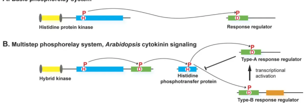
The identification of the CYTOKININ INDEPENDENT ONE (CKI1) gene from Arabidopsis thaliana (Kakimoto, 1996) was the long-awaited breakthrough, which initiated the molecular elucidation of the cytokinin signal trans-duction pathway in the following years. Its overexpression caused the typical cytokinin responses in tissue culture (Kakimoto, 1996) and in planta (Hwang and Sheen, 2001). CKI encodes for a hybrid kinase, suggesting it functions in a phosphorelay system. Phosphorelay, or two-component signalling systems, are prevalent in bacteria (West and Stock, 2001). In the simplest form, they consist of two conserved proteins: a histidine protein kinase and a re-sponse regulator protein that are phosphorylated at conserved His and Asp residues, respectively. Phospho-transfer from the histidine kinase to the response regulator results in activation of the latter (Fig. 1A). More complex versions of this two-component phosphotransfer involve multiple phosphotransfer steps, and often more than two proteins (Fig. 1B). Further evidence supporting the use of a phosphorelay system for cytokinin signalling came when additional genes with conserved His- and Asp-containing domains were identified, such as response regulators (RR), and histidine phosphotransfer proteins (HPt) (Mok and Mok, 2001). A forward genetic screen using tissue culture assays led to the identification of the cytokinin response 1-1 (cre1-1) mutation, allelic to the previously characterized woodenleg (wol) mutation (Ma¨ho¨nen et al., 2000; Inoue et al., 2001). The same gene was independently isolated and named as ARABIDOPSIS HISTIDINE KINASE 4 (AHK4) (Ueguchi et al., 2001). The CREI/WOL/AHK4 gene encodes for a true cytokinin receptor that, unlike CKI1, could bind and respond to cytokinins (Suzuki et al.,
2001; Yamada et al., 2001). The completion of the
Arabidopsis genome sequence allowed researchers system-atically to compile the potential signalling components, based on the characteristic signatures of domains impli-cated in phosphorelay signalling (Fig. 2; Hwang et al., 2002). Functional reconstitution of the pathway in an Arabidopsis cellular system established the core logic of the pathway (Fig. 1B, Hwang and Sheen, 2001). Pathway activation is initiated by autophosphorylation at a con-served His residue of the receptors, which is subsequently carried-over to a conserved Asp of the receiver domain. The phosphoryl group is then transferred to the functional Arabidopsis histidine phosphotransfer proteins (AHP) proteins (Imamura et al., 1999;Suzuki et al., 2000). AHP proteins can form dimers and shuttle between nucleus and cytosol in a cytokinin-independent manner (Punwani et al., 2010). From the AHPs, the phosphate gets passed-over to the nuclear-localized response regulators. Type-B Arabi-dopsis response regulators (type-B ARR) act as transcrip-tional activators, while type-A Arabidopsis response regulators (type-A ARR) negatively interfere with pathway activity, most likely by competing with type-B ARRs for access to the AHPs, and by acting as phosphate sinks (To et al., 2007). Transcription of type-A ARR is directly induced by the activated B-type ARRs (D’Agostino et al., 2000), which establishes a negative feedback loop to the pathway (Hwang and Sheen, 2001). Structurally and functionally, the type-C ARRs resemble type-A ARRs, as they contain a conserved receiver domain and their ectopic expression represses cytokinin output strongly. However, unlike type-A ARRs, their expression does not depend on cytokinin. No aberrant phenotypes have been observed with loss-of function mutants under normal growth conditions, however, since ARR22 expression was found to be induced by wounding, their functions may become relevant upon conditions of mechanical stress (Kiba et al., 2004;Gattolin et al., 2006;Hora´k et al., 2008).
Fig. 1. Schematic representation of the two-component and the multistep phosphorelay signalling systems. (A) The basic phosphorelay system uses a single phosphoryl transfer event between a His protein kinase and its cognate response regulator. (B) The multistep His-to-Asp phosphorelay system, in which a His-containing phosphotransfer protein (HPt) serves as a phosphoryl acceptor and donor between the hybrid protein kinase (HK) and the response regulators (RR). The activation of the type-B RRs results in transcriptional activation. Type-A RRs attenuate signalling activity. Transcriptional activation of repressive type-A RRs establishes a negative feedback loop to the pathway.
Functional studies reveal redundant and gene-specific functions of family members
For each step in the signalling cascade, multiple genes are encoded in the Arabidopsis genome (Fig. 2; Table 1). The question arises as to which degree the expansion of each gene family reflects functional diversification? On the one hand, functional studies in planta and in heterologous systems supported the core function of different family members in the signalling circuitry; on the other hand, gene-specific functions emerged as well. Sequence comparisons within protein families reveal stretches of poor conservation (Fig. 2). For example, beyond their conserved domains, both type-A and type-B ARRs harbour stretches of sequences that exhibit little similarity within the protein family. Such variable parts can be expected to mediate
gene-specific functions. Furthermore, the analysis of the tran-scription profiles of cytokinin signalling components in planta uncovered complex patterns (Ferreira and Kieber, 2005), which reflects diversity in expression. Thus, gene-specific functions appear to depend both on protein sequence and expression patterns.
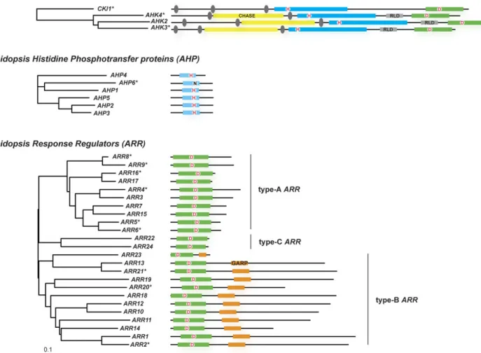
In general, loss-of-function mutations in single genes resulted in only subtle phenotypes, revealing the functional redundancy within gene families (Table 1). An exception is the hybrid kinase CKI1; it’s loss caused female gametophyte lethality (Pischke et al., 2002; Heja´tko et al., 2003; Deng et al., 2010). To obtain stronger phenotypes, plants were generated that harbour loss-of-function mutations in several to all genes belonging to a family, comprising the true receptors (Higuchi et al., 2004; Nishimura et al., 2004; Fig. 2. Compilation of cytokinin signalling components from Arabidopsis, and their phylogenetic relationship. On the left, the
phylogenetic relationship of Arabidopsis family members is shown for the HKs with a well-defined role in cytokinin signalling, for the AHPs, and the ARRs. Alignments were created using the MAFFT-E-INS-i algorithm (Katoh and Toh, 2008), based on full-length protein sequences retrieved from TAIR (Swarbreck et al., 2008). Unrooted Neighbor–Joining trees (Saitou and Nei, 1987) were calculated using a BLOSUM62 matrix. The scale bar represents 0.1 amino acid substitutions per site. On the right, the proteins are indicated as black lines. Conserved domains are indicated as boxes. The cytokinin-binding CHASE domains are drawn as yellow boxes, transmembrane segments as grey ovals. Receptor-like domains (RLD) as a grey box. Kinase/transmitter (in HKs) and transmitter domains (in AHPs) with the conserved His residue that gets phosphorylated are depicted as blue boxes; receiver domains, with the conserved Asp residue (D), are indicated by a green box. The DNA-binding GARP domain of the type-B ARRs is indicated by an orange box. The GARP domain of ARR23 is truncated. Asterisks denote genes products with a documented specific function within cytokinin signalling, as discussed in the text.
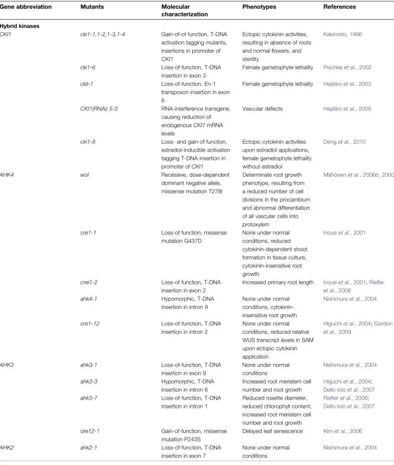
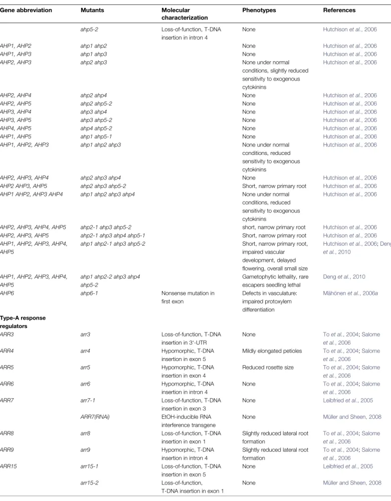
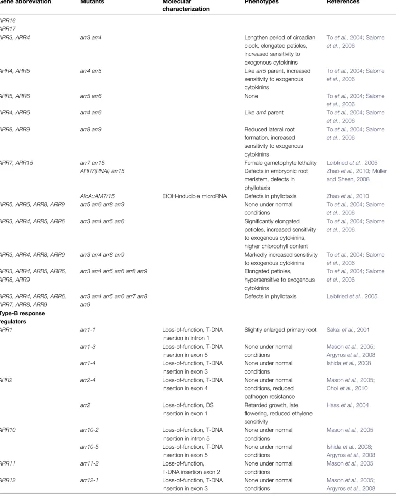
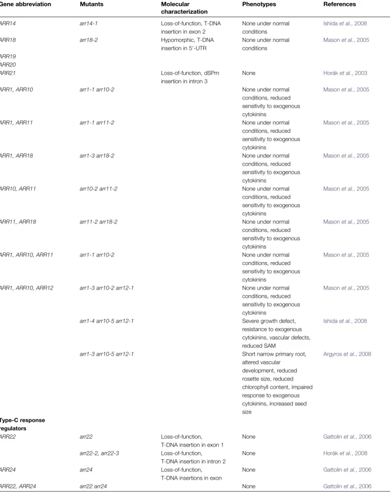
Table 1. List of mutants affecting cytokinin signalling genes, including higher-order mutants
Gene abbreviation Mutants Molecular
characterization
Phenotypes References
Hybrid kinases
CKI1 cki1-1,1-2,1-3,1-4 Gain-of-of function, T-DNA activation tagging mutants, insertions in promoter of CKI1
Ectopic cytokinin activities, resulting in absence of roots and normal flowers, and sterility
Kakimoto, 1996
cki1-6 Loss-of-function, T-DNA insertion in exon 3
Female gametophyte lethality Pischke et al., 2002
ckil-1 Loss-of-function, En-1 transposon insertion in exon 6
Female gametophyte lethality Heja´tko et al., 2003
CKI1(RNAi) 5-3 RNA-interference transgene, causing reduction of endogenous CKI1 mRNA levels
Vascular defects Heja´tko et al., 2009
cki1-8 Loss- and gain-of function, estradiol-inducible activation tagging T-DNA insertion in promoter of CKI1
Ectopic cytokinin activities upon estradiol applications, female gametophyte lethality without estradiol
Deng et al., 2010
AHK4 wol Recessive, dose-dependent
dominant negative allele, missense mutation T278I
Determinate root growth phenotype, resulting from a reduced number of cell divisions in the procambium and abnormal differentiation of all vascular cells into protoxylem
Ma¨ho¨nen et al., 2006b, 2000
cre1-1 Loss-of function, missense mutation G437D
None under normal conditions, reduced cytokinin-dependent shoot formation in tissue culture, cytokinin-insensitive root growth
Inoue et al., 2001
crei1-2 Loss-of function, T-DNA insertion in exon 2
Increased primary root length Inoue et al., 2001;Riefler et al., 2006
ahk4-1 Hypomorphic, T-DNA insertion in intron 9
None under normal conditions, cytokinin-insensitive root growth
Nishimura et al., 2004
cre1-12 Loss-of-function, T-DNA insertion in intron 2
None under normal conditions, reduced relative WUS transcript levels in SAM upon ectopic cytokinin application
Higuchi et al., 2004;Gordon et al., 2009
AHK3 ahk3-1 Loss-of-function, T-DNA
insertion in exon 9
None under normal conditions
Nishimura et al., 2004
ahk3-3 Hypomorphic, T-DNA insertion in intron 6
Increased root meristem cell number and root growth
Higuchi et al., 2004; Dello Ioio et al., 2007 ahk3-7 Loss-of-function, T-DNA
insertion in intron 1
Reduced rosette diameter, reduced chlorophyll content, increased root meristem cell number and root growth
Riefler et al., 2006; Dello Ioio et al., 2007
ore12-1 Gain-of-function, missense mutation P243S
Delayed leaf senescence Kim et al., 2006
AHK2 ahk2-1 Loss-of-function, T-DNA
insertion in exon 7
None under normal conditions
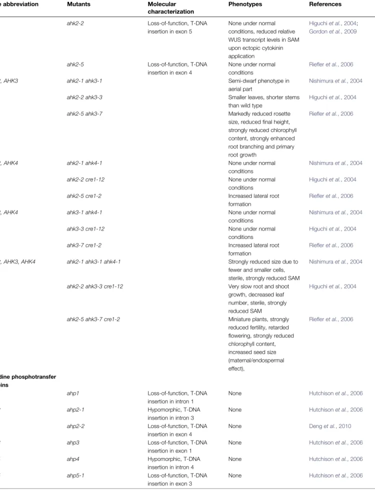
Table 1. Continued
Gene abbreviation Mutants Molecular
characterization
Phenotypes References
ahk2-2 Loss-of-function, T-DNA insertion in exon 5
None under normal conditions, reduced relative WUS transcript levels in SAM upon ectopic cytokinin application
Higuchi et al., 2004; Gordon et al., 2009
ahk2-5 Loss-of-function, T-DNA insertion in exon 4
None under normal conditions
Riefler et al., 2006
AHK2, AHK3 ahk2-1 ahk3-1 Semi-dwarf phenotype in
aerial part
Nishimura et al., 2004
ahk2-2 ahk3-3 Smaller leaves, shorter stems
than wild type
Higuchi et al., 2004
ahk2-5 ahk3-7 Markedly reduced rosette
size, reduced final height, strongly reduced chlorophyll content, strongly enhanced root branching and primary root growth
Riefler et al., 2006
AHK2, AHK4 ahk2-1 ahk4-1 None under normal
conditions
Nishimura et al., 2004
ahk2-2 cre1-12 None under normal
conditions
Higuchi et al., 2004
ahk2-5 cre1-2 Increased lateral root
formation
Riefler et al., 2006
AHK3, AHK4 ahk3-1 ahk4-1 None under normal
conditions
Nishimura et al., 2004
ahk3-3 cre1-12 None under normal
conditions
Higuchi et al., 2004
ahk3-7 cre1-2 Increased lateral root
formation
Riefler et al., 2006
AHK2, AHK3, AHK4 ahk2-1 ahk3-1 ahk4-1 Strongly reduced size due to fewer and smaller cells, sterile, strongly reduced SAM
Nishimura et al., 2004
ahk2-2 ahk3-3 cre1-12 Very slow root and shoot growth, decreased leaf number, sterile, strongly reduced SAM
Higuchi et al., 2004
ahk2-5 ahk3-7 cre1-2 Miniature plants, strongly reduced fertility, retarded flowering, strongly reduced chlorophyll content, increased seed size (maternal/endospermal effect),
Riefler et al., 2006
Histidine phosphotransfer proteins
AHP1 ahp1 Loss-of-function, T-DNA
insertion in intron 1
None Hutchison et al., 2006
AHP2 ahp2-1 Hypomorphic, T-DNA
insertion in intron 3
None Hutchison et al., 2006
ahp2-2 Loss-of-function, T-DNA insertion in exon 4
None Deng et al., 2010
AHP3 ahp3 Loss-of-function, T-DNA
insertion in exon 1
None Hutchison et al., 2006
AHP4 ahp4 Hypomorphic, T-DNA
insertion in intron 4
None Hutchison et al., 2006
AHP5 ahp5-1 Loss-of-function, T-DNA
insertion in exon 3
Table 1. Continued
Gene abbreviation Mutants Molecular
characterization
Phenotypes References
ahp5-2 Loss-of-function, T-DNA insertion in intron 4
None Hutchison et al., 2006
AHP1, AHP2 ahp1 ahp2 None Hutchison et al., 2006
AHP1, AHP3 ahp1 ahp3 None Hutchison et al., 2006
AHP2, AHP3 ahp2 ahp3 None under normal
conditions, slightly reduced sensitivity to exogenous cytokinins
Hutchison et al., 2006
AHP2, AHP4 ahp2 ahp4 None Hutchison et al., 2006
AHP2, AHP5 ahp2 ahp5-2 None Hutchison et al., 2006
AHP3, AHP4 ahp3 ahp4 None Hutchison et al., 2006
AHP3, AHP5 ahp3 ahp5-2 None Hutchison et al., 2006
AHP4, AHP5 ahp4 ahp5-2 None Hutchison et al., 2006
AHP1, AHP5 ahp1 ahp5-1 None Hutchison et al., 2006
AHP1, AHP2, AHP3 ahp1 ahp2 ahp3 None under normal
conditions, reduced sensitivity to exogenous cytokinins
Hutchison et al., 2006
AHP2, AHP3, AHP4 ahp2 ahp3 ahp4 None Hutchison et al., 2006
AHP2 AHP3, AHP5 ahp2 ahp3 ahp5-2 Short, narrow primary root Hutchison et al., 2006 AHP1 AHP2, AHP3 AHP4 ahp1 ahp2 ahp3 ahp4 None under normal
conditions, reduced sensitivity to exogenous cytokinins
Hutchison et al., 2006
AHP2, AHP3, AHP4, AHP5 ahp2-1 ahp3 ahp5-2 short, narrow primary root Hutchison et al., 2006 AHP2, AHP3, AHP5 ahp2-1 ahp3 ahp4 ahp5-1 Short, narrow primary root Hutchison et al., 2006 AHP1, AHP2, AHP3, AHP4,
AHP5
ahp1 ahp2-1 ahp3 ahp5-2 Short, narrow primary root, impaired vascular development, delayed flowering, overall small size
Hutchison et al., 2006;Deng et al., 2010
AHP1, AHP2, AHP3, AHP4, AHP5
ahp1 ahp2-2 ahp3 ahp4 ahp5-2
Gametophytic lethality, rare escapers seedling lethal
Deng et al., 2010
AHP6 ahp6-1 Nonsense mutation in
first exon
Defects in vasculature: impaired protoxylem differentiation
Ma¨ho¨nen et al., 2006a
Type-A response regulators
ARR3 arr3 Loss-of-function, T-DNA
insertion in 3‘-UTR
None To et al., 2004;Salome et al., 2006
ARR4 arr4 Hypomorphic, T-DNA
insertion in exon 5
Mildly elongated petioles To et al., 2004;Salome et al., 2006
ARR5 arr5 Hypomorphic, T-DNA
insertion in exon 4
Reduced rosette size To et al., 2004;Salome et al., 2006
ARR6 arr6 Hypomorphic, T-DNA
insertion in intron 4
None To et al., 2004;Salome et al., 2006
ARR7 arr7-1 Loss-of-function, T-DNA
insertion in exon 3
None Leibfried et al., 2005
ARR7(RNAi) EtOH-inducible RNA interference transgene
None Mu¨ller and Sheen, 2008
ARR8 arr8 Loss-of-function, T-DNA
insertion in exon 1
Slightly reduced lateral root formation
To et al., 2004;Salome et al., 2006
ARR9 arr9 Hypomorphic, T-DNA
insertion in intron 4
Slightly reduced lateral root formation
To et al., 2004;Salome et al., 2006
ARR15 arr15-1 Loss-of-function, T-DNA
insertion in exon 5
None Leibfried et al., 2005
arr15-2 Loss-of-function, T-DNA insertion in exon 1
Table 1. Continued
Gene abbreviation Mutants Molecular
characterization
Phenotypes References
ARR16 ARR17
ARR3, ARR4 arr3 arr4 Lengthen period of circadian
clock, elongated petioles, increased sensitivity to exogenous cytokinins
To et al., 2004;Salome et al., 2006
ARR4, ARR5 arr4 arr5 Like arr5 parent, increased
sensitivity to exogenous cytokinins
To et al., 2004;Salome et al., 2006
ARR5, ARR6 arr5 arr6 None To et al., 2004;Salome
et al., 2006
ARR4, ARR6 arr4 arr6 Like arr4 parent To et al., 2004;Salome
et al., 2006
ARR8, ARR9 arr8 arr9 Reduced lateral root
formation, increased sensitivity to exogenous cytokinins
To et al., 2004;Salome et al., 2006
ARR7, ARR15 arr7 arr15 Female gametophyte lethality Leibfried et al., 2005 ARR7(RNAi) arr15 Defects in embryonic root
meristem, defects in phyllotaxis
Zhao et al., 2010;Mu¨ller and Sheen, 2008
AlcA::AM7/15 EtOH-inducible microRNA Defects in phyllotaxis Zhao et al., 2010 ARR5, ARR6, ARR8, ARR9 arr5 arr6 arr8 arr9 None under normal
conditions
To et al., 2004;Salome et al., 2006
ARR3, ARR4, ARR5, ARR6 arr3 arr4 arr5 arr6 Significantly elongated petioles, increased sensitivity to exogenous cytokinins, higher chlorophyll content
To et al., 2004;Salome et al., 2006
ARR3, ARR4, ARR8, ARR9 arr3 arr4 arr8 arr9 Markedly increased sensitivity to exogenous cytokinins
To et al., 2004;Salome et al., 2006
ARR3, ARR4, ARR5, ARR6, ARR8, ARR9
arr3 arr4 arr5 arr6 arr8 arr9 Elongated petioles, hypersensitive to exogenous cytokinins
To et al., 2004;Salome et al., 2006
ARR3, ARR4, ARR5, ARR6, ARR7, ARR8, ARR9
arr3 arr4 arr5 arr6 arr7 arr8 arr9
Defects in phyllotaxis Leibfried et al., 2005
Type-B response regulators
ARR1 arr1-1 Loss-of-function, T-DNA
insertion in intron 1
Slightly enlarged primary root Sakai et al., 2001
arr1-3 Loss-of-function, T-DNA insertion in exon 5
None under normal conditions
Mason et al., 2005; Argyros et al., 2008 arr1-4 Loss-of-function, T-DNA
insertion in exon 3
None under normal conditions
Ishida et al., 2008
ARR2 arr2-4 Loss-of-function, T-DNA
insertion in exon 4
None under normal conditions, reduced pathogen resistance Mason et al., 2005; Choi et al., 2010 arr2 Loss-of-function, DS insertion in exon 1
Retarded growth, late flowering, reduced ethylene sensitivity
Hass et al., 2004
ARR10 arr10-2 Loss-of-function, T-DNA
insertion in intron 5
None under normal conditions
Mason et al., 2005
arr10-5 Loss-of-function, T-DNA insertion in exon 5
None under normal conditions
Ishida et al., 2008; Argyros et al., 2008
ARR11 arr11-2 Loss-of-function,
T-DNA insertion exon 2
None under normal conditions
Mason et al., 2005
ARR12 arr12-1 Loss-of-function, T-DNA
insertion in exon 3
None under normal conditions
Mason et al., 2005; Argyros et al., 2008
Table 1. Continued
Gene abbreviation Mutants Molecular
characterization
Phenotypes References
ARR14 arr14-1 Loss-of-function, T-DNA
insertion in exon 2
None under normal conditions
Ishida et al., 2008
ARR18 arr18-2 Hypomorphic, T-DNA
insertion in 5’-UTR
None under normal conditions Mason et al., 2005 ARR19 ARR20 ARR21 Loss-of-function, dSPm insertion in intron 3
None Hora´k et al., 2003
ARR1, ARR10 arr1-1 arr10-2 None under normal
conditions, reduced sensitivity to exogenous cytokinins
Mason et al., 2005
ARR1, ARR11 arr1-1 arr11-2 None under normal
conditions, reduced sensitivity to exogenous cytokinins
Mason et al., 2005
ARR1, ARR18 arr1-3 arr18-2 None under normal
conditions, reduced sensitivity to exogenous cytokinins
Mason et al., 2005
ARR10, ARR11 arr10-2 arr11-2 None under normal
conditions, reduced sensitivity to exogenous cytokinins
Mason et al., 2005
ARR11, ARR18 arr11-2 arr18-2 None under normal
conditions, reduced sensitivity to exogenous cytokinins
Mason et al., 2005
ARR1, ARR10, ARR11 arr1-1 arr10-2 None under normal
conditions, reduced sensitivity to exogenous cytokinins
Mason et al., 2005
ARR1, ARR10, ARR12 arr1-3 arr10-2 arr12-1 None under normal conditions, reduced sensitivity to exogenous cytokinins
Mason et al., 2005
arr1-4 arr10-5 arr12-1 Severe growth defect, resistance to exogenous cytokinins, vascular defects, reduced SAM
Ishida et al., 2008
arr1-3 arr10-5 arr12-1 Short narrow primary root, altered vascular
development, reduced rosette size, reduced chlorophyll content, impaired response to exogenous cytokinins, increased seed size
Argyros et al., 2008
Type-C response regulators
ARR22 arr22 Loss-of-function,
T-DNA insertion in exon 1
None Gattolin et al., 2006
arr22-2, arr22-3 Loss-of-function, T-DNA insertion in intron 2
None Hora´k et al., 2008
ARR24 arr24 Loss-of-function,
T-DNA insertions in exon
None Gattolin et al., 2006
Riefler et al., 2006), AHPs (Hutchison et al., 2006; Deng et al., 2010), type-A ARRs (Hutchison et al., 2006; Salome et al., 2006; To et al., 2007), and type-B ARRs (Mason et al., 2005; Argyros et al., 2008; Ishida et al., 2008). The results from these studies confirmed the exten-sive redundancy within each gene family: phenotype severity and insensitivity to cytokinin increased with the number of affected family members. In addition, gene-specific func-tions were uncovered (Table 1). In principle, these can be explained by the differential expression patterns, or by the specific protein functions of the genes involved. In the first scenario, loss of the family members with the highest expression levels in a given tissue is expected to result in the strongest phenotype. For example, AHK4’s functional contribution for the root system (Ueguchi et al., 2001; Higuchi et al., 2004; Nishimura et al., 2004; Riefler et al., 2006) reflects its relatively higher overall expression levels in root tissue (Ueguchi et al., 2001; Higuchi et al., 2004). In the transition zone of the primary root meristem however, AHK3 expression (Nishimura et al., 2004) and function prevails (Dello Ioio et al., 2007). In the shoot apical meristem (SAM), AHK2 and AHK4, but not AHK3, mediate cytokinin-dependent WUSCHEL (WUS) expres-sion (Gordon et al., 2009), which correlates with the low abundance of AHK3 in the SAM (Nishimura et al., 2004). Similarly, the strong phenotypes of a plant defective in three of the 11 type-B ARRs, ARR1, ARR10, and ARR12 (Mason et al., 2005; Yokoyama et al., 2007; Ishida et al., 2008) correlated with their relatively high and widespread expres-sion (Mason et al., 2004). Similarly, some of the type-A type-ARR mutant combinations exhibited specific phenotypes, which are consistent with the expression patterns of the affected genes. For example, ARR8 and ARR9 are expressed strongly throughout the root, and disrupting their loci affects lateral root number in seedlings but does not affect shoot development (To et al., 2004). Disruption of ARR7 and ARR15 functions interferes with embryonic root meristem establishment in the early embryo (Mu¨ller and Sheen, 2008) and causes disturbed phyllotaxis in the SAM (Zhao et al., 2010), in agreement with their unique expression patterns in these tissues. The reported female gametophyte lethality of an arr7 arr15 double mutation (Leibfried et al., 2005) could also reflect the expression patterns of ARR7 and ARR15 during this phase of de-velopment. The gene pairs ARR8/ARR9 and ARR7/ARR15 represent closely related sister genes (Fig. 2) and arose from the most recent Arabidopsis genome duplication (Vision et al., 2000). Commonly, such sister genes share more redundant functions compared with more divergent pairs (Lynch and Conery, 2000). By analogy, analysing the missing double-mutant combinations of the ARR gene pairs ARR16/ARR17, ARR19/ARR20, or ARR13/ARR21 (Fig. 2) could reveal interesting phenotypes. However, as an excep-tion to the rule, the closely related type-B ARR1 and ARR2 genes (Fig. 2) share few functions (Hass et al., 2004;Mason et al., 2005; Choi et al., 2010), despite their overlapping expression patterns (Mason et al., 2004). Recently, the unique ability of ARR2 to confer cytokinin-mediated
pathogen resistance has been mapped to the variable C-terminal domain beyond the conserved DNA-binding domain (Fig. 2). Via this portion of the protein, the transcription factor ARR2 is recruited by TGA3 to the cis-elements of the PR1 promoter, leading to its transcriptional activation. Under the same conditions, ARR1 did not interact with TGA3 (Choi et al., 2010). Gene-specific functions depending on the distinct biochemical functions of the affected genes were also described for other signalling components (see asterisks in Fig. 2). Similar to AHK2, AHK3, and AHK4, CKI1 codes for a hybrid kinase, which activates cytokinin signalling. However, it lacks the CHASE (cyclase/His kinase-associated sensing extracellular) domain involved in cytokinin binding (Fig. 2; Anantharaman and Aravind, 2001; Mougel and Zhulin, 2001). Indeed, cki plants still respond to exogenously applied cytokinins similar to the wild-type plants, supporting a specific func-tion of CKI1, which activates signalling independent of cytokinins (Heja´tko et al., 2009; Deng et al., 2010). Characterization of the wol allele of AHK4 revealed that phosphotransfer is bi-directional. Compared with wild-type AHK4, the WOL gene product has an amino acid change in the putative extracellular, cytokinin-binding region and lacks cytokinin binding activity, which reveals the inherent phosphatase activity of AHK4 that normally depletes the circuitry of phosphoryl groups in the absence of ligands (Ma¨ho¨nen et al., 2006b). AHK2 and AHK3 appear to lack a similar activity. AHK3, in turn, has been shown to exhibit unique functions in mediating cytokinin’s function in leaf senescence (Kim et al., 2006; Riefler et al., 2006). Neither mutants of the other receptors, AHK2 and AHK4, nor their overexpression affected leaf senescence (Kim et al., 2006). Within the family of AHPs, AHP6 stands out by lacking a conserved His residue in its receiver domain (Fig. 2), which renders it unable to accept an activating phosphoryl group (Ma¨ho¨nen et al., 2006b). Hence, it was named pseudo AHP. ahp6 mutant plants show defects in protoxylem differentiation, reflecting an increase in cytokinin signalling levels. AHP6 negatively interferes with pathway activity, most likely by competing with AHP1–5 for interaction with the activated receptors (Ma¨ho¨nen et al., 2006a).
More gene-specific protein functions were uncovered by overexpression studies done in planta or heterologous systems. Compared with the analysis of different mutants, the use of ubiquitous promoters equalizes the expression patterns among the genes studied and, therefore, allows a direct comparison of the phenotypic effects caused by the coding sequences. For example, differential sensitivity of the cytokinin receptors AKH4 and AHK3 to biologically active cytokinins has been described (Yonekura-Sakakibara et al., 2004; Spı´chal et al., 2004; Romanov et al., 2006), opening the possibility of controlling the sensitivity to the available active cytokinins by regulating the expression pattern of the receptors. As the relative abundance of each receptor varies in different tissues, this possibility might be realized in planta. While overexpression of various type-A ARRs confirmed their negative role with respect to signalling activity (Fig. 1B; Hwang and Sheen, 2001; Osakabe et al.,
2002;Kiba et al., 2003;Lee et al., 2007;To et al., 2007;Ren et al., 2009), differences in phenotypes were also recorded. For example, overexpression of ARR5 affected lateral root formation, whereas the closely related ARR6 had little effect. Instead, it interfered with elongation of the primary root, which was not significantly changed by ARR5 (Ren et al., 2009). ARR4 caused hypersensitivity to red light, and specifically interacted with Phytochrome B (Sweere et al., 2001). ARR16 appears unique among type-A family mem-bers in mediating cytokinin’s function in leaf senescence (Ren et al., 2009). These phenotypical differences are probably caused by the variable C-terminal domains of the type-A ARRs (Fig. 2). As cytokinin-dependent protein stability has been reported to be differentially affecting different members (To and Kieber, 2008;Ren et al., 2009), this mechanism may further contribute to the different phenotypes observed. Overexpression experiments with type-B ARRs resulted in increased sensitivity to cytokinin, consistent with their role as transcriptional activators in the signalling cascade (Fig. 1B;Hwang and Sheen, 2001; Sakai et al., 2001; Imamura et al., 2003; Tajima et al., 2004). In addition, different type-B ARRs caused differences in phenotypes, both in the tissues affected and in the severity of the abnormalities (Imamura et al., 2003; Hass et al., 2004; Tajima et al., 2004; Choi et al., 2010). Since little difference was observed in the DNA-binding affinity of the different B-type ARRs in vitro (Sakai et al., 2000; Hosoda et al., 2002; Imamura et al., 2003), and different B-type ARRs could all mediate transcriptional activation of a synthetic cytokinin reporter in vivo (Mu¨ller and Sheen, 2008), the unique functions of different type-B ARRs are probably encoded by their variable C-terminal extensions. This was exemplified with ARR2 (Choi et al., 2010), where the selection of cytokinin target genes is altered by the specific recruitment of DNA-binding cofactors. In sum-mary, a number of gene-specific functions have been reported within families of the cytokinin signalling pathway, ranging from the receptors to the transcription factors. In cases where specificity is encoded by the protein sequences, the causative determinants in the protein sequence map to the variable amino acid residues of the different protein families, which mediate interaction with specific partners (Choi et al., 2010), or exhibit altered biochemical activities (Ma¨ho¨nen et al., 2006a, b). In vitro, the potential for differential protein interactions of cytokinin signalling genes has been uncovered by systematic screens (Dortay et al., 2006, 2008). We are still far from a comprehensive view of how the individual protein signature of cytokinin signalling components contributes to signalling specificity. However, the identified specific functions provide a sound foundation to refine our understanding in a stepwise way. The contribution of differential expression to signalling specific-ity will be discussed further in the following sections. The expression level of signalling components affects the pathway activity
At least one member of a hybrid kinase, AHP and B-type ARR must be expressed beyond a minimal threshold level
for a cell to activate the signalling pathway. Increasing the expression levels of these positively acting components amplifies the signalling activity, while expression of the negatively acting type-A ARR and AHP6 attenuates it (Fig. 3). Therefore, the efficiency of phosphotransfer corre-lates with the expression levels of signalling components, which represents a potential mechanism as to how to tailor the sensitivity to a given stimulus. Tissue-specificity, feed-back regulation from cytokinin signalling and other signal-ling inputs are responsible for the spatio-temporal expression profiles of the signalling components. Feedback regulation has been documented for the expression of type-A type-ARRs and type-AHP6. Type-type-A type-ARRs represent immediate-early transcriptional target genes that negatively interfere with signalling activity. Such a negative feedback loop may serve to smooth-out fluctuations in signalling or to shut off pathway activity more abruptly after transient stimuli. Like type-A ARRs, AHP6 suppresses pathway activity. How-ever, its transcription is repressed by cytokinin signalling (Ma¨ho¨nen et al., 2006a). This probably serves to generate sharper boundaries between signalling and non-signalling domains in a tissue.
Spatiotemporal expression map of signalling components defines a sensitivity landscape
In addition to feedback regulation, the complex expression patterns of hybrid kinases, AHPs, and ARRs suggest elaborate transcription regulation, which integrates tissue-specific, and multiple signalling inputs. Thus, a picture emerges where expression of cytokinin signalling compo-nents predefines a spatiotemporal map of signalling sensi-tivity. This allows localized signal amplification and attenuation, depending on the functional requirements.
Recent studies of cytokinin function at the cellular resolution have opened the door to study the functional relevance as to how cytokinin output is influenced by the expression profiles of signalling genes (Fig. 4; Leibfried et al., 2005; Mu¨ller and Sheen, 2008; Gordon et al., 2009; Moubayidin et al., 2010; Zhao et al., 2010). While knowledge of all present signalling genes in a given cell defines its sensitivity and competence, the actual signalling response ultimately depends on the local concentration of active cytokinins, which is difficult to determine, due to the small size of active ligands and their diversity. Thus, the key to analysing cytokinin‘s function in the context of the embryonic root-meristem specification was the construction of a synthetic promoter, which does not rely on secondary signalling or tissue-specific information. Rather, its activity reflects the cytokinin signalling output in the nucleus, that is, transcriptional activation mediated by type-B ARRs (Mu¨ller and Sheen, 2008). Reporter activity was observed in the hypophysis, precursor of the root stem cell system. Upon asymmetric cell division of the hypophysis, the resulting basal cell lineage down-regulated cytokinin output, while the apical cell maintained activity. This differential signalling output, which is essential for the development of the nascent root meristem, depends on auxin to activate the
transcription of the type-A ARR7 and ARR15 directly (Fig. 4C). Auxin signalling thus interferes with cytokinin signal-ling in a defined domain by inducing negative regulators, which suppresses the efficiency of phosphorelay. An analo-gous mechanism operates in the shoot apical meristem (Fig. 4D). However, interestingly, the relationship is reversed: here, auxin represses the transcription of ARR7 and ARR15, leading to an increase in cytokinin signalling activity. Again, this cross-talk appears functionally relevant as interfering with ARR7 and ARR15 levels causes irregu-larities in phyllotaxis, the ordered arrangement of lateral shoots (Zhao et al., 2010). In addition, WUS directly attenuates transcription of type-A ARR5, ARR6, ARR7, and ARR15 (Leibfried et al., 2005), again enhancing cytokinsignalling activity. In the same context, an in-crease in receptor levels further contributes to inin-creased perception in a spatially defined domain (Gordon et al., 2009). Therefore, in the shoot apical meristem, both a de-crease in the abundance of negative regulators (Leibfried et al., 2005;Zhao et al., 2010), and an increase in expression of a cytokinin receptor (Gordon et al., 2009), a positive regulator, contributes to enhance the sensitivity to
cytoki-nins. Cytokinin signalling in the transition zone of the adult root meristem counteracts proliferation by promoting differentiation (Dello Ioio et al., 2007). Thus, exact signal-ling levels are critical for establishing the balance between growth and differentiation. During the early phases, when net growth of the root is required, the proliferation-inducing phytohormone Gibberellin selectively represses the transcription of type-B ARR1, hereby lowering the sensitiv-ity to available cytokinins and hence signalling levels (Fig. 4E). At the later stages, when Gibberellin levels decrease, ARR1 transcription and, consequently, cytokinin signalling increases, which tips the balance towards differentiation (Moubayidin et al., 2010). Here, competence to respond to cytokinin is regulated by controlling the expression of a type-B ARR. These studies have revealed the functional importance of modulating pathway activity by controlling the expression levels of signalling components.
More detailed studies will probably follow to demon-strate how other signalling pathways determine the sensitiv-ity landscape by amplifying or attenuating the cytokinin response. During female gametophyte development, cytoki-nin-signalling levels are controlled by the hybrid kinase Fig. 3. Cartoon to illustrate how different expression levels of intracellular cytokinin signalling components affects the signalling activity, given a constant concentration of active ligands. On the left, signalling output is blocked due to prevalence of negative regulators: pseudo AHP6 and type-A ARRs. On the right, signalling output is maximized due to up-regulation of positive-acting signalling components: HKs, AHPs, type-B ARRs. Cytokinins are indicated as grey circles. Up-facing arrows indicate high concentrations, down-facing arrows low concentrations. Conserved domains are depicted as inFig. 2.
CKI1 (Pischke et al., 2002; Heja´tko et al., 2003), indepen-dent of the bona fide cytokinin receptors AHK2, AHK3, and AHK4 (Deng et al., 2010). CKI1 activates cytokinin signalling independent of cytokinins (Hwang and Sheen, 2001;Yamada et al., 2001;Heja´tko et al., 2009;Deng et al., 2010). Accordingly, pathway activity depends on the transcriptional profile of CKI1, and it will be interesting to identify the signals responsible. One candidate signal is auxin, which has been reported to be in involved in patterning of the embryo sac (Pagnussat et al., 2009). By inducing CKI1 expression, these signals can directly activate the cytokinin-signalling pathway, bypassing the need of cytokinins. Cytokinin’s function in root gravitropism could, potentially, also be influenced by auxin (Aloni et al., 2004). Transcription of the type-C ARR ARR22 is induced by wounding (Gattolin et al., 2006), which could reflect an
additional case of how cytokinin output is determined by other signals. Recently, GeBP and GeBP-like transcription factors were identified that influence the cytokinin response by indirectly affecting the expression levels of type-A ARRs (Chevalier et al., 2008). The concept of organizing cross-talk among hormone signalling pathways by controlling the expression levels of key signalling components is further exemplified by several well-documented cases where cytoki-nin signalling influences other signalling pathways (Laplaze et al., 2007;Dello Ioio et al., 2008;Ruzicka et al., 2009).
The cellular response to cytokinin signalling qualitatively differs in the examples discussed above. In the nascent root meristem (Mu¨ller and Sheen, 2008) and during female gametophyte development (Pischke et al., 2002; Heja´tko et al., 2003; Deng et al., 2010), interfering with cytokinin function affects cell specification while, in the root, Fig. 4. The regulation of cytokinin signalling components affects the signalling output in different plant organs. Cartoons of an adult Arabidopsis plant (A), and a seedling (B). Schematic views of early embryos at the globular and transition stage (C), the shoot apical meristem (D), and the primary root (E) are drawn on the left. Cytokinin output is indicated in green, based on the expression of the synthetic TCS reporter in (C) (Mu¨ller and Sheen, 2008) and (D) (Zhao et al., 2010), or inferred from the expression patterns of ARR1 and ARR12 and phenotypic analysis of the corresponding mutants (Moubayidin et al., 2010) in (D). (C) Down-regulation of cytokinin output in the basal cell (bc) of a transition-stage embryo is caused by auxin, which induces the transcription of ARR7 and ARR15 (Mu¨ller and Sheen, 2008). In the shoot meristem (D), several mechanisms operate in parallel to up-regulate cytokinin sensitivity: auxin stimulates expression of AHK4 (Gordon et al., 2009), and represses transcription of ARR7 and ARR15 (Zhao et al., 2010), while WUS represses transcription of ARR5, ARR6, ARR7, and ARR15 (Leibfried et al., 2005). In the primary root (E), Gibberellin signalling represses ARR1 expression at the early stages (3 days post germination, dpg). Later, around 5 dpg, the mechanism is released, allowing expression of ARR1 and increased cytokinin signalling output (Moubayidin et al., 2010). TZ, transition zone; EDZ, elongation and differentiation zone. Refer also to the body text for further explanations.
cytokinin has been shown to counteract auxin-promoted proliferation in the transition zone (Dello Ioio et al., 2008). In the shoot meristem, cytokinin’s role is to maintain the size and function of the stem-cell pool (To and Kieber, 2008; Gordon et al., 2009; Werner and Schmu¨lling, 2009; Zhao et al., 2010). How can the same signalling pathway elicit these different responses? As discussed above, the specific set of cytokinin signalling proteins present in the cell may affect the signalling outcome. Along with the type-B ARR transcription factors, CYTOKININ RESPONSE FACTORS (CRF) have been reported to control a subset of immediate–early cytokinin target genes, further increas-ing the specificity of the signallincreas-ing response (Rashotte et al., 2006). In addition, the cellular status of the signal-receiving cell can affect how a signal is interpreted. In the case of cytokinin signalling, cytokinin-responsive promoters may rely on secondary regulatory input to confer transcriptional activation. For example, while transcription of type-A ARRs is sensitive to cytokinin, the transcription patterns only partly overlap, reflecting the dependence on additional input (To et al., 2004;Leibfried et al., 2005). Transcriptome changes in response to exogenous cytokinin treatment were characterized in different settings (Che et al., 2002; Hoth et al., 2003;Rashotte et al., 2003;Brenner et al., 2005). The overlap among cytokinin-target genes between the different experiments was relatively small, providing further argu-ments that, in addition to cytokinin signalling, tissue- and context-specific inputs are important. Transcription control of the ARR7 and ARR15 genes demonstrates how the cellular context influences promoter activity: the same cis-regulatory sequence integrates activating input by the auxin pathway in the embryonic root meristem (Mu¨ller and Sheen, 2008), and repressive auxin input in the shoot meristem (Zhao et al., 2010). Future analyses aimed at characterizing the relevant cytokinin target genes during female gametophyte development, root meristem specifica-tion and maintenance, and shoot meristem homeostasis will provide the necessary basis to address further the question of target-gene specificity in well-defined developmental contexts at high resolution.
Concluding remarks
Signalling circuitries allow cells to change the gene expres-sion profile specifically in response to cues originating from their outside. The limited number of signalling system contrasts with the numerous and diverse responses they elicit. On the one hand, gene-specific functions, both coded by cis-regulatory and coding sequence, contribute to specific responses. This also applies to the cytokinin signalling pathway—its relatively simple core pathway logic becomes increasingly complex with the multiple family members, each with a complex expression pattern and the potential to interact with diverse partners. On the other hand, the cellular context of signalling affects the cellular response triggered. Specifically, the integration of tissue-specific in-formation and other signals by the cis-regulatory regions of
potential target genes renders transcriptome changes context-dependent. Progress made in the last decade in the under-standing of the cytokinin signalling pathway, and how it is specifically implemented in the developmental context, has been remarkable, and will continue at a fast pace. Recent studies (Ma¨ho¨nen et al., 2006a; Dello Ioio et al., 2008; Mu¨ller and Sheen, 2008;Gordon et al., 2009;Heja´tko et al., 2009; Deng et al., 2010; Moubayidin et al., 2010; Zhao et al., 2010) have demonstrated the importance of precisely tuning the levels of cytokinin signalling during develop-ment, which results in an elaborate spatiotemporal signal-ling landscape. The main focus here was on work using the reference plant Arabidopsis, however, increasing effort is being devoted to study cytokinin signalling in crop plants (Giulini et al., 2004;Yonekura-Sakakibara et al., 2004; Ito and Kurata, 2006) and plants with different life cycles, which allows the role of cytokinin in the evolution of the plant body plan to be studied (Pils and Heyl, 2009; Hellmann et al., 2010).
Acknowledgements
I thank C Baroux for comments on the manuscript and H Scho¨b for comments on the figures.
References
Aloni R, Langhans M, Aloni E, Ullrich CI. 2004. Role of cytokinin in the regulation of root gravitropism. Planta 220, 177–182.
Amasino R. 2005. 1955: Kinetin arrives: the 50th anniversary of a new plant hormone. Plant Physiology 138, 1177–1184.
Anantharaman V, Aravind L. 2001. The CHASE domain: a predicted ligand-binding module in plant cytokinin receptors and other
eukaryotic and bacterial receptors. Trends in Biochemical Sciences 26, 579–582.
Argyros RD, Mathews DE, Chiang YH, Palmer CM, Thibault DM, Etheridge N, Argyros DA, Mason MG, Kieber JJ, Schaller GE. 2008. Type-B response regulators of Arabidopsis play key roles in cytokinin signalling and plant development. The Plant Cell 20, 2102–2116.
Brenner WG, Romanov GA, Ko¨llmer I, Bu¨rkle L, Schmu¨lling T. 2005. Immediate-early and delayed cytokinin response genes of Arabidopsis thaliana identified by genome-wide expression profiling reveal novel cytokinin-sensitive processes and suggest cytokinin action through transcriptional cascades. The Plant Journal 44, 314–333.
Che P, Gingerich DJ, Lall S, Howell SH. 2002. Global and hormone-induced gene expression changes during shoot development in Arabidopsis. The Plant Cell 14, 2771–2785.
Chevalier F, Perazza D, Laporte F, Le He´nanff G, Hornitschek P, Bonneville JM, Herzog M, Vachon G. 2008. GeBP and GeBP-like proteins are noncanonical leucine-zipper transcription factors that regulate cytokinin response in Arabidopsis. Plant Physiology 146, 1142–1154.
Choi J, Huh SU, Kojima M, Sakakibara H, Paek KH, Hwang I. 2010. The cytokinin-activated transcription factor ARR2 promotes plant immunity via TGA3/NPR1-dependent salicylic acid signalling in Arabidopsis. Developmental Cell 19, 284–295.
Cline M, Wessel’ T, Iwamura H. 1997. Cytokinin/auxin control of apical dominance in Ipomoea nil. Plant and Cell Physiology 38, 639–667.
D’Agostino IB, Deruere J, Kieber JJ. 2000. Characterization of the response of the Arabidopsis response regulator gene family to cytokinin. Plant Physiology 124, 1706–1717.
Davidson EH, Erwin DH. 2006. Gene regulatory networks and the evolution of animal body plans. Science 311, 796–800.
Dello Ioio R, Linhares FS, Scacchi E, Casamitjana-Martinez E, Heidstra R, Costantino P, Sabatini S. 2007. Cytokinins determine Arabidopsis root-meristem size by controlling cell differentiation. Current Biology 17, 678–682.
Dello Ioio R, Nakamura K, Moubayidin L, Perilli S, Taniguchi M, Morita MT, Aoyama T, Costantino P, Sabatini S. 2008. A genetic framework for the control of cell division and differentiation in the root meristem. Science 322, 1380–1384.
Deng Y, Dong H, Mu J, Ren B, Zheng B, Ji Z, Yang WC, Liang Y, Zuo J. 2010. Arabidopsis histidine kinase CKI1 acts upstream of histidine phosphotransfer proteins to regulate female
gametophyte development and vegetative growth. The Plant Cell 22, 1232–1248.
Dortay H, Gruhn N, Pfeifer A, Schwerdtner M, Schmu¨lling T, Heyl A. 2008. Toward an interaction map of the two-component signalling pathway of Arabidopsis thaliana. Journal of Proteome Research 7, 3649–3660.
Dortay H, Mehnert N, Bu¨rkle L, Schmu¨lling T, Heyl A. 2006. Analysis of protein interactions within the cytokinin-signalling pathway of Arabidopsis thaliana. FEBS Journal 273, 4631–4644.
Ferreira FJ, Kieber JJ. 2005. Cytokinin signalling. Current Opinion in Plant Biology 8, 518–525.
Gan S, Amasino RM. 1995. Inhibition of leaf senescence by autoregulated production of cytokinin. Science 270, 1986–1988. Gattolin S, Alandete-Saez M, Elliott K, Gonzalez-Carranza Z, Naomab E, Powell C, Roberts JA. 2006. Spatial and temporal expression of the response regulators ARR22 and ARR24 in Arabidopsis thaliana. Journal of Experimental Botany 57, 4225–4233. Giulini A, Wang J, Jackson D. 2004. Control of phyllotaxy by the cytokinin-inducible response regulator homologue ABPHYL1. Nature 430, 1031–1034.
Gordon SP, Chickarmane VS, Ohno C, Meyerowitz EM. 2009. Multiple feedback loops through cytokinin signalling control stem cell number within the Arabidopsis shoot meristem. Proceedings of the National Academy of Sciences, USA 106, 16529–16534.
Hass C, Lohrmann J, Albrecht V, et al. 2004. The response regulator 2 mediates ethylene signalling and hormone signal integration in Arabidopsis. EMBO Journal 23, 3290–3302. Heja´tko J, Ryu H, Kim GT, et al. 2009. The histidine kinases CYTOKININ-INDEPENDENT1 and Arabidopsis HISTIDINE KINASE2 and 3 regulate vascular tissue development in Arabidopsis shoots. The Plant Cell 21, 2008–2021.
Heja´tko J, Pernisova M, Eneva T, Palme K, Brzobohaty´ B. 2003. The putative sensor histidine kinase CKI1 is involved in female gametophyte development in Arabidopsis. Molecular Genetics and Genomics 269, 443–453.
Hellmann E, Gruhn N, Heyl A. 2010. The more, the merrier: cytokinin signalling beyond Arabidopsis. Plant Signalling and Behavior 5, (in press).
Higuchi M, Pischke MS, Maho¨nen AP, et al. 2004. In planta functions of the Arabidopsis cytokinin receptor family. Proceedings of the National Academy of Sciences, USA 101, 8821–8826.
Hora´k J, Grefen C, Berendzen KW, Hahn A, Stierhof YD, Stadelhofer B, Stahl M, Koncz C, Harter K. 2008. The Arabidopsis thaliana response regulator ARR22 is a putative AHP phospho-histidine phosphatase expressed in the chalaza of developing seeds. BMC Plant Biology 8, 77.
Hosoda K, Imamura A, Katoh E, Hatta T, Tachiki M, Yamada H, Mizuno T, Yamazaki T. 2002. Molecular structure of the GARP family of plant Myb-related DNA binding motifs of the Arabidopsis response regulators. The Plant Cell 14, 2015–2029.
Hoth S, Ikeda Y, Morgante M, Wang X, Zuo J, Hanafey MK, Gaasterland T, Tingey SV, Chua NH. 2003. Monitoring genome-wide changes in gene expression in response to endogenous cytokinin reveals targets in Arabidopsis thaliana. FEBS Letters 554, 373–380. Hutchison CE, Li J, Argueso C, et al. 2006. The Arabidopsis histidine phosphotransfer proteins are redundant positive regulators of cytokinin signalling. The Plant Cell 18, 3073–3087.
Hwang I, Chen HC, Sheen J. 2002. Two-component signal transduction pathways in Arabidopsis. Plant Physiology 129, 500–515. Hwang I, Sheen J. 2001. Two-component circuitry in Arabidopsis cytokinin signal transduction. Nature 413, 383–389.
Imamura A, Hanaki N, Nakamura A, Suzuki T, Taniguchi M, Kiba T, Ueguchi C, Sugiyama T, Mizuno T. 1999. Compilation and characterization of Arabidopsis thaliana response regulators implicated in His-Asp phosphorelay signal transduction. Plant and Cell Physiology 40, 733–742.
Imamura A, Kiba T, Tajima Y, Yamashino T, Mizuno T. 2003. In vivo and in vitro characterization of the ARR11response regulator implicated in the His-to-Asp phosphorelay signal transduction in Arabidopsis thaliana. Plant and Cell Physiology 44, 122–131.
Inoue T, Higuchi M, Hashimoto Y, Seki M, Kobayashi M, Kato T, Tabata S, Shinozaki K, Kakimoto T. 2001. Identification of CRE1as a cytokinin receptor from Arabidopsis. Nature 409, 1060–1063. Ishida K, Yamashino T, Yokoyama A, Mizuno T. 2008. Three type-B response regulators, ARR1, ARR10 and ARR12, play essential but redundant roles in cytokinin signal transduction throughout the life cycle of Arabidopsis thaliana. Plant and Cell Physiology 49, 47–57.
Ito Y, Kurata N. 2006. Identification and characterization of cytokinin-signalling gene families in rice. Gene 382, 57–65.
Jeon J, Kim NY, Kim S, Kang NY, Novak O, Ku SJ, Cho C, Lee DJ, Lee EJ, Strnad M, Kim J. 2010. A subset of cytokinin two-component signalling system plays a role in cold temperature stress response in Arabidopsis. Journal of Biological Chemistry 285, 23371–23386.
Kakimoto T. 1996. CKI1, a histidine kinase homolog implicated in cytokinin signal transduction. Science 274, 982–985.
Katoh K, Toh H. 2008. Recent developments in the MAFFT multiple sequence alignment program. Briefings in Bioinformatics 9, 286–298. Kiba T, Aoki K, Sakakibara H, Mizuno T. 2004. Arabidopsis response regulator, ARR22, ectopic expression of which results in phenotypes similar to the wol cytokinin-receptor mutant. Plant and Cell Physiology 45, 1063–1077.
Kiba T, Yamada H, Sato S, Kato T, Tabata S, Yamashino T, Mizuno T. 2003. The type-A response regulator, ARR15, acts as a negative regulator in the cytokinin-mediated signal transduction in Arabidopsis thaliana. Plant and Cell Physiology 44, 868–874. Kim HJ, Ryu H, Hong SH, Woo HR, Lim PO, Lee IC, Sheen J, Nam HG, Hwang I. 2006. Cytokinin-mediated control of leaf longevity by AHK3 through phosphorylation of ARR2 in Arabidopsis.
Proceedings of the National Academy of Sciences, USA 103, 814–819.
Kudo T, Kiba T, Sakakibara H. 2010. Metabolism and long-distance translocation of cytokinins. Journal of Integrative Plant Biology 52, 53–60.
Laplaze L, Benkova´ E, Casimiro I, et al. 2007. Cytokinins act directly on lateral root founder cells to inhibit root initiation. The Plant Cell 19, 3889–3900.
Lee DJ, Park JY, Ku SJ, Ha YM, Kim S, Kim MD, Oh MH, Kim J. 2007. Genome-wide expression profiling of Arabidopsis RESPONSE REGULATOR 7(ARR7) overexpression in cytokinin response. Molecular Genetics and Genomics 277, 115–137.
Leibfried A, To JP, Busch W, Stehling S, Kehle A, Demar M, Kieber JJ, Lohmann JU. 2005. WUSCHEL controls meristem function by direct regulation of cytokinin-inducible response regulators. Nature 438, 1172–1175.
Lynch M, Conery JS. 2000. The evolutionary fate and consequences of duplicate genes. Science 290, 1151–1155.
Ma¨ho¨nen AP, Bishopp A, Higuchi M, Nieminen KM, Kinoshita K, Tormakangas K, Ikeda Y, Oka A, Kakimoto T, Helariutta Y. 2006a. Cytokinin signalling and its inhibitor AHP6 regulate cell fate during vascular development. Science 311, 94–98.
Ma¨ho¨nen AP, Higuchi M, Tormakangas K, Miyawaki K, Pischke MS, Sussman MR, Helariutta Y, Kakimoto T. 2006b. Cytokinins regulate a bidirectional phosphorelay network in Arabidopsis. Current Biology 16, 1116–1122.
Ma¨ho¨nen AP, Bonke M, Kauppinen L, Riikonen M, Benfey PN, Helariutta Y. 2000. A novel two-component hybrid molecule regulates vascular morphogenesis of the Arabidopsis root. Genes and Development 14, 2938–2943.
Mason MG, Li J, Mathews DE, Kieber JJ, Schaller GE. 2004. Type-B response regulators display overlapping expression patterns in Arabidopsis. Plant Physiology 135, 927–937.
Mason MG, Mathews DE, Argyros DA, Maxwell BB, Kieber JJ, Alonso JM, Ecker JR, Schaller GE. 2005. Multiple type-B response regulators mediate cytokinin signal transduction in Arabidopsis. The Plant Cell 17, 3007–3018.
Meyerowitz EM. 2002. Plants compared to animals: the broadest comparative study of development. Science 295, 1482–1485.
Miller CO. 1956. Similarity of some Kinetin and red light effects. Plant Physiology 31, 318–319.
Miller CO. 1958. The relationship of the Kinetin and red-light promotions of lettuce seed germination. Plant Physiology 33, 115–117.
Miller CO, Skoog F, Von Saltza MH, Strong F. 1955. Kinetin, a cell division factor from deoxyribonucleic acid. Journal of the American Chemical Society 77, 1392.
Mizuno T. 2005. Two-component phosphorelay signal transduction systems in plants: from hormone responses to circadian rhythms. Bioscience, Biotechnology, and Biochemistry 69, 2263–2276. Mok DW, Mok MC. 2001. Cytokinin metabolism and action. Annual Review of Plant Physiology and Plant Molecular Biology 52, 89–118. Moubayidin L, Perilli S, Dello Ioio R, Di Mambro R,
Costantino P, Sabatini S. 2010. The rate of cell differentiation controls the Arabidopsis root meristem growth phase. Current Biology 20, 1138–1143.
Mougel C, Zhulin IB. 2001. CHASE: an extracellular sensing domain common to transmembrane receptors from prokaryotes, lower eukaryotes and plants. Trends in Biochemical Sciences 26, 582–584. Mu¨ller B, Sheen J. 2008. Cytokinin and auxin interaction in root stem-cell specification during early embryogenesis. Nature 453, 1094–1097.
Nishimura C, Ohashi Y, Sato S, Kato T, Tabata S, Ueguchi C. 2004. Histidine kinase homologs that act as cytokinin receptors possess overlapping functions in the regulation of shoot and root growth in Arabidopsis. The Plant Cell 16, 1365–1377.
Osakabe Y, Miyata S, Urao T, Seki M, Shinozaki K, Yamaguchi-Shinozaki K. 2002. Overexpression of Arabidopsis response regulators, ARR4/ATRR1/IBC7 and ARR8/ATRR3, alters cytokinin responses differentially in the shoot and in callus formation. Biochemical and Biophysical Research Communications 293, 806–815.
Pagnussat GC, Alandete-Saez M, Bowman JL, Sundaresan V. 2009. Auxin-dependent patterning and gamete specification in the Arabidopsis female gametophyte. Science 324, 1684–1689. Perilli S, Moubayidin L, Sabatini S. 2010. The molecular basis of cytokinin function. Current Opinion in Plant Biology 13, 21–26. Pils B, Heyl A. 2009. Unraveling the evolution of cytokinin signalling. Plant Physiology 151, 782–791.
Pischke MS, Jones LG, Otsuga D, Fernandez DE, Drews GN, Sussman MR. 2002. An Arabidopsis histidine kinase is essential for megagametogenesis. Proceedings of the National Academy of Sciences, USA 99, 15800–15805.
Punwani JA, Hutchison CE, Schaller GE, Kieber JJ. 2010. The subcellular distribution of the Arabidopsis histidine phosphotransfer proteins is independent of cytokinin signalling. The Plant Journal 62, 473–482.
Rashotte AM, Carson SD, To JP, Kieber JJ. 2003. Expression profiling of cytokinin action in Arabidopsis. Plant Physiology 132, 1998–2011.
Rashotte AM, Mason MG, Hutchison CE, Ferreira FJ, Schaller GE, Kieber JJ. 2006. A subset of Arabidopsis AP2 transcription factors mediates cytokinin responses in concert with
a two-component pathway. Proceedings of the National Academy of Sciences, USA 103, 11081–11085.
Ren B, Liang Y, Deng Y, Chen Q, Zhang J, Yang X, Zuo J. 2009. Genome-wide comparative analysis of type-A Arabidopsis response regulator genes by overexpression studies reveals their diverse roles and regulatory mechanisms in cytokinin signalling. Cell Research 19, 1178–1190.
Richmond AE, Lang A. 1957. Effect of Kinetin on protein content and survival of detached Xanthium leaves. Science 125, 650–651. Riefler M, Novak O, Strnad M, Schmu¨lling T. 2006. Arabidopsis cytokinin receptor mutants reveal functions in shoot growth, leaf senescence, seed size, germination, root development, and cytokinin metabolism. The Plant Cell 18, 40–54.
Romanov GA, Lomin SN, Schmu¨lling T. 2006. Biochemical characteristics and ligand-binding properties of Arabidopsis cytokinin receptor AHK3 compared to CRE1/AHK4 as revealed by a direct binding assay. Journal of Experimental Botany 57, 4051–4058. Ruzicka K, Simaskova M, Duclercq J, Petrasek J, Zazimalova E, Simon S, Friml J, Van Montagu MC, Benkova´ E. 2009. Cytokinin regulates root meristem activity via modulation of the polar auxin transport. Proceedings of the National Academy of Sciences, USA 106, 4284–4289.
Sakai H, Honma T, Aoyama T, Sato S, Kato T, Tabata S, Oka A. 2001. ARR1, a transcription factor for genes immediately responsive to cytokinins. Science 294, 1519–1521.
Sakai H, Aoyama T, Oka A. 2000. Arabidopsis ARR1 and ARR2 response regulators operate as transcriptional activators. The Plant Journal 24, 703–711.
Salome PA, To JP, Kieber JJ, McClung CR. 2006. Arabidopsis response regulators ARR3 and ARR4 play cytokinin-independent roles in the control of circadian period. The Plant Cell 18, 55–69.
Saitou N, Nei M. 1987. The Neighbor–Joining method: a new method for reconstructing phylogenetic trees. Molecular Biology and Evolution 4, 406–425.
Santner A, Calderon-Villalobos LI, Estelle M. 2009. Plant hormones are versatile chemical regulators of plant growth. Nature Chemical Biology 5, 301–307.
Skoog F, Miller CO. 1957. Chemical regulation of growth and organ formation in plant tissues cultured in vitro. Symposia of the Society for Experimental Biology 54, 118–130.
Spı´chal L, Rakova NY, Riefler M, Mizuno T, Romanov GA, Strnad M, Schmu¨lling T. 2004. Two cytokinin receptors of Arabidopsis thaliana, CRE1/AHK4 and AHK3, differ in their ligand specificity in a bacterial assay. Plant and Cell Physiology 45, 1299–1305.
Suzuki T, Miwa K, Ishikawa K, Yamada H, Aiba H, Mizuno T. 2001. The Arabidopsis sensor His-kinase, AHK4, can respond to cytokinins. Plant and Cell Physiology 42, 107–113.
Suzuki T, Sakurai K, Imamura A, Nakamura A, Ueguchi C, Mizuno T. 2000. Compilation and characterization of histidine-containing phosphotransmitters implicated in His-to-Asp phosphorelay in plants: AHP signal transducers of Arabidopsis thaliana. Bioscience, Biotechnology, and Biochemistry 64, 2486–2489.
Swarbreck D, Wilks C, Lamesch P, et al. 2008. The Arabidopsis Information Resource (TAIR): gene structure and function annotation. Nucleic Acids Research 36, D1009–D1014.
Sweere U, Eichenberg K, Lohrmann J, Mira-Rodado V, Baurle I, Kudla J, Nagy F, Schafer E, Harter K. 2001. Interaction of the response regulator ARR4 with phytochrome B in modulating red light signalling. Science 294, 1108–1111.
Tajima Y, Imamura A, Kiba T, Amano Y, Yamashino T, Mizuno T. 2004. Comparative studies on the type-B response regulators revealing their distinctive properties in the His-to-Asp phosphorelay signal transduction of Arabidopsis thaliana. Plant and Cell Physiology 45, 28–39.
To JP, Deruere J, Maxwell BB, Morris VF, Hutchison CE, Ferreira FJ, Schaller GE, Kieber JJ. 2007. Cytokinin regulates type-A type-Arabidopsis response regulator activity and protein stability via two-component phosphorelay. The Plant Cell 19, 3901–3914.
To JP, Haberer G, Ferreira FJ, Deruere J, Mason MG, Schaller GE, Alonso JM, Ecker JR, Kieber JJ. 2004. Type-A Arabidopsis response regulators are partially redundant negative regulators of cytokinin signalling. The Plant Cell 16, 658–671.
To JP, Kieber JJ. 2008. Cytokinin signalling: two-components and more. Trends in Plant Science 13, 85–92.
Ueguchi C, Sato S, Kato T, Tabata S. 2001. The AHK4 gene involved in the cytokinin-signalling pathway as a direct receptor molecule in Arabidopsis thaliana. Plant and Cell Physiology 42, 751–755.
Vision TJ, Brown DG, Tanksley SD. 2000. The origins of genomic duplications in Arabidopsis. Science 290, 2114–2117.
Werner T, Schmu¨lling T. 2009. Cytokinin action in plant development. Current Opinion in Plant Biology 12, 527–538.
West AH, Stock AM. 2001. Histidine kinases and response regulator proteins in two-component signalling systems. Trends in Biochemical Sciences 26, 369–376.
Wickson M, Thimann K. 1958. The antagonism of auxin and kinetin in apical dominance. Physiologia Plantarum 11, 62–74.
Yamada H, Suzuki T, Terada K, Takei K, Ishikawa K, Miwa K, Yamashino T, Mizuno T. 2001. The Arabidopsis AHK4
histidine kinase is a cytokinin-binding receptor that transduces cytokinin signals across the membrane. Plant and Cell Physiology 42, 1017–1023.
Yokoyama A, Yamashino T, Amano Y, Tajima Y, Imamura A, Sakakibara H, Mizuno T. 2007. Type-B ARR transcription factors, ARR10 and ARR12, are implicated in cytokinin-mediated regulation of protoxylem differentiation in roots of Arabidopsis thaliana. Plant and Cell Physiology 48, 84–96.
Yonekura-Sakakibara K, Kojima M, Yamaya T, Sakakibara H. 2004. Molecular characterization of cytokinin-responsive histidine kinases in maize. Differential ligand preferences and response to cis-zeatin. Plant Physiology 134, 1654–1661.
Zhao Z, Andersen SU, Ljung K, Dolezal K, Miotk A,
Schultheiss SJ, Lohmann JU. 2010. Hormonal control of the shoot stem-cell niche. Nature 465, 1089–1092.