Letter
Martin J.D. Clift*, Sabine Frey, Carola Endes, Vera Hirsch, Dagmar A. Kuhn, Blair D. Johnston,
Peter Wick, Alke Petri-Fink and Barbara Rothen-Rutishauser
Assessing the impact of the physical properties of
industrially produced carbon nanotubes on their
interaction with human primary macrophages
in vitro
Abstract: Currently it is not fully understood how carbon
nanotubes (CNTs) may affect human health. Despite this,
CNTs are produced at a tonne mass scale yearly. Due to
their large production and intended use within a variety
of applications it is imperative that a clear understanding
of the hazard potential of CNTs is gained. The aim of this
study therefore was to assess the impact of five different
industrially produced CNTs which varied in their physical
properties on the viability of human monocyte derived
mac-rophages (MDM), and subsequently, at sub-lethal
concen-trations (0.005–0.02 mg/mL), their ability to cause oxidative
stress and a pro-inflammatory response in these important
immune cells over a 24-h period. None of the CNTs caused
significant cytotoxicity up to 0.02 mg/mL after 24 h. Only
the long multi-walled CNTs (MWNCTs) caused a significant,
dose-dependent (0.005–0.02 mg/mL) reactive oxygen
spe-cies production, whilst bundled MWCNTs showed a
signifi-cant tumor necrosis factor alpha release after 24 h exposure
at 0.02 mg/mL. No effects were observed for either tangled
MWCNTs or short MWCNTs. It can be concluded from the
findings of the present study that the industrially produced
CNTs studied can cause hazardous effects in vitro that may
be associated with their physical properties.
Keywords: carbon nanotubes (CNTs); industrial CNTs;
monocyte derived macrophages (MDM); nanofiber-cell
interaction; oxidative stress; pro-inflammatory response.
*Corresponding author: Martin J.D. Clift, BioNanomaterials, Adolphe Merkle Institute, University of Fribourg, Rte de L’Ancienne Papeterie, 1723, Marly 1, Fribourg, Switzerland, Phone: +41 (0)26 300 95 17; Fax: +41 (0)26 300 96 24, E-mail: [email protected]
Sabine Frey, Carola Endes, Vera Hirsch, Dagmar A. Kuhn and Blair D. Johnston: BioNanomaterials, Adolphe Merkle Institute, University of Fribourg, Rte de l’Ancienne Papeterie, 1723, Marly 1, Fribourg, Switzerland
Peter Wick: Empa, Swiss Federal Laboratories for Material Science and Technology, Materials-Biology Interactions Laboratory, Lerchenfeldstrasse 5, CH-9014, St. Gallen, Switzerland Alke Petri-Fink: BioNanomaterials, Adolphe Merkle Institute, University of Fribourg, Rte de l’Ancienne Papeterie, 1723, Marly 1, Fribourg, Switzerland; and Department of Chemistry, University of Fribourg, Chemin du Museé 9, 1700 Fribourg, Switzerland Barbara Rothen-Rutishauser: BioNanomaterials, Adolphe Merkle Institute, University of Fribourg, Rte de l’Ancienne Papeterie, 1723, Marly 1, Fribourg, Switzerland; and Respiratory Medicine, Bern University Hospital, Inselspital, Freiburgstrasse, CH-3010 Bern, Switzerland
Introduction
Carbon nanotubes (CNTs) are currently produced on a
mass scale basis (11–1,000 tonnes per year [1]) and are
potentially advantageous for numerous consumer,
indus-trial and medical applications due to their unique
physi-cal properties [2, 3]. Human exposure to CNTs during
their production is considered inevitable, as it is during
their use within any potential application [4]. Although
alternative routes exist, the main exposure route for
CNTs to enter the human body is considered to be the
lung, via inhalation [5–7]. If CNTs enter into the human
lung, it is accepted that they may activate the innate
immune response and interact with important immune
cells (i.e., macrophages) at the epithelial airway barrier
[8]. This interaction can subsequently trigger a variety of
intracellular signaling cascades leading to potentially
adverse effects detrimental to human health [9, 10].
Over the past few years, in an aid to try and understand
the potential risk posed by these advantageous materials
towards human health, a growing literature base has
pro-duced a vast array of risk assessment (hazard × exposure)
[11] studies of the CNT-cell interaction and the subsequent
biochemical effects caused [12, 13].
Despite the many studies performed, a clear
under-standing as to the specific exposure levels of CNTs towards
humans is not evident. In addition to the landmark study
by Maynard and colleagues in 2004 which estimated
workers within a CNT manufacturing plant to be exposed
to 53 µg/m
3of aerosolized CNTs [5], only a recent NIOSH
report has managed to suggest a human CNT exposure
limit of 1 µg/m
3(previously considered as 7 µg/m
3[14]).
These limits however, were founded upon on a number of
high-profile studies using, debatably, overload doses in
vivo [15–22]. It is imperative therefore that these findings
are confirmed within a realistic occupational setting (i.e.,
CNT manufacturing plant).
In terms of hazard assessment, a plethora of studies,
from both an in vitro and in vivo perspective have
investi-gated how a variety of different CNT types (predominantly
single-walled and multi-walled CNTs (MWCNTs) may elicit
an adverse biological effect [13]. Due to their increased
strength and durability owing to the multiple graphene
layers, and thus high applicability towards industrial
exploitation within a range of applications, the main
focus of this research has been towards MWCNTs [3]). The
most important finding conveyed from such studies has
been that, based on the Stanton theory of fibers [23], if
CNTs are both extremely long and stiff, as well as being
biopersistent, then they are able to elicit ‘asbestos-like’
effects; i.e., the formation of inflammatory granulomas
on the peritoneal cavity of female C57Bl mice after 7 days
exposure at a final dose of 50 µg per animal [24]. From
these findings, it is evident that the production and use
within consumer applications of CNTs with these specific
physical properties should not be considered further due
to their implications on human health. Many other CNTs
are being produced however, with a variety of
alterna-tive physical properties (e.g., bundled, tangled and short
CNTs). Equivocal results regarding their (potential) hazard
however, have been reported. In 2010, Johnston and
col-leagues published an extensive overview of the literature
associated with CNT-cell interactions/effects,
highlight-ing that CNTs can cause a range of biological effects [e.g.,
cytotoxicity, oxidative stress and (pro-)inflammatory
reac-tions] both in vitro and in vivo [13]. Yet, despite such an
exhaustive review of the literature, no clear correlation
between CNT physical characteristics and a specific
bio-logical response – apart from those CNTs fitting the fiber
paradigm [i.e., long ( > 15 µm), high aspect ratio ( ≥ 1:3) and
biopersistent] [24–27] – was noted. The inability to show
any clear findings in regard to the physical properties of
bundled, tangled and short CNTs was attributed to
dis-crepancies in (i) the production method (e.g., chemical
vapor deposition vs. arc discharge) of CNTs [3], and thus
(ii) levels of contaminant metals in the CNT sample [28–
30], (iii) the dose and exposure method used [31] and (iv)
the biological system employed (i.e., in vitro vs. in vivo)
[8]. By controlling such variables, and in an attempt to
only consider the sole biological impact of CNT physical
properties [32], the aim of this study was to focus upon
the cellular response of five physically different and
cur-rently industrially produced CNTs (Table 1). To achieve
this, human monocyte derived macrophages (MDM) were
employed as a model, immune cell in vitro system. MDM
were exposed via suspension culture to each different
CNT type [all produced via the chemical vapor
deposi-tion (CVD) method] and subsequently assessed for their
Table 1 Overview of the key physico-chemical characteristics of the single-walled (bundled) CNTs (SWCNTs), multi-walled (bundled) CNTs (MWNCTs), long MWCNTs (NTL), tangled MWCNTs (NTT) and short MWCNTs (NTS) used in the present study.
CNT Morphology Supplier Length, µm Width, nm Endotoxin Contaminant metals SWCNT Bundled Yangtze
Nanotechnology (CHINA)
0.5–3a 20a ND (% wt): Ni (5.5), Y (0.7)b
MWCNT Bundled Cheap Tubes
(Bayer) (DE) 1–10
b 5–30b NDe (% wt): Fe (0.05), Mg (0.01), Ni (0.12),
Co ( < 0.001)b
MWCNT Long, Straight, Stiff
(NTL) Mitsui & Co. (JAPAN) 56 (max)
c 165 (max)c NDc (µg/g): Fe (37.3), Cu (1.2), V (ND),
Ni (6.2), Zn (ND), Co (3.4)c
MWCNT Tangled (NTT) NanoLab (USA) 5–20c 10 (max)c NDc (µg/g): Fe (13.4), Cu (1), V (ND), Ni (5),
Zn (7.5), Co (ND)c
MWCNT Short and straight
(NTS) Nanostructured and Amorphous Materials Inc. (USA)
0.5–2d 5–10d ND (µg/g): Cd ( < 0.1), Co ( < 0.1), Cr ( < 0.1),
Cu ( < 0.1), Fe (24.2), Mn (50.3), Ni (21.6), Ti (0.4), V ( < 0.1), Zn (5.3)d
All data presented is compiled from previous research studies (aWick et al. [33]; bThurnherr et al. [34]; cPoland et al. [24]; dMurphy et al. [26]
and eClift et al. [10]) that investigated the length, width, supplier, morphology, endotoxin levels and contaminant metals for each different
influence upon MDM viabi lity (cytotoxicity) and ability to
produce reactive oxygen species (ROS), as well as to
stim-ulate pro-inflammatory cytokine release. It was
hypothe-sized that CNT morpho logy alone would drive any adverse
effects observed.
Results
Cytotoxicity
No significant cytotoxicity (p > 0.05), as measured by the
release of the cytosolic enzyme lactate dehydrogenase
(LDH), was observed for any of the five different CNT samples
(as described per their key physico-chemical characteristics
in Table 1) after 24-h suspension exposure up to a nanofiber
concentration of 0.02 mg/mL (Figure 1A). Furthermore, no
significant LDH release was observed at the concentration
of 160 ppm or 0.04 mg/mL for the dispersants Pluronic
F127 and Tween 80, respectively, over the 24 h exposure
period. No significant cytotoxic (p > 0.05) effect was noted
for the 5% bovine serum albumin (i.e., BSA Media) solution
used to disperse the NTL, NTT and NTS MWCNTs. It is also
important to note that no significant (p > 0.05) LDH
adsorp-tion [35] was observed between the LDH enzyme and the
CNTs (i.e., eliciting a false positive/negative) or dispersants
tested (data not shown). Additional morphological
assess-ment of MDM via conventional light microscopy following
exposure to all CNTs and dispersants also showed no
quali-tative signs of cytotoxicity, supporting the quantiquali-tative data
shown in Figure 1A (Figure 1B).
Oxidative stress
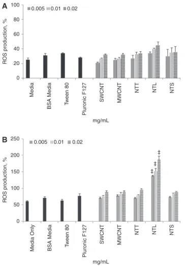
In a cell-free environment, no significant (p > 0.05) ROS was
observed for any of the CNTs or dispersants tested when
compared to the negative control (Figure 2A). In the
pres-ence of MDM, only the long MWCNTs (NTL) showed a
signif-icant (p < 0.05), dose-dependent increase in the production
of ROS following 24 h exposure to 0.005–0.02 mg/mL
(Figure 2B). No significant (p > 0.05) levels of oxidative
stress were noted in any of the other CNT or dispersants
tested when compared to the negative control (Figure 2B).
Pro-inflammatory response
Only the bundled MWCNTs, dispersed in Pluronic F127 (160
ppm), at 0.02 mg/mL showed a significant pro-inflammatory
response (p < 0.05), with an increased production in the
cytokine tumor necrosis factor-α (TNF-α) observed after
24 h (Figure 3). No significant (p > 0.05) pro-inflammatory
cytokine release was found for any of the other four CNTs
tested. It is also important to note that, compared to the
negative control, no significant (p > 0.05) (Figure 3) increase
in the production of TNF-α by MDM was observed for either
Pluronic F127 or Tween 80 at 160 ppm and 0.04 mg/mL,
respectively. The 5% dispersant solution (i.e., BSA Media)
for the NTL, NTT and NTS MWCNT samples also caused no
significant pro-inflammatory response in vitro (Figure 3). It
is also important to note that no significant (p > 0.05) protein
adsorption was observed between the TNF-α protein and
the CNTs (i.e., eliciting a false positive/negative) or
disper-sants tested (data not shown).
Discussion
The aim of this study was to perform a controlled in vitro
investigation of the biological impact of a series of
indus-trially produced CNTs that differed in their physical
prop-erties upon a primary in vitro model immune cell [i.e.,
monocyte derived macrophages (MDM)].
Assessment of the ability for each CNT type to affect
macrophage viability clearly showed no cytotoxic effects
up to 0.02 mg/mL following suspension exposure for
24 h. These findings were supported by conventional
light microscopy that showed the MDM to maintain their
morphological aspects compared to untreated primary
human macrophages. Furthermore, the findings of the
present study are in support of those of Murphy et al. [27],
who showed the NTT, NTL and NTS samples to be
non-cytotoxic (as determined via LDH release) to the THP-1
macrophage cell-line at administered concentrations
(suspension exposure) ranging from 0.02 to 0.05 µg/cm
2.
Additionally, Clift et al. [36] reported that the MWCNTs
also elicited no significant LDH release within MDM up to
0.03 mg/mL, as was also seen for SWCNTs [37]. Engulfment
of CNTs by MDM could not be detected due to the limited
resolution of conventional light microscopy, although
previous research by Clift and colleagues has shown that
the MWCNTs can locate within membrane-bound vesicles
inside MDM after 24 h suspension exposure [36], whilst
the SWCNTs used in the present study are internalized and
present within the cytosol of MDM [37]. It is also prudent
to note that Murphy et al. [27], as well as Poland et al. [24]
showed frustrated phagocytosis of the NTL sample in both
THP-1 and peritoneal macrophage cells, whilst the NTT
and NTS samples were both observed to be internalized
within vesicles by these phagocytic immune cells [27].
At sub-lethal concentrations CNTs have been shown
to induce the production of ROS which leads in an
inflam-matory response via the release of pro-inflaminflam-matory
cytokines in vitro [13, 32]; thus fitting the oxidative stress
paradigm – the most widely accepted paradigm concerning
the potential for nano-objects to cause adverse biological
effects [10]. ROS are produced from the respiratory chain
in the mitochondria by generating adenosine
tri-phos-phate (ATP) and can induce, even at low concentrations,
TNF-α release from the cell. This mechanism can
subse-quently lead to restoring the redox balance in the cell via
removing the oxidative species present [38]. In the current
study, only the NTL sample, in a concentration-dependent
manner, was found to cause any ROS production, although
0.005 100
A
B
i
ii
iv
v
vi
vii
iii
80 60 40 LDH Releas e, % 20 mg/mL 0 Media BSA Media Tw een 80 Pluronic F127 SWCNT MWCNT NTT NTL NTS 0.01 0.02Figure 1 (A) Indicates the level of cytotoxicity, as determined by the release of the cytosolic enzyme lactate dehydrogenase (LDH), of human blood isolated monocyte derived macrophages (MDM) after exposure to single-walled CNTs (SWCNTs), multi-walled CNTs (MWNCTs), long MWCNTs (NTL), tangled MWCNTs (NTT) and short MWCNTs (NTS) for 24 h at 37°C, 5% CO2 at nanofiber concentrations of 0.005, 0.01 and
0.02 mg/mL. In addition, the cytotoxic effects of the CNT dispersants Pluronic F127 (160 ppm), Tween 80 (0.04 mg/mL) and bovine serum albumin (0.5%) (BSA Media) are also shown. Data is normalized to the effects of the positive control (0.2% Triton ×100). Data presented is the mean ± standard error of the mean (SEM) (n = 3). (B) Shows conventional light microscopy images of the MDM exposed to the different CNT samples (i; Media only, ii; BSA Media, iii; SWCNTs, iv; MWCNTs, v; NTL, vi; NTT, vii; NTS) after 24 h at 37°C, 5% CO2. In each image the
0.005 100
B
A
80 50 100 150 200 250 0 60 40 R OS production, % R OS production, % 20 mg/mL 0 Medi a BSA Medi a Tw een 80 Pluronic F127 SWCNT MWCNT NTT NTL NTS 0.01 0.02 ‡ ‡ ‡ 0.005 0.01 0.02 mg/mLMedia Only BSA Media Tw
een 80
Pluronic F127
SWCNT MWCNT
NTT NTL NTS
Figure 2 (A) Shows the ability for the single-walled CNTs (SWCNTs), multi-walled CNTs (MWNCTs), long MWCNTs (NTL), tangled MWCNTs (NTT) and short MWCNTs (NTS) at nanofiber concentrations of 0.005, 0.01 and 0.02 mg/mL, as well as the dispersants Pluronic F127 (160 ppm), Tween 80 (0.04 mg/mL) and bovine serum albumin (0.5%) (BSA Media) to produce reactive oxygen species (ROS), as determined by the DCFH-DA assay via spectrometry, in a cell-free environment. (B) Illustrates the ability for the SWCNTs, MWCNTs, NTT, NTL and NTS at 0.005, 0.01 and 0.02 mg/mL, as well as the dispersants Pluronic F127 (160 ppm), Tween 80 (0.04 mg/mL) and bovine serum albumin (0.5%) to cause ROS in the presence of MDM via flow cytometry (BD LSR Fortessa). For both the cell-free and MDM experiments tert-butyl hydrogen peroxide (tbHP) (1:12,000 of 70% solution in PBS) was used. Data is normalized as a percentage against the ROS produced from the positive control (tbHP). Data presented is the mean ± standard error of the mean (SEM) (n = 3). *indicates p < 0.05. 0.005 0.50 0.45 0.40 0.35 0.30 0.25 0.20 0.15 0.10 0.05 0 TNF-α , ng/mL 0.01 0.02 ‡ ‡ mg/mL Medi a BSA Medi a Tw een 80 Pluronic F127 SWCNT LP S MWCNT NTT NTL NTS
Figure 3 Shows the pro-inflammatory response for the single-walled CNTs (SWCNTs), multi-single-walled CNTs (MWNCTs), long MWCNTs (NTL), tangled MWCNTs (NTT) and short MWCNTs (NTS) at nanofi-bre concentrations of 0.005, 0.01 and 0.02 mg/mL, as well as the dispersants Pluronic F127 (160 ppm), Tween 80 (0.04 mg/mL) and bovine serum albumin (0.5%) (BSA Media) to cause the production of the pro-inflammatory cytokine tumor necrosis factor-α (TNF-α). Lipopolysaccharide (LPS) was used as a positive control (0.1 mg/mL). Data presented is the mean ± standard error of the mean (SEM) (n = 3). *indicates p < 0.05.
only in the presence of MDM. No significant ROS
produc-tion was observed in a cell-free environment for any of the
CNTs tested. The high ROS production caused by the NTL
when interacting with the MDM can be attributed to the
process known as ‘frustrated phagocytosis’ [39], and not
to the increased redox potential via the high iron content,
as previously confirmed by Poland et al. [24]. Frustrated
phagocytosis occurs when macrophages are unable to
successfully internalize biopersistent fibres longer than 15
µm and with a diameter < 5 µm [39, 40]. The constant flux
of the immune cell attempting to phagocytose the fibre
generally causes the release of both oxidative and (pro-)
inflammatory mediators that can be detrimental towards
the normal homeostasis of the cell [39]. A similar effect
was also reported with the same NTL MWCNTs by Brown
and colleagues with the THP-1 macrophage cell-line after
CNT suspension exposure of up to 62.5 µg/mL for 4 h
[39]. Brown et al. [39] further showed the NTL MWCNTs
to cause a significant secretion of the pro-inflammatory
cytokine TNF-α by these immune cells, a finding that was
not observed within the present study. A possible reason
for the lack of TNF-α production observed from the MDM
could be due to the different cell types used (i.e., human
primary vs. a cell line), or the ELISA analysis being
per-formed after 24 h post-exposure samples and not within
the optimal expression/secretion of the protein [41], as it
was performed in the study by Brown et al. [39].
In the present study only the MWCNTs elicited any
sig-nificant TNF-α response from the MDM, despite the lack
of any ROS production. These findings are consistent with
those previously found with these MWCNTs [36] when
com-pared to the ability for cellulose nanowhiskers to elicit a
TNF-α response in both MDM monocultures and a
sophis-ticated triple cell co-culture model of the epithelial airway
barrier. Interestingly however, the SWCNTs showed no
significant pro-inflammatory characteristics to the MDM
after 24 h exposure up to 0.02 mg/mL. The fact that these
findings are not significant is in contrast to a recent study
[37], although, the biological effects shown in both cases
are the same. In the present study the lack of any statistical
significance can be attributed to the high variance between
repetitions, especially at the highest concentration tested.
It is important to note though, that there is a clear
dose-dependent effect shown with the SWCNTs and that whilst
this is not considered statistically significant, that this
bio-logical response can be considered influential upon
cellu-lar homeostasis when compared to the values shown with
the negative control (MDM exposed to cell culture media
only). In regards to the NTT and NTS MWCNTs results,
the observation that neither sample showed a
signifi-cant increase in either ROS or TNF-α production support
those reported by Murphy et al. [27], in which it was found
that after exposure of NTT and NTS to THP-1 cells for 24 h
caused no significant oxidative stress or pro-inflammatory
reaction after suspension exposure up to 0.05 µg/cm
2over
a 24-h period [27]. It is worth noting that the lack of ROS
formation, and potentially the subsequent
(pro-)inflam-matory response of immune cells to either the NTT or the
NTS could be masked by the formation of surface groups
following the extended sonication period (4 h). Whilst this
is not the case with the NTL sample (due to the increased
ROS formation shown), the intensity and duration of the
sonication period, which has been shown to be optimal in
suspending these CNT samples [24], could cause structural
surface defects that in turn do not elucidate to true hazard
potential of these CNTs. Structural defects to the surface
of CNTs remains an important issue [28, 29], as previously
shown by Kagan et al. [42], and requires further, in-depth
investigation to understand how they may contribute to
the potential adverse health effects of CNTs.
Concerning the pro-inflammatory response shown
with the MWCNTs, the finding that CNTs may cause an
inflammatory response, but no oxidative stress within
cells compliments the findings of Shvedova et al. [18].
Shvedova and colleagues reported an initial high,
dose-dependent release of TNF-α after 24 h following
inha-lation exposure (4 consecutive days 5 h/day) or single
pharyngeal aspiration of SWCNTs into mice, yet no
depletion of the intracellular thiol glutathione (GSH),
a key marker used to assess the oxidative potential of
nanomaterials was noted. It was however, observed
by Shvedova et al. [18] that GSH levels lowered after 7
and 28 days post-exposure, suggesting that deleterious
effects in vivo are delayed compared to those recorded
from in vitro studies; a phenomenon aptly shown by
Rothen- Rutishauser et al. [43] who compared the
oxida-tive and (pro-)inflammatory effects of MWCNTs in vitro
and in vivo. The in vivo study by Shvedova and colleagues
however used SWCNTs, which have been shown to cause
a limited (pro-)inflammatory response when compared
to that induced by MWCNTs in vitro and in vivo [13]. In a
recent study Bussy et al. [44] exposed MWCNT (MWCNT
treated with long lasting (7 weeks) ultrasonication in
water (S-MWCNT) and MWCNT untreated (L-MWCNT)) to
the murine macrophage cell-line (RAW 264.7) for 6 and
24 h. It was found that after 6 h an increased
pro-inflam-matory response was evident. Over time however, and
concomitant with an increasing expression (measured
at the gene level) of superoxide dismutase-2 (SOD-2) and
heme oxygenase-1 (HO-1) after exposure to S-MWCNT, the
level of TNF-α release by these macrophage cells in vitro
was significantly decreased. These findings suggest that
the pro-inflammatory response caused by MWCNTs is
often oxidant mediated, in contrast to the results shown
in the present study. A possible reason for such effects
seen could be attributed to the surfactant used to
origi-nally disperse the MWCNT sample. Although all CNTs
had the same final dispersant (i.e., ‘biological buffer’),
the MWCNTs and SWCNTs were dispersed using
chemi-cal surfactants [33, 34, 45]. Specifichemi-cally the MWCNTs were
dispersed in 160 ppm of Pluronic F127 in MilliQ H
2O [34].
This has been shown not to cause any ROS or TNF-α
pro-duction amongst numerous other tests (e.g., GSH
deple-tion and IL-8 producdeple-tion in epithelial cells [37]) on its own,
and could, in theory, be attributed to masking any ROS
formed by the MWCNTs due to its abundance upon the
surface of the MWCNTs. The TNF-α increase could
there-fore not be a simple result of the MWCNTs themselves,
but due to the specific interaction (active entry
mecha-nism) of the MWCNTs with the MDM, in which MWCNTs
gain entry into the MDM and localise within
membrane-bound compartments, as previously shown by Clift et al.
[36]. Both the NTT and NTS samples however, were also
internalized via a proposed active mechanism (i.e., found
to be present within vesicles inside macrophage cells
[24, 27]) although showed no pro-inflammatory effects.
The NTT and NTS samples were originally dispersed in a
0.5% bovine serum albumin (BSA) solution. It is possible
that, the BSA ‘coating’ has a further protective effect for
both the NTT and NTS samples compared to the Pluronic
‘coated’ MWCNTs, which may, within highly acidic
envi-ronments degrade from the CNT surface and allow for
direct interaction of the MWCNTs with the immune cell
machinery. Further research would have to be performed
to confirm this hypothesis however, and determine the
long-term, in situ stability of differently dispersed CNTs
in vitro and how this may contribute to an adverse
bio-logical impact of CNTs.
In summary therefore, the present study has shown
that for different biological endpoints, different
physi-cal aspects of industrial CNTs can be associated with the
onset of a severe, negative biological effect in vitro. Whilst
some of the effects noted can be clearly associated with
the physico-chemical characteristics of the CNTs (i.e., ROS
production of NTL MWCNTs), other reactions (e.g., TNF-α
production by MWCNTs in MDM) are more complicated. In
the latter, not only does the material play a role in the
bio-logical impact, but the manner in which it interacts with
the biological system is important. In conclusion therefore,
from the data presented within the present manuscript,
it is possible to state that the specific morphology of the
CNTs can contribute to the adverse biological responses
observed, yet are not entirely responsible for the observed
biological impact of CNTs upon immune cells in vitro.
Materials and methods
Chemical and reagents
All chemicals and reagents used were purchased from Sigma-Aldrich (Switzerland), unless otherwise stated.
CNT physico-chemical characteristics
A series of five different physico-chemical CNTs, that are industri-ally available and produced via chemical vapor deposition (CVD), were used in this study; single-walled bundled CNTs (SWCNTs), multi-walled bundled CNTs (MWCNTs), tangled MWCNTs (NTT), long MWCNTs (NTL), and short MWCNTs (NTS). The characteristics of the different CNTs used have previously been reported in the literature by Wick et al. [33] (SWCNT), Thurnherr et al. [34] (MWCNT), Poland et al. [24] (NTT, NTL) and Murphy et al. [26] (NTS) and are detailed in Table 1. The key physico-chemical characteristics of each CNT are described in Table 1. The length, diameter, and morphology of each CNT were obtained in all four studies by transmission electron microscopy (TEM) and scanning electron microscopy (SEM). Deter-mination of metal contaDeter-mination was achieved via inductive cou-pled plasma mass spectrometry (ICP-MS) for the SWCNTs, NTT and NTL, respectively [24, 33]. Thurnherr et al. [34] assessed the metal contaminations of the MWCNTs by inductive coupled plasma optical emission spectrometry (ICP-OES). Information related to the metal contaminants contained within the NTS sample was provided by the suppliers (Table 1). No endotoxin content, assessed by the limulous
amoebocyte lysate (LAL) test, is known to be present within any of
the CNT samples [10, 24, 26].
Preparation of CNT samples
In order to have a well-dispersed stock solution, SWCNTs were dis-persed in the surfactant Tween 80 at a concentration of 0.04 mg/mL,
whereas the MWCNTs were dispersed in Pluronic F127 at a concentra-tion of 160 ppm [33]. Dry powder samples of the NTT, NTL, and NTS were prepared at a concentration of 1 mg/mL using the method previously reported by Poland et al. [24]. Each dry powder sample was suspended in Rosewell Park Memorial Institute (RPMI 1640) medium supplemented with 0.5% bovine serum albumin (BSA), 1% L-glutamine (L-G) (100 U/ mL) and 1% penicillin/streptomycin (P/S) (0.1 µg/mL) (hereby referred to as ‘BSA Media’), sonicated for 4 h (Bransonic, Branson, Switzerland) and then sterile filtered to obtain a well dispersed solution.
All five CNTs were then suspended in RPMI 1640 compli-mented with 10% fetal calf serum (FCS), 1% L-G and 1% P/S (hereby referred to as ‘biological buffer’) at nanofibre concentra-tions of 0.005, 0.01, and 0.02 mg/mL. To control for the effects of suspending the CNTs in FCS, samples at concentrations of 0.005– 0.02 mg/mL were also prepared in RPMI 1640 supplemented with only 1% L-G and 1% P/S, and no FCS content. Samples were sub-sequently sonicated (Bransonic, Branson, Switzerland) for 10 min prior to being investigated in terms of their ability to cause cytotox-icity, pro-inflammatory cytokine stimulation and reactive oxygen species production following exposure to human blood monocyte derived macrophages (MDM).
Cell culture
MDM were isolated from human buffy coat (Blutspendezentrum, Bern, Switzerland) using a density gradient centrifugation as previ-ously described by Lehmann et al. [46]. In order to confirm a pure MDM population, the blood isolated leukocytes were stained with magnetic beads specific for the MDM surface protein CD14 and passed through a magnetic filter [47]. CD14+ MDM were subsequently
cultured for 6 days at 37°C, 5% CO2 in the presence of 10 ng/mL of
macrophage colony-stimulating factor (M-CSF) in the biological
buffer.
CNT cell exposure
After the 6-day culture period, MDM were seeded into a 12-well plate (TPP, CH) at a density of 1 × 10⁶ cells/mL (1 mL volume per well) and
cultured at 37°C, 5% CO2 for 24 h. Following the incubation period, MDM were exposed (via suspension exposure) to each different CNT sample at a nanofiber concentration of 0.005, 0.01, or 0.02 mg/mL for 24 h at 37°C, 5% CO2. After the exposure period, the cellular
superna-tant was collected and either stored at 4°C or –80°C for subsequent biochemical analysis. MDM were then prepared for an assessment of their cellular morphology via light microscopy, as described below.
Cellular morphology
MDM were washed once with 1 × phosphate buffered saline (PBS) and then fixed using 3% paraformaldehyde (PFA) in PBS (Merck, Switzerland) for 15 min at room temperature. After fixation, the PFA-PBS solution was removed and MDM were washed once with 1 × PBS. MDM were then imaged using a conventional light microscope (AE200, Motic, Switzerland) containing a digital camera (Nikon, Switzerland) at a magnification of 40 × (N.A. 0.5).
Biochemical analysis
Cytotoxicity
Assessment of lactate dehydrogenase release
The cytosolic enzyme lactate dehydrogenase (LDH) was measured to provide an indication of the cell membrane permeability and associated potential cytotoxicity of the different CNT samples to the MDM. LDH was measured in MDM supernatants using a diagnos-tic detection kit (Roche, Switzerland). As a positive control, Triton ×100 at a concentration of 0.2% in PBS (Fluka, Switzerland) was used.
Assessment of LDH adsorption
To determine if each CNT sample was interfering with the LDH enzyme and thus eliciting a false-negative or positive result [48] an assessment of the ability for CNT to adsorb or to concentrate the LDH enzyme was performed using the method previously described by Clift et al. [35].
Reactive oxygen species production
Reactive oxygen species (ROS) are a natural by-product of oxygen production and are important for intracellular signaling pathways. Since CNTs have been shown to cause ROS production independent of their interaction with mammalian cells (i.e., in a cell-free environ-ment) [39], the CNTs used in the present study were assessed for their ROS production in both a cell-free environment and also following exposure to MDM. On both occasions, an adapted version of the pro-tocol previously described by Wilson et al. [49] and Foucard et al. [50], the fluorescent probe 2′,7′-dichlorfluorecein-diacetate (DCFH2 -DA) which is degraded by ROS, cleaving the molecule to DCF which subsequently elicits a fluorescent signal [51], was used. Furthermore, the compound horse-radish peroxidase was used to amplify any ROS signal present [43].
ROS production in a cell-free environment
Briefly, in a 96-well plate (white, solid bottom plate [Berthold Tech-nologies, Germany]) 200 µL of HBSS-HRP solution (Hanks Buffered Salt Solution (Gibco, Switzerland) containing Horse-radish peroxi-dase (3 mg/mL) (Fluka, Switzerland) at a 1:32 ratio] was mixed with 1 µL DCFH2-DA solution (DCFH+) [DCFH2-DA (Invitrogen, Switzerland)
diluted 1:4 with methanol to 1 mM]. Subsequently, this solution was further diluted 1:10 with either sodium hydroxide (0.01 mM) (NaOH, 2.5 mg in 1 L of MilliQ water) or 1µ L DCFH- solution [methanol, NaOH (0.01 mM) and 1 × PBS mixed at a ratio of 1:4:15], respectively. The fluorescent intensity of each well in the 96 well-plate was then meas-ured using a fluorimeter (TriStar LB 941, Berthold Technologies, Germany) at a wavelength of 488 nm. After this baseline measure-ment, a total of 45 µL of either the CNT samples (prepared in
biologi-cal buffer) or the positive control [tert-butyl hydrogen peroxide (70%
solution) (tBHP, CH) mixed with 1 × PBS (1:12,000)] was then added to the corresponding wells in the 96-well plate. Each well was then measured in 1-min intervals over a 10-min period using the fluorim-eter (TriStar LB 941, Berthold Technologies, Germany) at a wave-length of 488 nm. Following acquisition, data was subsequently
calculated and expressed as a percentage of the negative control (biological buffer) value.
ROS production in MDM
ROS formation within MDM exposed to each different CNT sample was performed using fluorescence activated cell sorting (FACS) (LSR Fortessa, BD Biosciences, Switzerland). MDM a density of 1 × 10⁵ cells/
mL were seeded in 5 mL FACS tubes (BD Biosciences, Switzerland) and cultured for 24 h at 37°C, 5% CO2. MDM were then washed;
sam-ples were centrifuged for 8 min at 1300 rpm to form a cell pellet. The remaining supernatant was discarded and MDM were re-suspended with biological buffer containing CD14+ at a 1:50 dilution (BD
Bio-sciences, Switzerland). Samples were then incubated for 1 h at 37°C, 5% CO2. MDM were then washed twice: centrifugation for 8 min at
1300 rpm, removal of supernatant and re-suspension of cells in fresh
biological buffer. The supernatant was discarded and MDM were
exposed to the DCFH+ solution (DCFH diluted 1:4 in methanol and then 1:10 in 1 × PBS) for 30 min at 37°C, 5% CO2. After the
incuba-tion period samples were washed once, and then re-suspended with 500 µL of FACS buffer (1 × PBS containing 1% BSA and 0.1% sodium azide). Using consistent parameters the geometric mean fluorescent intensity (GMFI) was analysed immediately at each time point using two-colour flow cytometry (LSR Fortessa, BD Biosciences, Switzer-land). Fluorescent signals were collected in logarithmic mode (4 decade logarithmic amplifier) and cell numbers per channel in lin-ear mode. To identify the MDM population, an electronic gate was placed around the forward and side scatter modes with 10,000 gated events acquired for analysis. The fluorescent amplifiers of the detec-tor filters were adjusted to ensure that the negative cell population appeared in the first logarithmic decade. An electronic marker was then placed at the limit of the negative control to express all positive cell populations in the final three logarithmic decades. Compensa-tion for spectral overlap was performed automatically using the BD FACSDiva computational software. Following acquisition, data was subsequently calculated and expressed as a percentage of the nega-tive control (biological buffer) value.
Pro-inflammatory response
Tumor necrosis factor-alpha release
Tumor necrosis factor (TNF)-α is the primary cytokine released by macrophages in response to inflammatory stimuli. TNF-α regulates the activation of other immune cells, apoptosis, cell proliferation, and the release of other cytokines. The concentration of the released pro-inflammatory cytokine TNF-α (in MDM supernatant) was deter-mined using an enzyme-linked immunosorbant assay (ELISA) (R&D Systems, Switzerland). Lipopolysaccharide (LPS) (0.1 mg/mL) was used as a positive control.
TNF-α adsorption
Adsorption of the CNTs to the TNF-α protein, or vice-versa, was per-formed to exclude any false-positive or false-negative results. Briefly, each different CNT sample, at a nanofiber concentration of 0.005, 0.01, or 0.02 mg/mL, was incubated with TNF-α (10 ng/mL) at volume of 500 µL in an environment of 37°C, 5% CO2 for 1 h. After the
incuba-tion period, all samples were then assessed for their TNF-α content by ELISA (R&D Systems, Switzerland).
Data and statistical analysis
The data is presented as mean ± standard error of the mean (SEM). All data was found to be normally distributed (data not shown). To deter-mine statistical significance between the qualitative data sets, a one-way ANOVA was used with a Tukey’s post-hoc test (SPSS, IBM, USA). Results were considered significant if p < 0.05. A confidence interval of 95% was accepted.
Acknowledgments: The authors would like to
acknowl-edge the Adolphe Merkle Foundation, an Empa internal
grant, as well as the Swiss National Science Foundation,
the Swiss National research Programme 64 and the Swiss
Nanoscience Institute (SNI) within the National Center of
Research (NCCR) in Nanoscale Science for their financial
support. The authors also thank Vicki Stone (Heriot-Watt
University, Edinburgh, UK), Craig A. Poland (Institute of
Occupational Medicine, Edinburgh, UK) Ken Donaldson
and Rodger Duffin (Edinburgh University, UK) for their
kind donation of the NTT, NTL and NTS samples.
Conflict of interest statement: The authors declare no
conflict of interest. The authors are entirely
responsi-ble for the data contained within and the writing of the
manuscript.
Received July 29, 2013; accepted October 9, 2013; previously pub-lished online November 6, 2013
References
1. Piccinno F, Gottschalk F, Seeger S, Nowack B. Industrial production quantities and uses of ten engineered nanoma-terials in Europe and the world. J Nanopart Res 2012;14:1109. 2. Kostarelos K, Bianco A, Prato M. Promises, facts and challenges
for carbon nanotubes in imaging and therapeutics. Nat Nanotech 2009;4:627–33.
3. Robertson J. Realistic applications of CNTs. Mater Today 2004;7:46–52.
4. NIOSH. General safe practices for working with engineered nanomaterials in research laboratories. Cincinnati, OH: U.S. Department of Health and Human Services, Centers for Disease Control, National Institute for Occupational Safety and Health, DHHS (NIOSH) Publication No. 2012–147, 2012.
5. Maynard AD, Baron PA, Foley M, Shvedova AA, Kisin ER, Castranova V. Exposure to carbon nanotube material: aerosol release during the handling of unrefined single-walled carbon nanotube material. J Tox Environ Health Part A 2004;67:87–107.
6. Mueller L, Gasser M, Raemy DO, Herzog F, Brandenberger C, Schmid O, et al. Realistic exposure methods for investigating the interaction of nanoparticles with the lung at the air-liquid interface in vitro. InSci J (Nanotech) 2011;1:30–64.
7. Oberdorster G, Stone V, Donaldson K. Toxicology of
nanoparticles: a historical perspective. Nanotox 2007;1:2–25. 8. Rothen-Rutishauser B, Blank F, Muehlfeld C, Gehr P. In vitro
models of the human epithelial airway barrier to study the toxic potential of particulate matter. Exp Opin Drug Metab Toxicol 2008;4:1075–89.
9. Van Berlo D, Clift MJ, Albrecht C, Schins RP. Carbon nanotubes: an insight into the mechanisms of their potential genotoxicity. Swiss Med Wkly 2012;142:w13698.
10. Clift MJ, Foster EJ, Vanhecke D, Studer D, Wick P, Gehr P, et al. Investigating the interaction of cellulose nanofibers derived from cotton with a sophisticated 3D human lung cell coculture. Biomacromol 2011;12:3666–73.
11. Timbrell J. Principles of biochemical toxicology. CRC Press, UK: Taylor and Francis, 1999.
12. Wick P, Clift MJ, Rosslein M, Rothen-Rutishauser B. A brief summary of the past 20 years of carbon nanotubes in science:
a health and safety perspective. Chemsuschem 2011;4: 905–11.
13. Johnston HJ, Hutchison GR, Christensen FM, Peters S, Hankin S, Aschberger K, et al. A critical review of the biological mechanisms underlying the in vivo and in vitro toxicity of carbon nanotubes: the contribution of physico-chemical charac-teristics. Nanotox 2010;4:207–46.
14. NIOSH Current Intelligence Bulletin 65. Occupational exposure to carbon nanotubes and nanofibres. Cincinnati, OH: U.S. Department of Health and Human Services, Centers for Disease Control, National Institute for Occupational Safety and Health, DHHS (NIOSH) Publication No. 2013–145, 2013.
15. Lam CW, James JT, McCluskey R, Hunter RL. Pulmonary toxicity of single-wall carbon nanotubes in mice 7 and 90 days after intratracheal instillation. Toxicol Sci 2004;77:126–34. 16. Shvedova AA, Kisin ER, Mercer R, Murray AR, Johnson VJ,
Potapovich AI, et al. Unusual inflammatory and fibrogenic pulmonary responses to single-walled carbon nanotubes in mice. Am J Physiol Lung Cell Mol Physiol 2005;289: L698–708.
17. Muller J, Huaux F, Moreau N, Misson P, Heilier JF, Delos M, et al. Respiratory toxicity of multiwall carbon nanotubes. Toxicol Appl Pharmacol 2005;207:221–31.
18. Shvedova AA, Kisin E, Murray AR, Johnson VJ, Gorelik O, Arepalli S, et al. Inhalation vs. aspiration of single-walled carbon nanotubes in C57BL/6 mice: inflammation, fibrosis, oxidative stress, and mutagenesis. Am J Physiol Lung Cell Mol Physiol 2008;295:L552–65.
19. Ma-Hock L, Treumann S, Strauss V, Brill S, Luizi F, Mertler M, et al. Inhalation toxicity of multi-wall carbon nanotubes in rats exposed for 3 months. Toxicol Sci 2009;112:468–81.
20. Pauluhn J. Subchronic 13-week inhalation exposure of rats to multiwalled carbon nanotubes: toxic effects are determined by density of agglomerate structures, not fibrillar structures. Toxicol Sci 2010;113:226–42.
21. Porter DW, Hubbs AF, Mercer RR, Wu N, Wolfarth MG, Sriram K, et al. Mouse pulmonary dose- and time course-responses induced by exposure to multi-walled carbon nanotubes. Toxicol 2010;269:136–47.
22. Mercer RR, Hubbs AF, Scabilloni JF, Wang L, Battelli LA, Friend S, et al. Pulmonary fibrotic response to aspiration of multiwalled carbon nanotubes. Part Fibre Toxicol 2011;8:21.
23. Stanton MF, Layard M, Tegeris A, Miller E, May M, Kent E. Carcinogenicity of fibrous glass: pleural response in the rat in relation to fiber dimension. J Nat Can Inst 1977;58:587–603. 24. Poland CA, Duffin R, Kinloch I, Maynard A, Wallace WA,
Seaton A, et al. Carbon nanotubes introduced into the abdominal cavity ofmice show asbestoslike pathogenicity in a pilot study. Nat Nanotech 2008;3:423–8.
25. Donaldson K, Murphy FA, Duffin R, Poland CA. Asbestos, carbon nanotubes and the pleural mesothelium: a review of the hypothesis regarding the role of long fibre tetention in the parietal pleura, inflammation and mesothelioma. Part Fibre Toxicol 2010;7:5.
26. Murphy FA, Poland CA, Duffin R, Al-Jamal KT, Ali-Boucetta H, Nunes A, et al. Length-dependent retention of carbon nanotubes in the pleural space of mice initiates sustained inflammation and progressive fibrosis on the parietal pleura. Am J Path 2011;178:6.
27. Murphy FA, Schinwald A, Poland CA, Donaldson K. The mechanism of pleural inflammation by long carbon nanotubes: interaction of long fibers with macrophages stimulates them to amplify proinflammatory responses in mesothelial cells. Part Fibre Toxicol 2012;9:8.
28. Fenoglio I, Greco G, Tomatis M, Muller J, Raymundo-PinÌfero E, Beguin Fo, et al. Structural defects play a major role in the acute lung toxicity of multiwall carbon nanotubes: physicochemical aspects. Chem Res Tox 2008;21:1690–7.
29. Muller J, Huaux Fo, Fonseca A, Nagy JB, Moreau N, Delos M, et al. Structural defects play a major role in the acute lung toxicity of multiwall carbon nanotubes: toxicological aspects. Chem Res Tox 2008;21:1698–705.
30. Tian F, Cui D, Schwarz H, Estrada GG, Kobayashi H. Cytotoxicity of single-wall carbon nanotubes on human fibroblasts. Toxicol In Vitro 2006;20:1202–12.
31. Stone V, Johnston H, Schins RP. Development of in vitro systems for nanotoxicology: methodological considerations. Crit Rev Toxicol 2009;39:613–26.
32. Donaldson K, Aitken R, Tran L, Stone V, Duffin R, Forrest G, et al. Carbon nanotubes: a review of their properties in relation to pulmonary toxicology and workplace safety. Toxicol Sci 2006;92:5–22.
33. Wick P, Manser P, Limbach LK, Dettlaff-Weglikowska U, Krumeich F, Roth S, et al. The degree and kind of agglom-eration affect carbon nanotube cytotoxicity. Tox Letts 2007;168:121–31.
34. Thurnherr T, Su DS, Diener L, Weinberg G, Manser P, Pfaender N, et al. Comprehensive evaluation of in vitro toxicity of three large-scale produced carbon nanotubes on human Jurkat T cells and a comparison to crocidolite asbestos. Nanotox 2009;3: 319–38.
35. Clift MJ, Rothen-Rutishauser B, Brown DM, Duffin R, Donaldson K, Proudfoot L, et al. The impact of different nanoparticle surface chemistry and size on uptake and toxicity in a murine macrophage cell line. Toxicol Appl Pharm 2008;232:418–27.
36. Clift MJ, Gehr P, Rothen-Rutishauser B. In vitro testing for nanotox-icology: a valid alternative? Arch Toxicol 2011;85:723–31. 37. Clift MJ, Endes C, Vanhecke D, Wick P, Gehr P, Schins RP, et al.
A comparative study of different in vitro lung cell culture systems to assess the most beneficial tool for screening the potential adverse effects of carbon nanotubes. Toxicol Sci (in Press). 38. Donaldson K, Stone V, Borm PJ, Jimenez LA, Gilmour PS, Schins RP,
et al. Oxidative stress and calcium signaling in the adverse effects of environmental particles (PM10). Free Rad Biol Med 2003;34:1369–82.
39. Brown DM, Kinloch IA, Bangert U, Windle AH, Walter DM, Walker GS, et al. An in vitro study of the potential of carbon nanotubes and nanofibres to induce inflammatory mediators and frustrated phagocytosis. Carbon 2007;45:1743–56. 40. Donaldson K, Tran CL. Inflammation caused by particles and
fibers. Inhal Toxicol 2002;14:5–27.
41. Locksley RM, Killeen N, Lenardo MJ. The TNF and TNF receptor superfamilies: integrating mammalian biology. Cell 2001;104:487–501.
42. Kagan VE, Konduru NV, Feng W, Allen BL, Conroy J, Volkov Y, et al. Carbon nanotubes degraded by neutrophil myeloper-oxidase induce less pulmonary inflammation. Nat. Nanotech 2010;5:354–9.
43. Rothen-Rutishauser B, Brown DM, Piallier-Boyles M, Kinloch IA, Windle AH, Gehr P, et al. Relating the physicochemical charac-teristics and dispersion of multiwalled carbon nanotubes in different suspension media to their oxidative reacitivity in vitro and inflammation in vivo. Nanotox 2010;4:331–42.
44. Bussy C, Pinault M, Cambedouzou J, Landry MJ, Jegou P, Mayne-L’hermite M, et al. Critical role of surface chemical modifications induced by length shortening on multi-walled carbon nanotubes-induced toxicity. Part Fibre Toxicol 2012;9:46. 45. Thurnherr T, Brandenberger C, Fischer K, Diener L, Manser P,
Maeder-Althaus X, et al. A comparison of acute and long-term effects of industrial multiwalled carbon nanotubes on human lung and immune cells in vitro. Tox Letts 2011;200:176–86. 46. Lehmann A, Brandenberger C, Blank F, Gehr P,
Rothen-Rutishauser B. A 3D model of the human epithelial airway barrier. In: Yarmush ML, Langer RS, editors. Alternatives to animal testing. Artech House 2010:239–60.
47. Steiner S, Muller L, Popovicheva OB, Raemy DO, Czerwinski J, Comte P, et al. Cerium dioxide nanoparticles can interfere with the associated cellular mechanistic response to diesel exhaust exposure. Tox Letts 2012;214:218–25.
48. Worle-Knirsch JM, Pulskamp K, Krug HF. Oops they did it again! Carbon nanotubes hoax scientists in viability assays. Nano Letts 2006;6:6.
49. Wilson MR, Lightbody JH, Donaldson K, Sales J, Stone V. Interactions between ultrafine particles and transition metals in vivo and in vitro. Toxciol Appl Pharm 2002;184:172–9.
50. Foucaud L, Wilson MR, Brown DM, Stone V. Measurement of reactive species production by nanoparticles prepared in biologically relevant media. Tox Letts 2007;174:1–9. 51. Pal AK, Bello D, Budhlall B, Rogers E, Milton DK. Screening
for oxidative stress elicited by engineered nanomaterials: evaluation of acellular DCFH Assay. Dose-Response 2012;10:308–30.