The Induction of Meningeal Inflammation
by
Components of the
Pneumococcal Cell Wall
Elaine Tuomanen, Hans Liu, Bruno Hengstler, Oto Zak, and Alexander Tomasz
From the Laboratory of Microbiology, The Rockefeller University, New York; and the Research Department,
Pharmaceuticals Division, elBA-GEIGY, Basel, Switzerland Pneumococcal cell wall induces meningeal inflammation in rabbits injected
intracister-nally with >105cell equivalents. Both of the major cell wall components, teichoic acid
and peptidoglycan, contribute to this inflammatory activity although responses differ depending on the chemical nature, size, and complexity of these fractions. Challenge with teichoic acid (membrane or wall associated) results in greater inflammation at 5 hr than at 24 hr. Degraded teichoic acid is inactive. In contrast, the inflammation caused by whole cell wall or high-molecular-weight peptidoglycan-containing fractions increases in inten-sity from 5 to 24 hr. Peptidoglycan fractions lose activity at 24 hr when hydrolyzed to disaccharide-stem peptide moieties. Generation of free cellwallcomponents in cerebrospinal fluid as, for example, during treatment with antibiotics that are bacteriolytic as well as bactericidal, could contribute to increased inflammation in the subarachnoid space.
The cell wall of pneumococci, located under a layer of capsular polysaccharide, remains surprisingly ac-cessible to and reactive with the host environment [1]. We have shown that meningeal inflammation in rabbits is induced when whole pneumococci, with or without capsular polysaccharide, reach a density of >105cfu/ml of CSF. This inflammatory response
is remarkably similar to the inflammation follow-ing challenge with 105 cell equivalents of isolated cell wall, but not of isolated capsule [2]. Because the cell wall is a complex macromolecule with many possi-ble sites of interaction with several host defense sys-tems, the identification of which cell wall compo-nent(s) is active in inducing inflammation during pneumococcal meningitis is of considerable impor-tance.
The pneumococcal cell wall is composed of two major polymers: a peptidoglycan and a ribitol-phosphate teichoic acid of unusually complex struc-ture that contains phosphorylcholine [3]. The
inter-Received for publication September 17, 1984, and in revised form November 29, 1984.
This work was supported in part by a Parker B. Francis Fel-lowship to E. T. and by grants no. ROI-AI-16794 from the Na-tional Institute of Allergy and Infectious Diseases and no. BRSG-SO-7-RR-07065 from the National Institutes of Health.
We thank Drs. H. F. Chambers, M. Taueber, A.L.Smith, and M. Chase for review of the manuscript; and M. Geller for secretar-ial assistance.
Please address requests for reprints to Dr. Elaine Tuomanen, The RockefellerUniversity, Box 152, 1230York Avenue, New York, New York 10021
859
action of C-reactive protein and of certain myeloma proteins with pneumococci occurs through the rec-ognition of the teichoic acid-choline residues [4]. The teichoic acid component of the pneumococcal cell wall can also trigger the alternative pathway of complement activation in vitro [5], while both teichoic acid and peptidoglycan seem to have the ca-pacity to bind the C3b component of complement [6].
Itis not known which elements of the complex structure of the cell wall are responsible for the vari-ous interactions between the pneumococcus and the invaded host in vivo.The studies to be described here represent an attempt to answer this question in the case of the host response characteristic of pneumo-coccal meningitis. We tested whole pneumopneumo-coccal cell wall and various biochemically defined macro-molecular components of the cell wall for specific activity in provoking an inflammatory response when introduced intracisternally into the subarach-noid space of rabbits.
Materials and Methods
Streptococcus pneumoniae. Strain Au is an en-capsulated Rockefeller University laboratory strain (type II). Strains R6and lyt 4-4 (an
autolysin-defi-cient tolerant transformant of strain R6)are
unencap-sulated strains originally derived from strain Au.
Meningitis model. Male chinchilla rabbits weigh-ing 2 kg (Thome Farm, Biebarach der Riss, Federal Republic of Germany) were prepared according to
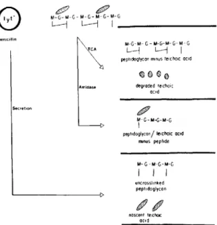
Figure 1. Summary of pneumococcal surface compo-nents that were isolated as described in Materials and Methods. Preparation H consists of disaccharide pen-tapeptide subunits derived from lysozyme treatment of preparation G. G = N-acetylglucosamine; M =
N-acetylmuramic acid; hatched area =teichoic acid;
-= stem peptide;TeA = trichloroacetic acid extraction; amidase = treatment with N-acetylmuramic acid-L-alanine amidase; lyt- = pneumococcus deficient in en-dogenous amidase activity; secretion = components secreted into medium upon treatment of strain lyt 4-4 with
0.024 JAg of penicillin/ml.
the method of Dacey and Sande [7] as described pre-viously [2]. Unless otherwise noted, a set of four rab-bits was tested for each bacteria or bacterial com-ponent inoculated. Each comcom-ponent was compared for activity on a cell equivalent and on a weight ba-sis. Immediately upon withdrawal, CSF samples were analyzed for cfu and cytochemistry as described else-where [2].
Preparation of pneumococcal cell surface com-ponents. The preparation of a wide variety of cell surface components is described in the following sec-tions. Because small variations in preparative tech-niques can yield components of significantly differ-ent natures, preparative methods are described in detail. Figure 1 schematically depicts these cell wall products.
1. Capsular polysaccharide. Several chemically different pneumococcal capsular polysaccharides were used. The polyvalent (14) pneumococcal poly-saccharide vaccine (Pneumovax'P; Merck Sharp& Dohme, Rahway, NJ) and type II and type III
cap-~
,
M-G-M-G-M-G-M-G-M-G l---i L---j I _ -penicillin Secretion TCA Amidase M-G-M- G - M-G-M- G- M- G L-j L.---j Ipeptidoglycan minusteicho«ocic
degraded telcholc ccid
IJ11fJlP
'f-G-M-G-M-G peptldoglycon/ lelcho,c aCid
Il'Ilnus peptide M- G-M-G-M-G I I I uncrossl,nked peptldaglycan ncscent telcholc ocrd
sular polysaccharides (Merck) were each dissolved in pyrogen-free saline at concentrations of 2-5 mg/ml and were dialyzed against several changes of the same solution. (Dialysis membrane was prepared by boiling, soaking in 0.5M EDTA, and then rins-ing in pyrogen-free saline.)
2. Pneumococcal cell wall. Cell wall with cho-line-containing teichoic acids (preparation A).
Strain R6was grown in one-liter batches of a
chemi-cally defined medium [8] to a cell concentration of 1"\15 x 108cfu/ml. The cells were harvested by
cen-trifugation and resuspended in 5 ml of saline, and the resulting suspension was submerged in a boiling water bath for 15 min to inactivate the autolytic en-zyme. The suspension was then transferred to a cuvette of the Mickle disintegrator (Hampton, Mid-dlesex, England) mixed with an equal volume of glass beads (Ballotini no. 13; l00-J,lm diameter; 3M, St. Paul, Minnesota), and shaken at maximum ampli-tude at 4 C for 3 hr. An airspace equal to the total volume of the suspension was kept in the cuvettes to allow efficient disruption. Next, the glass beads were separated out by sedimentation, the suspension was centrifuged at 3,000gfor 3 min (Sorvall RC2B; Sorvall, Newtown, Conn) to remove unbroken cells, and the supernatant was centrifuged at 10,000gfor 30 min to sediment out the cell wall material. Inspec-tion by phase-contrast microscopy revealed only amorphous debris in such preparations. This crude cell wall material was extracted with 2% SDS at 90-100 C for 30 min. Detergent was removed with six cycles of washings by centrifugation in distilled water. The cells, resuspended in 0.1 M Tris-HCI buffer (pH 8.0) containing 1 mMMgCb, weretreated at 37 C with pancreatic DNase I (50 ug/ml) plus RNase (100ug/ml) for 2 hr followed by trypsin (100 ug/ml) plus 10 mMCaCb for 10-12 hr. (All enzyme preparations were of crystalline grade and were ob-tained from Worthington Biochemicals, Freehold, NJ.) Cell wall was sedimented by centrifugation (10,000gfor 30 min) and resuspended in 5 ml of 2% SDS at 90-100 C in a water bath for 30 min. Deter-gent was removed by eight to 10 cycles of washing, first in 1M NaCI solution and then in distilled wa-ter, and the purified wall was lyophilized. The com-position of this cell wall material has been described [9].
On occasion, the following additional steps of purification were introduced. Cell wall (10 mg) was extracted with 10-ml portions of the following rea-gents at room temperature for 30 min each: 0.1M
Pneumococcal autolysin is an N-acetylmuramic acid-t-alanine amidase capable of degrading pneu-mococcal cell wall to two types of components separable on the basis of their different molecular sizes; the first, a high-molecular-weight glycan-Figure 2. Top:Elution profile of the products of ami-dase activity on pneumococcal cell wall. Cell wall was hydrolyzed with N-acetylmuramic acid-t-alanine amidase and prepared as described in Materials and Methods. Peak I (fractions 46-60) represents high-molecular-weight glycan containing all the cell wall teichoic acid (labeled with pH]methyl choline; 0), as well as glycan with residu-al pep tides (labeled with [3H]lysine; .). Peak II (frac-tions 105-150) represents lower molecular-weight peptides released from the cell wall by the amidase (labeled with PH]lysine only). Bottom: Elution profile of purified Forssman antigen. Partially purified pH]methyl cho-line-labeled Forssman antigen was passed through a Sepharose-TEPC 15 myeloma protein affinity column as described in Materials and Methods. Because the Forss-man antigen was retained on the column through its phos-phorylcholine residue, impurities could be removed by washes with buffer (no pH]methyl choline appears in fractions 1-15). Purified Forssman antigen was specifi-cally eluted from the column upon the addition of phos-phorylcholine (PC) to the eluting buffer ([3H]methyl choline appears in fractions 17-21).
=f. 150
t
150 n PC 100 FRACTION NUMBER 50 10 20 30 FRACTION NUMBER Buffer 1500 2 ll. U ... Q. 500 ~ s::10002"
&1000 ~ ~ u :...
::; ;f 500I
1500EDTA, 8 MLiCI solution, and finally, acetone. Cell wall was washed with six cyclesof distilled water and relyophilized.
Cell wall with ethanolamine-containing teichoic acid (preparation B). Ethanolamine-containing cell wall was prepared by the same procedure described above except that the bacteria were grown in medium in which the normal choline component (5 ug/ml) was replaced by ethanolamine (20 IAg/ml). Such pneumococci produce cell wall in which the choline component of teichoic acid is biosynthetically replaced by ethanolamine [10].
Peptidoglycan free of teichoic acid (preparation C). Pneumococcal cell wall (10 mg) was extracted with 5 ml of 5% trichloroacetic acid (TCA) at 100 C for 30 min. This procedure resulted in the complete solubilization (and degradation) of cell wall teichoic acid with only nJ10OJo of peptidoglycan released [9].
Insoluble peptidoglycan was separated by centrifu-gation, washed with eight changes (10 ml each) of distilled water, and lyophilized.
Degraded cell wall teichoic acid (preparation D). The TCA extract described above (preparation C) contained teichoic acid in degraded form, as evi-denced by its heterogeneity and lower molecular size [9]. The extract was shaken with an equal volume of ethyl ether to remove TCA. The water-phase (4.5 ml) was then dialyzed against four liters of distilled water for two days and finally lyophilized.
Radioactively labeled cell wall (preparation E). Strain R6was grown in the chemically defined
medium supplemented with either [3H]methyl cho-line (2 IACi and 5 IAg/ml of medium) or L-[4,53H]ly-sine (51ACi and 221Ag/ml of medium). Cell wall was prepared as described in preparation A above. The specific radioactivities were in the range of 5.0-7.0 IACi/mg of the dry weight of the cell walls.
3. Cell wall degradation products. Amidase product: peptide-poorglycan-teichoicacid complex (preparation F). Two batches of pneumococcal cell wall (80 mg of preparation A) were each resuspended in 3.5 ml of saline containing 10roMof K2 H P 04
(pH 7.4; SP). One batch received 100 IAI [3H]methyl choline-labeled cell wall (nJ105 cpm total) and the
other, 100 IAI [3H] lysine-labeled cell wall as tracers. Each suspension received 0.3lAg of pneumococcal autolysin (400IA1containing approximately four units of enzyme activity) prepared by a previously pub-lished procedure [10]. The suspensions were in-cubated at 30 C for 48 hr during which time en-zymatic solubilization was monitored [11].
teichoic acid complex containing all the cell wall teichoic acid and glycan with some of the stem pep-tides still attached and second, a lower molecular-weight mixture of most of the cell wall peptides [9-11]. Cell wall hydrolysates were centrifuged (15,000gfor 30 min) and the supernatants lyophi-lized, then dissolved in 1 ml of saline and layered on a G75 Sephadex column (Pharmacia Fine Chem-icals, Piscataway, NJ; 2.5
x
40 em) that was then eluted with saline (flow rate, 60 mllhr; fraction size, 1.2 mI). The void volume was determined by blue dextran, and 100-J.d portions of the fractions were assayed for radioactivity in 4 ml of Ultraflourv by using a scintillation spectrophotometer (Nuclear Chicago, Hartsdale, NY). Figure 2 (top) shows the elution profile. Fractions 40-60, representing the high-molecular-weight glycan-teichoic acid complex (peak I) were pooled, lyophilized, dissolved in dis-tilled water, desalted by passing through a column of Sephadex G-I0 (2.5 X 30 em), and lyophilized (yield, 11.91 mg).Peptidoglycan oligomers free of teichoic acid (preparation 0). Strain lyt 4-4 was grown in the syn-thetic medium supplemented with yeast extract [8]. At the cell concentration of 108cfu/ml, the bacteria
were filtered (Millipore, Bedford, Mass; 0.45-f.illl pore size) and resuspended in the same volume of prewarmed, chemically defined medium [12] modi-fied by omission of leucine, a nutritionally essential amino acid for pneumococci. Another modification was the lowering of the concentration of lysine from 230ugto 8j.tg,as well as lowering the concentration of choline from 5j.tg to 1 j.tg/ml. After incubation at 37 C for 30 min, penicillin G (0.1 j.tg/ml) was added. Fifteen minutes later 3 j.tCi of [3H]methyl-choline/ml were added to one portion of the culture and 3 j.tCi of ['Hllysine were added to another por-tion, and the incubation was continued for 2 hr. Bac-teria were then removed by centrifugation and the supernatants sterile filtered (Millipore;0.45-j.tmpore size) and dialyzed extensively against distilled water at 4 C. The dialysates wereconcentrated by vaccuum dialysis in a collodion bag apparatus (Schleicher and Schuell, Keene, NH).
Under the experimental conditions described, pneumococci secrete into the medium two kinds of cell wall polymers: nascent uncrosslinked peptidogly-can (labeled with radioactive lysine) and nascent teichoic acid (labeled with radioactive choline) [13]. These two polymers are not covalently linked to one another, and they may be separated by affinity
chro-matography on columns of Sepharose coupled with myeloma TEPC-15, an immunoglobulin with high affinity for the choline-phosphate residues that are present in the pneumococcal teichoic acid [14]. Mu-rine ascites fluid containing TEPC-15 myeloma pro-tein was supplied by M. Potter of the National In-stitutes of Allergy and Infectious Diseases (Bethesda, Md). The TEPC-15 protein was isolated by using a phosphorylcholine affinity column [15]. The mye-loma protein was coupled to CNBr-activated Sepharose 4B (Pharmacia) by using a published pro-cedure [16]. TEPC-15 Sepharose (5-ml bed volume) was equilibrated with 0.1M sodium borate (pH 8.0) containing 0.15MNaCl. The concentrated dialyzed
extracts (1 mI) were loaded onto the column and the columns were washed with the buffered borate so-lution. All the [3H]lysine-Iabeled material (pep-tidoglycan) was recovered in this step. The material was dialyzed against distilled water and lyophilized. Quantitative adsorption to vancomycin-Sepharose columns and additional analytical data indicate that this material was made up of strands of uncross-linked pneumococcal peptidoglycan [13].
Lysozyme product (preparation H). Uncross-linked pneumococcal peptidoglycan (10j.tgof prep-aration G) was resuspended in 1 ml of 0.1MK2 H P 04
buffer (pH 7.0), treated with egg-white lysozyme (1
j.tg; Sigma, St. Louis) at 37 C for 24 hr, and used directly for intracisternal inoculation. The lyso-zyme-cell wall digest was tested in the rabbit model in parallel with a solution of lysozyme that was five times more concentrated (5ugof protein/ml of 0.1 MK2 H P 04buffer) and that contained no cell wall.
Cell wallteichoicacidfree ofpeptidoglycan (prep-aration I). [3H]Methylcholine-labeled cell wall teichoic acid was recovered from the TEPC-15 columns (preparation G) in 80070-90% yield by spe-cific elution with 2mM phosphorylcholine in the
borate buffer. The elution was monitored for radio-activity by assaying O.1-ml fractions in 4 ml of Ultrafluor. Analytical work with this material indi-cates that it is made up of a mixture of cell wall teichoic acid (free of peptidoglycan) and membrane teichoic acid (Forssman antigen), in a ratio of 'Vl:l [13]. After dialysis against distilled water, the elu-ates were lyophilized.
4. Forssman antigen (preparationJ). Pneumo-coccal Forssman antigen was prepared from a 20-liter culture of strain R6grown in synthetic medium
by using a modification [17] of the original proce-dure of Goebel et al. [18]. A small (100 ml) culture
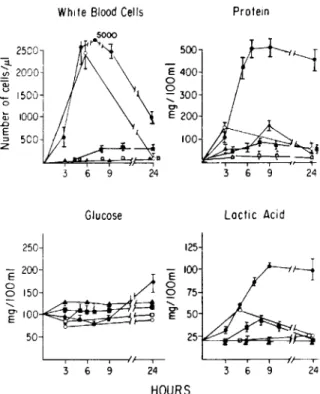
WhiteBlood Cells Protein Results ~ 25COl ~ 2000j '0 1500I Ci; 1000 ..0 E
s
500 6 9 Glucose 24 500 E 400 o Q... 300 C" E 200 100 Lactic AcidTitration of inflammatory activity of purified pneumococcal cell wall and capsular polysaccha-ride. The CSF cytochemical profile induced by
pneumococcal cell wall with choline-containing teichoic acid (preparation A) was compared with the profile obtained with capsular polysaccharide (fig-ure 3). Both cell wall and capsular polysaccharide induced inflammatory changes in CSF at high doses (2 mg/0.2 ml; 'V108cell equivalents). Cell wall was
active, however, in doses as small as 0.2~g/0.2ml ('V105cell equivalents), whereas capsular
polysaccha-Figure 3. Titration of CSF inflammatory response to isolated cell wall and capsular polysaccharide. Groups of two rabbits were challenged with cell wall (all closed sym-bols) or capsular polysaccharide (all open symsym-bols). Each component was instilled at three concentrations (suspend-ed in 0.2 ml saline): 2 mg (circle), 2IJg(square), 0.2IJg
(triangle). The CSF was then sampled over the next 24 hr and the cytochemistry determined as described in the text. Each point represents the mean ± SE.
A B F
c
5
10
15
20
HOURS
Figure 4. CSF white blood cell (WBC) response to cell wall components. A group of four rabbits was challenged with 2IJgof one of the following preparations: choline-containing cell wall (preparation A, ..); ethanolamine-containing cell wall (preparation B, D); amidase product (preparation F, .); crosslinked peptidoglycan stripped of teichoic acid (preparation C, X); peptidoglycan oligomers without teichoic acid (preparation G, 0);or lysozyme digest (preparation H, ..6.). Lysozyme alone produced only minimal inflammation (80 ± 20 WBC/1Jl)
at 5 hr. Determinations of WBC/IJI of CSF were made as described in the text.
16
/4
1.Lo:
/2
u ~ No
10
x u CD 3:8
-
0 ~6
Q) ..c E ~ z4
2
'---...,----.---r-~r---r-' 3 6 9 24 125J E 100 0 Q 75 ... C" 50 E 25 ~ 3 6 9 24 HOURS <,E
100 50 250 E 200g
150of R6was also grown in parallel in synthetic medium
containing [3H]methylcholine (5~Ciand 5~g/mlof medium) for use as a tracer in the purification of the choline-containing Forssman antigen. The pelleted bacteria from this culture were pooled with the pellet of the 20-liter culture at the beginning of the extraction procedure. The final water extract yielded 9 mg of dry material, out of which1'\.12.6mg was protein. Most of this protein impurity was re-moved during passage of the material (4.5 mg in 1.5 ml SP) through a Sepharose-myeloma TEPC-15 pro-tein affinity column (8 ml). After washing the column with 3.5 bed volumes of SP, the Forssman antigen was eluted with 10 ml of SP containing 5
mMphosphorylcholine. Elution was monitored by determining radioactivity in the eluant (figure 2, bot-tom). Fractions 17-21 were pooled and, after exten-sivedialysis against distilled water, the material was lyophilized (yield, 1.5 mg of dry material).
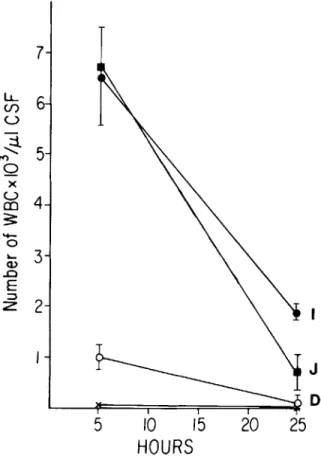
Figure 5. CSF leukocyte response to cell wall teichoic acid, membrane teichoic acid, or capsular polysaccharide. A group of four rabbits was challenged with21-lgof one of the following preparations: cell wall teichoic acid (preparation I, .); Forssman antigen (membrane teichoic acid, preparation J, _); degraded teichoic acid (prepa-ration D, 0);or capsular polysaccharide (X). CSF de-terminations were made as described in Figure 4.
or of teichoic acid only modestly augmented this activity.
CSF inflammatory response to teichoic acid. The activity of the mixture of cell wall teichoic acid (free of peptidoglycan) and Forssman antigen (prepara-tion I)is depicted in figure 5. The peak of activity for teichoic acid-containing preparations was at 5 hr after inoculation, in contrast to 24 hr after in-oculation for peptidoglycan-containing prepara-tions. Teichoic acid proved to have the highest spe-cific activity of all cell wall fractions on a weight basis. CSF leukocyte density 5 hr after inoculation was 10times higher following instillation ofteichoic acid than following instillation of the most active teichoic acid-free cell wall preparations. Virtually all of the activity associated with the teichoic acid was lost upon degradation to small fragments with ride was not active at doses of 200, 20, or 2 Ilg/ml.
The magnitude of the CSF leukocytosis varied directly with the concentration of cell wall intro-duced, e.g., leukocytes appeared both sooner and in greater number as the concentration of cell wall was raised. The minimum concentration of cell wall that could induce changes in each component of the CSF response varied: lactic acid, 20Ilg/0.2ml; leukocyte, 2Ilg/0.2ml; protein,0.2Ilg/0.2ml. CSF glucose con-centration failed to decrease with any nonliving in-oculum.
Comparison of peptidoglycan-containing frac-tions ofcell wallfor activity in inducing CSF inflam-mation. All pneumococcal peptidoglycan fractions (preparations A, B, C, F, and G), regardless of size or complexity, induced a CSF inflammatory re-sponse 5 hr after intracisternal instillation (figure 4). The initial leukocyte concentration ranged between 200-600 cells/ill, and> 60070 of these were PMNLs. The leukocyte response shifted to a predominance of lymphocytes by 24 hr after inoculation. This is the same response observable during meningeal in-flammation induced by live pneumococci. Protein and lactic acid concentrations increased in parallel with the leukocytes.
While peptidoglycan-containing fractions pro-duced similar inflammatory responses in the first few hours after intracisternal inoculation, the evolution of inflammation over 24 hr was quite different for the various fractions. Fractions containing large, soluble cell wall particles were the most potent in-ducers of leukocytosis whether they contained cho-line (preparation A) or ethanolamine (preparation B; figure 4). The activity of preparation A was un-changed even when extensively purified to remove divalent cations (EDTA treatment) and adsorbed teichoic acid (LiCI treatment). Removal of most of the stem peptides by amidase (as induced by peni-cillin; preparation F) or teichoic acid by hot TCA (preparation C) reduced the magnitude of the leu-kocytosis. These responses to soluble cell wall frac-tions remained similar to whole cell wall, however, in that the leukocyte numbers were higher at 24 hr than at 5 hr after inoculation. Activity 24 hr after inoculation was dramatically reduced in the case of peptidoglycan oligomers that were soluble and un-crosslinked and their lysozyme-degraded low-molec-ular-weight products (preparations G and H). Thus, the larger size of the polysaccharide backbone of the peptidoglycan oligomers seemed critical to the main-tenance of a CSF inflammatory response beyond 5 hr; the presence of a complete peptide network
7
1.L6
(f)u
~
5
rot') 0 xu
4
en3:
-
0 ~3
Q) ~ E ::J2
z
5
10
15
HOURS
20
J D25
hot TCA (figure 5; preparation D). Forssman anti-gen represents the lipid-containing membrane-associated teichoic acid. Titration of the activity of this component in the rabbit model yielded results very similar to preparation I (figure 5).
Phosphorylcholine is a characteristic component of pneumococcal teichoic acid. When this compo-nent is changed to phosphorylethanolamine several surface characteristics of the organism change, e.g., lack of daughter cell separation and sensitivity to endogenous autolysin [19]. The possibility that the reactivity of cell wall preparations in CSF would be altered by this substitution was studied by compari-son of cell wall derived from organisms grown in medium containing choline or containing ethanola-mine. Although ethanolamine-containing cell wall (preparation B) was less active in the initial 5 hr of the inflammatory response, both preparations A and B were equally active when assessed at 24 hr (P
>
.05; figure 4). Thus, a specificalteration in the com-position of teichoic acid had little effect on the CSF inflammatory reaction.Discussion
Pneumococcal cell wall appears to be a potent acti-vator of meningeal inflammation in vivo. These studies demonstrated that each of the major
com-ponents of the cell wall- peptidoglycan and teichoic acid - could contribute to the induction of inflam-mation. Each had specific activity high enough to produce an inflammatory reaction if given at doses
~105cell equivalents. In contrast, the specific inflam-matory activity of capsular material was 1,OOO-feld lower, a level insufficient to provoke inflammation at concentrations of capsule found in CSF during meningitis [20]. These findings are summarized in table 1.
Teichoic acid had the highest specific activity (on a weight basis) of all the cell wall fractions with the peak activity occurring 5 hr after instillation. In con-trast, peptidoglycan-containing fractions invoked in-creases in concentrations of CSF leukocytes, protein, and lactic acid that were greater 24 hr after inocu-lation.
Inflammatory activity occurred whether the cell wall fraction was soluble or not. Activity was markedly reduced, however,if either the peptidogly-can or the teichoic acid was extensively degraded (preparations H and D). This finding, taken together with both the high activity of the affinity-purified fractions (preparations G, I, and J)and the absence of activity in column effluents, strongly supports the contention that the cell wall components, and not an undefined contaminant, are responsible for in-ducing the inflammation.
Table 1. CSF inflammatory activity of pneumococcal surface components in a rabbit model of meningitis.
Inflammatory activity Preparation + A + B Purified + A + C D H + F + G + I + J Surface component Cell wall Choline-teichoic acid Ethanolamine-teichoic acid Without divalent cation, adsorbed
teichoic acid, or adsorbed lipoteichoic acid
Without teichoic acid Peptidoglycan Degraded teichoic acid Degraded peptidoglycan Glycan-teichoic acid polymers Penicillin-treated lyt: cells
Uncrosslinked peptidoglycan Nascent teichoic acid Pneumococcal plasma membrane
Forssman antigen
Pneumococcal capsular polysaccharide Pneumovax
Type III
NOTE. Relative activity of surface components, 5 hr after inoculation, in inducing abnormal CSF cytochemistry. Components were introduced at rv106cell equivalents. (+) =active; (-) =inactive.
The inflammatory activity of cell wall and several of its fractions has significant biological relevance in that these fractions could be generated in CSF dur-ing the course of mendur-ingitis. For instance, because both choline and ethanolamine are present in CSF [21], teichoic acid containing either amino alcohol could be produced during pneumococcal growth in vivo. The inflammatory activity of choline- and ethanolamine-containing cell wall are similar, how-ever, and the course of inflammation would not be expected to change if this shift in growth conditions wereto occur. Other fractions of pneumococcal cell wall could also be generated during the course of meningitis in vivo, particularly during antibiotic ther-apy. The peptidoglycan-teichoic acid complex pro-duced by the action of the pneumococcal autolytic enzyme (amidase) on whole cell wall (preparation F) appears in culture supernatants following treat-ment of pneumococci with fJ-Iactam antibiotics [1, 22]. This complex is also generated during sponta-neous autolysis of stationary-phase cells [1]. Nascent peptidoglycan (preparation G) and teichoic acid (preparation I) are secreted, unassociated with each other, during fJ-Iactam treatment of tolerant pneu-mococci [13], such as the South African pneumo-coccal strain 8249 that is multiply resistant [23]. Forssman antigen (preparationJ)is readily secreted upon treatment of all pneumococci with fJ-Iactam antibiotics [24].Itis accessible to host interactions even in living, encapsulated cells as evidenced by both the production of antibody to Forssman anti-gen upon challenge with whole organisms [25) and its putative role as a pneumococcal adhesin [26]. Thus, whole pneumococcal cell wall and several of its components are good candidates for initiation or augmentation of inflammation during the course of meningitis in vivo.
The mechanism whereby cell wall or.cell wall frac-tions act as chemotaxins is as yet undefined. The in-flammatory response could ensue in either of two ways: first, by activation of host defense systems that generate chemotaxins, or second, by direct interac-tion between cell wall and host cells.
In the first case, a likely mechanism may involve complement [27] or C-reactive protein (CRP). The alternative pathway of complement is specifically ac-tivated by teichoic acid and to a lesser extent by large peptidoglycan units [5, 6, 28]. The inflammatory re-sponse would, perhaps, be expected to appear ear-lier and might be more intense if complement were activated in CSF. This may explain in part the rapid
onset and high-specificactivity ofteichoic acid-con-taining cell wall components. CRP has been demon-strated in CSF [29] and is known to promote com-plement activation by interacting with teichoic acid-associated phosphorylcholine [4, 30, 31]. The finding that cell wall containing either ethanolamine or choline produces similar inflammatory responses in rabbits is consistent with the fact that rabbit CRP reacts equally well with either of these two cell wall preparations [32].Itmust be noted, however, that complement is not known to fix to small peptidogly-can units [33], yet these components are effective at inducing inflammation. In addition, CSF contains very little complement even late in the course of meningitis [34, 35].Itwould be more reasonable, therefore, to predict that while complement-dependent chemotaxins may contribute to the in-flammatory response in meningitis, they may not be the only mechanism for its generation.
In the second case, noncomplement-derived chemotaxins have been described in CSF [36,37] and are potentially attributable to direct interaction be-tween bacterial components themselvesand host cells [38, 39]. Cell wall could potentially interact directly with CSF monocytes and stimulate production of chemotaxins (e.g., leukotriene B4 ) without involving
complement. Preliminary data from our studies in complement-depleted animals are compatible with this hypothesis. Such an interaction presumes the ex-istence of a receptor for cell wallcomponents on host cells. Such a host cell receptor had been proposed to be on the surface of epithelial cells for the mem-brane analogue of cell wall teichoic acid - Forssman antigen [26].Itis conceivable that receptors for this amphipathic molecule or for other cell wall compo-nent(s) may be present on the meningeal membrane lining all or part of the CSF space.
Our findings strongly suggest that the inflamma-tory reaction in pneumococcal meningitis derives, at least in part, from component(s) of the bacterial cell wall rather than from the capsule. The genera-tion of an inflammatory response in the course of natural infection servesto control bacterial multipli-cation. Our evidencesuggests,however, that bacterial lytic products such as cell wall released during drug-induced autolysis, may represent a reservoir of in-flammatory products that could contribute to fur-ther injury to host tissues including, perhaps, mortality despite effectiveCSF sterilization. We sug-gest that exploring avenues of antibacterial therapy that are bactericidal but nonbacteriolytic may lead
to methods of treatment that diminish the overall host inflammatory response and may, conceivably, improve the overall morbidity and mortality of pneu-mococcal meningitis.
References
1. Tomasz A. Surface components ofStreptococcus pneumo-niae. Rev Infect Dis 1981;3:190-211
2. Tuomanen E, Tomasz A, Hengstler B, Zak O. The relative role of bacterial cell wall and capsule in the induction of inflammation in pneumococcal meningitis. J Infect Dis 1985;151:535-40
3. Mosser JL, Tomasz A. Choline-containing teichoic acid as a structural component of pneumococcal cell wall and its role in sensitivity to lysis by an autolytic enzyme. J BioI Chern 1970;245:287-98
4. Szu SC, Clarke S, Robbins JB. Protection against pneumo-coccal infection in mice conferred by phosphocholine-binding antibodies: specificityof the phosphocholine bind-ing and relation to several types. Infect Immun 1983;39:993-9
5. Winkelstein JA, Tomasz A. Activation of the alternative com-plement pathway by pneumococcal cell wall teichoic acid. J Immunol 1978;120:174-8.
6. Hummell DS, Berninger RW,Tomasz A, Winkelstein JA. The fixation of C3b to pneumococcal cell wall polymers as a result of activation of the alternative complement path-way. J Immunol 1981;127:1287-9
7. Dacey RG, Sande MA. Effect of probenecid on cerebrospi-nal fluid concentrations of penicillin and cephalosporin derivatives. Antimicrob Agents Chemother 1974;6:437-41 8. Lacks S, Hotchkiss RD. A study of the genetic material de-termining an enzyme activity inPneumococcus. Biochim
Biophys Acta 1960;39:508-18
9. Holtje JV, Tomasz A. Specific recognition of choline residues in the cell wall teichoic acid by the N-acetylmuramyl-L-alanine amidase of pneumococcus. J BioI Chern 1975;250:6072-6
10. Tomasz A, Westphal M, Briles EB, Fletcher P. On the physi-ological functions of teichoic acids. J Supramol Struct 1975;3
11. Holtje JV, Tomasz A. Purification of the pneumococcal N-acetylmuramyl-L-alanine amidase to biochemical homogeneity. J BioI Chern 1976;251:4199-207 12. Tomasz A. Studies on the competence (for genetic
transfor-mation) ofDiplococcus pneumoniae using a synthetic
medium. Bacteriological Proceedings 1964:29 13. Fischer H, Tomasz A. Production and release of
peptidogly-can and wall teichoic acid polymers in pneumococci treated with beta lactam antibiotics. J BacterioI1984;157:507-13 14. Potter M, Lieberman R. Common individual antigenic de-terminants in five of eight BALB/c IgA myeloma proteins that bind phosphoryl choline. J Exp Med 1970;132:737-51. 15. Chesebro B, Metzger H. Affinity labeling of a phosphoryl-choline binding mouse myeloma protein. Biochemistry 1972;11:766-71
16. Cuatrecases P. Protein purification by affinity chromatog-raphy. J BioI Chern 1970;245:3059-65
17. Briles EB, Tomasz A. Pneumococcal Forssman antigen: a choline-containing lipoteichoic acid. J BioI Chern 1973;248:6394-7
18. Goebel WF, Shedlovsky T, Lavin GI, Adams MH. The het-erophile antigen of pneumococcus. J BioI Chern 1943;148:1-15
19. Tomasz A. Biological consequences of the replacement of choline by ehtanolamine in the cell wall of pneumococcus: chain formation, loss of transformability and loss of au-tolysis. Proc Natl Acad Sci USA 1968;59:86-93 20. Nolan CM, Ulmer WC Jr. Enzyme immunoassay of the
cap-sular polysaccharide ofStreptococcus pneumoniae type
III in cerebrospinal fluid in experimental meningitis. J Med Microbiol 1980;13:551-60
21. Letner C, ed. Geigy Scientific tables. 8th ed. Basel, Switzer-land: CIBA-GEIGY, 1981:168-9
22. Tomasz A, Holtje JV. Murein hydrolases and the lytic and killing action of penicillin. In: Microbiology 1977. Washing-ton, DC: American Society for Microbiology, 1977:209-15 23. Liu H, Zighelboim S, Tomasz A. Penicillin tolerance in
mul-tiply drug-resistance natural isolates of Streptococcuspneu-moniae (abstract no. 506]. In: Program and abstracts of
the 21st Interscience Conference on Antimicrobial Agents and Chemotherapy. Washington, DC: American Society for Microbiology, 1981.
24. Horne D, Hakenbeck R, Tomasz A. Secretion of lipids in-duced by inhibition of peptidoglycan synthesis in strep-tococci. J Bacteriol 1977;132:704-17
25. Wicken AJ, Knox KW. Lipoteichoic acids: a new class of bac-terial antigen. Science 1975;187:1161-7
26. Ofek I, Beachey EH, Jefferson W, Campbell GL. Cell mem-brane-binding properties of Group A streptococcal lipoteichoic acid. J Exp Med 1975;141:990-1003 27. Ernst JD, Hartiala KT, Goldstein 1M, Sande MA.
Comple-ment (C5)-derived chemotactic activity accounts for ac-cumlation of polymorphonuclear leukocytes in cerebrospi-nal fluid of rabbits with pneumococcal meningitis. Infect Immun 1984;46:81-6
28. Hummell DS, SwiftAJ, TomaszA, Winkelstein JA. Activa-tion of the alternative complement pathway by pneumo-coccal lipoteichoic acid. Infect Immun 1985;47:384-7 29. Corrall CJ, Pepple JM, Moxon ER, Hughes WT. C-reactive
protein in spinal fluid of children with meningitis. J Pedi-atr 1981;99:365-9
30. Kaplan MH, Volanakis JE. Interaction of C-reactive protein complexes with the complement system. J Immunol 1974;112:2135-47
31. Mold C, Nakayama S, Holzer TJ, Gewurz H, du Clos TW. C-reactive protein is protective against Streptococcuspneu-moniae infection in mice. J Exp Med 1981;154:1703-8
32. Oliveira EB, Gotschlich EC, Liu T-Y. Comparative studies on the binding properties of human and rabbit C-reactive proteins. J Immunol 1980;124:1396-1402
33. Dinarello CA, Elin RJ, Chedid L, Wolff SM. The pyroge-nicity of the synthetic adjuvant muramyl dipeptide and two structural analogues. J Infect Dis 1978;138:760-7 34. Zwahlen A, Nydegger UE, Vaudaux P, Lambert P-H,
Wald-vogel FA. Complement-mediated opsonic activity in nor-mal and infected human cerebrospinal fluid: early response during bacterial meningitis. J Infect Dis 1982;145:635-46 35. Simberkoff MS, Moldover NH, Rahal J J Jr. Absence of
de-tectable bactericidal and opsonic activities in normal and infected human cerebrospinal fluids. J Lab Clin Med 1980;95:362-72
36. Nolan CM, Clark RA, Beaty NH, Experimental pneumo-coccal meningitis. III. Chemotactic activity in cerebrospi-nal fluid. Proc Soc Exp BioI Med 1975;150:134-6 37. Ward PA, Lepow IH, Newman LJ. Bacterial factors
chemotac-tic for polymorphonuclear leukocytes. Am J Pathol 1968;52:725-36
38. Peterson PK, Wilkinson BJ, Kim Y, Schmeling D, Douglas SD, Quie PO, Verhoef J. The key role of peptidoglycan in the opsonization ofStaphylococcus aureus.J Clin In-vest 1978;61:597-609
39. Ogawa T, Kotani S, Fukuda K, Tsukamoto Y, Mori M, Kusumoto S, Shiba T. Stimulation of migration of human monocytes by bacterial cell walls and muramyl peptides, Infect Immun 1982;38:817-24 .