RESEARCH OUTPUTS / RÉSULTATS DE RECHERCHE
Author(s) - Auteur(s) :
Publication date - Date de publication :
Permanent link - Permalien :
Rights / License - Licence de droit d’auteur :
Institutional Repository - Research Portal
Dépôt Institutionnel - Portail de la Recherche
researchportal.unamur.be
University of Namur
Comparison of the clonogenic survival of A549 non-small cell lung adenocarcinoma cells after irradiation with low-dose-rate beta particles and high-dose-rate X-rays
Wéra, A.-C.; Borlon, C.; Nuttens, V.E.; Lucas, S.; Riquier, H.; Michiels, C.; Feron, O.
Published in:
International Journal of Radiation Biology
DOI:
10.3109/09553002.2012.643274
Publication date: 2012
Document Version Peer reviewed version
Link to publication
Citation for pulished version (HARVARD):
Wéra, A-C, Borlon, C, Nuttens, VE, Lucas, S, Riquier, H, Michiels, C & Feron, O 2012, 'Comparison of the clonogenic survival of A549 non-small cell lung adenocarcinoma cells after irradiation with low-dose-rate beta particles and high-dose-rate X-rays', International Journal of Radiation Biology, vol. 88, no. 3, pp. 253-257. https://doi.org/10.3109/09553002.2012.643274
General rights
Copyright and moral rights for the publications made accessible in the public portal are retained by the authors and/or other copyright owners and it is a condition of accessing publications that users recognise and abide by the legal requirements associated with these rights. • Users may download and print one copy of any publication from the public portal for the purpose of private study or research. • You may not further distribute the material or use it for any profit-making activity or commercial gain
• You may freely distribute the URL identifying the publication in the public portal ?
Title page
Title
Comparison of the clonogenic survival of A549 non-small cell lung adenocarcinoma cells after irradiation with low-dose-rate beta particles and high-dose-rate X-rays
Authors
Anne-Catherine Wéra1a, Céline Borlon1a, Vincent E. Nuttensa, Hélène Riquierb, Olivier Feronc, Carine Michielsb, Stéphane Lucasa.
1
These authors equally contributed to this work
a
PMR, NARILIS, University of Namur - FUNDP
b
URBC, NARILIS, University of Namur - FUNDP
c
FATH, Université Catholique de Louvain (UCL)
Corresponding author
Anne-Catherine Wéra PMR, FUNDP, Rue de Bruxelles, 61, 5000 Namur BELGIUM Tél. +32-81725479 Fax +32-81725474 Email: anne-catherine.wera@fundp.ac.be
Running Title
A549 cell irradiation with low-dose-rate beta radiation
Key Words
: beta-radiation, surviving fraction, radiosensitivity parameters, lung cancer cells, hyper-radiosensitivityAbstract
Purpose: lung cancer is the leading cause of cancer-related death. Among the new modalities to treat cancer, internal radiotherapy seems to be very promising. However, the achievable dose-rate is two orders of magnitude lower than the one used in conventional external radiotherapy, and data has to be collected to evaluate the cell response to highlight the potential effectiveness of low-dose-rate beta particles irradiation. This work investigates the phosphorus beta irradiation (32P) dose response on the clonogenicity of human A549 non-small cell lung adenocarcinoma cells and compares it to high-dose-rate X-irradiations results.
Materials and methods: cell survival was evaluated by a colony forming assay 8 days after low-dose-rate 32P beta irradiations (0.8 Gy/h) and high-dose-rate X-ray irradiations (0.855 Gy/min).
Results: survival curves were obtained for both types of irradiations, and showed hyper-radiosensitivity at very low doses. Radiosensitivity parameters were obtained by using the linear-quadratic and induced-repair models.
Conclusions: Comparison with high-dose-rate X-rays shows a similar surviving fraction, confirming the effectiveness of beta particles for tumor sterilization.
Introduction
Lung cancer is the leading cause of cancer-related death. Besides traditional treatments, new modalities are under investigation; internal radiotherapy, like radio-immunotherapy (RIT), is a promising example. RIT is a biologically targeted radio-pharmaceutical treatment in which a radioactive isotope is chemically bound to a target-specific monoclonal antibody or antibody fragments with a high degree of specificity for one particular tumor associated antigen (Pohlman et al. 2006). RIT requires dealing with three interdependent factors: the tumor targeted by the treatment, the antibody and the radionuclide. Howell et al. (Howell et al. 1994) and Rao et al. (Rao & Howell 1993) have shown that one of the most promising radioisotope for RIT is 32P owing to its half life (14.3 days) and the tissue penetration of its beta radiation (8 mm maximum). Initially, this radioisotope was mainly used for the therapy of bone pain and metastases, in ovarian cancer and for myeloproliferative diseases (Pattillo et al. 1995; Berlin 2000; Pandit-Taskar et al. 2004; Tennvall & Brans 2007). More recently, studies on the use of 32P as colloid or microsphere for solid tumors were performed; they showed not only an improvement in the patient’s quality of life, but also in their survival (Gao et al. 2008; Wang et al. 2008). However, 32P was not used in RIT due to the difficulty of the radiolabeling. Abraham and colleagues used decapeptides labeled with 32P for the colorectal cancer therapy (Abraham et al. 2007; Abraham et al. 2008) and showed that these molecules avidly bind to different adenocarcinoma cell lines with a permanent incorporation of radioisotopes into cellular proteins at a rate over 100 times greater than in cell lines derived from a variety of other cancers or from normal organs. The use of such small molecules allows for their elimination in the urine, as their molecular weight
is lower than the limit of the filtering kidney. Moreover, they can penetrate deeper into the tumor, which is, in combination with the long penetrating range of the 32P beta particles, highly attractive for the treatment of solid tumors. In parallel, investigations into the use of radioactive nanoparticles containing several radioactive atoms are currently underway to improve the therapeutic effectiveness of RIT and to enhance diagnostic sensitivity in medical imaging. Theoretical studies confirm the benefit of using radiolabeled beta emitter nanoparticles, such as 90Y2O3 or 32P2O3, to treat solid and poorly
vascularized tumors by RIT (Bouchat et al. 2007; Nuttens et al. 2008). Nevertheless, the currently achievable dose-rate in RIT is one or two orders of magnitude lower than the one obtained by external X-ray radiotherapy.
One major question still remains: is it possible to obtain adequate cell sterilization with beta radiation with a dose-rate that is one or two orders of magnitude lower than with traditional X-rays? The aim of this work is to try to answer this question by studying the clonogenic survival of human A549 adenocarcinoma non-small cell lung cells irradiated with beta radiation emitted by 32P (0.8 Gy/h) and to compare the results with data obtained with X-ray irradiation (0.855 Gy/min).
Materials and Methods
Cell culture and irradiation
A549 (ATCC-LGC Standards, Molsheim, France) cells were grown in MEM (Minimum Essential Medium) (Invitrogen, Merelbeke, Belgium) containing 10% fetal calf serum (FCS) (Invitrogen). Both beta and X-ray irradiations were performed 24 hours after plating.
32
P irradiations were performed at 37°C with a Na2H32PO4 solution (PerkinElmer,
Waltham, Massachusetts, USA) as 32P source in serum-free CO2-independent medium
(Invitrogen) supplemented with 1 mM L-glutamine (Sigma, Bornem, Belgium). 32P is a pure beta-emitting radionuclide with a physical half-life of 14.3 days. The maximum and mean beta particle energies are, respectively, 1.75 and 0.695 MeV. The source was calibrated to have a specific activity of 12 mCi/ml at the irradiation day. The irradiation duration and the radioactive volume were adapted to reach respectively the required dose and dose-rate. After incubation, cells were washed twice with phosphate-buffered saline (PBS) (Invitrogen) and replaced in MEM + FCS. The cellular uptake of 32P radionuclide was evaluated using a scintillation counter (Beckman Coulter Inc., Fullerton, CA, USA) after cell solubilization in 250 µl NaOH and neutralization with 250 µl HCl.
A549 cells were exposed to X-rays (RT250, Philips Medical Systems, Bruxelles, Belgium) at 0.855 Gy/min (51.3 Gy/h) in the same condition as for 32P.
Dosimetry simulation
The deposited dose was calculated by MCNPX (Monte Carlo N-Particle eXtended) simulations (Pelowitz 2005) (code version 2.5.0) according to the calculation method
proposed by Schaart et al. (Schaart et al. 2002) to assess the dose deposited by 32P in a solution of Na2H32PO4 on A549 cells. It treats a three dimensional configuration of
materials in geometric cells bounded by first- and second-degree surfaces and fourth-degree elliptical tori (Pelowitz 2005). Working with charged particles as electrons in very small geometric cells leads to some uncertainties that are inherent in the condensed history method. Indeed, Schaart et al. have shown that this leads to significant errors in the absorbed dose (Schaart et al. 2002). They proposed a new method, used here proper evaluation. The calculations were performed using the track length estimator tally F4, combined with a dose response function. The ITS (Integrated Tiger Series) energy indexing algorithm was used (Debug Information Card: DBCN 17j 1). The photon and electron energy cut-offs were set to 1 keV as particles with less energy can not pass through the culture medium and reach the biological cells to damage them. The geometry of the experiment was a culture well whose bottom was covered by A549 cells (one confluent monolayer). The plastic well was filled with a radioactive 32P aqueous solution. The cell chemical composition was taken from the National Institute of Standards and Technology data base (NIST).
The cell thickness was experimentally measured by confocal microscopy after immunofluorescence staining. An average thickness of 3.2 µm 0.1 µm was obtained over more than 30 measurements.
Knowing the cell thickness, it was possible to evaluate the dose delivered by using the Tally T value. The obtained values were 7.047 10-2 and 6.200 10-1 MeV/g/nps for 6-well and 24-well plates, respectively. This corresponds to the mean deposited energy (MeV)
for an incident particle (nps) per mass unit (g). This value can be converted to dose-rate (D) in Gy/h using: T A k D . .
where A is the source activity (mCi) that gives the number of emitted beta particles per unit time, and k is the unit conversion constant (equal to 21.34) to obtain the unit of dose-rate (J/kg/h or Gy/h) from the multiplication of the tally by the activity (MeV/g/s).
The T value is specific to the geometry used in these experiments. As the source activity is 12 mCi/ml, the amount of radioactive solution added to the medium has been adjusted to obtain a dose-rate of 0.8 Gy/h. The exposition time has been adapted to reach the total targeted dose.
The cellular uptake of 32P into exposed cells after the irradiation was experimentally measured: it was less than 0.05 % for each dose. This result allowed us to neglect the residual activity incorporated into the cells and to consider the total dose equal to the one obtained by the simulations.
Colony-Forming Assay
Two thousand A549 cells were seeded in 6-well plates. Control cells underwent exactly the same manipulations without the addition of the radioisotope for 32P or without X-ray irradiation. 8 days post-irradiation, the number of visible colonies (containing > 50 cells) was counted after staining cells with crystal violet. The surviving fraction was obtained by calculating the ratio of the colony number for the irradiated cells to the colony number for control cells.
Survival curve analysis
The results obtained from the colony forming assays were analyzed with the Linear-Quadratic (LQ) model and the Induced-Repair model (IndRep). The LQ model allows the determination of the two radiosensitivity parameters α and β:
²)
( D D
e
S
where S is the surviving fraction and D is the deposited dose. The LQ model considers cell killing as the result of single or double hit events into DNA. In different studies, a deviation between experimental data and the LQ model was observed. This deviation is due to hyper-radiosensitivity (HRS) which has been reviewed in (Raaphorst & Boyden 1999; Joiner et al. 2001; Leskov et al. 2003; Marples et al. 2004). To obtain a better approximation of the data at low doses, the Induced-Repair model has been suggested to match this HRS as well as the induced radioresistance observed by increasing doses until 1 Gy. ² ) ) 1 ( 1 ( e Dc D D D r s r
e
S
The IndRep model is an adaptation of the LQ model to allow the α parameter to vary with the dose (Short & Joiner 1998). The model defines four parameters: αr and β obtained
with the LQ model; αs and Dc characterizing the HRS.
The surviving fractions were fitted using Origin 7.5 (OriginLab) using a chi-square minimization method.
Results
Survival curves for 32P irradiation
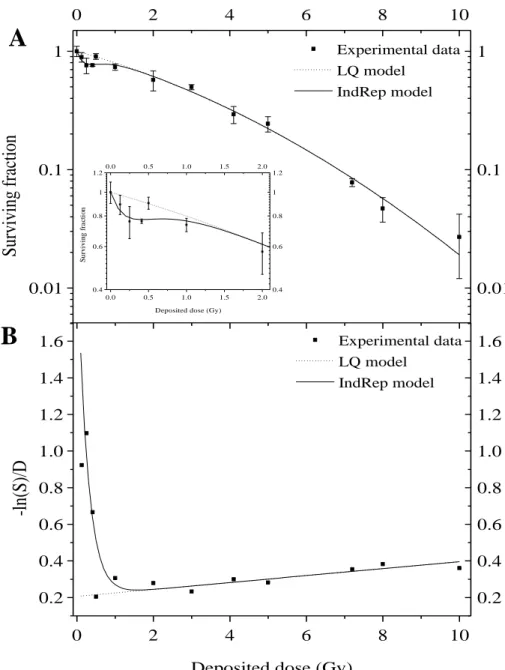
The irradiations were performed at 0.8 Gy/h for doses ranging from 0.125 to 10 Gy. This dose-rate is within the usual range used in RIT and can be considered as a low-dose-rate. Four independent irradiation experiments with at least 4 replicates each were performed (Figure 1A). The experimental data were fitted with both LQ and IndRep models. The LQ model gave parameters equal to 0.21 ± 0.02 Gy-1 and 0.019 ± 0.002 Gy-2 for α and β respectively. The ratio α/β is 11.0 ± 2.1 Gy. At low doses from 0.125 to 0.5 Gy, a deviation between experimental data and the LQ model was observed (inset of Figure 1A). This deviation is due to HRS and is highlighted by linearization of the surviving fraction (Figure 1B). A better approximation of the data at low doses was obtained with the IndRep model (see fit in Figures 1A and 1B). The values obtained for αs and Dc are,
respectively, 2.1 ± 1.2 Gy-1 and 0.27 ± 0.10 Gy. The ratio αs/αr is equal to 10.4 ± 6.9,
indicating that cells are about ten times more sensitive at very low doses than at higher doses.
Survival curves for X-ray irradiation
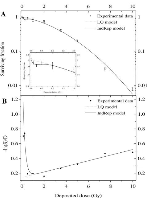
The surviving fraction of A549 cells after X-ray irradiation (0.855 Gy/min) is shown in Figure 2A. The experimental data were fitted with both LQ and IndRep models. The parameters obtained with LQ model are 0.14 ± 0.02 Gy-1 and 0.038 ± 0.003 Gy-2 for α and β respectively. At low doses from 0.125 to 0.5 Gy, a deviation between experimental data and the LQ model was observed (inset of Figure 2A). This deviation, highlighted by linearization (Figure 2B), is due to HRS and is comparable to the deviation observed for
beta particles. The data were fitted with the IndRep model giving a αs value of 1.8 ± 1.2
Gy-1 and a Dc value equal to 0.22 ± 0.11 Gy. A comparison was made between the
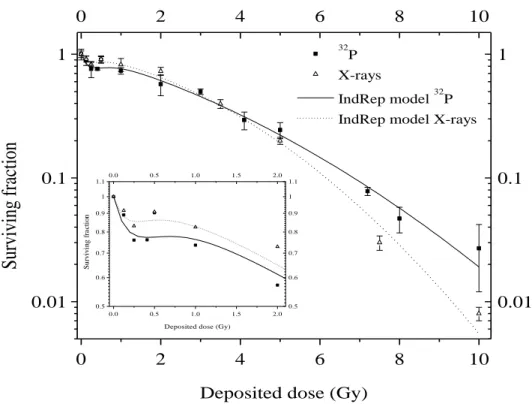
surviving fraction obtained with 32P and X-ray irradiations (Figure 3). We observed a nearly complete overlay between both curves, with the curve for X-ray irradiation being just slightly more shouldered. Although irradiations were performed with serum-free medium to avoid progression in the cell cycle, effects of irradiation time may occur, and if so, influence the surviving fraction. Table I presents the value obtained for the
parameters from the two models for each type of irradiation. The surviving fraction at 2 Gy and the mean inactivation dose (
D
) are also presented. Fertil et al. (Fertil & Malaise 1985) have previously shown that the initial part of the surviving fraction curve, which is well characterized by r andD
, was specific of the cell line. Following theirclassification, the results obtained with beta and X-rays radiation show that A549 cells belong to the group of cell lines derived from tumors exhibiting low radioresponsiveness.
Discussion
This work compares the clonogenic survival of A549 cells after low-dose-rate beta irradiations to high-dose-rate X-rays irradiations. For the 32P exposures, a low-dose-rate of 0.8 Gy/h was chosen. This dose-rate is in the range of those actually used in radionuclide therapy in which beta particles are involved.
The surviving fraction curve showed that 60% of the cells were still able to grow colonies at 2 Gy, this fraction decreased to less than 3 % at 10 Gy. For X-rays, 65% and less than 1% of survival was observed for 2 Gy and 10 Gy respectively.
For the two types of radiations, a deviation from the LQ model was observed at low doses. This low dose hypersensitivity was highlighted in many tumor cell lines and was called hyper-radiosensitivity (HRS) (Joiner et al. 2001). HRS is usually displayed for doses below 0.5 Gy. Several explanations have been proposed by Joiner et al. (Joiner et al. 2001). This phenomenon seems to precede the occurrence of a relative resistance to cell killing by radiation over the dose range of 0.5-1 Gy. Enns et al. (Enns et al. 2004) have also evidenced a HRS for A549 cells but with gamma irradiation. They have shown that HRS is due to apoptotic death and happens during the first cell cycle post-irradiation. The surviving fraction curves obtained in this work are characteristics of sparsely ionizing radiation, such as X-rays or beta particles. Fertil & Malaisse have demonstrated the correlation between in vitro radiosensitivity and clinical radioresponsiveness (Fertil & Malaise 1985). Following their classification, a radiosensitivity parameter α around 0.20 Gy-1 corresponds to cell lines derived from tumor exhibiting low radioresponsiveness. The surviving fraction results obtained with beta particles on A549 cells were compared to the ones obtained with X-ray irradiation: beta particles led to a similar decreasing
survival, while the dose-rate was 65 times lower. The surviving fraction at 2 Gy (0.61) obtained for 32P was slightly smaller than the one obtained with X-rays (0.65). Our data are also comparable to those reported in the literature. Munshi et al. obtained a surviving fraction of 0.61 0.004 at 2 Gy with high-dose-rate gamma radiation from 137Cs (Munshi et al. 2006). Moreover, Bromley et al. obtained parameter of 0.2432 Gy-1 and 0.0257 Gy-2 for and respectively for A549 cells irradiated using 6 MV X-rays produced by a linear accelerator (Bromley et al. 2009). These values are close to the ones obtained in this work (0.21 Gy-1 and 0.019 Gy-2).
The little difference between the results obtained with beta radiation at low dose-rate and high dose-rate X-rays may be surprising, as it is generally thought that the radiation effects increase with the dose-rate. However, many studies have shown what was termed an inverse dose-rate effect (Gridley DS 2005; Sgouros et al. 2007) i.e. the observation that low-dose-rate (LDR) leads to reduced survival compared to high-dose-rate (HDR). Among the different possible explanations for this observation, a lower activation of the DNA damage sensor ataxia-telangiectasia mutated (ATM) for cells irradiated at LDR compared to HDR was evidenced (Collis et al. 2004). These results suggest that the low ATM activation and the resulting lower rate of phosphorilated histone H2AX (γ-H2AX)
led to a lower cell survival, possibly due to an ineffective activation of cell cycle checkpoint and repair mechanisms.
The results obtained in this work are of potential interest when the response of normal tissues is considered: the range of beta particles of 1.75 MeV in lung tissues is about 8 mm. For 250 keV X-rays, only 11% are absorbed within this distance. X-rays will thus
use of a beta emitter for lung tumor treatment should allow normal tissues to be spared, as non-targeted irradiation can be avoided due to the radiation range.
Further studies are warranted to confirm the potential of using LDR beta particles for tumor sterilization while sparing normal tissues.
Acknowledgments
O. Feron is Research Director from FNRS (National Funds for Scientific Research, Belgium). H. Riquier is recipient of a Télévie fellowship.
Declaration of interest
References
Abraham JM, Sato F, Cheng YL, Paun B, Kan T, Olaru A, Jin Z, Yang J, Agarwal R, David S and others 2007. Novel Decapeptides that Bind Avidly and Deliver Radioisotope to Colon Cancer Cells. PloS one 2: -.
Abraham JM, Cheng Y, Hamilton JP, Paun B, Jin Z, Agarwal R, Kan T, David S, Olaru A, Yang J and others 2008. Generation of small 32P-labeled peptides as a potential approach to colorectal cancer therapy. PloS one 3: e2508.
Berlin NI 2000. Treatment of the myeloproliferative disorders with P-32. European
Journal of Haematology 65: 1-7.
Bouchat V, Nuttens VE, Lucas S, Michiels C, Masereel B, Feron O, Gallez B, Vander Borght T 2007. Radioimmunotherapy with radioactive nanoparticles: first results of dosimetry for vascularized and necrosed solid tumors. Medical physics 34: 4504-4513.
Bromley R, Oliver L, Davey R, Harvie R, Baldock C 2009. Predicting the clonogenic survival of A549 cells after modulated x-ray irradiation using the linear quadratic model. Physics in medicine and biology 54: 187-206.
Collis SJ, Schwaninger JM, Ntambi AJ, Keller TW, Nelson WG, Dillehay LE, Deweese TL 2004. Evasion of early cellular response mechanisms following low level radiation-induced DNA damage. The Journal of biological chemistry 279: 49624-49632.
Enns L, Bogen KT, Wizniak J, Murtha AD, Weinfeld M 2004. Low-dose radiation hypersensitivity is associated with p53-dependent apoptosis. Molecular Cancer
Research 2: 557-566.
Fertil B, Malaise EP 1985. Intrinsic radiosensitivity of human cell lines is correlated with radioresponsiveness of human tumors: analysis of 101 published survival curves.
International journal of radiation oncology, biology, physics 11: 1699-1707.
Gao W, Liu L, Teng GJ, Feng GS, Tong GS, Gao NR 2008. Internal radiotherapy using 32P colloid or microsphere for refractory solid tumors. Annals of nuclear
medicine 22: 653-660.
Gridley DS WJ, Slater JM 2005. Low-dose/low-dose-rate radiation: a feasible strategy to improve cancer therapy ? Cancer Therapy 3: 105-130.
Howell RW, Goddu SM, Rao DV 1994. Application of the linear-quadratic model to radioimmunotherapy: further support for the advantage of longer-lived radionuclides. Journal of Nuclear Medicine 35: 1861-1869.
Joiner MC, Marples B, Lambin P, Short SC, Turesson I 2001. Low-dose hypersensitivity: current status and possible mechanisms. International journal of radiation
oncology, biology, physics 49: 379-389.
Leskov KS, Klokov DY, Li J, Kinsella TJ, Boothman DA 2003. Synthesis and functional analyses of nuclear clusterin, a cell death protein. The Journal of biological
chemistry 278: 11590-11600.
Marples B, Wouters BG, Collis SJ, Chalmers AJ, Joiner MC 2004. Low-dose hyper-radiosensitivity: a consequence of ineffective cell cycle arrest of
radiation-Munshi A, Tanaka T, Hobbs ML, Tucker SL, Richon VM, Meyn RE 2006. Vorinostat, a histone deacetylase inhibitor, enhances the response of human tumor cells to ionizing radiation through prolongation of gamma-H2AX foci. Molecular cancer
therapeutics 5: 1967-1974.
NIST http://physics.nist.gov/PhysRefData/XrayMassCoef/tab2.html
Nuttens VE, Wera AC, Bouchat V, Lucas S 2008. Determination of biological vector characteristics and nanoparticle dimensions for radioimmunotherapy with radioactive nanoparticles. Applied Radiation and Isotopes 66: 168-172.
Pandit-Taskar N, Batraki M, Divgi CR 2004. Radiopharmaceutical therapy for palliation of bone pain from osseous metastases. Journal of Nuclear Medicine 45: 1358-1365.
Pattillo RA, Collier BD, Abdeldayem H, Ozker K, Wilson C, Ruckert ACF, Hamilton K 1995. Phosphorus-32-Chromic Phosphate for Ovarian-Cancer .1. Fractionated Low-Dose Intraperitoneal Treatments in Conjunction with Platinum Analog Chemotherapy. Journal of Nuclear Medicine 36: 29-36.
Pelowitz DB 2005. MCNPX User's Manual, Version 2.5.0. Los Alamos National
Laboratory report LA-CP-05-0369.
Pohlman B, Sweetenham J, Macklis RM 2006. Review of clinical radioimmunotherapy.
Expert review of anticancer therapy 6: 445-461.
Raaphorst GP, Boyden S 1999. Adaptive response and its variation in human normal and tumour cells. International journal of radiation biology 75: 865-873.
Rao DV, Howell RW 1993. Time-dose-fractionation in radioimmunotherapy: implications for selecting radionuclides. Journal of Nuclear Medicine 34: 1801-1810.
Schaart DR, Jansen JT, Zoetelief J, de Leege PF 2002. A comparison of MCNP4C electron transport with ITS 3.0 and experiment at incident energies between 100 keV and 20 MeV: influence of voxel size, substeps and energy indexing algorithm. Physics in medicine and biology 47: 1459-1484.
Sgouros G, Knox SJ, Joiner MC, Morgan WF, Kassis AI 2007. MIRD continuing education: Bystander and low dose-rate effects: are these relevant to radionuclide therapy? Journal of Nuclear Medicine 48: 1683-1691.
Short SC, Joiner MC 1998. Cellular response to low-dose irradiation. Clinical oncology
(Royal College of Radiologists (Great Britain)) 10: 73-77.
Tennvall J, Brans B 2007. EANM procedure guideline for 32P phosphate treatment of myeloproliferative diseases. European journal of nuclear medicine and molecular
imaging 34: 1324-1327.
Wang XM, Yin ZY, Yu RX, Peng YY, Liu PG, Wu GY 2008. Preventive effect of regional radiotherapy with phosphorus-32 glass microspheres in hepatocellular carcinoma recurrence after hepatectomy. World Journal of Gastroenterology 14: 518-523.
Figure 1: A549 cell surviving fraction 8 days after beta irradiation from 32P (0.8 Gy/h). The experimental data were obtained with conventional colony forming assays.
A: Surviving fraction curve performed with dose ranging from 0.125 to 10 Gy. LQ model (dashed line) and IndRep model (straight line) were applied to experimental data (filled square). Results are expressed as means standard deviation. The inset is an enlargement of the data obtained at low doses.
B: Experimental data (filled square), LQ model (dashed line) and IndRep model
0.01 0.1 1 0 2 4 6 8 10 0.01 0.1 1 Experimental data LQ model IndRep model 0.0 0.5 1.0 1.5 2.0 0.4 0.6 0.8 1 1.20.0 0.5 1.0 1.5 2.0 0.4 0.6 0.8 1 1.2 S ur vi va l f ra ct io n
Deposited dose (Gy)
S
urviva
l fr
ac
ti
on
0 2 4 6 8 10 0.2 0.4 0.6 0.8 1.0 1.2 1.4 1.6 0.2 0.4 0.6 0.8 1.0 1.2 1.4 1.6 Experimental data LQ model IndRep model-ln(S
)/D
Deposited dose (Gy)
A
B
0.01 0.1 1 0 2 4 6 8 10 0.01 0.1 1 Experimental data LQ model IndRep model 0.0 0.5 1.0 1.5 2.0 0.4 0.6 0.8 1 1.2 0.0 0.5 1.0 1.5 2.0 0.4 0.6 0.8 1 1.2 S ur vi va l fr ac ti onDeposited dose (Gy)
S urviving fr ac ti on S u rv iv in g f ra ct io n
Figure 2: A549 cell surviving fraction 8 days after X-ray irradiation (0.855 Gy/min). The experimental data were obtained with conventional colony forming assays.
A: Surviving fraction curve performed with dose ranging from 0.125 to 10 Gy. LQ model (dashed line) and IndRep model (straight line) were applied to experimental data (open triangle). Results are expressed as means standard deviation. The inset is an enlargement of the data obtained at low doses.
B: Experimental data (filled circle), LQ model (dashed line) and IndRep model (straight line) linearization. 0.01 0.1 1 0 2 4 6 8 10 0.01 0.1 1 Experimental data LQ model IndRep model 0.0 0.5 1.0 1.5 2.0 0.4 0.6 0.8 1 1.2 0.0 0.5 1.0 1.5 2.0 0.4 0.6 0.8 1 1.2 S ur viv al fr ac tio n
Deposited dose (Gy)
S urviva l fr ac ti on 0 2 4 6 8 10 0.2 0.4 0.6 0.8 1.0 1.2 0.2 0.4 0.6 0.8 1.0 1.2 Experimental data LQ model IndRep model -ln(S )/D
Deposited dose (Gy)
A
B
0.01 0.1 1 0 2 4 6 8 10 0.01 0.1 1 Experimental data LQ model IndRep model 0.0 0.5 1.0 1.5 2.0 0.4 0.6 0.8 1 1.2 0.0 0.5 1.0 1.5 2.0 0.4 0.6 0.8 1 1.2 S ur vi va l fr ac ti onDeposited dose (Gy)
S urviving fr ac ti on S u rv iv in g f ra ct io n
Figure 3: Comparison of 32P experimental data (filled square), X-ray irradiation experimental data (open triangle), IndRep model for 32P (straight line) and IndRep model for X-rays (dashed line). Results are expressed as means standard deviation.
0 2 4 6 8 10 0.01 0.1 1 0 2 4 6 8 10 0.01 0.1 1 0.0 0.5 1.0 1.5 2.0 0.5 0.6 0.7 0.8 0.9 1 1.1 0.0 0.5 1.0 1.5 2.0 0.5 0.6 0.7 0.8 0.9 1 1.1 S ur vi va l fr ac ti on
Deposited dose (Gy)
32
P X-rays
IndRep model 32P
IndRep model X-rays
S
urviva
l fr
ac
ti
on
Deposited dose (Gy) 0.01 0.1 1 0 2 4 6 8 10 0.01 0.1 1 Experimental data LQ model IndRep model 0.0 0.5 1.0 1.5 2.0 0.4 0.6 0.8 1 1.20.0 0.5 1.0 1.5 2.0 0.4 0.6 0.8 1 1.2 S ur vi va l f ra ct io n
Deposited dose (Gy)
S
urviving fr
ac
ti
on
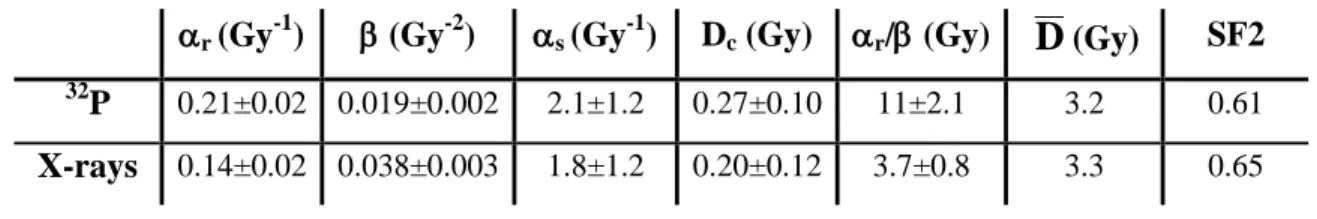
S u rv iv in g f ra ct io nTable I: Parameters obtained from the LQ and IndRep model fit of the survival fraction of A549 cells. Survival fractions were obtained with conventional clonogenic assays performed 8 days after 32P beta irradiation (0.8 Gy/h) or X-irradiation (0.855 Gy/min). Parameters and uncertainties were obtained from chi-square minimization fitting method of the experimental data.
r (Gy-1) (Gy-2) s (Gy-1) Dc (Gy) r/(Gy)
D
(Gy) SF2 32P 0.21±0.02 0.019±0.002 2.1±1.2 0.27±0.10 11±2.1 3.2 0.61