HAL Id: tel-01140663
https://tel.archives-ouvertes.fr/tel-01140663
Submitted on 9 Apr 2015HAL is a multi-disciplinary open access archive for the deposit and dissemination of sci-entific research documents, whether they are pub-lished or not. The documents may come from teaching and research institutions in France or abroad, or from public or private research centers.
L’archive ouverte pluridisciplinaire HAL, est destinée au dépôt et à la diffusion de documents scientifiques de niveau recherche, publiés ou non, émanant des établissements d’enseignement et de recherche français ou étrangers, des laboratoires publics ou privés.
Gait kinematic analysis of the osteoarthritic knee :
pre-and post- total knee arthroplasty
Dafina Bytyqi
To cite this version:
Dafina Bytyqi. Gait kinematic analysis of the osteoarthritic knee : pre- and post- total knee arthro-plasty. Biomechanics [physics.med-ph]. Université Claude Bernard - Lyon I, 2015. English. �NNT : 2015LYO10020�. �tel-01140663�
N˚
Année 2015THÈSE
présentée
devant
l’UNIVERSITÉ CLAUDE BERNARD - LYON I
préparée en cotutelle avec l’ UNIVERSITÉ DE PRISHTINA
pour l’obtentiondu DIPLÔME DE DOCTORAT
(arrêté du 7 août 2006)
Présentée et soutenue publiquement le
25 Fevrier 2015
par
Dafina BYTYQI
TITRE : Gait Kinematic Analysis of the Osteoarthritic Knee;
Pre- and Post – Total Knee Arthroplasty
Directeur de thèse: Monsieur le Professeur Philippe NEYRET
Co-directeur de thèse : Madamme la Professeure Natyra KARAHODA GJURGJEALA
JURY:
Monsieur le Professeur Dominique SARAGAGLIA Monsieur le Professeur Frédéric FARIZON
Madame la Professeure Laurence CHEZE
1
UNIVERSITE CLAUDE BERNARD - LYON 1
Président de l’Université
Vice-président du Conseil d’Administration
Vice-président du Conseil des Etudes et de la Vie Universitaire Vice-président du Conseil Scientifique
Directeur Général des Services
M. François-Noël GILLY
M. le Professeur Hamda BEN HADID M. le Professeur Philippe LALLE M. le Professeur Germain GILLET M. Alain HELLEU
COMPOSANTES SANTE
Faculté de Médecine Lyon Est – Claude Bernard
Faculté de Médecine et de Maïeutique Lyon Sud – Charles Mérieux
Faculté d’Odontologie
Institut des Sciences Pharmaceutiques et Biologiques Institut des Sciences et Techniques de la Réadaptation
Département de formation et Centre de Recherche en Biologie Humaine
Directeur : M. le Professeur J. ETIENNE Directeur : Mme la Professeure C. BURILLON Directeur : M. le Professeur D. BOURGEOIS Directeur : Mme la Professeure C. VINCIGUERRA Directeur : M. le Professeur Y. MATILLON Directeur : Mme. la Professeure A-M. SCHOTT
COMPOSANTES ET DEPARTEMENTS DE SCIENCES ET TECHNOLOGIE
Faculté des Sciences et Technologies Département Biologie
Département Chimie Biochimie Département GEP
Directeur : M. F. DE MARCHI
Directeur : M. le Professeur F. FLEURY Directeur : Mme Caroline FELIX Directeur : M. Hassan HAMMOURI
2 Département Informatique
Département Mathématiques Département Mécanique Département Physique
UFR Sciences et Techniques des Activités Physiques et Sportives Observatoire des Sciences de l’Univers de Lyon
Polytech Lyon
Ecole Supérieure de Chimie Physique Electronique Institut Universitaire de Technologie de Lyon 1 Ecole Supérieure du Professorat et de l’Education Institut de Science Financière et d'Assurances
Directeur : M. le Professeur S. AKKOUCHE Directeur : M. le Professeur Georges TOMANOV Directeur : M. le Professeur H. BEN HADID Directeur : M. Jean-Claude PLENET Directeur : M. Y.VANPOULLE Directeur : M. B. GUIDERDONI Directeur : M. P. FOURNIER Directeur : M. G. PIGNAULT
Directeur : M. le Professeur C. VITON
Directeur : M. le Professeur A. MOUGNIOTTE Directeur : M. N. LEBOISNE
3
Rapporteurs
Docteur Dominique Saragaglia
Professeur des Universités – Praticien HospitalierUniversité Joseph Fourier – Grenoble I
Grenoble
Docteur Frédéric Farizon
Professeur des Universités - Praticien Hospitalier
Université Jean Monnet - Saint – Étienne
4
Dedication / Dédicace
First of all, this thesis work is dedicated to my parents, who have always loved and supported me unconditionally and whose good examples have taught me to work hard for
the things that I aspire to achieve.
This work is also dedicated to my husband, who has been a constant source of support and encouragement during the challenges of graduate school and life.
5
Acknowledgements / Remerciements
Monsieur le Professeur Dominique Saragaglia
Pour l’honneur qu’il m’a fait pour sa participation à mon jury de thèse en qualité de rapporteur de mon travail, pour le temps consacré à la lecture de cette thèse.
Je suis particulièrement honorée de sa présence dans ce jury de soutenance. Je lui exprime ici ma plus haute considération.
Monsieur le Professeur Frédéric Farizon
Pour avoir accepté d’être rapporteur et membre de jury de cette thèse. Je le remercie pour l’intérêt et la considération qu’il a porté à ces travaux.
Je lui adresse mes sincères remerciements ainsi que ma profonde gratitude.
Monsieur le Professeur Philippe Neyret,
Je tenais à vous remercier de l'accueil que vous m'avez réservé et du temps que vous m'avez consacré lors de mon parcours professionnel.
Cette expérience sera très importante pour ma carrière et les tâches auxquelles vous m'avez associée m'ont vraiment permis de consolider mes connaissances et d'en développer de nouvelles.
Monsieur le Professeur Sébastien Lustig,
Sans vous, cette thèse n’aurait pas été possible. Je vous remercie pour votre patience et vos encouragements qui m’ont portée dans les moments difficiles, pour vos idées et suggestions
6
qui ont contribué à façonner mes compétences en recherche. Vos précieuces idées et commentaires contribuent grandement à cette thèse.
Madame la Professeure Natyra Karahoda Gjurgjeala
Thank you for you constant support and your valuable advices.
Your life and career pathway is an example to be followed.
Madame la Professeure Laurence Chèze
Vous avez su me guider sur le chemin de la recherche en biomécanique.
Votre écoute, vos conseils et remarques m’ont permis de mener à bien cette thèse.
- Je voudrais remercier toute l’équipe du centre Albert Trillat et de l’Hôpital de la Croix Rousse pour leur aide, disponibilité et gentillesse.
- Enfin et surtout, je voudrais remercier l'Ambassade de France au Kosovo pour la possibilité qu'ils m’ont donné d'étudier en France et pour leur soutien financier au cours de mon séjour.
7
ABSTRACT
Introduction. Patients with knee osteoarthritis tend to modify spatial and temporal parameters during walking to reduce the pain. There are common gait features which are consistently shown to be significantly linked to osteoarthritis severity such as knee adduction moment, knee flexion angle, stiffness and walking speed.
Total knee arthroplasty (TKA) is considered the gold standard treatment for end-stage knee osteoarthritis. Nearly a million of total knee prosthesis are implanted worldwide each year. However, reduced physical function of the knee is partly, but apparently not fully, remedied by surgery.
The purpose of this thesis was to investigate the in vivo, three dimensional knee kinematics during gait at the patients with knee osteoarthritis and the influence of total knee arthroplasty on restoration of normal kinematics.
Material and methods. Thirty patients with medial knee OA and a control group with age- matched subjects were prospectively collected for this study. From the same group 20 patients were re-assessed 11 months after undergoing a total knee arthroplasty with posterior stabilized prosthesis.
All subjects were assessed with a 3D, in vivo, real time device, KneeKGTM, while walking on a treadmill at a self-selected speed. The KneeKG is composed of passive motion sensors fixed on the validated knee harness, an infrared motion capture system (Polaris Spectra camera, Northern Digital Inc.), and a computer equipped with the Knee3DTM software suite (Emovi, Inc.). The whole procedure lasted 20-25 minutes. For each participant, the 4 biomechanical patterns consisting of the 3 knee angles: flexion-extension, abduction-adduction, internal-external tibial rotation and anterior-posterior translation, were calculated.
8
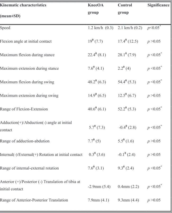
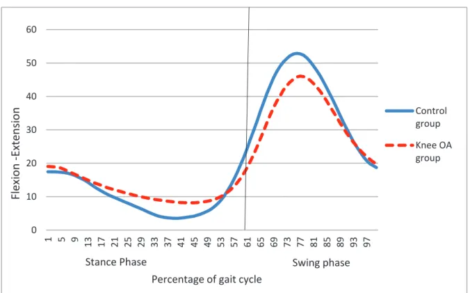
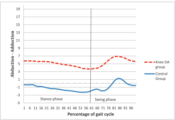
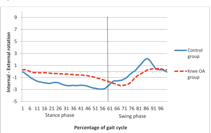
Results. The patients with knee OA had a reduced extension during stance phase (p<0.05) and a reduced flexion during push-off and initial swing phase (p<0.05). The adduction angle was consistently greater for the OA patients (p<0.05). The frontal laxity for OA patients was positively correlated with varus deformity (r=0.42, p<0.05). There was a significant difference (p<0.05) in the tibial rotation during the midstance phase; OA patients retained a neutral position while control group presented internal tibial rotation.
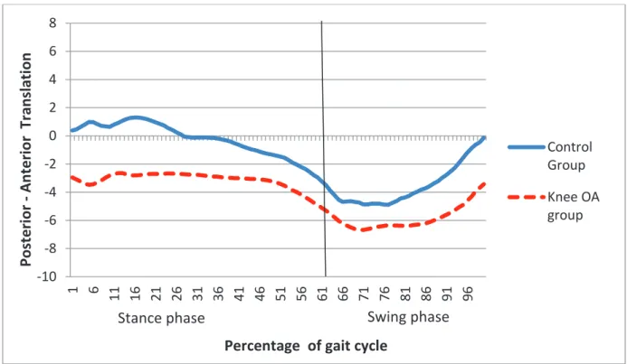
The patients walked faster after total knee arthroplasty comparing with pre-op assessment (p <0.05), but with lower speed comparing with control group. During walking, range of flexion/extension was improved significantly (p<0.05) after TKA but it still remained lower than control group. Eventhough there was a visible improvement in the movement in frontal plane after total knee arthroplasty, the difference did not reach the significance. The range of motion in axial plane did not change pre- and post arthroplasty, but remained lower than the matched control group (p<0.05). The maximum posterior translation during swing phase was significantly higher at post arthroplasty group comparing with control group , p<0.05.
Conclusion. Weight-bearing kinematics in medial OA knees differ from normal knee kinematics. Knee OA group showed an altered “screw-home” mechanism by decreased excursion in sagittal and axial tibial rotation and a posterior translation of the tibia.
Following TKA, patients had better clinical, spatiotemporal and kinametic parameters. They walked longer, faster and with a better range of motion. Despite improvements, the knee kinematics during gait in TKA group differed from healthy control group. They had a lower extension, lower range of axial rotation and an increased tibial posterior translation.
Future research should be focused on comparing different designs of prosthesis pre- and post operatively in a longer follow-up delay.
9
PLAN
ABSTRACT
CHAPTER I. INTRODUCTION
1. Introduction
2. Knee joint kinematics
A. Historical insights B. MRI studies
C. RSA studies
D. Fluoroscopy studies E. Optoelectronic markers F. Normal gait cycle G. Conclusion
3. Kinematics of osteoarthritic knee
A. Overview of the knee osteoarthritis
B. Kinematics of osteoarthritic knee-literature review
4. Kinematics of replaced knee
A. Overview of total knee arthroplasty
10 CHAPTER 2. IN VIVO KINEMATIC GAIT ANALYSIS OF THE OSTEOARTHRITIC KNEE
A.Introduction
B. Material and Methods
C. Results
D. Discussion
E. Conclusion
CHAPTER 3. GAIT ANALYSIS OF TOTAL KNEE ARTHROPLASTY; PRE – AND POST OP
A. Introduction
B. Material and Methods
C. Results
D. Discussion
E. Conclusion
CHAPTER 4. GENERAL CONCLUSIONS AND PERSPECTIVES
RÉSUMÉ
11
ABREVIATIONS
AP
Anterior-Posterior
ACL Anterior Cruciate Ligament
CG Control Group
CT
Computed Tomography
ER
External
Rotation
HKA
Hip-Knee-Ankle Angle
IR
Internal Rotation
LCS
Low Contact Stress
KJC
Knee Joint Center
MRI Magnetic Resonance Imaging
OA
Osteoarthritis
OP Operation
PCA
Principal Component Analysis
PCL Posterior Cruciate Ligament
PCR
Posterior Cruciate Retaining
PS
Posterior Stabilized
RSA RadioStereometric Analysis
UHMPE
Ultra-high-molecular-weight polyethylene
3D Three dimensional
12
C
HAPTER
1.
INTRODUCTION
1. Introduction
Knee osteoarthritis (OA) represents one of the most prevalent forms of osteoarthritis, with population-based studies estimating severe radiographic disease amongst 1% of 25-34 year olds, and 30% in those aged 75 and above [1]. The high incidence of medial compartment knee OA may be attributable to both anatomical and mechanical factors. Mechanically, functional activities such as gait oblige the medial compartment to bear greater loads than the lateral compartment [2].
Currently, the total knee arthroplasty appears to be the treatment of choice in end stage osteoarthritis in subjects older than 55 years, with severe pain and/or functional problems. Total knee arthroplasty has proven to be a successful and durable solution because the fundamental goal of total knee arthroplasty is to give the patients what they need for their everyday activities: pain relief, a good post-operative range of motion and stability [3, 4]; however, it is still not clear if the restoration of normal knee kinematics is possible.
Quantitative kinematic analysis using advanced motion capture technology has been used as an important tool for thorough understanding of joint function.
This work is focused on the three dimensional, in-vivo kinematics of the osteoarthritic knee before and after total knee arthroplasty during all phases of the gait cycle.
13
- The first part, the general overview and the literature review of the kinematics of the native, osteoarthritic and replaced knee.
- The second part, the kinematic data during gait of the osteoarthritic knee are compared with the native knee.
- The third part, a comparision of kinematic data of osetoarthritic knee, pre- and post total knee arthroplasty with the control group.
- The last part, a conclusion based on the obtained results is drawn and the perspectives for the next studies is presented.
14
2. Kinematics Of Native Knee
A. Historical insights
The description of the evolution of spatial position of rigid bodies, without the considerations of the forces involved, is called kinematics. The advancement of the study of locomotion remains dependent on the development of new tools for observation. Over the last several decades, there have been several fundamental advancements that have made a substantial impact on our understanding of the process of knee motion. Flexion and extension are the main movement types of the knee joint and they have been the subject of scientific investigations for over 100 years. Fundamental research on tibiofemoral kinematics was published as early as 1836 by the Weber brothers [5]. For the first time, the kinematics of the knee joint was described as a motion comprising rolling and gliding (Fig. 1). Since that first description, based upon direct visual observation of a cadaveric specimen, several methods have been used to examine the kinematics of the human knee. The axis of knee flexion and extension was derived from the geometry of the femoral condyles, as early as the late 19th century, by analysis of true sagittal plane sections through the femoral condyles. From these sagittal sections, it was clear that the femoral condyles were not circular, but were elongated. The femoral condyles were described as spirals, with the lateral condyle having a greater variation in curvature than the medial condyle. If the femoral condyles were circular, the axis of flexion and extension of the knee would be fixed at its center, like a hinge. The changing curvature of the condyles seen on sagittal sections results in an axis that moves as the knee flexes and extends. This was described as “the instant center of motion” moving along a predictable curved pathway during knee flexion. The instant center of motion (Reuleaux)
15
model was useful because it linked the shape of the condyles to the motion characteristics of the knee (Fig.2) [6].
a) b)
c)
Fig.1. The movement of the femur during flexion/extension. a) the movement of the femur relative to tibia if the movement was a pure rolling, the femur would roll off the tibia; b) the movement of the femur relative to tibia if the movement was a pure gliding, the femur would engage on the posterior rim of tibia; c) the physiologic movement of the femur relative to tibia generated by rolling and gliding [7]
16
Fig.2. Anatomically sagittal diagram of the medial and lateral femoral condyles. The axis of knee flexion and extension or “instant center” moves as the knee flexes, following a
predictable pathway [6]
However, the principal criticism of the instant center theory is that it assumes the flexion and extension movement lies exactly in the sagittal plane. Fick in 1911 reanalyzed the condyle shapes by using 3 rather than 2 dimensions and concluded that the flexion-extension of the knee did not lie in the sagittal plane but was offset by several degrees. This offset orientation of the flexion-extension would result in a single, fixed axis, rather than an instant center. The method of Reuleaux used to map the instant center of motion is correct only if the considered motion lies in a plane. If the plane of movement is offset, then the calculated axis appears to move. However, from Braune and Fisher in 1891,until at least the 1970s, researchers had based their calculations on this assumption.
17
being the inability to ascertain the location of the axes of rotation before performing kinematic analyses.
Contemporary movement toward the concept of a fixed flexion-extension axis began in the field of total knee arthroplasty. The dynamic growth in knee arthroplasty in the 1970s required that knee kinematics be understood for prosthetic design.
In 1983, Grood and Suntay [8] presented a joint coordinate system providing a geometric description of the three-dimensional rotational and translational motion between two rigid bodies, applied to the knee joint. With this model, the described joint displacements are dependent of the order in which the rotation components and translations occur.
Hollister [9] essentially described knee motion as pure rotations occurring around two axes: the so called ‘flexion-extension axis’ and the so called ‘longitudinal rotation axis’, with the understanding of the flexion extension axis not being exactly located in the coronal plane and the longitudinal axis not being exactly located in the sagittal plane. As a consequence, these mathematical ‘simple rotations’ meant in reality flexion-extension, varus-valgus and internal-external rotation of the knee joint.
With the improvement of the technology, more advanced tools were possible to be used such as MRI, CT, dual fluoroscopy, dual coil MRI, roentgen stereo photogrammetry, optoelectronic motion capture systems.
These methods have provided a more thorough understanding of the knee kinematics.
B. MRI studies
Hill, Iwaki and coworkers [10] applied MRI scans to 13 unloaded knees and 7 loaded knees for the description of the surface geometry and relative movements of the femur and the tibia. According to this study, during flexion in the unloaded knee, medially, the mean AP position of the femoral condyle did not change from 110° to -5°. Laterally, the femoral condyle rolled
18
forwards from 110° to 60°, a total of 13 mm, corresponding to 15° of femoral IR (tibial ER) as the knee extended. There was then 1 mm of forward femoral movement, equivalent to 1° of rotation, from 60° to 0°. Finally, the condyle again moved forward 3 mm as the femur internally rotated 4° to ‘screw home’. When load is applied it alters tibiofemoral motion in neutral tibial longitudinal rotation. The medial femoral condyle translates forwards about 4 mm between 10° and 45° flexion and the lateral femoral condyle moves backwards further than in the absence of load.
Todo et al. [11] also, analyzed MR images perpendicular to the flexion-extension axis, rather than sagittal images. They concluded that roll back, if present at all, is small, perhaps 2mm, and can be suppressed in either the medial or lateral compartment by the longitudinal rotation of the knee.
In a later paper in 2004, Pinskerova and Freeman [12] reworked these findings and concluded that medially the condyle hardly moves antero-posteriorly from 0˚ to 120˚ but the contact area transfers from an anterior pair of tibio-femoral surfaces at 10˚ to a posterior pair at about 30˚. Thus because of the shapes of the bones, the medial contact area moves backwards with flexion to 30˚ but the condyle does not. Laterally the femoral condyle and the contact area move posteriorly but to a variable extent in the mid-range causing tibial internal rotation to occur with flexion around a medial axis. From 120˚ to full flexion both condyles roll back onto the posterior horn so that the tibio-femoral joint subluxes.
Nakagawa and co-workers [11] studied unloaded high flexion in 20 Japanese volunteers with an open MRI. Active flexion was measured from 90°-133° and passive flexion from 90° to 162°. They found a mean posterior translation of the medial condyle and lateral condyle of 2 mm and 13 mm respectively from 90-130° of flexion. Pushing the knee further, from 133° to 162° of flexion, caused further posterior translation, medially by 4.5mm and laterally by 15 mm, subluxing the femur behind the tibia.
19
Johal and co-workers [13] studied the full range of motion of a “loaded squat” (wall supported squat sit) with 10 volunteers, using an interventional MRI. They observed posterior translation of the lateral femoral condyle of 22 mm from hyperextension to 120° of flexion. The medial condyle moved forward 1.7 mm from hyperextension to 30° of flexion and started translating posterior from 90° flexion onwards: 3.6 mm between 90° and 120°, and an additional 8.4 mm from 120° to full flexion. This differential translation on the medial and lateral side leads to an external rotation of 20° of the femur relative to the tibia during flexion.
Beyond 120° of flexion, posterior translation was equal on the medial and lateral side and no further tibiofemoral rotation was occurring.
Although MRI permits imaging of knee motion and has been useful in understanding the motion of the knee transferring the kinematics of these cyclical knee flexion-extension motions to walking or other activities of daily living has not yet been established.
C. RSA Studies
Karrholm et al. [14] confirmed the data published by Hill in an in vivo RSA experiment where volunteers performed a step-up activity from 50° to 65° of flexion to full extension. They also found anterior translation of the femoral condyle in early flexion (3 mm from 0° to 50° of flexion). Forced external rotation of the foot suppressed physiologic internal rotation of the tibia with flexion. The main weakness of this study is the variability of the motion that was performed and the fact that they only studied a single motion from moderate flexion to full extension and not a full motion cycle.
On the other hand, this procedure is done while the patient is lying in horizontal position hence it does not allow the 3-D kinematic analysis while performing normal daily activities.
20
D.
Fluoroscopy Studies
Fluoroscopy allows following knee motion in real time in vivo, with an obvious drawback of delivering two-dimensional images. A preliminary CT scan allows making a 3D bone model that can convert the 2D fluoroscopic image with a shape-matching technique.
Komistek and co-workers [15] used this technology to study gait and deep flexion activities in five volunteers. They found significantly less translation and rotation of the femur during gait than during deep flexion. They confirmed previous findings of less translation on the medial side than on the lateral side but the most important result of their work seems to be the impressive inter-individual variability. For deep flexion, the medial condyle translation ranged from +3 mm to -9 mm versus +1,4 to -30 mm for the lateral condyle. The average tibiofemoral rotation during flexion was 13°.
Bank’s group [16] used CT-derived bone models for model registration and added MRI derived articular surfaces for obtaining higher accuracy of the contact areas. They observed the greatest femoral external rotation during the squat activity, but reported no posterior subluxation of either femoral condyle in maximum knee flexion.
E. Optical tracking
In tracking based approaches, an appropriate marker is affixed to the patient limb and a tracking device traces the markers position. It is very useful to get information about knee kinematics during daily activities like walking, running and squatting. There are two approaches for the positioning of markers on the limbs [7]. Invasive approach is skeletal intracortical pins drilled in the limb with markers in surface. Invasive methods are more reproducible and accurate than non-invasive ones when it comes to recording knee kinematics, but they are usually less accessible and less safe, mainly due to risk of infection. For this reason, non-invasive methods with passive markers are widely used. One approach is
21
to place markers directly onto the skin, usually over bony anatomical landmarks. The other is to fix a set of at least three markers to each limb segment (rigid body), either directly or placed on a rigid structure. Both of these approaches allow representation of the motion of the body segment, but are subject to skin movement artefacts when movements out of the sagittal plane have to be assessed.
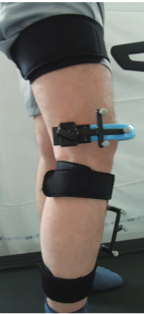
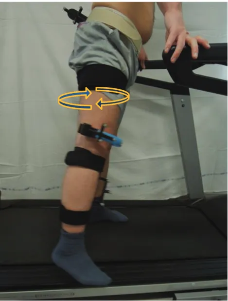
KneeKGTM
In order to reduce skin motion artefacts, a new non invasive knee attachment system has been developed by the Imaging and Orthopaedics Laboratory, University of Montreal Hospital Centre. The group developed a harness to be fixed quasi-statically on the thigh and calf, therefore reducing the skin motion artifact. This harness was shown to be accurate in obtaining 3D kinematic data and was validated for gait applications [17, 18]. The potential of the device to assess 3D knee kinematics in a variety of situations led to the commercialization of the device under the name KneeKGTM.
Another good point of this tool is that it allows a quick (20 min) assessment of knee kinematics and patient examination can be performed in a small assessment room with a treadmill. This advantage allows the use of KneeKGTM on the clinical settings.
22
F. Normal Gait Cycle
Walking and running are the most common human movements and probably the most complex. The individual walking pattern is a personal identity because each of us performs a characteristic and unique way of walking [19].
The gait cycle is defined as the period from heel contact of one foot to the next heel contact of the same foot. This cycle consists of two parts, stance and swing phase. On average, the gait cycle is about one second in duration with 60% in stance and 40% in swing.
It has eight sub-phases which assist in determining overall coordination of the limbs, and assessing functional tasks associated with walking (Fig.3).
Phase 1- Initail contact (Heel strike). The instant when the foot contacts the ground. The knee is extended and the ankle is neutral.
Phase 2 – Loading. The limb attempts to absorb shock caused by ground reaction forces, stabilize the limb to bear the weight of the body, and continue forward progression.
The knee flexes rapidly, reaching approximately 15˚ of flexion.
Phases 3 – Midstance. Represents the first half of single support, which occurs from the 10% to 30% periods of the gait cycle. It begins when the contralateral foot leaves the ground and continues as the body weight travels along the length of the foot until it is aligned over the forefoot. The knee extends until it reaches 3˚ of flexion by midway through terminal stance (Fig.4).
Phase 4- Terminal stance. The second half of the single support from 30 to 50% of the gait cycle and is defined as the time from heel rise until the other limb makes contact with the floor. During this phase, body weight moves ahead of the forefoot.
23
Phase 5 – Pre swing . From the time of initial contact with the contralateral limb to ipsilateral toe-off. During this period, the stance limb is unloaded and body weight is transferred onto the contralateral limb. Knee flexes from 7˚ to 40˚ of flexion.
Phase 6 - Initial swing phase. It begins at the moment the foot leaves the ground and continues until maximum knee flexion occurs, when the swinging extremity is directly under the body and directly opposite to the stance limb. The knee is at 60˚ of flexion.
Phase 7 – Mid swing. From the time the swing foot is opposite to the stance limb to when the tibia is vertical. Begins from maximum knee flexion (when the swing limb is under the body) until the swing limb passes the stance limb and the tibia becomes in a vertical position. Phase 8 – Terminal swing. Is the final phase of the swing period from 85% to 100% of the gait cycle. The tibia passes beyond perpendicular, and the knee fully extends in preparation for heel contact. The knee extends rapidly throughout mid- and terminal swing until peak extension is reached just before initial contact. Peak extension can range from 3˚ of hyperextension to 5˚ of flexion.
24
Fig. 4 Healthy knee flexion and extension during gait [21]
Lafortune et al. [22] studied knee kinematics during walking in 5 healthy volunteers using intracortical pins with clusters. They found out that the average pattern of flexion/extension of the tibiofemoral joint during walking was biphasic: a slight flexion followed by an extension during the stance phase and a large flexion also followed by an extension during the swing phase. The average pattern of abduction/adduction of the tibiofemoral joint (rotation around the floating axis) was uniphasic and was limited to 5˚. Twice during the stance phase, the tibiofemoral joint rotated internally -as heel strike occurred and again prior to toe-off. During the middle part of the stance phase, the tibiofemoral joint remained close neutral position. From toe-off until heel strike, the tibiofemoral joint rotated externally. Andriacchi et al. [23] studied the anterior posterior (AP) motion of the knee during walking. At heel strike with the knee at full extension, the tibia is at its maximum anterior position during the gait cycle. The next key event occurs at terminal extension where the tibia is located posteriorly again while the knee is near full extension. Thus, heel strike and terminal extension provide two events where the tibia is located over a range of AP positions relative to the femur.
25
G. Conclusion
On the basis of the published data, one can conclude the normal kinematic pattern of the human knee consists of internal rotation of the tibia relative to the femur with increasing flexion, following a greater posterior translation of the lateral femoral condyle than of the medial femoral condyle. On the other hand, gait analysis has allowed researchers and clinicians to better understand biomechanical factors of gait in healthy participants and those with lower limb pathology [24]. However, different methodologies seem to discover different kinematic patterns. Moreover, it remains unclear to what extent the natural kinematic patterns can be modified after post-traumatic or degenerative disease of the knee joint and to what extend the prosthetic surgery replaces the functional anatomy.
26
3. Kinematics Of Osteoarthritic Knee
A. Overview Of Knee Osteoarthritis
Osteoarthritis is a degenerative process that typically affects the synovial joints of the body. WHO estimates that osteoarthritis (OA) affects 9.6% of men and 18% of women older than 60 years of age [25]. Increases in life expectancy and ageing populations are expected to make OA the fourth leading cause of disability by the year 2020 [26]. It leads to social, psychological and economical burdens in patients with substantial financial consequences [27]. In France, the cumulated health costs resulting from OA almost doubled within 10 years (from 1993–2003) [28] .
In recent years, it is shown that not only cartilage, but also the subchondral bone, ligaments, the synovial fluid, and surrounding muscles are involved in the OA process. Although the exact aetiology is still unknown, OA is in general characterized by loss of articular cartilage, osteophyte formation, and subchondral bone sclerosis [29].
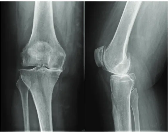
Knee OA represents one of the most prevalent forms of osteoarthritis, with population-based studies estimating severe radiographic disease amongst 1% of 25-34 year olds, and 30% in those aged 75 and above [1]. The diagnosis of the knee OA is made at first by clinical examination. Pain, morning stiffness, swelling and crepitus at one patient older than 50 years may be consequences of the knee osteoarthritis. During the examination, it is important to analyze the deformity of the lower limbs and its reductibility, the ligamentary status and the range of motion. Imaging modalities play an important role in the knee OA, since they confirm the diagnosis, detemine the involved compartment and evaluate the stage of the disease. Conventional radiography (Fig.5) is still today the technique of reference for evaluating knee OA. The main radiological signs are joint space narrowing corresponding to loss of cartilage; the osteophytes which represent marginal bone reaction to loss of cartilage;
27
subchondral bone reactions, geodes or bone condensation [30]. There are two main classifications of the structural changes associated with knee OA (Tab.1), Kellgren and Lawrence composed criteria for a 5-point grading scale using radiographic features [31]; and Ahlbäck scaled the knee OA from stage 0 (no OA) to stage V (bone defect/loss >10 mm, often with subluxation and arthritis of the other compartment) [32] .
Kellgren-Lawrence classification
Grade 0 Normal
Grade 1 Doubtful narrowing of joint space and possible osteophysic lipping Grade 2 Definite osteophytes and possible narrowing of the jointspace
Grade 3 Moderate multiple osteophytes, definite narrowing of joint space and some sclerosis,
possible deformity of the bone ends
Grade 4 Large osteophytes, marked narrowing of joint space, severe seclerosis, and definite
deformity of bone ends Ahlbäck classification criteria
Stage 0 No radiographic sign of arthritis
Stage I Narrowing of the joint space (JSN) (with or without subchondral sclerosis). JSN is defined by a space inferior to 3 mm, or inferior to the half of the space in the other compartment (or in the homologous compartment of the other knee)
Stage II Obliteration of the joint space Stage III Bone defect/loss <5 mm
Stage IV Bone defect/loss between 5 and 10 mm
Stage V Bone defect/loss >10 mm, often with subluxation and arthritis of the other compartment
28
According to Petersson et al., Ahlbäck and Kellgren and Lawrence classifications show a good correlation [33]. Moreover, Ahlbäck classification seems easy to apply and suitable for the assessment of medial compartment arthritis of the knee; thus, it seems particularly useful for the orthopaedic treatment of knee disorders [34].
Whilst the symptoms and diagnosis of the disease have been clearly defined in medical research, the underlying causes are not yet fully understood. It is thought that biomechanical factors play a key role in OA aetiology. Furthering the understanding of these factors could lead to better treatments and help reduce prevalence through preventative measures.
29
B. Kinematics Of Osteoarthritic Knee – Literature Review
Over the past 20 years, there have been numerous investigations into the effects of OA on knee kinematics and also into what biomechanical factors may be causing the initiation of the disease. Kinematic data allow physicians to obtain and process accurate objective measurements of sophisticated movement such as human walking.
Hamai et al. [35] studied knee kinematics in 12 subjects with medial OA during three activities using dynamic imaging and model-image registration with CT-derived models. They concluded that knees with medial OA differed from normal knees. First, they displayed a femoral internal rotation bias of about 80 compared to normal knees: femoral external rotations of 40 and 150 during squatting (200 and 1000 flexion, respectively) were less in knees with medial OA than the 120 and 240 observed in normal knees. Second, the natural screw-home movement was not observed in knees with medial OA, perhaps because they did not reach full extension in squatting or stair climbing. Finally, femoral condylar contact in knees with medial OA did not exhibit significant AP translation between 300 and 800 for either squatting or stair climbing.
Saari et al. [36] studied the kinematics of the knee during weight-bearing active extension in 14 patients with medial osteoarthrosis (OA) and in 10 controls using dynamic radiostereometry. They found out that between 500 and 200 of extension, the OA knees showed decreased internal tibial rotation corresponding to less posterior displacement of the lateral femoral flexion facet center.
Matsui et al. [37] evaluated the rotational deformity at 150 osteoarthritic knees compared with 31 control knees using CT scans. Results of the this study indicate that the varus deformity in OA of the knee is associated with significant rotational deformity. The tibia tended to locate in an externally rotated position in the knees with severe varus deformity.
30
Siston et al. [38] performed a study where a surgical navigation system measured normal passive kinematics from 7 embalmed cadaver lower extremities and in vivo intraoperative passive kinematics on 17 patients undergoing primary total knee arthroplasty. Osteoarthritic knees displayed a decreased screw-home motion and abnormal varus/valgus rotations between 100 and 900 of knee flexion when compared to normal knees. The anterior–posterior motion of the femur in osteoarthritic knees was not different than in normal knees.
In 2001, Kaufman et al. [39] performed a large study on 139 adults diagnosed with early stage knee OA and compared their gait characteristics against those of 20 healthy control. They found that, for level walking, the OA sufferers had 6° less peak knee flexion than healthy controls. OA subjects also showed significantly lower knee extensor moments which were conjectured to be a method of minimising pain in the joint. Cadence was also significantly reduced during walking for the OA subjects. The stair ascent and descent exercises did not show any differences in the range of joint motion, however speed and knee extensor moment was found to be significantly smaller for OA sufferers for both activities. Briem and Snyder-Mackler in 2009 [40], looked at the inter-limb differences in 32 patients with moderate medial knee OA. Asymmetry was seen between affected and unaffected knees for flexion and adduction. Knee on the involved side had a significantly smaller flexion and greater adduction angle than on the uninvolved side during weight acceptance.
Mundermann, Dyrby and Andriacchi in 2005 [41] performed a gait analysis of 42 patients with bilateral medial knee osteoarthritis and they observed that all patients with knee OA made initial contact with the ground with the knee in a more extended position than that of the control subjects.
Nagano et al. (2012) [42] in their kinematic gait study of 45 patients with different stages of knee OA and 13 healthy subjects, also found out similar results. The flexion angle at the time of foot contact was significantly less in patients with severe and moderate osteoarthritis than
31
in normal subjects. The abduction angle at the 50% stance phase was significantly less in patients with severe osteoarthritis than in normal subjects. The excursion of axial tibial rotation was significantly less in patients with early osteoarthritis than in normal subjects. On the other hand, Heiden, Lloyd and Ackland [43] revealed different results in their study of gait parameters of 54 patients with knee OA. Gait differences in the knee osteoarthritis patients were greater knee flexion at heel strike and during early stance along with reductions in the peak external knee extension moment in late stance[43].
Childs et al. [44] compared a group of 24 knee OA sufferers against a group of 24 healthy controls. Gait recording was performed using an electromagnetic system, a force plate and a surface EMG system with subjects performing both walking and step descent tasks. During the walking task, they also found that OA sufferers had a higher knee angle at heelstrike and also had a lower knee flexion range of motion in the loading response phase of stance . Following on from the work of Astephen in 2004 [45], Deluzio and Astephen in 2007 [46] used a group of 50 end-stage knee OA patients to look at the gait waveform data of three variables; knee flexion angle, flexion moment and knee adduction moment. As with the previous investigation, a force plate and optoelectronic system was used to collect gait data and a control group of 63 healthy subjects was also used. The authors then used principle component analysis (PCA) to compare the two groups. OA patients knees were less flexed throughout the gait cycle than the controls, and also they had less range of motion in the joint. OA subjects were also shown to have a smaller range of flexion moment during gait and a lower flexion moment during the first half of the stance phase. A lower adduction moment in early stance was also shown in the OA subjects.
Astephen et al. [47] in 2011 investigated the associations between joint biomechanics and neuromuscular control for moderate OA, looking at the differences between radiographic changes and pain severity. Data were collected on a group of 40 OA patients (with a range of
32
severities) using an optoelectronic system, force plate and EMG system. Radiographic OA severity was found to be correlated with knee adduction moment during stance and maximum knee flexion angle over the whole cycle with higher knee adduction moments and lower maximum flexion angles associated with more severe OA.
In 2007, Landry et al. [48] used the PCA technique previously developed [55] to look at the effect of walking speed on OA. An optoelectronic system and force plate was used to collect data, and 41 patients with radiographic grade 1-3 on the KL scale were compared against 43 asymptomatic patients. Two gait speeds were analysed: selected and 150% of self-selected speed. They found that the OA patients had similar stride characteristics and joint kinematics to the control group. This does not agree with the majority of the literature.
Lewek, Rudolph and Snyder-Mackler in 2004 [49] studied control of frontal plane knee laxity during gait in patients with medial compartment knee osteoarthritis. Twelve subjects with genu varum and medial compartment knee osteoarthritis (OA group) and 12 age-matched uninjured subjects underwent stress radiography to determine the presence and magnitude of frontal plane laxity. All subjects also went through gait analysis with surface electromyography of the medial and lateral quadriceps, hamstrings, and gastrocnemius to calculate knee joint kinematics and kinetics and co-contraction levels during gait. The OA group showed significantly greater knee instability, medial joint laxity, greater medial quadriceps - medial gastrocnemius (VMMG) co-contraction and greater knee adduction moments than the control group. Also, the OA group had a knee flexion significantly less than excursion of the healthy control subjects.
More recently, in 2013, Baert et al. [50] assesed the gait adaptions of the knee with early and established OA in comparision with a control group. Fourteen female patients with early knee OA, 12 female patients with established knee OA and 14 female control subjects participated in the study. The gait parameters were acquired using 3D LED motion analysis
33
system. None of the kinematic variables were significantly different between the early OA and control group. Early OA patients showed significantly less knee adduction in stance phase and more maximal knee extension in late stance than established OA patients. In stance phase, established OA patients showed significantly more knee adduction and less late stance maximal knee extension than controls.
Conclusion:
This review of the existing literature on the links between osteoarthritis andgait kinematics has highlighted several areas of interest. There are common gait features which are consistently shown to be significantly linked to osteoarthritis severity such as knee adduction moment, knee flexion angle, stiffness and walking speed.
Most previous studies of changes in OA patients during gait were focused on spatiotemporal parameters [51, 52]; demonstrating that knee OA patients walked slower, with a reduced stride length, and a lower single-limb support compared to controls. Some studies have examined kinematic alterations during phase-specific gait cycle and reported a decreased knee excursion during flexion, decreased peak flexion during stance phase and increased knee flexion at heel strike [41, 47]. Although the kinematic changes in sagittal plane have been enlightened, there are still contradictions. On the other hand there are only few studies that observed the changes in frontal and axial planes and anterior-posterior (AP) translation which still remain unclear.
34
Authors Subjects Method Activity Studied
Saari T et al 2005 14 patients medial knee OA 10 control group Dynamic
radiostereometry Active extension Matsui et al.
2005
150 medial OA knees
31 control knees CT scans Passive Flexion-Extension Siston et al 2006 17 patients OA 7 cadaver normal knees a surgical
navigation system Passive flexion- extension Hamai S et al 2009 12 subjects medial knee OA CT scans kneeling, squatting, stair climbing Kaufman et al. 2001 139 patients knee OA 20 control group Reflective markers, six video cameras
-walking -stair ascent -stair descent Briem et Snyder-Mackler 2009 32 patients with medial knee OA -interlimb differences Optoelectric
motion analysis -gait, stance phase Mündermann, Dyrby and Andriacchi 2005 42 patients with medial knee OA 42 control group
Reflective markers -gait, stance phase
Heiden , Lloyd, Ackland 2009 54 patients knee OA 30 control group Reflective cluster
markers -gait, stance phase Deluzio,
Astephen 2007
50 patients knee OA
63 control group Optoelectric system -gait Nagano et al. 2012 45 patients knee OA 13 control group Reflective cluster markers
-gait, foot contact and 50% of stance phase
35 Childs et al. 2003 24 patients knee OA 24 control group Electromagnetic motion analysis system
-gait, stance phase Landry et al. 2007 41 patients knee OA 43 control group optoelectronic system -gait Lewek, Rudolph and Snyder-Mackler 2004 12 patients medial knee OA 12 control group -stress radiography -surface electro- myography
-gait, stance phase
Baert et al. 2013
14 female early knee OA (Early OA) 12 female established knee OA (Estab. OA) 14 female control group (CG)
3D motion analysis
(LED) -gait
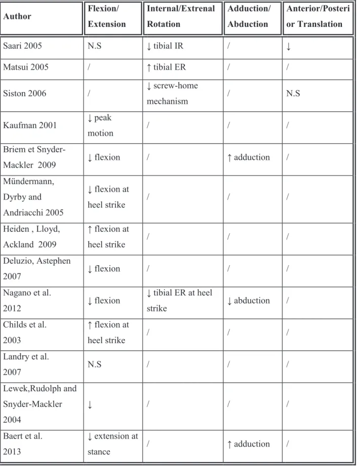
36 Author Flexion/ Extension Internal/Extrenal Rotation Adduction/ Abduction Anterior/Posteri or Translation Saari 2005 N.S ↓ tibial IR / ↓ Matsui 2005 / ↑ tibial ER / / Siston 2006 / ↓ screw-home mechanism / N.S Kaufman 2001 ↓ peak motion / / / Briem et
Snyder-Mackler 2009 ↓ flexion / ↑ adduction /
Mündermann, Dyrby and Andriacchi 2005 ↓ flexion at heel strike / / / Heiden , Lloyd, Ackland 2009 ↑ flexion at heel strike / / / Deluzio, Astephen 2007 ↓ flexion / / / Nagano et al. 2012 ↓ flexion ↓ tibial ER at heel strike ↓ abduction / Childs et al. 2003 ↑ flexion at heel strike / / / Landry et al. 2007 N.S / / / Lewek,Rudolph and Snyder-Mackler 2004 ↓ / / / Baert et al. 2013 ↓ extension at stance / ↑ adduction /
Tab. 3. Reported results of the studies comparing knee kinematics within OA groups or with a control group.
37
4. Kinematics Of Replaced Knee
A. Overview Of Knee Replacement
Nearly a million of total knee prostheses are implanted worldwide each year. Approximately 600,000 knee replacements are performed every year in the United States of America [53], more than 70,000 in France [54].
Attempts to replace the knee joint with an arthroplasty have been made for at least 140 years. In 1860, Verneuil suggested “the interposition of tissues between resected bone to prevent fusion”. Jules Emelie Péan, one of the leading surgeons in Paris, described in 1894 the first attempt with an articular prosthesis of metal of humerus. In 1938, Venable and Struck presented an imortant work when they significantly improved the quality of vitallium and certain steel alloys. This raised the success rate for all metal implants and meant a significant step forward in the development of implant surgery.
It was not until the late 60s that the first series of replacements of the knee with metal implants emerged and the early 70s saw the rise of prostheses of the knee.
In 1971, Gunston [55] importantly recognized that the knee does not rotate on a single axis like a hinge but rather the femoral condyles roll and glide on the tibia with multiple instant centres of rotation. His polycentric knee replacement had early success with its improved kinematics over hinged implants but failed because of inadequate fixation of the prosthesis to bone.
The Total Condylar prosthesis was designed by Insall at the Hospital for Special Surgery in 1973. This prosthesis concentrated on mechanics with the intention to create a knee replacement with kinematic characteristics as similar as possible to the normal knee.
38
Nowadays, there is a wide diversity of total knee prosthesis designs, developed with specific properties and with a specific patient group in mind and therefore each one has its own theoretical advantages and disadvantages. However, there are some main implant variations. A set of knee-prosthesis components consists of: an anatomically shaped distal femoral component made of Cobalt-Chrome alloy which is a very hard and durable material, allowing it to withstand the massive loads and cycles a knee endures on a daily basis; a tibial component that is usually composed of two main pieces, the tibial tray made of Titanium or Cobalt-Chrome and the tibial bearing component made of ultra high molecular weight polyethylene (UHMWPE); and the optional patellar component is also made of plastic (UHMWPE).
The medial collateral ligament and lateral collateral ligament are critical in holding the joint in place and producing joint motion. Implantation of prosthesis typically requires removal of the anterior cruciate ligament and, depending on the prosthesis design, may also involve removal of the posterior cruciate ligament. The posterior cruciate ligament (PCL) is an important structure that stabilizes the knee joint. The cruciate retaining prosthesis enables the surgeon to preserve the ligament. It has a small groove that helps the ligament continue to provide flexion. When PCL is removed, the posterior stabilized prosthesis is used. These implants employ a “cam and post” system that substitutes for the posterior ligament and provides support on the posterior part of the knee (Fig.6).
39
A) B)
Fig.6. A) PCLR prosthesis and B) PCLS prosthesis, note the post and cam mechanism of the PCLS implant, which prevents the posterior translation of the tibia.
For fixation, most predominantly knee prostheses are cemented to the bone with polymethylmethacrylate but some other types of cementless prosthesis, instead of cement, have a special surface that encourages bone to grow into the implant for fixation.
Current TKR devices can be subdivided into two groups based on different fundamental design principles: fixed-bearing knees, in which the UHMWPE insert is snapped or press fitted into the tibial tray, and mobile-bearing designs which facilitate movement of the insert relative to the tray (Fig.7).
a) b)
40
HLS KneeTec Prosthesis (Tornier), the prosthesis used in our study, is a posterior stabilized total knee replacement (Fig.8) with rotatory platform without conservation of cruciate ligaments. Its distinctive feature is the system of posterior stabilization: a third condyle in the midline of the intercondylar notch that permits progressive contact with the tibial cam from 35° flexion, thus ensuring stability very early on in flexion. This causes roll-back of the femoral condyles that optimizes quadriceps function, reduces load on the extensor system and improves flexion. Finally, the third condyle constitutes an additional point of tibiofemoral contact, which permits better load distribution on the polyethylene [56].
The trochlear groove is convex-dome with a constant radius along its trajectory, giving better congruence on engagement and therefore stability throughout all movement of the knee. The tibial axis of rotation of the mobile platform is located posterior to the midline to restore a movement close to anatomic rotation. An anterior circular rail allows for 30˚ range of axial rotation.
Fig.8 The HLS KneeTec Prosthesis
41
B. Kinematics of Replaced Knee
There is a long-standing controversy on which type of total knee prosthesis provides better kinematics and clinical outcome. Several studies have analyzed the kinematics of total knee prosthesis, comparing different designs or with a control group during passive movements. Dennis et al., in 1998 [57], have studied the effect of implant designs in range of motion after total knee arthroplasty. The range of motion of twenty patients with posterior cruciate retaining (PCR) prosthesis; 20 patients with posterior substituting (PS) prosthesis and 20 control group were obtained using video fluoroscopy. The normal knee group exhibited superior flexion over either TKA subgroup, whether measured under passive non-weightbearing or active weight-bearing conditions. Maximal mean postoperative flexion for PCR and PS TKA groups was similar when evaluated under passive non-weightbearing conditions. When measured under weight-bearing conditions, patients implanted with PS TKA exhibited significantly greater mean range of motion than those with PCR TKA.
Siston et al., in 2006 [38], measured intraoperatively using surgical navigation system passive kinematics of 17 patients pre- and post- TKA and compared it with that of 7 normal cadaver knees. Throughout the range of flexion, no systematic relationship of varus/valgus rotation angle with flexion was present in any knee following TKA, resulting in a motion pattern significantly different from normal knees. The screw-home motion following TKA was significantly less than the screw-home motion in the OA knees. AP femoral translation was significantly different in the knees following TKA compared to normal knees. Following TKA, the femur translated anteriorly on the tibia until approximately 600 of flexion before beginning a posterior translation.
Yoshiya et al., in 2005 [58] , realized a study where in vivo comparison of flexion kinematics for posterior cruciate- retaining (PCR) and posterior stabilized (PS) total knee arthroplasty (TKA) was performed. In the PCR TKA, an anterior femoral translation from 300 to 600 of
42
flexion was observed in the weight-bearing condition. In contrast, flexion kinematics for the PS TKA was characterized by the maintenance of a constant contact position under weight-bearing conditions and posterior femoral rollback in passive flexion.
In 2008, Cates et al. [59] also analyzed the kinematics from full extension to maximum flexion for 30 subjects (15 PS, 15 PCR) using fluoroscopy. Ranges of motion were not statistically different between the 2 implant designs. In comparing the PCR group to the PS group at each flexion angle (ie, at 0°, 30°, 60°, 90°, and 120°), no significant differences were found in axial rotation angles. The PCR knees had a significantly larger mean axial rotation angle than the PS knees at maximum flexion.
Other studies analyzed the kinematics of the replaced knee during daily activities as walking, stair climbing, knee bending.
Haas et al., in 2002 [60], investigated in vivo kinematics of 10 subjects with either posterior stabilised (PS) or posterior cruciate substituting (PCS) mobile bearing TKAs during gait and during a knee bend from 0° to 90° flexion. This study showed that the kinematic patterns for subjects having either a PS or PCS mobile bearing TKA were similar during gait but subjects having a PS TKA experienced more posterior femoral rollback of the lateral condyle during the deep-knee bend.
In 2011, Hatfield et al. [61] investigated 3D kinematic and kinetic gait patterns of 42 patients with severe knee osteoarthritis, collected 1 week prior and 1 year post-TKA. Overall and midstance knee adduction moment magnitude decreased while knee flexion angle magnitude increased during swing. Increases in the early stance knee flexion moment and late stance knee extension moment were found, indicating improved impact attenuation and function. A decrease in the early stance knee external rotation moment indicated alteration in the typical rotation mechanism.
43
Moro-oka et al., in 2007 [62], compared knee kinematics in 9 patients with bi-cruciate preserving total knee arthroplasty (ACL/ PCL knees) and 5 patients with posterior cruciate ligament (PCL) preserving total knee arthroplasty. They studied treadmill gait, stair stepping, and maximum flexion activities using lateral fluoroscopy and shape matching. The ACL/PCL knees showed greater knee flexion, greater tibial internal rotation and greater posterior condylar translation than the PCL knees all through the gait cycle. For the stair activity, posterior translations of the lateral condyle were significantly greater in the ACL/PCL knees from 300 to 700 flexion.
Argenson et al., in 2006 [63], studied in vivo kinematics of the femorotibial joint during a deep knee bend using fluoroscopy for 20 subjects having a TKA designed for deep flexion. The average weight-bearing range of motion was 1250. On average, subjects in this study experienced 5.40 more internal rotation of normal axial rotation.
In 2007, van der Linden et al. [64] evaluated the knee kinematics in functional activities seven years after TKA. Nineteen patients with knee osteoarthritis were assessed using electro-goniometry before surgery, 18–24 months and seven years after total knee surgery. Maximum knee angle during the swing phase while walking on a level surface increased at 18–24 months after surgery but decreased again seven years after surgery. Patients used a significantly greater range of motion of the knee during ascending and descending a flight of stairs, seven years after surgery compared to 18–24 months after surgery.
Recently, in 2012, Joglekar et al. [65] investigated the gait of 18 subjects with either a PS or PCR TKA and sacrificed PCL and compared with the normal contralateral knee using a passive reflective arrays and an optoelectric system. The data for the operated knees for stair descent demonstrated significant differences in maximum overall knee flexion angle; maximum stance phase knee flexion angle; maximum swing phase knee flexion angle;
44
average swing phase knee flexion angle with the PS knees showing higher flexion angles than the PCR knees.
One of the few studies that included coronal and transverse planes is that of McClleland et al., in 2011 [66], who compared the knee kinematics of 40 patients following TKR and 40 unimpaired controls during comfortable and fast walking speeds using three dimensional motion analysis.
In the sagittal plane, the TKR group walked with less knee flexion during stance and swing phases. The TKR group also had less knee flexion at initial contact during fast speed walking but the reduced knee flexion at initial contact during comfortable speed walking did not reach significance. There were no differences in the coronal plane kinematics between groups. In the transverse plane, the TKR group walked with significantly less internal rotation and significantly more external rotation compared to the control group.
Conclusion.
Evidence from multiple studies included in this review indicates that kneekinematics after total knee arthroplasty differ from normal healthy controls. TKA patients walked with less total range of knee motion than their control counterparts.
The range of flexion during the loading phase of stance was also reduced compared to controls. Peak knee flexion during weight acceptance and knee flexion excursion are less in the operated knee after TKA than in healthy controls.
Substantial differences were also identified in the characteristics of the knee replacement prostheses designs used in the various studies. There is ongoing discussion in the literature about the biomechanical effects of either retaining or resecting the PCL in TKA.
However, there is a lack in literature in analyzing the knee kinematics during gait in frontal, transverse and axial plane. Also most of the studies compare designs of the prosthesis or the TKA group with the healthy control subjects but there are only few studies that investigated the pre- and post-TKA knee kinematics.
45
Authors Subjects Methods Activity
studied Prosthesis design
Dennis et al. 1998 40 patients with TKA 20 control group Video fluoroscopy Passive flexion-extension PCR vs PS prosthesis Siston et al. 2006 17 patients with TKA 7 cadaver knees Surgical navigation system Passive flexion-extension PS Prosthesis Yoyisha et al. 2005 20 patients with bilateral TKA Fluoroscopy Passive and weight-bearing Flexion-extension PCR prosthesis in one knee PC prosthesis on the other knee Cates et al. 2008 30 patients
with TKA Fluoroscopy
Flexion-extension PS prosthesis PCR prosthesis Hatfield et al. 2011 42 patients pre- and post -TKA
Electromyography, infrared reflective markers Gait PS prosthesis Kitagawa et al. 2010 10 subjects pre- and post TKA Fluoroscopy Flexion/Extens ion PCR- prosthesis Saari et al. 2005 39 patients with TKA 18 control group Skin reflective
markers Gait PCR and PS prosthesis
Van de Linden et al. 2007
19 patients pre – and post TKA (after 18 months and 7 years) electrogoniometry Gait, level walking, ascend and descend LCS prosthesis
46
Yue et al. 2011
11 patients pre- and post TKA 22 control group Fluoroscopy Flexion/Extens ion PCR prosthesis Joglekar et al. 2012 18 patients with TKA Control: contralateral knee Reflective markers Gait: stair ascend, descend PS prosthesis PCR prosthesis with sacrifing PCL McClleland et al. 2011 40 patients with TKA 40 control group Reflective markers
motion analysis Gait PS prosthesis
47 Author Flexion/ Extension Internal/Extrenal Rotation Adduction/ Abduction Anterior/Posteri or Translation TKA vs normal healthy knees
Denniss et al. 1998 ↓ flexion - - -
Siston et al. 2006 - ↓ screw-home
mechanism ↑ varus ↑ femoral anterior translation McClleland et al. 2011 ↓ flexion ↓ tibial IR ↑ tibial ER - -
Saari et al. 2005 ↓ extension - ↓ abduction
↑ adduction -
Yue et al. 2011 - ↓ tibial IR N.S
↓ posterior femoral translation TKA vs pre – OP Hatfield et al. 2011 ↑ flexion - ↓ adduction - Van de Linden et al. 2007 ↑ flexion - - -
Yue et al. 2011 - ↓ tibial IR N.S
↓ posterior femoral translation Kitagawa et al. 2010 - N.S N.S PCR vs PS prosthesis
Yoyisha et al. 2005 - - - ↑ anterior shift
Cates et al. 2008 N.S ↑ range - N.S
Joglekar et al.
2012 ↓ flexion - - -
Tab.5. Reported results of the studies comparing knee kinematics pre- and post-TKA, within TKA groups or with a control group.
48
C
HAPTER
2.
GAIT
KNEE
KINEMATIC
ALTERATIONS
IN
MEDIAL
OSTEOARTHRITIS:
3D
ASSESSEMENT
The study described in this chapter has been presented at EFORT 2014 and published. D Bytyqi , B Shabani, S Lustig, L Cheze, N Karahoda Gjyrgjeala, P Neyret (2014). Gait Knee Kinematic Alterations in Medial Osteoarthritis: 3D assessment. International
Orthopaedics, 38(6):1191-8, DOI 10.1007/s00264-014-2312-3.
A. Introduction
Knee function can be quantified with either patient-based scales (questionnaires) or performance based measures. Seeking to improve knee function evaluation, numerous studies have analysed movement of intact, pathological and treated knees. Quantitative kinematic analysis has been used as an important tool for thorough understanding of joint function [67]. Kinematics of osteoarthritic (OA) knees has been evaluated using surgical navigation systems, magnetic resonance imaging (MRI), and computed tomography (CT) [35, 38, 68] but these techniques could not be used to study weight-bearing activities, and their results may be affected by the lack of weight contribution.
With advances in motion capture technology, three dimensional knee motion during weight-bearing is now avaible. Gait, as the most common activity of daily living, has been analyzed to clarify the biomechanical characteristics of OA knees.
Most previous studies about changes in OA patients during gait were focused on spatiotemporal parameters [51, 69]; demonstrating that knee OA patients walked slower, with a reduced stride length, and a lower single-limb support compared to controls. Some studies
49
have examined kinematic alterations during phase-specific gait cycle and reported a decreased knee excursion during flexion, decreased peak flexion during stance phase and increased knee flexion at heel strike [24, 41, 70]. Although the kinematic changes in sagittal plane have been enlightened, the changes in frontal and axial planes and anterior-posterior (AP) translation remain unclear. Some studies reported an increase in knee adduction angle at initial contact and midstance and a smaller external tibial rotation angle at inital contact [40, 42] but, both studies were concentrated only in stance phase. Because the AP translation is small in magnitude and can be affected by the choice of assessement system, only few studies have analyzed this parameter [36, 38]. The quasi-statical fixation of our assessement system on the bones allows us to be one of the first studies to examine the anterior-posterior translation during walking [18, 71].
The objective of this study was to use three dimensional motion analyses (KneeKGTM) to identify the changes in the kinematic variables of patients with osteoarthritic knee during a complete gait cycle and to correlate them with clinical characteristics.
We hypothesised that the OA knee patients exhibit an altered knee kinematics in sagittal, frontal and axial plane.
![Fig. 18 The surgical technique of our center of implantation of total knee arthroplasty [82]](https://thumb-eu.123doks.com/thumbv2/123doknet/14388891.700862/74.892.111.786.117.960/fig-surgical-technique-center-implantation-total-knee-arthroplasty.webp)
