ORIGINAL PAPER
Total hip replacement with a collarless polished cemented
anatomic stem: clinical and gait analysis results
at ten years follow-up
Arthur Grzesiak&Kamiar Aminian&Estelle Lécureux&
Florence Jobin&Brigitte M. Jolles
Received: 1 November 2013 / Accepted: 3 November 2013 / Published online: 19 December 2013 # Springer-Verlag Berlin Heidelberg 2013
Abstract
Purpose The aim of this study was to determine outcomes of total hip replacement (THR) with the Lemania cemented femoral stem.
Methods A total of 78 THR patients were followed and compared to 17“fit”, healthy, elderly and 72 “frail” elderly subjects without THR, using clinical outcome measures and a portable, in-field gait analysis device at five and ten years follow-up.
Results Forty-one patients (53 %), mean age 83.4 years, avail-able at ten years follow-up, reported very good to excellent satisfaction. Mean Harris Hip and Western Ontario and McMaster Universities Osteoarthritis Index (WOMAC) scores were 81.2 and 10.5 points, respectively, with excellent radio-logical preservation of proximal femur bone stock. Spatial and
temporal gait parameters were close to the fit group and better than the frail group.
Conclusions Lemania THR demonstrated very good, stable clinical and radiological results at ten years in an older patient group, comparable to other cemented systems for primary THR. Gait analysis confirmed good walking performance in a real-life environment.
Keywords Total hip replacement . Cemented stem . Anatomic femoral stem . Gait analysis . Outcome . Geriatric population
Introduction
Total hip replacement (THR) is an established surgical treat-ment for advanced hip osteoarthritis, with a low complication rate and survival rates of 94 % at 10 years and 78–87 % at 25 years [1]. Different stem philosophies, surface finishes, cementing and surgery techniques, and a wide choice of implants are available.
Encouraging short-term results prompted widespread use of several cemented prostheses, but long-term results proved less than satisfactory [2, 3]. A clinical need and economic demand to improve implants has encouraged the development of a less expensive implant with an equal or higher survival rate. The Lemania™ femoral stem (Symbios, Yverdon-les-Bains, Switzerland), introduced in 1999, proposes a biome-chanical advantage and is produced at 25 % lower cost than equivalent cemented stems. The smooth, polished, collarless and symmetrical stainless steel stem has a unique design that fills most of the proximal femur metaphysis and better dis-perses loads to bone [4]. Outcomes with the Lemania stem have not yet been reported.
Traditional outcome measures to assess THR include the Harris Hip Score (HHS) [5], the Western Ontario and
A. Grzesiak
:
F. Jobin:
B. M. JollesDepartment of Orthopaedic Surgery and Traumatology, Centre Hospitalier Universitaire Vaudois (CHUV) and University of Lausanne, Lausanne, Switzerland
K. Aminian
Laboratory of Movement Analysis and Measurement, Interinstitutional Center of Translational Biomechanics, Ecole Polytechnique Federale de Lausanne, 1015 Lausanne, Switzerland E. Lécureux
Department of Musculoskeletal Medicine, Centre Hospitalier Universitaire Vaudois (CHUV) and University of Lausanne, Lausanne, Switzerland
A. Grzesiak
:
E. LécureuxCHUV and University of Lausanne, Rue du Bugnon 46, 1011 Lausanne, Switzerland
F. Jobin
:
B. M. Jolles (*)CHUV, Site Hôpital Orthopédique, Avenue Pierre Decker 4, 1011 Lausanne, Switzerland
McMaster Universities Osteoarthritis Index (WOMAC) [6] and general quality of life questionnaires such as the EuroQol in five dimensions (EQ-5D) [7]. These scores correlate poorly with the real walking performance of patients, which can be assessed objectively with gait analysis [8]. Most gait analysis techniques are expensive, complicated and require a gait laboratory with specially trained staff or interfere with the patient’s gait [9]. Hence, lighter, portable, easy-to-use, ambulatory gait recording systems have been developed [10].
We determined subjective and objective outcomes of THR with the cemented Lemania stem, using traditional outcome measures and a portable, in-field gait analysis device at five and ten years follow-up.
Materials and methods
In this prospective cohort study, osteoarthritis patients aged ≥65 years requiring THR were recruited at a large, urban hospital from March 1999 to October 2000. Patients provided informed consent prior to study enrolment. Gait performance was compared to two reference populations: (1)“fit elder-ly”—17 selected individuals (mean age 77.5 years, range 71– 88) with no health problems and (2)“frail elderly”—72 pa-tients (mean age 82.0 years, range 63–98) hospitalised at a rehabilitation centre for three weeks for health problems un-related to the lower limb, who performed the gait trial prior to discharge. The Research Ethics Board of our institution ap-proved this study.
Primary THR for primary or secondary coxarthrosis was performed by a senior surgeon using a standardised operative technique. Patients received the collarless cemented Lemania stem and the Hilock™ (Symbios, Yverdon-les-Bains, Switzerland) press-fit cup using a posterolateral approach. The femoral diaphysis was washed under pressure with phys-iological serum after implantation of the cement stopper (Cemstop™, Teknimed, France) using the OrthoLav™ sys-tem (Stryker, Kalamazoo, MI, USA). Retrograde ssys-tem cemen-tation was performed under drain aspiration with gentamicin-impregnated cement AKZ™ (Howmedica, Mahwah, NJ, USA). Patients were mobilised to a bedside seat on the first post-operative day and began walking with crutches, without weight-bearing restrictions, on the second day. Patients re-ceived physical therapy for one week and walked with crutches for six weeks.
Clinical follow-up Patients were evaluated by an independent observer before and one, five and ten years after THR. Preop-erative HHS and WOMAC scores were calculated. Clinical examinations included physical examination, documentation of complications and HHS, WOMAC and EQ-5D question-naires. Pain and stiffness in the operated limb were evaluated
using visual analogue scales (VAS). Patients also completed a satisfaction questionnaire.
Radiological follow-up Radiological assessment after each follow-up visit included anteroposterior and axial view ra-diographs of the pelvis. All radiological analyses were per-formed by the same independent trained observer. Cup incli-nation and anteversion, caput-collum-diaphyseal (CCD) an-gle and prosthesis neck anteversion were calculated. The femur was analysed for osteolysis zones, defined as newly developed radiolucent lines >2 mm at the cement-bone in-terface. Stem subsidence was measured as a vertical increase of >2 mm of radiolucency created by distal migration of the prosthesis shoulder from any overlapping cement in Gruen zone 1. Calcar bone resorption, diaphyseal cortical hypertro-phy and fractures of the cement mantle were assessed. The modified Brooker classification was used to describe hetero-topic ossifications [11].
Objective gait analysis Objective gait analysis at five and ten years after surgery used the Physilog® ambulatory in-field system (BioAGM, La Tour-de-Peilz, Switzerland). A lightweight (300 g) portable recorder was placed on the waist belt, with four miniature gyroscopes (ADXRS150, Analog Devices Inc., Munich, Germany) attached to each calf and thigh with Velcro. This system is easy to use and shows good agreement with a gait analysis laboratory reference system and the HHS [10].
We measured lower limb movement while patients walked 30 metres in a corridor, at a self-selected normal speed. Two walking trials were recorded and averaged for analysis. Sensor signals were digitised and stored by the Physilog recorder for offline 2D gait analysis [10].
Statistical analysis Non-parametric tests were used due to the asymmetrical distribution of most variables. The Wilcoxon signed rank test was used for paired comparison (i.e. same patient at different times). The Wilcoxon rank sum test was used for unpaired comparison (i.e. reference population versus patients). A p value <0.05 was considered significant.
Results
Eighty-four patients received a Lemania THR. In the first year, four patients died for reasons unrelated to the surgery, one was lost to follow-up and one declined to participate retroactively. The mean age at surgery of the 78 remaining patients (74 % women) was 75.6 years (range 62–91).
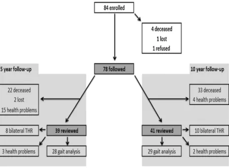
We reviewed 39 patients (72 % women) clinically and radiologically at five years (range 5.1–7.0) follow-up (Fig. 1). Of these patients, 22 (28 %) had died for reasons
unrelated to the THR, two (3 %) were lost to follow-up and 15 (19 %) were not examined due to health reasons unrelated to the surgery. Gait was analysed in 28 patients with a mean age of 79.2 years (range 67–96). Patients who received a contra-lateral THR during the follow-up period or could not perform
the walking trials due to health reasons unrelated to the THR were excluded from gait analysis. At ten years (range 10.8– 11.8) follow-up, 41 patients (81 % women) were reviewed clinically and radiologically. Gait was analysed in 29 patients with a mean age of 83.4 years (range 72–93) (Fig.1).
Fig. 1 Flow chart presenting the examined patients and patients lost to follow-up during the study
Table 1 Clinical outcomes fol-lowing THR
a
Wilcoxon signed rank test, testing the difference between the variable at 5 and 10 years follow-up
Outcome measure Preoperative
(n =78) 5 years follow-up (n =39) 10 years follow-up (n =41) p valuea WOMAC
Total score, mean (range) 63.0 (31–94) 4.9 (0–23) 10.5 (0–50) p <0.001 Pain subscore, mean (range) 17.3 (10–20) 0.8 (0–6) 0.7 (0–8)
HHS
Total score, mean (range) 43.8 (24–56) 84.7 (43–100) 81.2 (49–100) p <0.16 Distance walked subscore, mean (range) 4.2 (2–5) 8.7 (5–11) 8.1 (0–11)
Pain subscore, mean (range) 10.9 (10–20) 40.1 (10–44) 40.3 (10–44) EQ-5D
VAS, mean (range) 87.5 (75–95) 74.5 (40–100) p =0.003 Level 1 (no problems), %
Mobility 67 50
Self-care 92 86
Usual (daily) activities 69 71
Pain/discomfort 87 82
Anxiety/depression 100 82
Level 2 (some problems), %
Mobility 33 50
Self-care 8 11
Usual (daily) activities 31 27
Pain/discomfort 13 18
Clinical results The EQ-5D, HHS and WOMAC results are summarised in Table 1. The difference between five and ten years follow-up was statistically significant for the WOMAC total score and EQ-5D VAS score, with better scores at five years follow-up.
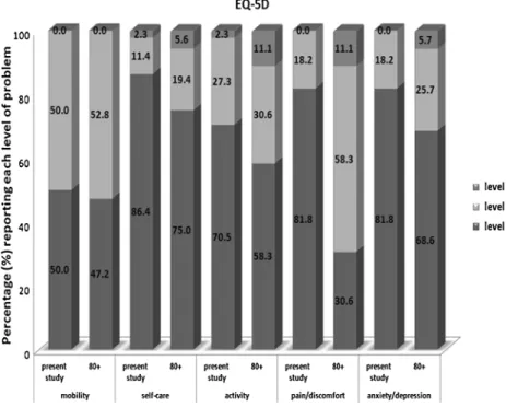
For the EQ-5D, most patients reported no problems (level 1) for the various domains, some reported some prob-lems (level 2) and one patient (2.3 %) reported level 3 probprob-lems for self-care and usual activity. The EQ-5D scores are compared to EQ-5D norm scores in the general population≥80 years [7] in Fig.2.
Total HHS are shown in Fig.3. The mean walked distance was a maximum of 500 metres pre-operatively (4.2 points, range 2–5) and improved to >500 m at five years (8.7 points, range 5––11) and ten years (8.2 points, range 0–11) follow-up. The difference between preoperative and five years follow-up was significant (p <0.05), but between five and ten years follow-up was not (p =0.16). At ten years follow-up, 34 % of THR were rated excellent, and 22 % were rated good.
At ten years follow-up, 95 % of patients were satisfied or very satisfied with the surgery. Pain relief was excellent: 34 patients (83 %) had no pain in the operated hip, five (12 %) had
Fig. 2 The EQ-5D score at 10 years follow-up, compared to the reference population of people age 80 years and older (80+) (EQ-5D norms table for national population [7]). Values are percentages
occasional mild pain (VAS=2 or 3) and one (2 %) complained of occasional moderate pain (VAS=5).
Radiological results At ten years follow-up, mean cup incli-nation was 49.9° (range 36–65°), and mean cup anteversion was 29.2° (range 12–46°). The mean CCD angle was 132.4° (range 123–140°), and mean anteversion of the stem neck was 10.1° (range−4 to 38°).
Radiolucent lines >2 mm were found around the stem in 11 cases (27 %). Stem subsidence occurred in four cases, of which one presented with stem subsidence >20 mm, a distal cement fracture and radiolucent lines at the stem-cement interface. This woman had few symptoms (VAS pain = 0, VAS stiffness = 0) and a good clinical outcome with total WOMAC score of 24 points. There were no cases of calcar resorption greater than grade I and five cases (12 %) of cortical hypertrophy (one patient in Gruen zone 3, three patients in zone 5 and one patient in zones 3 and 5). Hetero-topic ossifications were identified in seven (17 %) patients— six with Brooker grade I and one with Brooker grade III. All patients with the above-described radiological changes were asymptomatic.
Gait analysis Mean spatial and temporal gait parameters at five and ten years follow-up and for the fit elderly and frail elderly reference populations are summarised in Table 2. There was a significant difference between all groups for each parameter (p <0.001). A significant deterioration of every gait parameter was observed between five and ten years follow-up. With advancing age, THR patients walked with slower ca-dence and speed and shortened stride length. Single and double stance phases increased with advancing age. Range of motion (ROM) for each limb segment also decreased with age.
Discussion
Clinical outcome measures and gait analysis results were very good at five and ten years following THR with the cemented Lemania stem. Gait analysis was performed while patients walked freely for 30 metres in a corridor, and the results were compared to reference populations representing two ex-tremes—a fit elderly and a frail elderly population. No other study on outcomes with this anatomic femoral stem has been published to date.
Table 2 Spatial and temporal gait parameters
Double support and stance are expressed as percentage of gait cycle time. Significant differences were observed between each group (Wilcoxon p value < 0.001)
Variable Reference group, fit elderly Patients, 5 years follow-up Patients, 10 years follow-up Reference group, frail elderly Cadence (steps/min) 112.63±10.04 102.77±11.05 99.09±11.88 75.63±14.78 Stance (%) 59.02±2.09 61.52±2.49 62.48±2.45 67.79±5.41 Double support (%) 18.03±4.18 23.03±4.97 24.96±4.91 36.06±10.55 Stride length (m) 1.28±0.11 1.13±0.18 1.00±0.15 0.81±0.17 Speed (m/s) 1.20±0.16 0.97±0.20 0.83±0.15 0.52±0.17 Shank (°) 70.57±4.07 65.83±7.95 58.99±6.49 47.80±9.15 Thigh (°) 42.32±4.41 41.82±5.92 34.24±5.20 26.65±5.86 Knee (°) 52.29±5.03 53.85±6.71 46.39±6.40 35.34±7.84
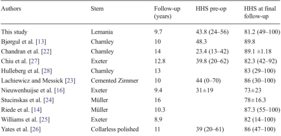
Table 3 Comparison of HHS af-ter THR, at long-af-term follow-up, in the literature
Values are mean (range) in points or mean ± SD
Authors Stem Follow-up
(years)
HHS pre-op HHS at final follow-up
This study Lemania 9.7 43.8 (24–56) 81.2 (49–100)
Bjørgul et al. [13] Charnley 10 48.3 89.8
Chandran et al. [22] Charnley 14 23.4 (13–42) 89.1 ±1.18 Chiu et al. [27] Exeter 12.8 39.8 (20–62) 82.3 (42–92)
Hulleberg et al. [28] Charnley 13 83 (29–100)
Lachiewicz and Messick [23] Cemented Zimmer 10 44 (0–70) 86 (30–100)
Nieuwenhuijse et al. [16] Exeter 9.4 31±19 73±23
Stucinskas et al. [24] Müller 16 78±16.3
Riede et al. [14] Müller 10.3 87.3 (55–100)
Williams et al. [25] Exeter 8.9 82 (14–100)
Ten years following THR, the EQ-5D score was excellent, with a mean VAS of 74.5 points, compared to 58 points in the age-matched healthy reference population [7]. Our study’s
patients reported fewer difficulties for each dimension: 82 % presented with no pain versus 31 % of the reference popula-tion, and 71 % reported no difficulty in usual activities versus 58 % of the reference population. HHS and WOMAC scores improved significantly compared to baseline (Fig. 3). The magnitude of HHS improvement was comparable to that
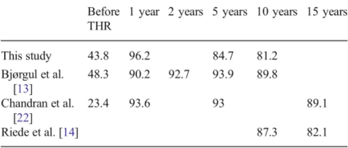
reported for other prostheses with similar follow-up (Table3). Both HHS and WOMAC scores declined slowly during the follow-up period (Fig.3). Patients remained pain free over the long term, despite a gradual functional decline. Other studies have noted a gradual decline in HHS following THR (Table4), at an average of 0.7 points per year with no medical compli-cations [12]. Deterioration of functional scores appears to correspond to a decline in general health due to aging. The use of instruments other than the HHS to assess activity level and walking ability in THR patients has been recommended [13,14].
The Lemania stem demonstrated good radiological results at ten years follow-up, with radiolucent lines at the cement-bone interface in 11 patients (27 %), all asymptomatic. Linear radiolucency and isolated bone lysis at the cement-bone inter-face have been reported in up to 30 % of cases at long-term follow-up [14] and do not necessarily indicate loosening [15]. We found stem subsidence in four asymptomatic patients, which is often the case in this type of prosthesis design [16]. This finding validates the design of the Lemania stem to optimally fill the proximal metaphysis and improve proximal load transfer to the bone.
Table 4 Long-term evolution of the HHS following THR in the literature Before
THR
1 year 2 years 5 years 10 years 15 years
This study 43.8 96.2 84.7 81.2 Bjørgul et al. [13] 48.3 90.2 92.7 93.9 89.8 Chandran et al. [22] 23.4 93.6 93 89.1 Riede et al. [14] 87.3 82.1
Values are mean in points
Objective gait analysis is useful for locomotor function evaluation after THR. Most studies evaluated gait at short-term follow-up, with contradictory findings. Petersen et al. [17] compared operated and non-operated hips at 12 weeks post-operatively and reported an asymmetric gait pattern, with significantly less power produced by muscles around the operated hip. Nantel and colleagues [18] observed a return to normal walking one to four years after THR, where-as Perron et al. [19] failed to show normal gait at short-term follow-up. Kyriazis and Rigas [9] found significant differ-ences in gait at ten years follow-up in THR patients and controls, suggesting that patients who have undergone THR do not return to the same normal gait pattern as healthy, age-matched controls. We found significant differences be-tween the THR and reference groups in every analysed tem-poral and spatial parameter at five and ten years follow-up, with significant deterioration of every gait parameter be-tween five and ten years (Fig.4). Our study’s patients had
better gait performance than the frail elderly, but never achieved the level of the fit elderly patients. The differences were small and likely difficult to elucidate by physical ex-amination only. However, objective gait analysis demon-strated statistically significant differences and confirmed its utility as a complement to traditional evaluation methods.
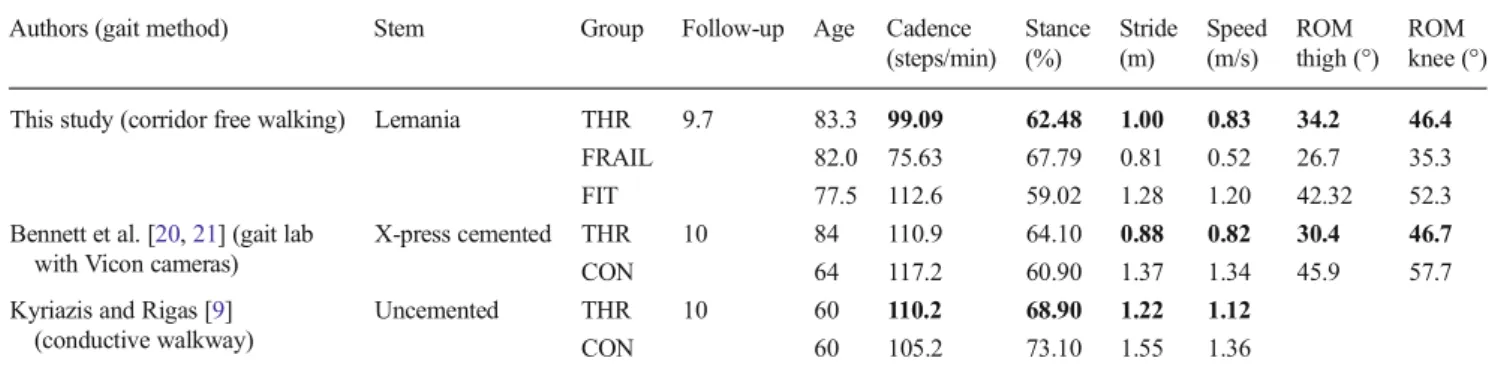
Three studies analysed gait parameters of THR patients at a follow-up of at least ten years (Table5) [9,20,21]. None had patients walking freely in a corridor (without cameras or specific walkways) as in our study. The observed decreases in cadence, stride length, speed and all ROM from five to ten years follow-up in our study are similar to those reported by Bennett et al. [20,21]. However, THR patients in our study walked faster and with a greater stride length (Table5). They also had a better thigh ROM, despite a similar mean age. In contrast, our patients had a worse gait pattern than that report-ed by Kyriazis and Rigas [9], with slower speed, slower cadence and shorter stride length (Table5). Comparison of these studies is difficult, due to the substantially younger
patient population in the study by Kyriazis and Rigas (60 years versus 83 years in our cohort). Our good clinical and radio-logical results suggest that the deterioration in gait pattern over time is related to advanced age rather than THR.
A strength of this study is that gait analysis was performed over a relatively long distance of 30 metres, with two walking trials, which allowed recording of real walking performance and minimised the effect of better initial gait values produced by a rested patient. Bennett et al. [20,21] performed their gait analysis at one self-selected speed on a walkway with six cameras. Kyriazis and Rigas [9] used a conductive walkway. More age-matched controlled trials with a standardised gait analysis are required for a better understanding of long-term changes in gait patterns after THR and to improve comparison of results.
In conclusion, THR with the Lemania stem demonstrated good, stable clinical and radiological results at ten years follow-up in a relatively old patient group. Our results are comparable to those of primary THR with other cemented systems such as Müller or Exeter. Patients demonstrated good walking performance ten years following Lemania THR. Temporal and spatial gait parameters were better than those of frail, matched patients and close to those of a fit, age-matched population, as measured in a real-life environment. Clinical outcome scores and gait parameters decrease progres-sively in patients≥80 years of age, likely due to the effect of advancing age rather than THR.
Acknowledgments The authors thank Professor Christophe Büla, M.D., Service of Geriatric Medicine and Geriatric Rehabilitation, University of Lausanne, for providing the gait analysis results from patients in the rehabilitation centre, and P. Morel for assistance with data collection. We also thank Angelina Poloni for administrative assistance and Dagmar Gross for assistance with preparation of the manuscript. The Interinstitutional Center of Translational Biomechanics (CBT-EPFL, Switzerland) provided funding for this project.
Conflict of interest B. Jolles is a consultant for Symbios Orthopédie SA. The remaining authors declare that they have no conflict of interest. Table 5 Comparison of chosen gait parameters to other studies
Authors (gait method) Stem Group Follow-up Age Cadence (steps/min) Stance (%) Stride (m) Speed (m/s) ROM thigh (°) ROM knee (°) This study (corridor free walking) Lemania THR 9.7 83.3 99.09 62.48 1.00 0.83 34.2 46.4
FRAIL 82.0 75.63 67.79 0.81 0.52 26.7 35.3
FIT 77.5 112.6 59.02 1.28 1.20 42.32 52.3
Bennett et al. [20,21] (gait lab with Vicon cameras)
X-press cemented THR 10 84 110.9 64.10 0.88 0.82 30.4 46.7
CON 64 117.2 60.90 1.37 1.34 45.9 57.7
Kyriazis and Rigas [9] (conductive walkway)
Uncemented THR 10 60 110.2 68.90 1.22 1.12
CON 60 105.2 73.10 1.55 1.36
Boldface indicates statistical difference between THR group and control group. Values for follow-up and age are mean in years
THR total hip replacement group, FRAIL our reference population of 72 elderly frail patients, FIT our reference population of fit patients, CON control group, ROM range of motion
References
1. Berry DJ, Harmsen WS, Cabanela ME, Morrey BF (2002) Twenty-five-year survivorship of two thousand consecutive primary Charnley total hip replacements: factors affecting survivorship of acetabular and femoral components. J Bone Joint Surg Am 84-A: 171–177
2. Espehaug B, Furnes O, Engesaeter LB, Havelin LI (2009) 18 years of results with cemented primary hip prostheses in the Norwegian Arthroplasty Register: concerns about some newer implants. Acta Orthop 80:402–412
3. Murray DW, Carr AJ, Bulstrode CJ (1995) Which primary total hip replacement? J Bone Joint Surg Br 77:520–527
4. Rubin PJ, Rakotomanana RL, Leyvraz PF, Zysset PK, Curnier A, Heegaard JH (1993) Frictional interface micromotions and anisotrop-ic stress distribution in a femoral total hip component. J Biomech 26(6):725–739
5. Harris WH (1969) Traumatic arthritis of the hip after dislocation and acetabular fractures: treatment by mold arthroplasty. An end-result study using a new method of result evaluation. J Bone Joint Surg Am 51:737–755
6. Bellamy N, Campbell J, Stevens J, Pilch L, Stewart C, Mahmood Z (1997) Validation study of a computerized version of the Western Ontario and McMaster Universities VA3.0 Osteoarthritis Index. J Rheumatol 24:2413–2415
7. Rabin R, de Charro F (2001) EQ-5D: a measure of health status from the EuroQol Group. Ann Med 33:337–343
8. Lindemann U, Becker C, Unnewehr I, Muche R, Aminin K, Dejnabadi H, Nikolaus T, Puhl W, Huch K, Dreinhöfer KE (2006) Gait analysis and WOMAC are complementary in assessing func-tional outcome in total hip replacement. Clin Rehabil 20:413–420 9. Kyriazis V, Rigas C (2002) Temporal gait analysis of hip
osteoar-thritic patients operated with cementless hip replacement. Clin Biomech (Bristol, Avon) 17(4):318–321
10. Aminian K, Trevisan C, Najafi B, Dejnabadi H, Frigo C, Pavan E, Telonio A, Cerati F, Marinoni EC, Robert P, Leyvraz PF (2004) Evaluation of an ambulatory system for gait analysis in hip osteoar-thritis and after total hip replacement. Gait Posture 20:102–107 11. Brooker AF, Bowerman JW, Robinson RA, Riley LH Jr (1973)
Ectopic ossification following total hip replacement. Incidence and a method of classification. J Bone Joint Surg Am 55:1629–1632 12. Ritter MA, Thong AE, Davis KE, Berend ME, Meding JB, Faris PM
(2004) Long-term deterioration of joint evaluation scores. J Bone Joint Surg Br 86:438–442
13. Bjørgul K, Novicoff WM, Andersen ST, Brevig K, Thu F, Wiig M, Ahlund O (2010) No differences in outcomes between cemented and uncemented acetabular components after 12–14 years: results from a randomized controlled trial comparing Duraloc with Charnley cups. J Orthop Traumatol 11:37–45
14. Riede U, Lüem M, Ilchmann T, Eucker M, Ochsner PE (2007) The M.E Müller straight stem prosthesis: 15 year follow-up. Survivorship and clinical results. Arch Orthop Trauma Surg 127:587–592 15. Acklin YP, Berli BJ, Frick W, Elke R, Morscher EW (2001)
Nine-year results of Müller cemented titanium Straight Stems in total hip replacement. Arch Orthop Trauma Surg 121:391–398
16. Nieuwenhuijse MJ, Valstar ER, Kaptein BL, Nelissen RG (2012) The Exeter femoral stem continues to migrate during its first decade after implantation: 10–12 years of follow-up with radiostereometric anal-ysis (RSA). Acta Orthop 83:129–134
17. Petersen MK, Andersen NT, Mogensen P, Voight M, Søballe K (2011) Gait analysis after total hip replacement with hip resurfacing implant or Mallory-head Exeter prosthesis: a randomised controlled trial. Int Orthop 35:667–674
18. Nantel J, Termoz N, Vendittoli PA, Lavigne M, Prince F (2009) Gait patterns after total hip arthroplasty and surface replacement arthroplasty. Arch Phys Med Rehabil 90:463–469
19. Perron M, Malouin F, Moffet H, McFadyen BJ (2000) Three-dimensional gait analysis in women with a total hip arthroplasty. Clin Biomech (Bristol, Avon) 15:504–515
20. Bennett D, Humphreys L, O’Brien S, Kelly C, Orr JF, Beverland DE (2008) Gait kinematics of age-stratified hip replacement patients–a large scale, long-term follow-up study. Gait Posture 28:194–200 21. Bennett D, Humphreys L, O’Brien S, Orr J, Beverland DE (2009)
Temporospatial parameters of hip replacement patients ten years post-operatively. Int Orthop 33:1203–1207
22. Chandran P, Azzabi M, Miles J, Andrews M, Bradley J (2010) Furlong hydroxyapatite-coated hip prosthesis vs the Charnley cemented hip prosthesis. J Arthroplasty 25:52–57
23. Lachiewicz PF, Messick P (2003) Precoated femoral component in primary hybrid total hip arthroplasty: results at a mean 10-year follow-up. J Arthroplasty 18:1–5
24. Stucinskas J, Clauss M, Tarasevicius S, Wingstrand H, Ilchmann T (2012) Long-term femoral bone remodeling after cemented hip arthroplasty with the Müller straight stem in the operated and nonoperated femora. J Arthroplasty 27:927–933
25. Williams HD, Browne G, Gie GA, Ling RS, Timperley AJ, Wendover NA (2002) The Exeter universal cemented femoral com-ponent at 8 to 12 years. A study of the first 325 hips. J Bone Joint Surg Br 84:324–334
26. Yates PJ, Burston BJ, Whitley E, Bannister GC (2008) Collarless polished tapered stem: clinical and radiological results at a minimum of ten years’ follow-up. J Bone Joint Surg Br 90:16–22
27. Chiu KH, Shen WY, Cheung KW, Tsui HF (2005) Primary exeter total hip arthroplasty in patients with small femurs: a minimal of 10 years follow-up. J Arthroplasty 20:275–281
28. Hulleberg G, Aamodt A, Espehaug B, Benum P (2008) A clinical and radiographic 13-year follow-up study of 138 Charnley hip arthroplasties in patients 50–70 years old: comparison of university hospital data and registry data. Acta Orthop 79:609–617