HAL Id: tel-01751427
https://tel.archives-ouvertes.fr/tel-01751427
Submitted on 29 Mar 2018HAL is a multi-disciplinary open access
archive for the deposit and dissemination of sci-entific research documents, whether they are pub-lished or not. The documents may come from teaching and research institutions in France or abroad, or from public or private research centers.
L’archive ouverte pluridisciplinaire HAL, est destinée au dépôt et à la diffusion de documents scientifiques de niveau recherche, publiés ou non, émanant des établissements d’enseignement et de recherche français ou étrangers, des laboratoires publics ou privés.
Role of the Glycogen Synthase Kinase 3 for the Retinal
Development and Homeostasis
François Paquet-Durand
To cite this version:
François Paquet-Durand. Role of the Glycogen Synthase Kinase 3 for the Retinal Development and Homeostasis. Neurobiology. Université Paris Saclay (COmUE), 2018. English. �NNT : 2018SACLS060�. �tel-01751427�
Role of the Glycogen Synthase Kinase 3
in Retinal Development and Homeostasis
Rôle de la Glycogène Synthase Kinase 3 dans le
Développement et l'Homéostasie de la Rétine
Thèse de doctorat de l'Université Paris-Saclay
préparée à l’Institut des Neurosciences Paris-Saclay
UMR9197
École doctorale n°568 BIOSIGNE
Spécialité de doctorat: Sciences de la vie et de la santéThèse présentée et soutenue à Orsay, le 22 mars 2018, par
Elena Braginskaja
Composition du Jury:
Pr. Simon Saule Président
Pr. François Paquet-Durand Rapporteur
Pr. Thomas Lamonerie Rapporteur
Dr. Marie-Paule Felder-Schmittbuhl Examinatrice
Dr. Gaël Orieux Examinateur
Dr. Muriel Perron Directrice de thèse
Dr. Jérôme Roger Co-Directeur de thèse
NNT
:
20
18
S
A
CL
S
06
0
To My Parents,
To Lutz Neumann
I
ACKNOWLEDGEMENTS
My 4-year adventure in France comes to the end with the accomplishment of my Ph.D. studies. Before heading for an “old-new life” in my home country, I would like to thank all the people, who accompanied and supported me in my journey that started with the Erasmus exchange in 2014, followed by my Master internship and finally my Ph.D. thesis. I would like to start by expressing my gratitude to my thesis director Muriel Perron. Thank you so much for the scientific growth I experienced through all the years on your team: First during my Erasmus internship, then during my Master internship and the Ph.D. thesis. I am very grateful to you for all your support and guidance and I appreciate the time you dedicated to improving my manuscript.
Especially, I would like to deeply thank my thesis director Jérôme Roger. First, for the great opportunity and trust to work on this project during my Master internship and then continuing on Ph.D. thesis. Thank you for providing me with invaluable guidance, constructive feedback and supporting me at each stage of my work and beyond, even when it was the hardest. Thank you for the time dedicated to correcting my manuscript. Finally, I hope that our hard work could reward us with fruitful results in the (very) near future.
I wish to acknowledge Pr. Simon Saule for the honour of presiding my Jury. I would like to thank Pr. François Paquet-Durand and Pr. Thomas Lamonerie for accepting to evaluate my work as well as Dr. Marie-Paule Felder-Schmittbuhl and Dr. Gaël Orieux who have accepted to be my examinators.
Furthermore, I am grateful to Dr. Célio Pouponnot and Dr. Thierry Léveillard for having evaluated my work during my mid-term examination and to Dr. Thierry Léveillard for the tutorship of my Ph.D. work.
I would like to thank the all the team of the Ernst Ludwig Ehrlich Studienwerk, and especially Dr. Eva Lezzi and Pr. Vingron for all the support and regular exchange during the past years, and for all the seminars, workshops and meetings. Furthermore, I would like to thank ELES for the financial support that allowed me to continue my Ph.D. I would also like to thank the Studienstiftung des deutschen Volkes, in particular the Max-Weber Program Bayern for sponsoring seminars as well as scientific and linguistic workshops and summer schools during my Master and Ph.D. studies. A particular thought to all the great people I had the chance to meet and with whom I exchanged through these events.
I would like to thank the team of Pierre-Marie Lledo, and notably Françoise and Marie with whom I accomplished my second Erasmus internship. Thank you for the great exchange and everything I could learn from you.
I am grateful to all the people in the animal facility at Gif for taking care of the animals, and in particular Krystel, Valérie and Xavier. Also, I am thankful to our “neighbors”: Vero, Sandrine, Jean-Vianney, Kevin and Nathalie for their availability and kindness.
I would like to express my gratitude to our collaborators, members of Anand Swaroop’s lab at the NIH in the US lab. Thank you particularly for welcoming me in such a friendly atmosphere in the US during the ARVO conference 2017. Furthermore, I thank Robin from Alain Chédotal lab at the Vision Institute for our fruitful collaboration on one of my projects.
III
I deeply thank all members of the CERTO and SCaNR team for great teamwork, for help and discussion and a friendly atmosphere.
More personally, I would like to thank Odile for her friendliness and valuable advice during the meetings and Morgane for our discussions on the way to the lab 😉 as well as for all the valuable advice.
I profoundly thank Tatiana for always being there when needed, for our long discussions in Russian 😊 and all for all her encouragements.
Of course, I would like to express my gratitude to Christel. Thank you for being kind and supportive at all times.
I am grateful to Karine, my closest neighbor in our office 😊 Thank you for our agreeable discussions, and your friendliness and support!
I thank Sophie for her support with experiments, but also for her kindness and our nice discussions. I thank Elodie for her help, and in particular during my Master internship and my first Ph.D. year. I appreciated our agreeable discussions during the ERG experiment. I thank Alicia for always being joyful and smiling, Diana (although you could talk a little bit slower in Spanish for me 😉) and Divya.
Expressly, I wish to say a big “thank you” to Annaïg and Juliette. Annaïg, my «forerunner», thank you for everything, all the support, but also the fun we had during the years. I would explicitly like to mention your active participation in my journey of MT180 😊. And Juliette, certainly I thank you for all the valuable scientific advice and guidance (thank you for having taught me many molecular and cellular biology methods), but especially for your constant encouragements and your enormous support in most, if not all, difficult situations and moments I experienced!
Another big “thank you” I would like to express to the “other” men in our lab: Albert and Rodrigo 😊 Albert, thank you for being so easy-going, supportive and amicable! Thank you for your friendship and our discussions inside and outside the lab. Rodrigo, thank you for all the support and for simply being there, my friend! Furthermore, I thank Rahul, who has already left the lab, with whom my journey started on the 31st March 2014, for managing
the obstacles, but also for sharing plenty of good moments together.
Finally, I would like to thank all the students who were doing their internship with me during my Master and Ph.D.: Megan, Claire, Nareh, Louise, Alexana and Leah, but also Elise who came during the last month of my Ph.D. In particular, my friend Louise for her support during her (unfortunately) short stay in the lab, and Megan, Leah and Elise for their help with English.
V
My acknowledgements would not be complete without mentioning all my wonderful and very international family members and friends. I warmly thank all my friends I found in France. In particular, my very first friend, Benoît for having discovered Paris with me and for still keeping in touch. Guillaume, my faithful friend, for all our adventures in France and abroad. Koen for our German-Dutch exchange and the fun in the polyglot club that facilitated me my last weeks of work on this manuscript 😊. Giovanni and Tito, my “big brothers” in France, true friends and supporters during the years, who are always there. Furthermore, I thank all my other friends spread all across the globe for keeping in touch!
I deeply thank my family, my dearest parents, my brother Ilia, his wife Oksana as well as my niece Veronika and my nephew Netanel for their unwavering love and support despite the great distance between us. I thank all my other family members, most of whom are (very) far, for the regular contact and constant support!
And Steven. Thank you for all the joy you bring to my life and everything to come!
Last but not least (and the most important comes at the end) I would like to express my immense gratitude to Lutz, my pillar, my friend, my everything. Words are not enough to describe my gratitude to you. It is to you that I dedicate my thesis.
1
Contents
A Résumé détaillé ... 3
B Introduction ... 9
B.1 Visual perception ... 9
B.1.1 Anatomy of the eye ... 9
B.1.2 Organization of the retina... 9
B.2 Retinal dystrophies and available treatments ... 21
B.2.1 Photoreceptor dystrophies ... 21
B.2.2 Available treatments ... 27
B.3 Development of the vertebrate retina ... 33
B.3.1 Retinal specification ... 33
B.3.2 Retinal proliferation maintenance of progenitors ... 35
B.3.3 Retinal differentiation and the competence model ... 43
B.3.4 Posttranslational modifications ... 67
B.4 Glycogen synthase kinase 3 alpha and beta ... 71
B.4.1 GSK3 regulation and mode of action ... 71
B.4.2 GSK3 function in the nervous system ... 75
B.5 Objective of my Ph.D. project... 99
C Results ... 101
C.1 GSK3 function in retinal progenitors ... 101
C.1.1 Main results ... 103
C.1.2 Article n°1 ... 105
C.2 GSK3 function in retinal photoreceptors ... 181
C.2.1 Main results ... 183
C.2.2 Article n°2 ... 185
D Discussion and perspectives ... 279
D.1 GSK3 in the context of retinal development ... 279
D.1.1 Short-term perspectives ... 279
D.1.2 Long-term perspectives ... 289
D.2 GSK3 in the context of stress and disease... 295
3
A
Résumé détaillé
Le développement et le maintien des photorécepteurs sont principalement contrôlés par une combinaison de facteurs de transcription, nécessitant une régulation fine de leur activité selon les stades de développement ou le contexte environnemental. Outre la régulation du niveau de l’expression de ces facteurs, il existe une modulation fine de leur activité via des modifications post-traductionnelles (MPTs). Celles-ci ont la particularité de moduler rapidement la fonction des protéines dans la cellule. Ainsi, mon directeur de thèse et d’autres équipes ont montré par exemple que la SUMOylation de NRL et NR2E3, deux facteurs de transcription indispensables au développement des photorécepteurs, est nécessaire à leur fonction dans la différenciation des bâtonnets. La phosphorylation est un autre type de MPTs de première importance. Des expériences in vitro montrent que la phosphorylation de NRL est nécessaire à la régulation de l’expression de certains de ses gènes cibles comme la Rhodopsine, mais à ce jour la fonction des isoformes phosphorylées n’est pas connue in vivo. Cependant, et de manière intéressante, des mutations empêchant la phosphorylation de NRL conduisent à des rétinites pigmentaires chez l'homme. Plusieurs mutations conduisant à des rétinites pigmentaires chez l’homme ont été identifiées dans des gènes impliqués dans la régulation des MPTs ou bien altérant la capacité des protéines à être modifiées. Malgré l’importance de ces processus de régulation post-traductionnelle, les mécanismes sous-jacents et les conséquences fonctionnelles sont encore peu étudiés et en particulier in vivo. Dans ce contexte, mon projet de thèse se focalise sur des enzymes essentielles au développement et au fonctionnement du système nerveux, les Glycogène Synthase Kinases 3 (GSK3), qui modifient l’activité de leurs protéines cibles par phosphorylation. Ces kinases sont impliquées dans la régulation de nombreuses voies de signalisation et leur dérégulation a été observée dans de nombreuses pathologies neurodégénératives du système nerveux central. Ce lien potentiel entre les GSK3 et la neurodégénérescence, ainsi que l’importance des MPTs dans les photorécepteurs, nous a logiquement amené à nous interroger sur leurs rôles dans la survie des photorécepteurs. L'objectif de ma thèse était d'étudier le rôle de ces kinases au cours du développement et de l'homéostasie rétinienne. En utilisant les souris invalidées de manière conditionnelle spécifiquement dans les progéniteurs rétiniens (Gsk3fl/flGsk3fl/fl-Cre), j’ai montré que
l'absence totale de Gsk3 et de Gsk3 très tôt au cours du développement rétinien entraîne une microphtalmie chez l'adulte, due à des défauts sévères de prolifération et de différenciation conduisant à la mort des progéniteurs. Ces données suggèrent que les kinases GSK3s sont essentielles au maintien des progéniteurs rétiniens.
5
Les deux kinases jouent des rôles redondants puisque l'expression d'un seul allèle Gsk3 est suffisante pour prévenir le phénotype de microphtalmie (Gsk3fl/+Gsk3fl/fl-Cre ou
Gsk3fl/flGsk3fl/+-Cre). Cependant, une analyse phénotypique approfondie dans ce
contexte génétique (un seul allèle Gsk3) a révélé une forte augmentation du nombre de cellules ganglionnaires déplacées (dRGCs) dans la couche nucléaire interne, qui apparaissent normalement en très faible nombre dans une rétine sauvage. Si la fonction de ces cellules reste encore à déterminer, j’ai pu montrer qu’elles sont produites en même temps que les autres cellules ganglionnaires. Le traçage des axones des cellules ganglionnaires par marquage antérograde a mis en évidence des projections dans un noyau ipsilatérale qui n’est pas visible chez les animaux sauvages. Dans l’ensemble, ces données suggèrent que les kinases GSK3s sont impliquées dans la genèse des dRGCs, dont les projections au cerveau sont particulières et dont la fonction est méconnue. Compte tenu du nombre important de dRGCs dans le modèle génétique que j’ai développé par rapport à la situation normale, ce modèle offre par conséquent un outil de choix pour étudier l’ontogenèse et la fonction de ces cellules.
Mes travaux de thèse se sont ensuite concentrés sur le rôle de GSK3 dans les photorécepteurs. En effet, des défauts de développement ou leur mort est l’une des principales causes de dégénérescence rétinienne. Afin de mieux comprendre la fonction de ces kinases dans la maintenance des photorécepteurs, j'ai utilisé des souris invalidées de manière conditionnelle pour Gsk3 et Gsk3 spécifiquement dans les précurseurs des photorécepteurs postmitotiques (Gsk3fl/fl
Gsk3fl/flCrx-Cre). L’analyse phénotypique a révélé que l’absence de GSK3 dans les photorécepteurs conduit à une altération de leur maturation et de leur fonction dès 14 jours après la naissance (P14), suivie de leur dégénérescence progressive à partir de P18. Ces données suggèrent que les kinases GSK3 sont nécessaires à la différenciation et à la survie des photorécepteurs. J’ai alors combiné des analyses transcriptomiques et des approches in vitro et in vivo pour élucider les mécanismes sous-jacents. Les analyses d’expression des principaux facteurs de transcription spécifiques des photorécepteurs ont permis de mettre en évidence que l’expression de NRL (facteur de transcription nécessaire au développement des photorécepteurs de type bâtonnet) est inchangée au niveau transcriptionnel mais augmentée au niveau protéique. J’ai montré in vitro que GSK3 phosphoryle NRL et que cette phosphorylation régule sa stabilité en induisant sa dégradation rapide par le protéasome. Mes données m’ont donc conduit à proposer un modèle selon lequel l’absence de GSK3 dans les photorécepteurs conduit à des défauts de phosphorylation de NRL, augmentant sa stabilité. J’ai également montré que l’absence de GSK3 dans les photorécepteurs conduit à la diminution d’expression d'un sous-ensemble de gènes cibles
7
de NRL, co-régulés par CRX, et impliqués dans le développement et l'homéostasie des photorécepteurs. Mes travaux suggèrent donc que GSK3, via la régulation de la stabilité de NRL, joue un rôle essentiel dans le contrôle de l’expression de gènes nécessaires à la maturation et l'homéostasie des photorécepteurs. Une telle perte de régulation de NRL pourrait être à l’origine de la dégénérescence des photorécepteurs observée chez les patients atteints de rétinites pigmentaires et porteurs de mutations dans NRL connues pour bloquer sa phosphorylation.
8
,
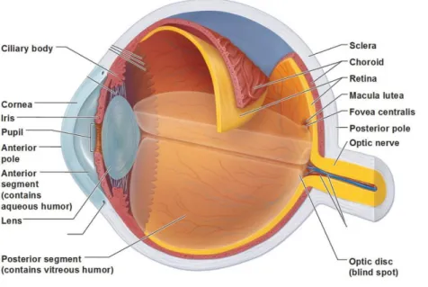
Figure 1. Anatomical organization of the human eye.
➢ The cornea, the most exterior part of the eye, is a protective barrier for the eye. It is mainly responsible for the refraction of the light. It is continuing with sclera in its posterior part.
➢ The second layer is the uvea that provides the eye with oxygen and nutrients. It contains the iris and the ciliary body in its anterior part and the choroid in its posterior part. The iris (greek rainbow) is a “pinhole” spanned around the pupil. It changes its diameter to regulate the amount of light reaching the retina. The ciliary body includes the ciliary muscle that controls the contraction of the lens and the ciliary epithelium that produces the aqueous humor.
➢ The lens plays a major role in the accommodation of the eye and changes its shape by contraction or relaxation of the ciliary muscles adjusting the refraction of light.
➢ The third layer at the back of the eye is the retina. The retina encompasses the retinal pigment
epithelium (RPE) and the neural retina. The function of the retina is to receive the light and transform
it into electrical signals that will be transmitted to the brain. These signals are then processed by brain areas dedicated to process vision information.
➢ Eye chambers embrace the anterior (between cornea and iris) and posterior (between iris, zonule fibers and lens) as well as the vitreous chamber (between the lens and the retina). Anterior and posterior eye chambers are filled with aqueous humor which flows from the posterior to the anterior eye chamber, whereas the vitreous chamber is filled with vitreous humor. The humor is able to refract the light and is therefore part of the optical system. Figure taken from www.eyephysicians.com.
9
B
Introduction
B.1
Visual perception
The surface of our planet Earth is supplied with an abundance of light from the sun. Light is an essential source of energy, but also a major information carrier from our environment. Vision is both a dominant and among the most valuable of our five senses, functioning primarily as a light perception system in animals. It allows us to integrate and process information from our environment, although we can only see “what the mind is prepared to comprehend”.
B.1.1 Anatomy of the eye
The eye is the organ that is responsible for mediating vision, which can be viewed as a connection between the brain and the outside world. It captures visual information and processes it before it is transferred via the optic nerve to the corresponding brain regions and transformed into an image. The eye is composed of three layers, the cornea, the uvea, and the retina, along with the lens and three chambers (Figure 1). This principal arrangement is similar between mice and humans (Figure 1). In the presented thesis manuscript, I will concentrate on the retina, the essential layer that lies at the back of the eye and is responsible for light capture, visual transduction, and processing. For a better understanding of the background and presented results, I will start with a description of the retina including the different retinal cells, with a focus on photoreceptors.
B.1.2 Organization of the retina
The retina is a multilayered tissue in the innermost part of the eye and encompasses the
retinal pigment epithelium (RPE) and the neural retina.
The RPE consists of a monolayer of pigmented epithelial cells with photoreceptors at their apical side and the choroid at their basal side. Due to its melanin synthesis and light absorption, along with its absorption of lipofuscin and photooxidation products, it has a protective function (Strauss, 2005) (Sakina et al., 2013). Furthermore, it establishes tight junctions to contribute to the formation of blood-retinal barrier and secretes immunosuppressive factors such as pro- or anti-inflammatory cytokines and growth factors (Strauss, 2005), both of which are crucial for the immune privilege of the eye. The RPE helps maintain photoreceptor function: It contributes to selective exchange of ions, water and metabolites between the blood vessels of the choroid and the photoreceptors, thus “nourishing” photoreceptors.
10
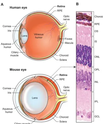
Figure 2. Structure of the human and the mouse eye and the neural retina.
(A) Scheme of the human and mouse eye.Light is focused by optical elements at the neural retina at the back
of the eye. In contrast to the mouse retina, the human retina possesses the macula and the fovea. Relative to the eye size, the lens is much larger in mouse than in the human eye. (B) Hematoxylin and eosin photograph of a mouse retinal section indicating the laminar organization of the retina. RPE, retinal pigment epithelium; OS, outer segment; IS, inner segment; ONL, outer nuclear layer; OPL, outer plexiform layer; INL, inner nuclear layer; IPL, inner plexiform layer; GCL, ganglion cell layer. Figure reproduced from (Veleri et al., 2015).
11
It also sheds and phagocytizes outer segments (OS) of photoreceptors. In fact, around 10 % of OS discs are renewed at the distal end and added to the proximal end by disc shedding whereby a complete renewal of OS occurs in 10 - 15 days (Levine and Green, 2004). The RPE also plays a critical role in the visual cycle by recycling photopigment molecules of photoreceptors after they have been exposed to light. Due to the importance of the RPE for maintenance of photoreceptors, loss of any one of these functions can lead to retinal degeneration, which is associated with vision loss (Strauss, 2005).
The neural retina has a laminar structure comprising seven different types of neurons whose cell bodies are organized into three nuclear layers (Figure 2B).
Laminar organization of the neural retina
➢ The outer nuclear layer (ONL) incorporates photoreceptor cell bodies.
➢ The outer plexiform layer (OPL) encloses synaptic connections established between photoreceptors, bipolar and horizontal cells.
➢ The inner nuclear layer (INL) is composed of the bipolar, horizontal, amacrine and Müller glial cell bodies. Notably, close to the inner plexiform layer, a part of the INL displays nuclei with a larger diameter than the other INL. These cell types have been identified as displaced ganglion cells (Bunt et al., 1974) (Drager and Olsen, 1980).
➢ The inner plexiform layer (IPL) encompasses synaptic connections established between bipolar, ganglion and amacrine cells. The IPL is much thicker than the OPL. ➢ The ganglion cell layer (GCL) includes ganglion cells and displaced amacrine
cells.
B.1.2.1 Cellular organization of the neural retina
In the following section I will describe the different cell types present in the retina (Figure 3), with a particular focus on retinal photoreceptors, the input cells, as well as ganglion cells, the output cells of the retina. These cell types were the focus of my Ph.D. project.
B.1.2.1.1 Retinal photoreceptors
➢ Photoreceptor subtypes
Photoreceptors, the main cells for vision and most abundant in the mammalian retina, can be divided into two main subtypes, rods and cones. They each have distinct features in terms of shape, distribution, the type of photopigment they contain, and their pattern of synaptic connections.
12
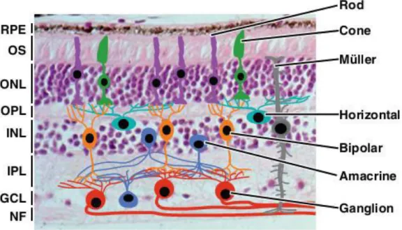
Figure 3. Schematic representation of the cellular organization in the neural retina.
Synaptic connections are established in the outer plexiform layer (OPL) and in the inner plexiform layer (IPL). RPE, Retinal pigment epithelium OS, outer segment; ONL, outer nuclear layer; INL, inner nuclear layer; GCL, ganglion cell layer; NF, optic nerve fiber. Figure reproduced from (Bassett and Wallace, 2012).
Figure 4. Schematic representation of the photoreceptor compartments.
Rod and cone photoreceptors have a distinct compartmentalized morphology, but both contain the same structure. Proteins are transported from the inner to the outer segment via the connecting cilium by intraflagellar transport. Figure taken from (Veleri et al., 2015).
13
• Rod photoreceptors are for scotopic vision under dim light conditions due to their high light sensitivity, but have low spatial resolution as well as a slow response and adaptation (Sato et al., 2015).
• Cone photoreceptors allow color vision and visual acuity under daylight conditions to distinguish features of visual environment at the expense of sensitivity (Sato et al., 2015).
➢ Photoreceptor structure
To sustain the high level of energy required for visual function, photoreceptors are highly differentiated and polarized neurons with a very high metabolic rate. They are composed of five structural and functional regions (Figure 4):
• The outer segment (OS) discs contact the RPE. They are rich in lipids and contain components of the phototransduction system. The OS structure differs between rods and cones. Rod OS have a stack of individualized coin-like membrane discs, whereas cone OS have stacked membranes resulting from continuous invagination of the cell membrane. The OS of both rods and cones consists of hundreds of stacked membranes discs carrying phototransduction proteins including opsins, the visual pigment that defines the photoreceptor subtype. Opsins are G-protein-coupled receptors. They are bound to the light-sensitive chromophore, 11-cis-retinal, that isomerizes to all-trans retinal upon photon capture, initiating the process of phototransduction, a cascade of biochemical events culminating in photoreceptor hyperpolarization and signal transmission. The well-organized OS is however devoid of any protein translation machinery and depends on the inner segment (IS) to synthesize the numerous components that are required for proper phototransduction in the OS.
• The IS are rich in cellular organelles necessary for protein synthesis, energy production and metabolism. In fact, the IS contains the endoplasmic reticulum, the Golgi apparatus and the mitochondria required for glycolysis, oxidative phosphorylation as well as ATP buffering.
• The cilium ensures intraflagellar transport (IFT), a high flow of membranes and proteins that have been synthesized in the IS for their utilization in the OS and is therefore crucial for the survival of photoreceptors. IFT is a bidirectional motility along axonemal microtubules.
• The nucleus is the control center localized in the ONL and surrounded by a very restrained cytoplasm. The different types of photoreceptors show specific chromatin
15
Rod nuclei are denser and have one large clump of heterochromatin, whereas cone nuclei display an irregularly shaped heterochromatin of one to three clumps (Carter-Dawson and LaVail, 1979). To improve light propagation through the retina and night vision, the rod chromatin in mice and other nocturnal mammals is inverted compared to conventional nuclei. Indeed, rod nuclei have heterochromatin centrally and euchromatin peripherally (Solovei et al., 2009).
• The axonal terminals develop a specialized structure called ribbon synapses. A synaptic ribbon is an electron-dense horseshoe-like organelle in the photoreceptor presynaptic cytoplasm and can be observed by electron microscopy. Axon terminals establish synaptic connections in the IPL with second order neurons, bipolar and horizontal cells to which they transmit signals in a graded fashion (tom Dieck and Brandstatter, 2006). Hereby, photoreceptors process large pools of fast-release vesicles tethered to the synaptic ribbons (Sterling and Matthews, 2005). Interestingly, rod photoreceptor terminals contain a single ribbon that has a horseshoe-shaped structure, called spherules (Rao-Mirotznik et al., 1995), whereas cone photoreceptor terminals are larger and contain multiple smaller synaptic ribbons in a single cone photoreceptor axonal terminal, called pedicles.
• Photoreceptor distribution and visual pigments organization in the retina In mammals, photoreceptors account in total for more than 70 % of retinal cells. The distribution or relative density of rods and cones within the retina is adapted to the “lifestyle” of species (Peichl, 2005). The density ratio of cones to rods ranges from 1:200 in the most nocturnal to 20:1 in a few diurnal species. The diurnal to nocturnal repartition of rods versus cones is not the only segregation criteria, as nocturnal rodents, such as mice, possess 97 % of rods and diurnal humans possess 85 % of rods (Jeon et al., 1998) (Swaroop et al., 2010).
Visual pigments are transmembrane proteins, G protein-coupled receptors localized in the
OS of photoreceptors. Opsin is composed of a protein moiety with seven α-helices and its ligand, the aldehyde form of vitamin A, referred to as chromophore retinal. Light rays must cross through non-light sensitive elements of the retina including the retinal vasculature and all retinal layers to reach visual pigments of photoreceptors. The latter are in charge of converting electromagnetic radiation of light photons into neuronal signals and stimulating all the machinery to conduct phototransduction. The vertebrate retina has only one type of rod photoreceptor, carrying the rhodopsin visual pigment with a maximum absorption wavelength of 500 nm.
16
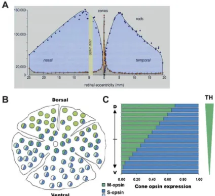
Figure 5. Spatial photoreceptor distribution and opsin expression in human and mouse retina.
(A) In human retina rods (blue) predominate cones (orange) except for the central region, corresponding to the
macula. The fovea, adjacent to the optic disc, marks position 0. (B) In mouse retina, M-cones (green) predominate in the dorsal retina and S-cones (blue) in the ventral retina. (C) M- and S-opsin expression follow opposite gradients, established by the action of the thyroid hormone (TH). Figure reproduced from (Hennig et al., 2008).
17
The type of cones varies between species due to the spectral sensitivity of different opsins. Most birds, fish, some insects, and reptiles are at least tetrachromatic, with at least four types of cones (Goldsmith, 2006) (Hofmann and Carleton, 2009) (Scholtyssek and Kelber, 2017). The fourth cone photoreceptor allows these animals to perceive UV light.(Hisatomi et al., 1996). Humans and Old-World monkeys are trichromats with three distinguishable opsins: L- (long, 564 nm), M- (medium, 533nm) and S- (short, 437 nm) wavelength cones to discriminate between red, green and blue colors, respectively (Hunt et al., 1998) (Lucas et al., 2003). New World monkeys and most mammals, including rodents are dichromats and possess two different cone types: M/L- and S- cones, with the exception of howler monkeys that are uniformly trichromatic (Kainz et al., 1998).
Both Humans and Old-World monkeys, but also New-World monkeys possess a central retinal region, the macula lutea (Hunt et al., 1998). In the center of the macula, cones are concentrated in a cone-enriched area, containing in its center a thin, pit-like, cone-only region, the fovea centralis, the region conferring the highest visual acuity (Figure 2A) (Figure 5A). Rods are excluded from this region and evenly distributed predominating cones elsewhere in the retina (Figure 5B).
In rodents, cones account for 3 % of retinal neurons. Diurnal nil grass rats which possess a cone-rich retina with 35 - 40 % of cones and a higher spatial activity than usual laboratory rodents, are an exception (Gaillard et al., 2008). In contrast to primates, the cone distribution generally differs between rodents. Mice do not possess a cone-rich area, the macula lutea, but their two cone subtypes are organized following inverse dorso-ventral gradients in the retina. Most cones co-express both and M-opsins throughout the retina, however S-cones predominate in the ventral retina, and M-S-cones are denser in the dorsal area (Figure 5B) (Applebury et al., 2000) (Lukats et al., 2005).
B.1.2.1.2 Neurons of the inner nuclear layer
• Bipolar cells receive signals from rods (thus named rod bipolar cells) or cones (thus named cone bipolar cells) and transfer them to cells in the inner retina or directly to the RGCs via their synaptic ribbon terminals (Euler and Schubert, 2015). They comprise at least 13 different subtypes and are excitatory neurons which use glutamate as a neurotransmitter (Euler and Wassle, 1995) (Helmstaedter et al., 2013). The signal from bipolar cells is modulated by other interneurons comprising amacrine and horizontal cells (Diamond, 2017) before being processed by ganglion cells whose axons bundle to form the optic nerve for transmission to the brain.
19
Horizontal cells regulate signal transmission from photoreceptors to bipolar cells by
releasing the inhibitory neurotransmitter -aminobutyric acid (GABA). This mechanism is known as dendritic lateral inhibition, and ensures a better contrast (Kramer and Davenport, 2015).
Amacrine cells constitute the most diversified retinal cell class with at least 28 subtypes.
These subtypes can be classified into two subgroups depending on the neurotransmitter they release: GABAergic or Glycinergic (Wassle et al., 1998) (Kolb et al., 2002). The majority of amacrine cells are located in the proximal part of the INL, and displaced amacrine cells, which encompass 10 different subtypes, are found in the GCL (Galvez et al., 1977) (Perry and Walker, 1980) (Perez De Sevilla Muller et al., 2007).
In contrast to dendritic lateral inhibition of horizontal cells, amacrine cells are responsible for lateral inhibition at axon terminals (Kolb et al., 2002).
Retinal ganglion cells (RGCs) convey visual information from the neural retina to the
brain. Their axons form the optic nerve to transfer signals to the visual cortex in the brain. RGCs have soma with a larger diameter and larger axons than most preceding neurons. More than 20 different subtypes have been identified which differ morphologically in their soma size, dendritic filed size, dendritic ramification and projection level. A recent study revealed even more subgroups of RGCs, at least 32, distinguished based on their light responses and basic anatomical criteria (Baden et al., 2016). To implement binocular vision, RGCs project both to the ipsilateral and contralateral sides of the brain in mammals. In humans, the 45 % of RGCs that are ipsilateral ensure efficient binocular vision. In contrast, mice have very poor binocular vision because of the low amount of ipsilateral-projecting RGCs (3 %) residing exclusively in the ventrotemporal (VT) retina, whereas contralateral RGCs are distributed across the entire retina. Interestingly, the retina also contains
displaced retinal ganglion cells (dRGC) located in the INL. These cells are very rare,
representing only 2 % of RGCs in the retina (Buhl and Dann, 1988) (Nadal-Nicolas et al., 2014). It is believed that dRGCs are misplaced due to an ontogenic aberration rather than representing an independent class of RGCs (Doi et al., 1994) (Buhl and Dann, 1988). However, their exact origin, time of production, projection to the brain, and function have never been described in mammals in the literature due to the absence of suitable models.
21
B.1.2.1.3 Glial cells
Microglia are the principal resident immune cells of the central nervous system with
macrophagic function. Under physiologic conditions, microglia are required for the maintenance of synaptic structure (Wang et al., 2016). Under pathological conditions, such as in glaucoma, microglia become activated and redistributed within the retina (Neufeld, 1999). Müller cells represent the main glial cells in the mammalian retina. They are the only glial cells that share a common embryonic origin with retinal neurons (Turner and Cepko, 1987) (Holt et al., 1988). In response to stress, injury or in retinal diseases, Müller cells and microglia become activated (de Hoz et al., 2016).
B.2
Retinal dystrophies and available treatments
B.2.1 Photoreceptor dystrophies
Retinal neurodegenerative diseases are mainly due to progressive dysfunction of photoreceptors, such as their inability to detect or transmit light to the brain. Photoreceptor degenerative diseases are genetically and clinically heterogeneous and constitute the major cause of blindness. The onset of degeneration can directly affect photoreceptor function and can originate from the RPE.
Retinal degenerative diseases comprise two main forms:
• monogenic (inherited, Mendelian) retinal diseases, such as Retinitis Pigmentosa • multifactorial (complex) diseases such as Age-related Macular degeneration (AMD) Monogenic retinal diseases can be in turn subdivided into non-syndromic types including Retinitis pigmentosa, Leber Congenital Amaurosis (LCA) and Macular Degeneration or syndromic diseases. The latter are characterized by pleiotropic clinical symptoms and affect multiple organs or functions. Examples are the Bardet-Biedl syndrome (affecting retinal function and many other functions) or the Usher Syndrome (affecting retinal and auditory function) (Berger et al., 2010) (Veleri et al., 2015). Up to date, around 300 genes have been mapped and 250 have been identified to cause retinal diseases (Figure 6). They can alter photoreceptor and/or RPE development and key functions such as the visual cycle, phototransduction, gene regulation or transport (Wright et al., 2010).
22
Figure 6. Mapped and identified Retinal Disease Genes between 1980 and 2017.
Progress in the discovery of causal genes for retinal degenerative diseases. In 1996, 19 genes were identified to cause retinal degenerative diseases, in 2017 the gene number reached 256. Figure reproduced from
https://sph.uth.edu/retnet/sum-dis.htm.
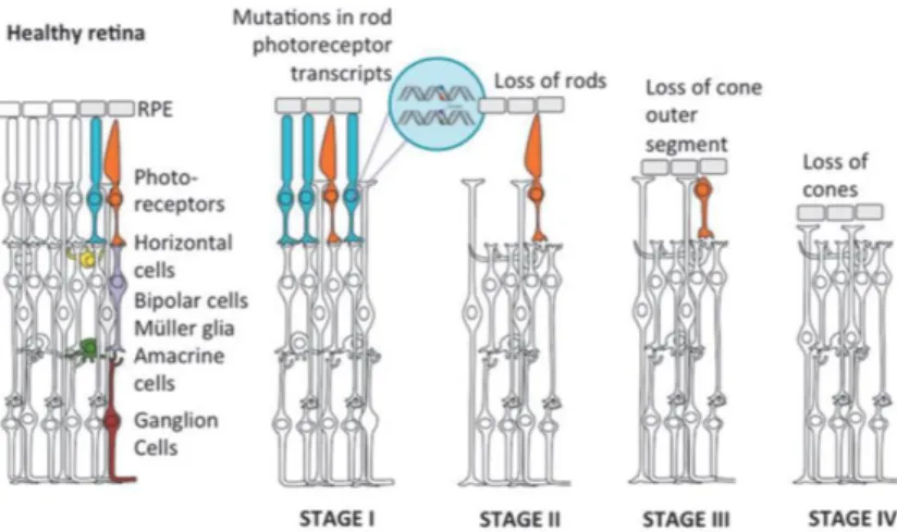
Figure 7. Retinitis pigmentosa as an example of retinal degeneration progression.
Representation of the progressive loss of the photoreceptors occurring in a retina with retinitis pigmentosa. Mutations mostly concern rod photoreceptors (blue) leading to a primary loss of rods (Stage I), followed by progressive cone death (orange, Stages II, III and IV). Figure taken from (Dalkara et al., 2016).
23
Retinitis Pigmentosa, the most common subtype of inherited photoreceptor degeneration,
has a prevalence of 1:3000 to 1:7000 people. There is a large genetic heterogeneity with over 50 genes for which mutations have been identified to cause the disease (Daiger et al., 2013), with autosomal dominant, recessive or X-linked inheritance (Wright et al., 2010). Some genes are mutated very frequently such as Rhodopsin mutations, which occur in 30 % of all cases (al-Maghtheh et al., 1993). The pathology is often characterized by an initial loss of night vision due to the loss of the rod photoreceptors. During disease progression, secondary loss of cone photoreceptors occurs as well, even when the mutation is in a rod-specific expressed gene (Figure 7) (Narayan et al., 2016). Consequently, the daylight vision also becomes affected and the visual field progressively decreases from the periphery to the center, resulting in tunnel vision and ultimately in complete blindness (Nash et al., 2015).
A specific form of Retinitis Pigmentosa is the Cone-rod dystrophy which concerns 1:40000 patients. This form is characterized by an initial loss of cone photoreceptors followed by, or with a concomitant, loss of rod photoreceptors.
Leber Congenital Amaurosis (LCA) and early-onset severe retinal dystrophy (EOSRD)
are the most severe congenital or early-onset diseases. LCA patients present clinical features such as involuntary eye movements, called nystagmus, photophobia, abnormal pupillary response and undetectable or severely abnormal electroretinogram. LCA has prevalence between 1:33,000 to 1:80,000 and accounts for more than 5 % of all inherited retinal degenerations. For instance, mutations in the major ciliary component gene nephrocystin-6 NPHP6, also known as CEP290 are responsible for up to 25 % of Leber Congenital Amaurosis forms and mutations in the RPE65 gene, encoding the enzyme in charge of chromophore recycling, occurs in around 15 % of cases (Morimura et al., 1998) (den Hollander et al., 2008) (Kumaran et al., 2017).
Macular Degeneration is characterized by a loss of the central vision caused by the
progressive death of cone photoreceptors located in the central, cone-only macula, mainly caused by the loss of RPE cells. It can be inherited in a Mendelian manner, and mutations in several genes are associated with the disease (Stone, 2007). The most common is Stargardt disease with a prevalence of 1:10000 patients and mostly caused by mutations in the ABCA4 gene (Allikmets et al., 1997). The ABCA4 gene encodes the cone and rod photoreceptor Rim protein which is a transmembrane transporter of vitamin A intermediates. ABCA4 malfunction leads to the accumulation of yellow-brown pigment granules composed of lipid-containing residues of lysosomal digestionin and around the macula. The retina of Stargardt patients displays cholesterol deposits from unshed photoreceptor OS in the macular region (Molday and Zhang, 2010).
25
Macular Degeneration can also manifest as a complex, multifactorial late-onset disease, then termed Age-related Macular Degeneration (AMD). AMD is the major cause of blindness in industrial countries, with a prevalence of 25 to 30 million people worldwide, and an incidence of 500,000 patients a year. As such, twice as many patients are suffering from AMD than from Alzheimer’s disease (Fletcher et al., 2014). AMD is characterized by formation of lipid- and protein- rich extracellular deposits named drusen beneath the macula (Wang et al., 2010) (Zarubina et al., 2016) (Neely et al., 2017), inflammation with macrophage and microglia infiltration accompanied by complement activation (Parmeggiani et al., 2012) and ultimately degeneration in the macula.
Importantly, since the pioneering discovery of a common polymorphism in the complement factor H gene (Klein et al., 2005), new genetic AMD-associated risk variants are steadily being identified with a total of 20 susceptibility loci found. Together they account for 40 - 60 % of the disease heritability (Zhan et al., 2013) (Fritsche et al., 2014).
In addition to genetics, environmental risk factors such as age, cigarette smoking, blood hypertension, high lipid levels, obesity, and low physical activity contribute to the disease progression. Therefore, AMD patients uniformly suffer from loss of cone photoreceptors in the central macula area, but can exhibit phenotypic heterogeneity (Fritsche et al., 2014). At advanced stages, the central vision is impaired because of the cone loss impairing the visual field.
There are two types of late AMD:
• The wet or exudative AMD (choroidal neovascularization) affects a minority of AMD patients. In wet AMD, abnormal blood vessels grow underneath the retina in the macula zone. The blood vessels leak and eventually damage the retina resulting in vision loss.
• The dry or atrophic AMD affects 80 - 90 % of AMD patients, which is, unlike wet AMD, a slowly progressing AMD form leading in its late stage to so-called geographic atrophy, typically defined as a round or oval area of atrophy of 175 m or more (Sacconi et al., 2017).
27
B.2.2 Available treatments
In total, blinding diseases affect a steadily increasing patient population, currently 34 million people worldwide. Therefore, effective treatment strategies are constantly being developed. They include cell replacement, antibody therapy, gene therapy, neuroprotection, optogenetics and retinal prosthesis (Santos-Ferreira et al., 2016). The most common therapy forms are briefly presented here (Figure 8):
• Antibody-based therapy against vascular endothelial growth factor (VEGF) is widely used to slow down the wet neovascular form of AMD (Krzystolik et al., 2002). Conversely, no existing therapy has been approved for geographic (dry) atrophy (Bandello et al., 2017).
• Cell-based therapy consists of replacing the dysfunctional or degenerated retinal cells, primarily RPE and photoreceptor cells using stem cells. The advantage of this therapeutic approach is its disease independence. Encouraging results have been obtained by transplanting human embryonic stem cell-derived (hESC) RPE and induced pluripotent stem cells (iPSC) RPE (Nazari et al., 2015) (Schwartz et al., 2016). Clinical trials in phase I/II have been initiated to transplant hESC-derived RPE showing medium-term to long-term safety in patients with AMD and Stargardt disease (Schwartz et al., 2015). iPSC-derived RPE has also been transplanted into AMD patients in 2014 and their safety seemed to be confirmed (Takahashi, 2016). However, a serious adverse event in a clinical trial participant, who received iPSC-based therapy, was reported in January 2018. This patient developed retinal edema requiring hospitalization for treatment. A recent study in rats with retinal degeneration demonstrated a better effect in terms of photoreceptor rescue and visual acuity when hESC-RPE cell sheets were grown on a human amniotic membrane compared to hESC-RPE delivered via cell suspension (Ben M'Barek et al., 2017). These findings could influence the production and delivery of hESC-RPE in future clinical trials. One of the special cases of cell therapy is the use of the
neurogenic potential of endogenous cells located in the RPE, the ciliary body or
the Müller cells to regenerate degenerative cells. However, such neurogenic potential and the stem cell properties are extremely limited and inefficient in mammals, in contrast to fish, Xenopus, and other non-mammalian vertebrates (Jayakody et al., 2015) (Hamon et al., 2016).
28
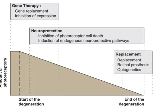
Figure 8. Main therapeutic approaches to treat retinal dystrophies.
Gene therapy has to be used at early stage of the degeneration since it requires viable photoreceptors. Conversely, retinal prosthesis and optogenetics aim at restore vision at later stage when most or all photoreceptors are lost. Neuroprotection has a broader application efficiency window. Figure adapted after (Trifunovic et al., 2012).
29
• Gene therapy uses viral vectors, including in most cases the adeno-associated virus (AAV) vectors, to enable delivery of a functional copy of a gene that is mutated in patients. Most frequently, gene therapy is applied via subretinal injections as most inherited retinal degenerations are caused by mutations in photoreceptors. Cells need to be alive for this approach and therefore an early diagnosis is required (Jayakody et al., 2015) (Dalkara et al., 2016). This approach has causal treatment effect, but, success has been limited to few patients due to the large genetic heterogeneity of retinal disorders. Hence, it cannot be applied to a large number of patients. The first gene therapy started with clinical trials in 2007 with the replacement of the RPE specific protein RPE65 to treat LCA, that accounts for 5 - 10 % of all LCA cases, and was a big success in subsequent clinical trials (Jacobson et al., 2006) (Maguire et al., 2008) (Amado et al., 2010) (Bennett et al., 2016) (Le Meur et al., 2018). RPE65 gene therapy has just been approved in December 2017 by the U.S. Food and Drug Administration under the name of LuxturnaTM to treat
patients with confirmed biallelic RPE65 mutation. While the price of the treatment has been announced at $425,000 per eye, the scientific breakthrough is undeniable. • Neuroprotection aims to administer pro-survival, apoptotic and
anti-inflammatory factors to stop or delay photoreceptor cell death. It can also be achieved by stimulating pro-survival pathways upregulated in degenerative retina but whose endogenous potential is not sufficient to rescue photoreceptors. Structural and functional rescue of retinal cells has been demonstrated for neurotrophic factors such as ciliary neurotrophic factor (CNTF), pigment epithelium-derived factor (PEDF) and glial cell line-epithelium-derived neurotrophic factor (GDNF). In contrast to gene therapy and similar to cell-based therapy, neuroprotective approaches are mutation independent and can be applied to treat a wide range of retinal degenerative diseases. A pioneering clinical trial to deliver CNTF using encapsulated cell implants is currently being pursued in phase III (Tao et al., 2002) (Sieving et al., 2006). This promising approach may have applications beyond diseases caused by genetic mutations. In addition, it avoids secondary effects due to repetitive intraocular injections. Gene therapy approaches aiming to deliver neurotrophic factors using viral vectors have similar advantages (Cuenca et al., 2014) (Dalkara et al., 2016). A prominent candidate is the rod-derived cone viability factor (RdCVF). It has been administered in the form of AAV-RdCVF and prolonged cone survival and function in Retinitis Pigmentosa mice (Byrne et al., 2015).
30
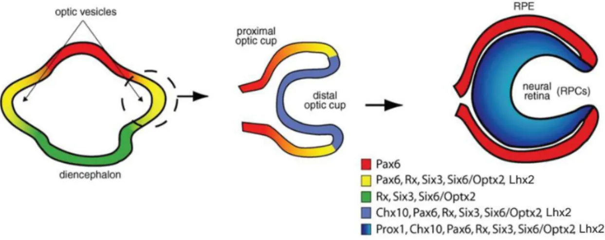
Figure 9. Eye and retina specification involving homeobox gene expression.
The eye field separates bi-laterally from the diencephalon to form the double-layered optic cup: The distal optic cup gives rise to the neural retina and proximal optic cup to the RPE. All transcription factors with the exception of Chx10 and Prox1 are broadly expressed before being restricted to RPCs. Pax6 is the only transcription factor expressed both in RPCs and in the RPE. Figure adapted after (Levine and Green, 2004).
31
Interestingly, additional drug candidates can originate from drug libraries conceived for other indications. For example, olaparib, commonly used for cancer treatment, has shown promising neuroprotective effects in models of retinitis pigmentosa by inhibiting the activity of poly-ADP ribose polymerase (PARP) (Sahaboglu et al., 2016) (Jiao et al., 2016).
• Optogenetics and prosthetic devices constitute an approach to treat severe photoreceptor degeneration with almost no functional cells remaining. For optogenetics, this is achieved via genetically encoded light sensors to photosensibilize retinal cells by expressing one or several genes such as channelrhodopsin from the green alga Chlamydomonas reinhardtii (Nagel et al., 2002). Promising results have been obtained in neurodegenerative mouse models (Cuenca et al., 2014). As for optogenetics, prosthetic devices (epiretinal or subretinal implants) use an artificial photosensor to capture light. The current is used to stimulate retinal cells within the remaining retinal circuit (Mills et al., 2017).
32
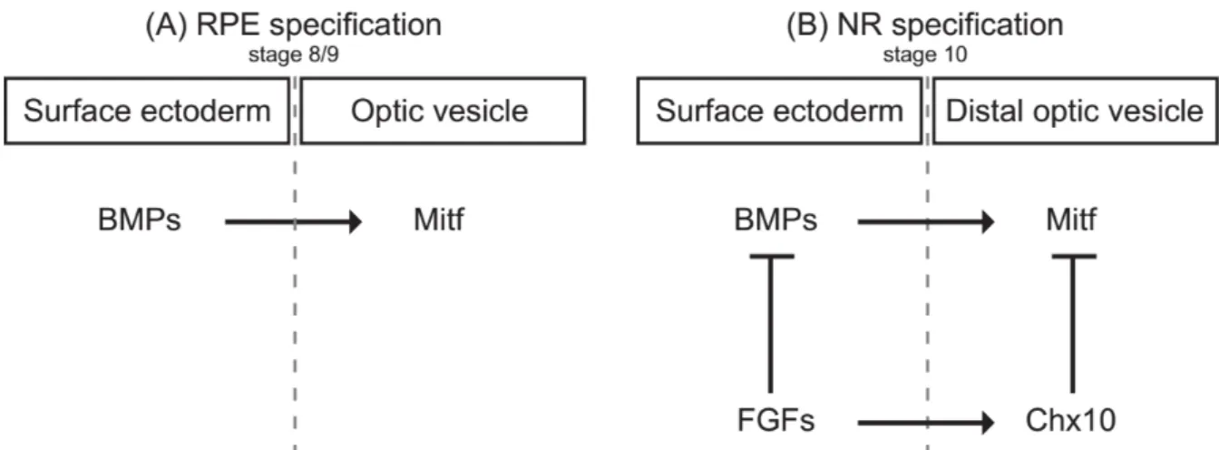
Figure 10. Proposed model for the regulation of the RPE and NR specification.
(A) To achieve RPE specification, BMP induces Mitf expression in the underlying optic vesicle. (B) NR
specification is initiated by FGF induction of Chx10 in the distal portion of the optic vesicle that inhibits Mitf expression. RPE: retinal pigment epithelium; NR: neural retina. Figure adapted after (Muller et al., 2007).
Figure 11. RPCs require Pax6 to exert their retinogenic potential.
Pax6 is required for the expression of different cell type specific transcription factors and generation of different
retinal cell types (left). Upon conditional deletion of Pax6 in RPCs, only NeuroD is expressed, leading to an exclusive generation of amacrine cells (middle). Homozygous Pax6 loss-of-function (Pax6-/-) results in
33
B.3
Development of the vertebrate retina
B.3.1 Retinal specification
Eye formation is initiated after the onset of gastrulation. The eye identity, named “eye field”,
is formed from the neuroepithelium within the midline of the anterior neural plate. The eye field separates bi-laterally to produce the optic primordia that evaginate to form the optic
vesicles (diencephalic evagination). They extend toward the surface ectoderm where the
lens placode develops as well as the primordia of the anterior structures of the eye, such as the cornea. At the same time, the optic vesicles invaginate to form the double-layered
optic cup (Figure 9).The inner layer of the optic cup gives rise to the neural retina and the
non-pigmented region of the ciliary body, while the outer layer becomes pigmented and forms the RPE as well as the pigmented region of the ciliary body (pigmented neuroepithelium) (Chow and Lang, 2001) (Sinn and Wittbrodt, 2013).
Specific transcription factors are decisive in neural retina versus RPE specification (Figure 10). Microphthalmia-associated transcription factor (MITF) triggers gene expression that is important for RPE cell differentiation (Mochii et al., 1998). MITF is itself induced by the bone morphogenetic protein (BMP) (Steinfeld et al., 2017). Another gene important for the RPE melanogenesis is Otx2 (Figure 10) (Martinez-Morales et al., 2003) (Beby and Lamonerie, 2013) (Fuhrmann et al., 2014).
In contrast, fibroblast growth factor (FGF) from the surface ectoderm is decisive for the formation of the neural retina and represses MITF (Pittack et al., 1997) (Nguyen and Arnheiter, 2000). This inhibition is mediated by the expression of the paired-like homeodomain transcription factor Chx10 (Vsx2) (Figure 10) (Horsford et al., 2005).
Interestingly, the homeodomain transcription factor Pax6 holds a dual role in retina versus RPE specification. In combination with MITF it acts as an anti-retinogenic, whereas in combination with retinogenic genes as a pro-retinogenic factor (Bharti et al., 2012).
34
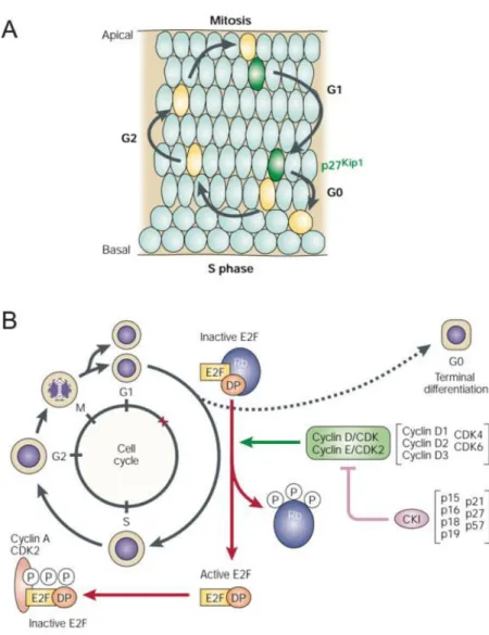
Figure 12. Cell cycle of RPCs.
(A) RPCs nuclei localization changes during the different cell cycle phases; mitosis occurs near the apical side
and S-phase near the basal side. P27/Kip1 is expressed in most RPCs that are exiting the cell cycle. (B) Different proteins intervene at cell cycle progression. The phosphorylation state of Rb dictates whether a RPC will pass the G1/S checkpoint for another cell cycle round or exit the cell cycle and differentiate. If a progenitor remains proliferative, Rb is phosphorylated by a cyclin-dependent kinase (CDK) complex, and thus becomes inactive allowing cell cycle progression. If the cell is going to exit the cell cycle and undergo differentiation, this phosphorylation is blocked by cyclin-kinase inhibitor proteins (CKIs). Hypophosphorylated Rb binds to the E2F/DP heterodimer to prevent transcription of E2F-regulated genes. Figure adapted after (Dyer and Cepko, 2001c).
35
B.3.2 Retinal proliferation maintenance of progenitors
B.3.2.1 Intrinsic factors controlling retinal proliferation
The homeobox factors Pax6, Rax, Six3, Optx2/Six6 are crucial for proliferation and multipotency of RPCs within the developing optic cup (Figure 9) (Levine and Green, 2004). To exemplify the importance of these transcription factors, I will focus on the homeodomain transcription factor Pax6, probably the most studied in eye development and highly conserved across species (Morgan, 2004). Throughout retinogenesis, it is expressed in RPCs (Remez et al., 2017) and has an essential role to regulate transcription factors to generate all retinal cell types (Marquardt et al., 2001). Conditional deletion of Pax6 in RPCs leads to exclusive generation of amacrine cells. However, recent work suggests that Pax6 also inhibits photoreceptor differentiation (Figure 11) (Remez et al., 2017)
During the cell cycle, nuclei of RPCs migrate within the retina, and this phenomenon is referred to as interkinetic nuclear migration (Del Bene, 2011). During the M-phase of the cell cycle, they are located at the apical site of the retina. The S-phase of the cell cycle occurs at the basal site of the retina, and the nuclear migration through the apico-basal axis occurs during the phases G1 and G2 (Figure 12A) (Dyer and Cepko, 2001c) (Del Bene, 2011).
The regulation of the cell-cycle and the participating molecules are evolutionarily conserved, highly coordinated, and precisely regulated. This accurate regulation is necessary to avoid dysplasia or any kind of malignancy (Dyer and Cepko, 2001c). Typically, the cyclins are responsible for cell cycle progression at different stages of the cell cycle and therefore promote cell proliferation. Cyclin D1 forms a complex with CDK4 to phosphorylate and inhibit Rb protein and thus regulates the cell cycle during G1/S transition. Cyclin D1 deficiency slows down the cell cycle time and enhances the cell cycle exit (Dyer and Cepko, 2001c).
Three of seven cyclin inhibitors described in literature (p27, p19, p57) are implicated in retinal development (Dyer and Cepko, 2001a) (Dyer and Cepko, 2001b) (Cunningham et al., 2002). They control the exit from the cell cycle of retinal progenitors in the phase G0/G1. In mouse retina, the main representative p27/Kip1 is expressed throughout retinogenesis in cells that exit the cell cycle and differentiate (Dehay and Kennedy, 2007).
Altogether, the interplay between cyclin D1 and p27 is tightly involved in retinal proliferation and cell cycle exit regulation of RPCs (Figure 12B).
36
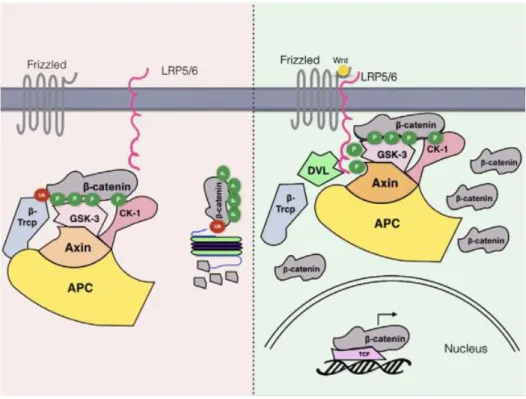
Figure 13. The Wnt signaling pathway.
In absence of Wnt ligands under basal conditions (left), a “destruction complex” of APC, Axin, CK and TrCP and GSK3 is formed. GSK3 becomes activated in this complex and phosphorylates -catenin at the N-terminal
“destruction box” on Thr41 and Ser 37 and 33 after priming phosphorylation of -catenin by casein-kinase 1 (CK1) at Ser 45. Phosphorylation of -catenin creates a recognition site for the E3 ubiquitin ligase TrCP
triggering its degradation by the ubiquitin proteasome system. Upon Wnt ligand stimulation that binds to
Frizzled receptors, Frizzled associates with low-density lipoprotein receptor 5/6 (LRP5/6) (right). Phosphorylation of LRP5/6, achieved by GSK3 and CK1, activates Dishevelled (DVL) and creates high-affinity binding sites for Axin so that the entire complex including GSK3 but excluding TrCP is recruited to the cell membrane. Thus, -catenin is stabilized and accumulates in the cell. Subsequently, -catenin translocates to the nucleus where it binds to TCF/LEF DNA-binding factors facilitating target gene expression involved in cell fate regulation. Figure taken from (Patel and Woodgett, 2017).
37
B.3.2.2 Extrinsic pathways controlling retinal proliferation
RPCs repeat several rounds of division to produce a pool of progenitors before they exit the cell cycle to give birth to different retinal cell types. Pathways to maintain this RPC pool mostly include Wnt, Shh, Notch as well as growth factor signaling. All these signaling pathways involve the regulation by a central node molecule, the Glycogen Synthase Kinase 3, that will be discussed in detail further in the manuscript.
➢ Wnt signaling
The Wnt signaling cascade, involving Wnt ligands (Wnts), evolutionary conserved glycoproteins, occurs in all phyla of the animal kingdom. It is among the most important signaling pathways controlling a large number of neurodevelopmental processes including cell proliferation, polarity, differentiation and neurogenesis during brain development (Figure 13) (Logan and Nusse, 2004).
There are approximately 20 different Wnt ligands binding to 15 different Frizzled receptor subtypes. Wnt binding to Frizzled receptors leads to the accumulation of -catenin and its nuclear translocation. In the nucleus, -catenin interacts with the TCF/LEF family of transcription factors and regulates their target genes. In absence of Wnt, -catenin is phosphorylated by a “destruction complex” composed of multiple proteins, including Axin2 and GSK3 , and targeted for degradation (Loh et al., 2016).
The pro-proliferative role of Wnt, which helps neural progenitors to maintain their undifferentiated state, is widely accepted. However, there is also evidence that Wnts can promote differentiation of stem cells (Otero et al., 2004) (Muroyama et al., 2002) (Inestrosa and Varela-Nallar, 2015). As such, Wnt ligands are involved in many steps and the use of reporter line and genetic tools allowed the identification of some of their function or expression pattern. The Wnt signaling pathway is particularly active in the peripheral embryonic and adult retina, including the CMZ in various species, such as zebrafish, Xenopus, chicken and mice (Denayer et al., 2008) (Kubo and Nakagawa, 2008). The use of transgenic mice reporting Wnt activity (TCF/Lef-LacZ) demonstrated that the highest levels of expression of the reporter gene was in the CMZ (Liu et al., 2003). Similar results were obtained in zebrafish and chicken (Dorsky et al., 2002) (Cho and Cepko, 2006). Specifically, Wnt activity in the retina is particularly well mirrored by Wnt2b expression whose expression is similar to the mentioned reporter gene expression in mice (Liu et al., 2003). Overexpression of Wnt2b inhibits neuronal differentiation and leads to the maintenance of retinal progenitors. Wnt2b loss-of-function suppresses proliferation in the CMZ triggering premature differentiation (Kubo et al., 2003) (Kubo et al., 2005).
38
Figure 14. The Hedgehog signaling pathway.
In absence of Hedgehog ligands (left), under basal conditions, the 12-pass transmembrane receptor Patched represses another 7-pass G-protein coupled receptor-like protein Smoothened. The role of Smoothened is to inhibit the proteolytic cleavage of the effectors of the Hedgehog pathway, the zinc-finger proteins Gli (cubitus interruptus, Ci in flies). Under basal conditions though, Smoothened is repressed allowing sequential phosphorylation of Gli proteins are within their carboxy-terminal domain by PKA, GSK3 and CK which target them for cleavage when bound to Kif7/Cos and SuFu via the protein E3-ligase TrCP. The cleaved Gli transcription factors are transcriptional repressors. The binding of Hedgehog ligand to its receptor Patched releases the inhibition of Smoothened by Patched (right). In its active state, Smoothened promotes dissociation from Kif7/Cos and SuFu releasing full length Gli proteins that translocate to the nucleus where they act as transcriptional activators of cell cycle-related target genes. Figure taken from (Patel and Woodgett, 2017).
39
Based on these results, Wnt2b seems to represent an important factor for neural proliferation.
Strikingly, results obtained in mouse RPCs upon an activation of inactivation of members of the canonical Wnt signaling pathway suggested an inhibitory function of the Wnt signaling on differentiation, without affecting cell cycle exit or proliferation (Ouchi et al., 2005) (Ouchi et al., 2011). Elimination of -catenin or ectopic activation did not alter the proliferation rate of RPCs (Fu et al., 2006).
In summary, Wnt seems to be crucial for maintaining stem cell potential in the CMZ, while it does not appear to have major pro-proliferative function in RPCs.
➢ Hedgehog signaling
Similar to Wnt, the Hedgehog signaling pathway, conserved across species, is a critical regulator of cell fate determination during development. In mammals, Hedgehog ligands include Indian Hedgehog, Desert Hedgehog, and Sonic Hedgehog (Shh). The binding of Hedgehog ligand to its receptor Patched releases the inhibition of Smoothened by Patched. Smoothened activation releases the Gli proteins that translocate to the nucleus where they act as transcriptional activators of cell cycle-related target genes (Figure 14). Most studied Hedgehog target genes are involved in cell cycle regulation, such as cyclins D and E, N-myc, Bcl2, Tbx2, but also its receptor Patched in a negative feedback loop (Patel and Woodgett, 2017).
Several studies have identified Shh signaling as implicated in proliferation of RPCs. For instance, in the embryonic mouse retina, Smoothened ablation in proliferating RPCs dramatically reduces RPCs since RPCs exhibit aberrant expression of cell cycle regulators and a delayed G1/S transition (Sakagami et al., 2009). Therefore, Shh has a profound impact on cell proliferation and progression of the cell cycle. On the contrary, mice with a constitutively active Hedgehog pathway exhibit persistent progenitors in peripheral retinal regions until 3 months of age, reminiscent of the ciliary marginal zone of lower vertebrates (Moshiri and Reh, 2004).
Hedgehog activation during early development in Xenopus or in chicken has been associated with CMZ stem cell proliferation, while inhibition of Hedgehog signaling via cyclopamine, a Smoothened antagonist, results in the opposite effect (Locker et al., 2006) (Moshiri et al., 2005). In summary, Hedgehog signals promote progenitor proliferation in the developing central nervous system.
40
Figure 15. The Notch signaling pathway.
Notch is a ligand-activated transmembrane receptor, expressed on the surface of cells (left), and triggered by direct juxtracrine physical cell-cell contacts, acts as a transcription factor (right). Notch ligands are transmembrane proteins themselves and include Delta-like 1 (Dll1), Jagged and Serrate from neighbouring cells bind. Upon ligand binding (right), two intracellular enzymes ADAM and -secretase are activated to cleave and release notch intracellular domain (NICD). NICD translocates to the nucleus where it binds to the CSL transcription factor to regulate gene expression including the bHLH transcriptional repressors Hes1 and Hes5. These repressors inhibit transcription of proneural genes, and thus promote proliferation of progenitors while inhibiting differentiation into neurons. GSK3 has been shown to bind and phosphorylate NICD probably leading to its degradation. Figure taken from (Patel and Woodgett, 2017).