Review
Iseult Lynch*, Arti Ahluwalia, Diana Boraschi, Hugh J. Byrne, Bengt Fadeel, Peter Gehr,
Arno C. Gutleb, Michaela Kendall and Manthos G. Papadopoulos
The bio-nano-interface in predicting nanoparticle
fate and behaviour in living organisms: towards
grouping and categorising nanomaterials and
ensuring nanosafety by design
Abstract: In biological media, nanoparticles acquire a coating
of biomolecules (proteins, lipids, polysaccharides) from their surroundings, which reduces their surface energy and confers a biological identity to the particles. This adsorbed layer is the interface between the nanomaterial and living systems and therefore plays a significant role in determining the fate and behaviour of the nanoparticles. This review summarises the state of the art in terms of understanding the bio-nano inter-face and provides direction for potential future research and recommendations for future priorities and strategies to support the safe implementation of nanotechnologies. The central premise is that nanomaterials must be studied as biological entities under the appropriate exposure condi-tions and that this should be implemented in study design and reporting for nanosafety assessment. The implications of the bio-nano interface for nanomaterials fate and behav-iour are described in light of four interlinked perspectives: the Coating concept; the Translocation concept; the Signal-ling concept, and the Kinetics concept. A key conclusion is that nanoparticles cannot be viewed as non-interacting species, but rather must be thought of, and studied as, bio-logical entities, where their interaction with the environment is mediated by the proteins and other biomolecules that adsorb to them, and the key parameter to characterise then becomes the nature, composition and evolution of the bio-nano interface.
Keywords: biological identity; biomolecule corona;
bio-nano interface; bio-nanomedicine; bio-nanoparticle; bio-nanosafety; synthetic identity.
*Corresponding author: Iseult Lynch, Department of Geography, Earth and Environmental Sciences, University of Birmingham, Edgbaston, Birmingham, B15 2TT, UK, E-mail: [email protected] Arti Ahluwalia: Research Center “E. Piaggio”, Faculty of Engineering, University of Pisa, via Diotisalvi 2, 56126 Pisa, Italy
Diana Boraschi: Institute of Protein Biochemistry, National Research Council, Via Pietro Castellino 111, 80131 Napoli, Italy
Hugh J. Byrne: Focas Research Institute, Dublin Institute of Technology, Camden Row, Dublin 8, Ireland
Bengt Fadeel: Institute of Environmental Medicine, Karolinska Institutet, Nobels väg 13, S-171 77 Stockholm, Sweden
Peter Gehr: Institute of Anatomy, University of Berne, Baltzerstrasse 2, CH-3000 Bern 9, Switzerland
Arno C. Gutleb: Department of Environment and
Agro-biotechnologies, Centre de Recherche Public – Gabriel Lippmann, 41, rue du Brill, L-4422 Belvaux, Luxembourg
Michaela Kendall: School of Chemical Engineering, University of Birmingham, Edgbaston, Birmingham, B15 2TT, UK
Manthos G. Papadopoulos: National Hellenic Research Foundation, 116-35 Athens, Greece
The bio-nano interface – providing a
biological identity to nanomaterials
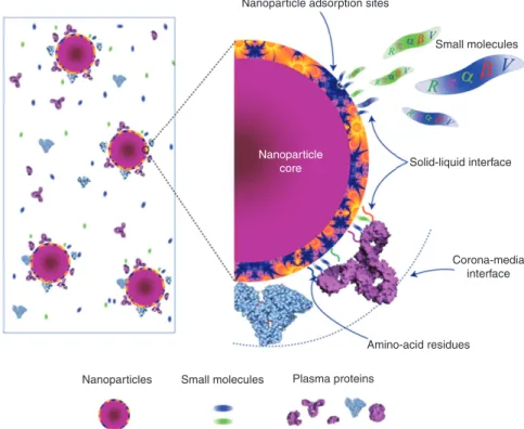
The role of nanoparticle interaction with biological mol-ecules as the key to nanomedicine and nanotoxicity has emerged recently [1], with the development of the idea of the nanoparticle-protein or biomolecule corona [2]. This is the dynamic layer of proteins and other biomolecules that adsorbs to nanoparticle surfaces immediately upon contact with living systems and is what organisms or cells “see” and interact with [3]. Unlike bulk biomateri-als, however, the fact that nanoparticles are sufficiently small that they can reach sub-cellular locations results in significant new potential impacts, specifically in terms of their interactions with biomolecules and biomembranes. The consequences of this for nanoparticle uptake and bio-distribution and nanoparticle-induced signalling impacts need to be considered. A recent review of nanoparticles interacting with biological systems suggests that nano-particles are mobile solids, combining the properties of
solids (for example, fluorescence in the case of quantum dots where the constituent components are non-fluores-cent) with the ability to thermally diffuse (a property of molecules) [4]. Their large surface area allows them to act as a scaffold for protein and biomolecule binding, leading nanoparticles to acquire a biological identity, or a bio-nano interface. The bio-nanoparticle protein or biomolecule corona hypothesis suggests that the biomolecules (pro-teins, lipids, nucleic acids etc.) that reside on the particle determine its biological identity and subsequent impacts [3]. The view is thus emerging that one should correlate the properties of the nanoparticle corona or its bio-nano interface to the biological activity/responses rather than simply the bare nanomaterial properties [5].
It is clear that the biological behaviour and conse-quences of nanoparticles are largely dictated by how they interface to biology. The idea of airborne particulates becoming coated with lung surfactant lipid following inhalation was postulated in 1990 [6]. Proteins and lipids in lung lining liquid at the air-liquid (alveolar fluid, hypo-phase) interface – the first biostructure an inhaled nano-particle encounters when deposited in the alveoli – were later observed to coat urban nanoparticles in a corona and cause nanoparticle aggregation [7–9].
This lung-surfactant corona was later proposed as an important protective mechanism mediating the health impacts associated with breathing in airborne particles and nanoparticles in urban air [10]. Despite early evidence of particle coronas in the lung identified using newly devel-oped atomic and molecular scale techniques [11], the bio-logical interface remains the least understood aspect about nanoparticles, and classification systems to characterise the outermost layers of the bio-nano interface, i.e., those biological signatures that are available to engage endoge-nous cellular machinery, are absent. By far the most studied component of the corona is the protein composition [7, 12],
but lipids, sugars and other species likely also play a role [13–15]. Thus, despite the importance of the bio-nano inter-face, and the fact that it potentially holds the key to both safe implementation of nanotechnologies and nanomedi-cine, efforts to characterize it are surprisingly scarce [16]. While the focus of this review is on biomolecules such as proteins and lipids, the ideas are equally applicable to nan-oparticles dispersed in environmental milieu, where decay-ing plant and animal matter results in so-called natural organic matter, typically composed of polysaccharides, interact with nanoparticles affecting their stability, dispers-ability and environmental fate and behaviour [17, 18, 19].
Significant advances have been made in the last 5 years, both in understanding of the importance of the bio-nano interface and in terms of methods and approaches to study it. Evidence for this wide scale acceptance of the concept of the nanoparticle biomolecule corona and the importance of the bio-nano interface comes from the fact that the OECD Sponsorship Programme has included characterisation of nanoparticles in biofluids as part of their list of endpoints at the end of 2010 [20].
Before getting into the details of the state of the art and recommendations for moving beyond this it is important to note that interactions between nanoparticles and bio-molecules, and the formation of the bio-nano interface, has consequences for both the nanoparticle surface itself, and potentially also for the proteins and other biomol-ecules contained in the biomolecule corona. A summary of some of these effects, and reviews or key publications relating to these effects, are given in Table 1.
The emerging discipline of nanotoxicology may be viewed essentially as the study of the undesirable inter-ference between man-made nanomaterials and cellu-lar nanostructures or nanomachines [35]. In parallel, the considerable allure of engineered nanoparticles for clinical applications is due to the fact that these artificial Table 1 Nanoparticle-biomolecule interactions and the formation of the nano interface has consequences for both the adsorbed bio-molecules and for the nanoparticle surface and dispersion.
Effect of adsorption on Biomolecules: Effect of interaction on Nanoparticles: – conformation changes → blocked or enhanced presentation
of active sites and subsequent functional changes [21, 22] – conferring a biological identity – altered interaction/uptake and biodistribution [23–25] – altered propensity for protein-protein interactions (e.g.,
fibrillation) [26] – altered surface characteristics and thereby stability and dispersability [8, 27] and potentially also dissolution potential (as per environmental macromolecules such as humic acids) although limited literature [28] – oxidative effects – lesions, post-translational effects,
etc. [29]. – reduced surface energy/reactivity [30] (possibly only temporarily) – depletion of medium components which can result in
indirect toxicity effects [8, 10, 31] – masking targeting or other bio-functional elements? (possibly only temporarily) [32] – altered kinetics (distribution, half-life, degradation, etc.)
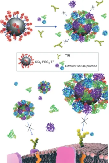
entities are designed to interact with biological systems at the nano-scale. Moreover, understanding and controlling the bio-nano-interface is equally important both from a nanomedicine and nanotoxicology point of view. In fact, a recent study reported that the adsorption of serum pro-teins obscures targeting ligands grafted to nanoparticles (in this case: transferrin) thereby preventing targeted uptake, as shown schematically in Figure 1 [32]. This dem-onstrates the importance of biomolecular interactions in determining nanoparticle uptake, uptake mechanism and fate and behavior in living systems. It is important to note that the size ratio between nanoparticle and pro-teins plays a vital role in determining nanomaterial-pro-tein (macromolecule) interactions, and indeed for many types of inherently small nanomaterials (e.g., Quantum
SiO2-PEG8-TF
TfR
Different serum proteins
Figure 1 The biomolecule corona masks targeting ligands: Trans-ferrin-functionalised nanoparticles lose their targeting capabilities when a biomolecule corona adsorbs on the surface. Schematic representation of loss of TfR targeting for Tf-conjugated nanoparti-cles in the presence of FBS proteins (endogenous Tf, where present, could also compete for TfR). Reproduced from [32]. Note that the particles in this schematic are intended to represent ~50 nm parti-cles, but that the nanoparticles and proteins are not drawn to scale.
dots which are typically < 5 nm) the nanomaterials may be smaller than the proteins, as discussed in Klein et al. [36] and demonstrated by Deng et al. in their study of the role of gold nanoparticle size on binding to fibrinogen, whereby small changes in nanomaterial size (from 8 nm to 10–12 nm to 15 nm) resulted in significant differences in how the protein and nanomaterials interacted [37]. The schematic figures in this manuscript (taken from the lit-erature) are not drawn to scale, but are intended only to illustrate the principles and concepts being described.
The fact that many endogenous transport and other processes utilise biomolecule clusters in the nanoscale, such as the lipoprotein complexes, e.g., chylomicrons ( > 100 nm) and High Density Lipoproteins (8–10 nm), and ribosomes (DNA and protein clusters; 25–30 nm), suggests that in many cases nanoparticles may simply be recog-nised as scaffolds onto which biomolecules can adsorb as part of the normal functioning of the biomolecules [38]. Indeed, most spherical nanoparticles studied to date have been shown to bind lipoproteins, often with a size and surface curvature influence, in addition to a composi-tional influence, such as from surface charge [21].
Building on this background and the current state of the art, we suggest some potential future research direc-tions and make recommendadirec-tions for future priorities and strategies to ensure that the importance of the bio-nano interface is recognised. Central to this is the fact that nanomaterials must be studied as biological enti-ties under the appropriate exposure conditions, and that this approach must be implemented in study design and reporting for nanosafety assessment. The implications of the bio-nano interface for nanomaterials fate and behav-iour are described from four primary perspectives: – the Coating concept,
– the Translocation concept, – the Signalling concept, – the Kinetics concept.
In this paper we considered that the principles outlined hold for all nanoparticles types, and thus the examples cited from the literature may refer to metallic, metal oxide, carbon based, polymer coated or polymeric nanoparti-cles, quantum dots etc. Clearly surface chemistry matters, as does size and shape, in terms of the finer details of how each different particle type behaves, but we believe that the principles hold generally and indeed that these could form the basis of a future classification strategy that is independent of nanomaterial composition or structure. The four primary perspectives presented here align well with, and indeed expand upon, the three “principles of nanotoxicology” described by Krug and Wick, namely the
Transport Principle, the Surface Principle and the Mate-rial Principle [39] and focus on the role of biomolecules in providing a crucial interface between nanoparticles them-selves (which are seen more as scaffolds for protein/bio-molecule binding) and biological systems. Indeed, a key conclusion is that nanoparticles must be thought of, and studied as, biological entities, where their interaction with the environment is mediated by the proteins and other biomolecules that adsorb to them, and the key parameter to characterise then becomes the nature, composition and evolution of the bio-nano interface.
The coating concept: Interactions of
nanoparticles with proteins, lipids,
polysaccharides, DNA/RNA, natural
organic matter, etc.
Scientists increasingly recognise that nanoparticles imme-diately absorb proteins and/or other biomolecules from their surroundings to lower their surface free energy [40] with important consequences for nanoparticle stability in dispersion [41], and interaction with biological systems. Protein binding to nanoparticles changes both the hydro-phobicity of the outermost surface (the bio-nano interface) and the effective surface charge, with even very positively charged nanoparticles typically presenting a neutral to slightly negative zeta potential in plasma or cell culture medium containing foetal calf serum. Note that for eco-toxicological studies, natural organic matter plays much the same role as proteins for in vitro and in vivo toxicology studies, modulating the nanoparticle surface (free energy) and thus the dispersibility of nanomaterials [18] and inter-action with biological systems. As a consequence, nano-particles dispersed in biofluids containing proteins, lipids, polysaccharides, etc. can have a very different dispersion profile than the same nanoparticles dispersed in refer-ence buffers, which can lead to very different effective or available doses of nanoparticles for interaction with living systems [3, 5]. The understanding that nanoparticles in a biological medium are remarkably strongly associated to a biomolecular layer (rich in proteins in vivo) drawn from their environment shifts the focus of studies and discus-sions away from the bare material identity to a new, more nuanced, conception in which particle size, shape and corona expression (collectively the bio-nano interface) are more likely to be the defining features of nanomaterial bio-logical identity and consequently fate and behaviour. Thus, in addition to describing the physico-chemical properties
Nanomaterials classification and grouping
Bio-nano interactions Coating translocation signalling kinetics Measurement principles for NM identification in different environments and
matrices
Identification of metrics relevant for safety
assessment Synthetic identity (context dependent)Biological identity
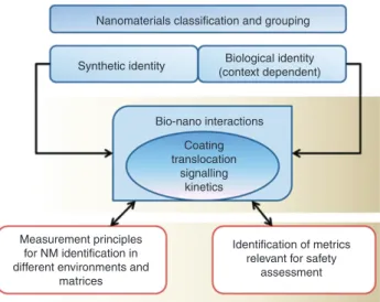
Figure 2 The importance of understanding the inter-relationship between the synthetic and biological identities of nanomaterials for hazard assessment, identification and classification of nanomateri-als). Figure re-drawn from the NanoSafety in Europe 2015–2025 report [43].
(the so-called “Synthetic identity”), a “biological” property should be added to describe the nanoparticles as they exist in the relevant exposure context [42], as shown in Figure 2.
The composition of the biomolecule corona, and the subsequent evolution as a particle moves from one biologi-cal environment to another [44], available dose and conse-quent biological interactions of the coated nanoparticles, have been found to depend on the specific details of the biofluid in which the nanoparticles are dispersed, which may account for much of the contradictory reports present in the literature for nominally identical materials to date [5]. Thus, the same (batch of) nanoparticles dispersed in different cell culture media (e.g., DMEM or RPMI) con-taining identical concentrations of Foetal Bovine Serum (FBS) from the same batch have been shown to result in quite different coronas, both in terms of their thickness and dynamics [25]. The authors of that study observed that DMEM elicits the formation of a large time-dependent protein corona, while RPMI shows different dynamics with reduced protein coating. These different coronas, which resulted from the different ionic strengths and salt com-positions of the media, had implications for uptake and impact, with the protein-nanoparticle complexes formed in RPMI being more abundantly internalized in cells as compared to protein-nanoparticle complexes formed in DMEM, consequently exerting overall stronger cytotoxic effects [25]. These results suggest that cell culture medium composition and ionic strength can alter adsorption of proteins onto the nanoparticle surface, which can impact on the particle agglomeration and potentially alter the
available dose of nanoparticles under the different expo-sure conditions. However, in the absence of characterisa-tion of the nanoparticles in the two different media, it is not possible to make any interpretation of the data on the basis of whether the different protein coronas result in different available doses, which could potentially explain the different observed impacts [5]. In other studies, the surface chemistry profiles of airborne particle samples collected in different continents, and exposed to the lung lining liquid of different subjects, showed surprising con-sistency and resulted in similar aggregation effects [9].
Similar data resulted from comparison of corona com-position and cellular uptake of nanoparticles dispersed in cell culture media containing 10% foetal calf serum that had either been heat inactivated (to remove the comple-ment proteins) or not heat inactivated [24]. Here also, the particles with the lower protein content in their coronas entered cells more effectively than those with higher protein content in their coronas [24, 45]. Related work has shown that the composition of the nanoparticle biomole-cule corona can depend on the ratio of nanoparticle surface area to available proteins, with the consequence that nano-particle coronas prepared under conditions typical for in
vitro testing (i.e., 3%–10% serum proteins in medium) may
not be representative of the corona that would form under
in vivo conditions (typically 55%–80% proteins) [30].
A study of the interaction of nanoparticles with sur-factant protein A (SP-A), a significant protein component of alveolar lining fluid, found different particle-protein interactions for each of eight different nanoparticles [46]. Interestingly, three variants of the same material (cerium dioxide nanoparticles) revealed different adsorption pat-terns despite the materials being nominally identical and indistinguishable in electron microscopy images [46]. This suggests that the biomolecule corona composition, or the details of the bio-nano interface, could be a very sen-sitive tool to distinguish subtle material differences and to predict biological impacts, once correlations between adsorbed biomolecules and signalling or other effects are confirmed. In contrast, surfactant protein D (SP-D) alters cellular uptake in a different way [47].
Multiple studies have assessed the effect of nanoparti-cle composition [38, 48, 49], size [49, 50], surface coating [51, 52], shape and other physico-chemical parameters on the nature and composition of the protein corona. Recent reviews have attempted to summarise the many factors that have been found to influence protein–nanoparticle interactions [53, 54], and have described these factors as falling into three categories: protein (or
macromolecule)-related (molecular weight, isoelectric point (pI), and
conformational flexibility), NM-related (species, size,
shape, charge, roughness, hydrophobicity, crystallization, defects, and functionalization), and medium-related (pH and ion strength) [54].
In the case of nanoparticles dispersed in the environ-ment, interaction with environmental organic matter and biological molecules has been shown to determine their subsequent effects and fate in a similar manner. Some studies have shown that nanoparticles can adsorb natural organic matter to form complexes and, as a consequence of such coating, they may become negatively charged, alter-ing their fate and transport in the aqueous environment [55, 56]. Humic acids are a major part of the natural organic matter contained in soil and fresh water. Their structure, which consists of a skeleton of alkyl/aromatic units cross-linked mainly by oxygen and nitrogen groups with the major functional groups being carboxylic acid, phenolic and alco-holic hydroxyls, ketone and quinine groups, allows them to behave as surfactants, with the ability to bind both hydro-phobic and hydrophilic materials [57]. As a consequence of this behavior, nanoparticle-humic acids interactions should be a factor of relevance in the study of the life cycle of nano-materials, and likely natural organic matter plays a similar role in the environment that proteins do for human health.
We thus propose that it is vital that nanomaterials be considered as biological entities, and studied as such.1 Key
steps in the short to medium term include understanding and eventually predicting which chemical, geometrical and physico-chemical parameters of nanomaterials lead them to preferentially adsorb which proteins, and con-necting the absorbed proteins with observed impacts, such as uptake, localization and signaling. For example, the role of opsonins and dysopsonins is well understood in terms of phagocytic recognition [58, 59]. However, much work is required to tease out the signaling pathways influenced by biomolecules contained at the bio-nano interface, whether these be functioning normally or expe-riencing altered functionality as a result of conformation changes induced by binding to the surface.
The translocation concept:
interactions of nanomaterials with
cells, tissues, barriers, etc.
The human body has four portals of entry for nanoparti-cles: three natural portals, the lung, the digestive tract and 1 http://www.skep-network.eu/Libraries/Network_ documents/ SKEP_Nanomaterials_in_REACH_Report.sflb.ashx.
the intact skin, and one artificial portal, the veins, into which nanoparticles can be injected. It has been shown that nanoparticle translocation into capillaries leads to their translocation into other organs [60–65], and evidence is emerging that the route of entry, and the biomolecules that form the initial corona at the site of entry (e.g., plasma proteins following injections versus lung surfactant pro-teins following inhalation), play a distinct role in deter-mining the organ biodistribution of nanoparticles [66]. For example, accumulation of TiO2 nanoparticles in the brain was 100-fold higher for particles entering via lung than for those injected directly into the bloodstream (Person-nal Communication W.G. Kreyling). Further evidence for the importance of the initial bio-nano interface, which is determined by the route of exposure, comes from a study of the interaction of magnetite nanoparticles (110–180 nm in diameter), coated with different polymers (starch, car-boxymethyldextran, chitosan, poly-maleic-oleic acid, phosphatidylcholine), with alveolar macrophages [67]. Cellular binding and uptake of nanoparticles by alveolar macrophages was increased for nanoparticles treated with SP-A, whereas albumin, the prevailing protein in plasma, led to a significant decrease. This study provides evidence that, after inhalation of nanoparticles, a different protein coating and thus different biological behavior may result compared to direct administration to the bloodstream [67].
A direct effect of biomolecule corona composition on nanoparticle biodistribution has been shown in an elegant study using radiolabelled gold nanoparticles of five different sizes (1.4, 5, 18, 80, and 200 nm) and 2.8 nm gold nanoparticles with opposite surface charges fol-lowing intravenous injection into rats [68]. Results indi-cated that both size and surface charge of nanoparticles strongly determined the biodistribution, with different charge particularly leading to significantly different accumulations in several organs. The authors concluded that the alterations of accumulation in the various organs and tissues, depending on nanoparticle size and surface charge, were mediated by dynamic protein binding and exchange [68].
When interacting with cells, nanomaterials come into close contact with the cell membrane, a dynamic structure that segregates the chemically distinct intra-cellular milieu (the cytoplasm) from the extraintra-cellular environment by coordinating the entry and exit of small and large molecules. Macromolecules are carried into the cells in membrane bound vesicles derived from the invagination and pinching-off of pieces of the plasma membrane to form endocytic vesicles. This process, termed endocytosis, has different features depending on the size and type of the molecule/structure internalized:
receptor-independent endocytosis (fluid-based endo-cytosis and macropinoendo-cytosis), and several types of receptor-dependent endocytosis (clathrin-dependent, lipid-raft-independent, lipid raft-dependent/caveolae-independent, and caveloae-dependent). These endog-enous processes are exploited by bacteria and viruses for invading cells, and it is likely that also nanoparticles may enter cells through them, although there are quite conflicting reports as to the exact mechanism(s) medi-ating cell entry by different nanoparticles [69–72]. Very likely this is a result of different bio-nano interfaces. A recent review of cellular uptake of nanoparticles describes work revealing active endocytosis mechanisms and pathways involved in their cellular uptake, and summarises the current state of knowledge: Interested readers are referred here for more details [73]. All endo-cytic pathways have a feature in common, in that the particle, which entered the cell, is finally located in an intracellular vesicle, typically the lysosome, as the final sub-cellular destination [74–76]. However, there are studies, which reported the intracellular localization of nanoparticles that were not membrane bound, suggest-ing alternative pathways for particles to enter, or that the nanoparticles can escape the endosomes, as a result of endosomolysis [77–80]. This alternative way of nanopar-ticle penetration into cells, i.e., through the cell mem-brane, has been called “adhesive interaction” (by van der Waals and other forces) and has been suggested as an alternative mechanism of passive entry [81]. Another recent publication offers convincing evidence for a passive mechanism whereby nanoparticles enter cells by membrane penetration on an experimental and cal-culation basis [80]. The fact that nanoparticles are found free in the cytoplasm following uptake via the adhesive mechanism is noteworthy, since free nanoparticles in the cytoplasm may cause cellular effects different from those contained in vesicles (which entered the cell probably by an endocytic mechanism). Moreover, the intracellu-lar trafficking may be different and also interaction with intracellular structures like the cytoskeleton, the orga-nelles and the nucleus may be via direct contact for free nanoparticles. These aspects may all contribute to the different reactions of the cell to different nanoparticles, and are worthy of further investigation.
Very significant modulatory effects of proteins and the bio-nano interface have been observed in terms of nanoparticle uptake by human macrophages, by undiffer-entiated and by PMA-differundiffer-entiated monocytic THP-1 cells of 50 nm and 100 nm fluorescently-labelled carboxyl- or amine- modified polystyrene nanoparticles from medium with and without (serum free medium) serum proteins
[59]. The amount of internalized nanoparticles, the uptake kinetics, and its mechanism were critically dependent on particle opsonization by serum proteins, with nanoparti-cles being rapidly internalized by cells in serum-free (SF) medium until they reached saturation kinetics, whereas in 10% human AB serum-enriched (SE) medium the nano-particle uptake rate was drastically reduced as the uptake was via receptor-mediated processes [59]. Thus, in terms of understanding nanoparticle uptake and biokinetics, the bare material surface is clearly the wrong parameter to use, and indeed uptake studies in the absence of an appropriate biomolecule environment report on uptake as a result of membrane damage in an artefactual situation [34], rather than on nanoparticle uptake utilising endog-enous pathways.
However, one important question that remains to be answered is whether the protein and biomolecule layer on the nanoparticle surface mediates the binding to cells through one or more specific active mechanisms or via non-specific interactions, whereby the biomol-ecules behave as a simple coating which reduces the surface energy of the nanoparticles. There are consider-able differences in interpretation in the literature to date regarding uptake mechanisms, and approaches such as poisoning the endocytic receptors using pharmacologi-cal inhibitors or silencing of selected proteins involved in specific endocytic pathways (e.g., clathrin or caveolin) suggest that the same nanoparticle might exploit differ-ent uptake mechanisms to differ-enter differdiffer-ent cell types or indeed the same cell type under different conditions [82]. Similar results have been reported for nanoparticles engi-neered for targeting the folate receptor: folate receptor-specific siRNA was used to reduce folate receptor levels and uptake of heparin-folate-paclitaxel nanoparticles by target cells, but silencing only reduced uptake to half of its value in the control cells, suggesting multiple path-ways of uptake [72]. A recent study of differently surface modified 100 nm polystyrene nanoparticles indicated that different specific protein coronas did not result in dif-ferential association of the particles to endothelial cells, suggesting that binding and cellular uptake by these cells may not be triggered by interaction of the protein corona with specific receptors [83]. The authors suggested that an assessment of the adsorptive capacity of nanoparticles could be useful in order to predict the magnitude of nano-particle cellular interactions. Much of the success to date in terms of nanoparticles for drug delivery has been the result of fortuitous absorption of endogenous transporter proteins, such as Apolipoprotein E, to nanoparticles, or deliberate functionalisation of nanoparticles with these proteins [84, 85].
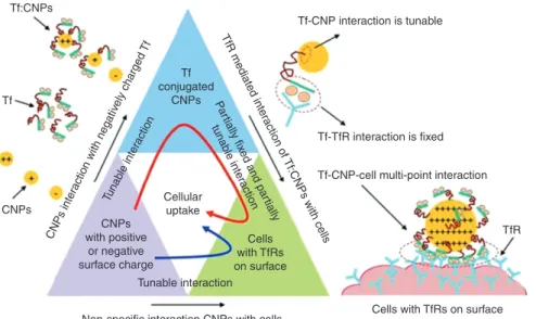
Once it is recognised that the biointerface is the key parameter to understand in terms of determining mecha-nisms of uptake of nanomaterials, a key question becomes how researchers can target nanoparticles to best utilise these pathways, and simultaneously avoid non-specific protein interactions, by designing the bio-nano inter-face. Several recent reviews highlight the various endog-enous uptake pathways available to nanomaterials, and examples of therapeutic strategies that could potentially utilise these pathways [86, 87]. Indeed, there has been considerable effort devoted to development of targeting nanoparticles, such as conjugation of 100-nm polysty-rene nanoparticles with glycocalicin, which significantly increased the particle adhesion on P-selectin-coated sur-faces and cellular uptake of nanoparticles by activated endothelial cells under physiological flow conditions [88], or functionalisation of cerium oxide nanoparticles with transferrin (Tf), to increase preferential uptake by trans-ferrin receptor (TfR) over-expressing human lung cancer cells (A549) and normal embryo lung cells (WI-38) [89]. A key finding from this work is that the strength of interac-tion between the nanoparticles and the targeting protein (Tf in this case) can be tuned by modifying the surface charge of the nanoparticles, and that binding energy values could be correlated with cellular uptake (as shown schematically in Figure 3) [89], in a first step towards quantitative structure-activity relationships (QSARs).
The task of modeling, and ultimately predicting, the distribution and fate of nanoparticles represents an interesting, and quite new challenge [76]. A flux-based approach, based on live cell imaging of fluorescent nanoparticle uptake and transport in a time resolved manner has shown that the nanoparticles rapidly local-ize to endosomes (1 h) and later to lysosomes (by 4 h), with no evidence of nanoparticles exiting from lys-osomes once they have arrived there [76]. The observed decrease in fluorescence over time was shown to corre-late with cell division, and with the nanoparticle load being split evenly between the two daughter cells. While vesicle recycling is a normal part of cell homeostasis, no recycling of fluorescent nanoparticles from vesicles was observed in the above study [76]. Indeed, another study has shown that relatively few 40 or 100 nm carboxylic-modified polystyrene nanoparticles are able to access endocytic recycling pathways, as judged by the lack of significant co-localization with Rab11, a key protein asso-ciated with recycling vesicles [90]. There is no evidence that corona-driven export processes exist for nanoparti-cles (unless specifically engineered to express an export signal – such as transferrin, as described below), and the chances of the appropriate specific intracellular corona
arising from non-specific protein binding during uptake are slim, [76] although potentially such signals could arise as a result of the nanoparticles binding peptides from the antigen processing pathways. Clearly there are some exceptions to this general rule in the case of spe-cialized cells such as biological barriers, where trans-location is a key function, such as the air-blood tissue barrier and the mucosal barriers where particle trans-cytosis, by epithelial and endothelial cells separated by the fused basement laminae of the two cells is common though a selective mechanism [60]. The penetration of nanoparticles through a cell (into and out, membrane bound) is called transcytosis or cytopempsis.
Single-walled carbon nanotubes (SWNT) were observed to undergo exocytosis in NIH-3T3 cells, the exo-cytosis rate closely matching the endoexo-cytosis rate with negligible temporal offset [91]. The exocytosis pathway was illustrated by superimposing example particle trajec-tories recorded in the near infrared onto the correspond-ing optical image of different cells [91]. Tf, which has a well-known recycling pathway, has been shown to induce exocytosis of gold nanoparticles to which it is physically adsorbed at a rate that was in linear correlation with the nanoparticle size [92]. Fifty nm was reported as the optimal cellular accumulation size for Tf-gold nanoparti-cles, due to the equilibration of the rates of uptake and exocytosis [92].
The signalling concept: interaction
of nanoparticles with major
intra-cellular chemical systems
The range and amount of nanoparticles humans are exposed to is quite significant (particularly from com-bustion), with the increasing production of engineered nanomaterials now contributing to this. The occupational setting is now considered as the most likely exposure route to engineered nanoparticles, but medical formula-tions may result in a wider population exposure [12]. The contribution of exposure to ultrafine particles such as those from combustion processes, to respiratory and skin diseases as well as more insidious and complex patholo-gies such as cancer and cardiovascular dysfunction, is now becoming apparent [93–95]. Their cumulative effects, or more likely the knock-on effect to neighbouring cells as well as more distant tissues and organs (i.e., paracrine sig-nalling and cell activation) plays a key role in their toxicity at the systemic level.
A key question to consider at the outset is whether nanoparticles “signal” or merely perturb signalling path-ways? Clearly sophisticated nanoparticles envisioned for biomedical applications may “signal” according to their deliberate functionalization and/or cargo of signalling molecules/drugs/genes, but it is important to consider Tf-CNP interaction is tunable
Tf-CNP-cell multi-point interaction
Cells with TfRs on surface Non-specific interaction CNPs with cells
Tunable interaction Tuna ble inte ract ion CN Ps inte ract ion with neg ativ ely charged Tf Pa rtially fix ed and par tially tuna ble inte ra ction TfR m edia te d inte ra ctio n o f Tf:CNPs with cells Cellular uptake CNPs with positive or negative surface charge Cells with TfRs on surface Tf conjugated CNPs Tf-TfR interaction is fixed TfR Tf:CNPs CNPs Tf
Figure 3 Schematic diagram of interaction forces acting at different stages of cellular uptake. The triangle blocks show the interaction pathways of cellular uptake of cerium oxide nanoparticles (CNPs). CNPs with strong positive charge show better adsorption of transferrin (Tf). The interaction between Tf and CNPs can be tuned by protonation; however, the interaction of Tf with TfR is fixed. CNPs with a strong positive charge lead to enhanced Tf adsorption and multiple interactions with the TfRs on the cell surface. Red curved arrow inside the trian-gle blocks indicates the receptor mediated cellular internalization pathway of positively charged CNPs, and blue curved arrow indicates the non-specific cellular internalization pathway of both positively and negatively charged CNPs. The red dashed circle represents the domain of multi-point interaction between Tf:CNP and TfRs on cell surface. From [89].
whether other, pristine or non-functionalized nanopar-ticles “signal”. The main thesis in the present review is that nanoparticles can signal by virtue of their acquired bio-corona of proteins, lipids, sugars, or other biomol-ecules and that studies conducted to date have not fully taken into account the “biological” identity of nanopar-ticles. There are certainly some (very few) examples of nanoparticles that appear to signal per se as a function of their specific size, for instance, the finding that single-walled carbon nanotubes (SWNTs) constitute a new class of universal K+ channel inhibitors that hamper channel
function by fitting into the pore and thus either hindering ion movement or alternatively preventing further confor-mational steps [96]. We believe that further studies, using systems biology approaches, may uncover more examples of nanoparticle-mediated signaling at doses of nanoparti-cles that are more realistic, and that the bio-corona and, hence, the bio-nano-interface is likely to be an important determinant of such signaling. This is in distinction to the vast majority of publications to date which apply exces-sive amounts of nanoparticles to cells and which focus only on crude measurements of cell death.
Mapping the research agenda for investigating nano-particle signalling effects, the research objectives can be broken down into three interlinked levels. Firstly, nano-particles attached to the cell surface (cell membrane) may trigger a cascade of signalling processes into the cell and throughout the cell. A key question which one should ask is whether nanoparticles need to enter a cell in order to trigger/cause any effects inside the cell. While researchers have long been concerned about distinguishing between nanoparticles taken up into cells versus those adhered to the membrane for quantification of uptake [76, 97], less research has been directed to understanding nan-oparticle-induced signalling from the cellular surface, which is a critical knowledge gap at present although several groups are now working on this and publications addressing this topic will appear in the literature in the near future [Personnal Communications from H. Hofmann (EPFL) and K.A. Dawson (UCD)]. Secondly, nanoparticles can signal once they are inside single cells. Here, impor-tant aspects that need to be addressed are the manner in which nanoparticles bypass the cell membrane, enter into the cell, localise to the mitochondria and other organelles and finally the nucleus, causing changes in phenotypic function and the activation of distress signals in the form of pro-inflammatory or other molecules. The third signal-ling level includes the pathways by which cells commu-nicate their response to interaction with nanoparticles to neighbouring cells and then to more distant tissues and organs through soluble mediators. A limited number of
studies demonstrate paracrine signalling from nanopar-ticles in cells to other non-exposed cells, suggesting that such effects may occur [98–101].
At present, much of our understanding of nanoparti-cle induced alterations of signal transduction is limited to a very small space and time window. The question of how these adaptive signals are propagated to and translated by remote tissues and organs, how the internal milieu is modulated by them, and the signalling pathways involved in the translocation of nanoparticles from their point of uptake to distant tissues have not yet been addressed. Much of what we know of the integrative pathophysiology of engineered nanoparticles comes from animal studies focused on translocation and bioaccumulation [68, 102]. Translating the results to humans is not straightforward, and as several studies on drug toxicity have shown, caution needs to be exercised when extrapolating results from animal models to human physiology, pathology or toxicity [103]. Moreover, animal models are not amena-ble to decomposition of signalling dynamics in differ-ent tissues and organs. Additionally, given the degree of redundancy in signalling pathways, decoupling effects of biomolecules bound to nanoparticles from those of their unbound counterparts will be challenging. However, systems biology approaches are making significant strides here [104–106].
The induction of oxidative stress is commonly viewed as a unifying concept for understanding the cytotoxic effects of nanoparticles, such as nano-sized metal parti-cles and their oxides as well as those produced by com-bustion (including from motor vehicles) [107]. However, it is also important to assess whether “oxidative stress” is merely a secondary event resulting inevitably from dis-ruption of biochemical processes and the demise of the cell, or a specific, non-random event that plays a role in the induction of cellular damage, e.g., apoptosis? For a further discussion of these issues, readers are referred to Shedova et al. [108]. The mechanism by which oxidative damage occurs appears to be via the inhibition of elec-tron transfer in the TCA (tricarboxylic acid) cycle and the consequent accumulation of TCA cycle intermediates. The TCA cycle is an essential metabolic network in all oxidative organisms and provides precursors for anabolic processes and reducing factors (NADH and FADH2) that drive the generation of energy. Reactive oxygen species (ROS) are formed in the mitochondria during respiration. It has been shown that a healthy metabolic network plays a key role in the defense against oxidative stress through the production of enzymes and electron acceptors which participate in the detoxification of ROS [109]. Interfer-ence in the mitochondrial cycle results in oxidative stress
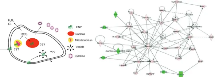
owing to an increase in ROS, which act as second messen-gers to induce a cell reaction that may eventually lead to cell death. How nanoparticles cause oxidative stress and subsequent apoptotic or necrotic signalling is not clearly understood, and furthermore the process is unlikely to be the only mechanism by which nanoparticles cause cell and tissue damage [110]. Probing signalling mecha-nisms at the cellular level should focus on delving into the mechanisms of ROS production by nanoparticles upon entering cells, and subsequent cellular adaptation at the nuclear level (changes in gene expression) (Figure 4). It may be worth considering whether the develop-ment of oxidative stress could be a first (unspecific) reac-tion mechanism to foreign material intrusion. In fact, a short-lived reaction is compatible with a normal innate defensive response, which resolves rapidly with limited cell damage that is readily repaired. On the other hand, the persistence of the reaction could lead to significant damage from apoptosis and genotoxicity, and eventually to cancer. Thus, understanding the origin and kinetics of the oxidative stress induced by nanoparticles and corre-lating it to pathological risk could provide vital insights in the future as to the impacts of nanomaterials. Note that this theme of transience or duration of response is picked up in the “kinetics concept” section, as full understand-ing of mechanisms will only be possible with a proper emphasis on rate and kinetics of the various biological processes.
A detailed investigation of the mechanism of toxicity induced by 50 nm amine-modified polystyrene nanopar-ticles following uptake by 1321N1 brain astrocytoma cells found that the nanoparticles are localized in lysosomes, whereupon the lysosomal membrane becomes destabi-lized, likely because of the nanoparticle’s positive charge, leading to release into the cytoplasm of both nanoparti-cles and proteolytic enzymes such as cathepsins [112]. This, in turn, results in damage to the mitochondria and activation of caspases 3 and 7, with consequent cleavage of PARP-1, ultimately resulting in the apoptotic death of the cells [112]. Ongoing work has shown that the kinetics of the lysosome membrane damage can be correlated with the digestion of the nanoparticle protein corona in the lyso-somes which allows the underlying amine groups on the nanoparticles to be re-exposed [113]. Smaller, fully ami-nated, dendritic polymer nanoparticles have been seen to cause endosomolysis prior to transfer to lysosomes, however, and have been seen to be later localized in the mitochondria. In both stages, oxidative stress has been seen to give rise to activation of caspases, inflammatory responses and subsequent apoptotic (and other kinds of) cell death [79, 114].
An important advance in understanding nanopar-ticle-induced signalling and toxicity has been achieved utilising an integrated proteomics approach, as routinely used to identify protein interaction pathways, to identify the toxicity pathways and networks that are associated
Figure 4 Left: Critical aspects of intracellular signalling: mechanisms of nanoparticle (ENP) entry, ROS induction, nuclear damage and ENP fate after cellular reaction (or death). Right: Protein interacting network 1. This representative network of differentially expressed proteins shows the protein inter-relationships and relevant signalling pathways. Two major sub-networks within the network are centered by NF-κB and YWHA2. Proteins in pink are up-regulated while proteins in green are down-regulated. Solid lines represent direct relationships. Dotted lines represent indirect relationships. Lines connecting the proteins indicate known inter-relationships from the IPA database. From [111]. Whether, in this particular case the impacts can be best described as “signalling” or “perturbation of signalling” pathways is open to debate, but the possibility that that signalling may take place at the bio-nano-interface and as such needs to be considered in the design, and interpretation, of nanosafety and nanointeraction studies is one the main messages of the present review.
with exposure of human bronchial epithelial cells to nanoscale titanium dioxide [111]. Utilizing 2-DE and MS, 46 proteins that were altered at protein expression levels were identified and mapped, using Ingenuity Pathway Analyses™ (IPA) canonical pathways and Ingenuity Pathway Analyses tox lists, to create protein-interacting networks and proteomic pathways. This provided the first preliminary protein-interacting network maps and may give novel insights into the biological responses and potential toxicity and detoxification pathways of titanium dioxide [111]. However, as with many early studies in an emerging field, there are some concerns regarding how much can be interpreted from this study which lacked appropriate controls [e.g., no bulk TiO2 or other (reference) material has been used for compari-son]. Another example of this approach uses proteomic techniques including two-dimensional electrophoresis/ mass spectrometry and protein microarrays to study the differentially expressed proteome and phosphopro-teome, respectively [115]. Here, systems biology analysis of the data revealed that unfolded protein-associated endoplasmic reticulum (ER) stress response was the predominant event in response to the presence of gold nanoparticles [115].
An alternative route to inflammation, directly linked to protein conformation and presentation at the bio-nano interface, has recently been suggested by Deng et al. [116]. Their study demonstrated that negatively surface charged nanoparticles can unfold fibrinogen and that the binding of fibrinogen to poly(acrylic acid) coated gold nanoparti-cles of 5 nm leads to interaction with the Mac-1 receptor and its activation, leading to a cytokine response through degradation of IκB, subsequent release of NF-κB and its translocation to the nucleus. Since plasmatic fibrino-gen has been reported to bind many different types of nanomaterials, including the metal oxides SiO2 and TiO2 [117], the authors proposed that fibrinogen-bound nano-particles are potentially pro-inflammatory. Note that, as indicated above for oxidative stress, activation of an inflammatory pathway does not necessarily correspond to toxicity, but is rather a defensive reaction and may cause no overt damage if transient, and thus an understanding of the kinetics and duration of the response to nanoparti-cles is vital.
An elegant approach to assessing nanoparticle impacts has been to look at the effect of citrate-reduced gold and silver nanoparticles on primary cultures of murine adrenal medullary chromaffin cells [118]. Car-bon-fiber microelectrode amperometry examination of exocytosis in nanoparticle-exposed cells revealed that nanoparticle exposure lead to decreased secretion of
chemical messenger molecules, of up to 32.5% at 48 h of gold nanoparticle exposure. Repeated stimulation of exo-cytosis demonstrated that these effects persisted during subsequent stimulations, meaning that nanoparticles do not interfere directly with the vesicle recycling machinery but also that cellular function is unable to recover follow-ing vesicle content expulsion [118]. Similar results were also observed in mast cells [118].
Beyond the level of the single cell, the means by which injured cells communicate their distress causing systemic and long-term damage is therefore a critical issue. The adaptive response of nanoparticle-injured cells is an over production of H2O2 with the consequent genera-tion of free radicals and the secregenera-tion of pro-inflamma-tory cytokines [119]. Recapitulating the systemic effects of nanomaterial toxicity in vitro requires properly scaled in
vitro and computational models so that the consequences
of localized nanoparticle induced injury to the whole body response can be systematically investigated. Cell-cell signalling, and signal propagation across the foetal barrier has recently been shown as the toxicity mecha-nism by which CoCr alloy metal nanoparticles induce a novel type of indirect genotoxic effect across cellular bar-riers [98, 120]. The CoCr nanoparticles were observed to cause DNA damage and tetraploidy in cells not directly exposed to the nanoparticles [98], i.e., on the other side of the barrier in an in vitro model system, without a sig-nificant passage or leakage of metal through the barrier. Such cell-cell signalling is protein mediated, and likely triggered by something present on the nanoparticle surface that interacts with cellular receptors, or induces signalling from lysosomes.
It is a fair criticism of the field to date that the major-ity of studies published have utilised unrealistically high doses of nanoparticles and that the “toxic effects” that have been reported, and the assays used to detect these effects, lack the sophistication that is needed to fully appreciate potential nanoparticle-induced signalling events. However, this current lack of evidence for nano-particle-induced signalling impacts is also a consequence of the fact that systems approaches are only beginning to be applied to these issues, and research to address this issue is underway. Data demonstrating nanoparti-cle induced signalling from nanopartinanoparti-cles located at cell membranes, from nanoparticles taken up into cells, and from nanoparticle-exposed cells to neighbouring cells will soon begin to appear in the literature as this convergence of nanosafety maturing as a discipline and more wide-spread application of systems biology approaches reaches fruition [Personnal Communications from H. Hofmann (EPFL) and K.A. Dawson (UCD)].
Connecting the nature of the bio-nano interface with downstream signalling impacts will be important in teasing out potential longer term consequences of expo-sure to nanomaterials, and to correlating impacts to the composition of the bio-nano interface and the underlying nanoparticle physico-chemical properties.
The Kinetics concept: Timescales
of interaction of nanoparticles with
biomolecules and cells
A deep understanding of the biological effects of nano-particles requires knowledge of the equilibrium and kinetic binding properties of proteins (and other biomol-ecules such as lipids and polysaccharides) that associ-ate with the particles, and especially under competitive binding conditions, such as those occurring in vivo [1]. The rates by which different proteins bind to and disso-ciate from nanoparticles, i.e., the time scales on which particle-associated proteins exchange with free proteins, are critical parameters determining their interaction with receptors, and biological responses generally. The biological outcome may also be different, depending on the relative exchange rates of proteins with nanoparti-cles and cellular receptors, respectively [3]. In addition, the particle-bound protein may have altered exchange rates with a cellular receptor. It is clear that, in under-standing how particles will interact with cells, these issues, currently almost unstudied, are amongst the most fundamental. Additionally, the corona may not imme-diately reach equilibrium when exposed to a biological fluid, and will evolve as the nanoparticle encounters new milieu, for example, when particles redistribute from one compartment or organ to another, such as upon receptor-mediated endocytosis from the extracellular environment into the primary endosomal cavity, or from the cytosol to the nucleus [44].
In addition to understanding the kinetics of forma-tion of the corona and its evoluforma-tion during nanoparti-cle uptake and translocation, it is vital to understand the fate of both the nanoparticle corona and the nano-particles themselves in their final sub-cellular loca-tions. In particular, there is emerging evidence that the bio-nano interface can be degraded upon localisa-tion of the nanoparticles in endosomes or lysosomes [113], and indeed even that some nanoparticles them-selves, including carbon nanotubes, may degrade in
situ in cells or in vivo [121]. Using fluorescently labelled
proteins in the nanoparticle corona, it has been possible to track nanoparticle localisation and corona digestion and to correlate this with the toxicity impacts observed [113]. Using amine-modified polystyrene nanoparticles, a detailed event sequence has been tracked showing how nanoparticle location and biological responses are connected – nanoparticles are localised in the lysosomes undergo acid-degradation of the corona, leading to re-expression of the positive charge on the nan-oparticles, which were previously masked by the pres-ence of the protein corona, and consequently disruption of the lysosomal wall, leading to a complex set of signal-ling responses [113]. For aminated dendrimers, a clear generation dependence is observed, reflecting the sys-tematic increase in the number of surface amino groups, implying a similar digestion in the endosomes [79].
The ultimate goal of nanosafety assessment is to be able to correlate the uptake rate, localisation and actual sub-cellular dose with the kinetics of the onset and prop-agation of the impacts observed. This requires a good understanding of the timing of signalling events that various assays report on, to ensure that experiments are designed with appropriate time-points such that desired effects can be observed. This is especially important for transitory or late-onset impacts, or for impacts that are down-stream of the initial signalling impacts. Cel-lular responses may not be linear with dose, however, and the process of saturation should be understood. Equally, understanding the intrinsic cellular protection mechanisms through antioxidants is critical to differ-entiating between responses of different cell types. To fully understand the impacts of different nanomaterials, it is important to identify the relevant cascade pathways and to link the rates of response to the physico-chemical properties of the nanoparticles, to their different bio-nano interfaces, and to the intrinsic characteristics of the cell-lines. For all processes, the rate of response is the most important characteristic parameter, and rel-evant rates include:
1. Uptake rates (endocytosis, adhesive interaction) and connection to bio-nano interface composition
2. Impact rates of membrane bound vs. free nanoparticles 3. Signalling rate of nanoparticles attached to cell
surface – identification and quantification of most relevant signalling markers at each time point
4. Rate of recovery of relevant signalling markers at each time point
5. Rate of ROS generation
6. Endosomolysis thresholds and rates
7. Rates of trafficking/translocation to other organelles (mitochondria, nuclei) and/or trancytosis
8. Uptake rates and size thresholds for organelles (mitochondria, nuclei)
9. Identification of impact pathways and rates (including activation/expression of relevant signalling proteins) 10. Identification of types and kinetics of cell-cell
signalling impacts
11. Identification of kinetics of evolution [122] and degradation of the bio-nano interface [114] and the underlying nanoparticle degradation both prior to uptake (i.e., in the exposure media and the influence of media composition on nanoparticle degradation rate for example [28]) and following uptake (for example in the lysosomes). As recently suggested by Shannahan et al. [123], the corona may only degrade after lysosomal localisation (of silver nanoparticles) resulting in acid-mediated oxidation and ultimately cell death due to toxic metal ions. Indeed corona degradation in the lysosomes has recently been demonstrated utilising loss of fluorescence signal from fluorescently-labelled serum proteins bound to nanoparticles over time [113]. Thus, elucidating the role of the bio-nano interface in modulating the ionic dissolution of metal and metal-oxide based nanoparticles is of particular importance given the widespread role of these materials in medicine. Coupled with this is a requirement to understand in detail where nanoparticles are located, in order to cor-relate impacts with particle localisation. Significant progress to this end has been achieved, and indeed it is now possible to use cellular proteins associated with the different vesicular structures of cellular uptake path-ways to report on kinetics of nanoparticle uptake and localisation. Thus, 40 nm carboxyl-modified polysty-rene nanoparticles were shown to first pass through an early endosome intermediate decorated with Rab5, but to rapidly transfer to late endosomes and ultimately lys-osomes labelled with Rab9 and Rab7, respectively [90]. Larger nanoparticles of 100 nm diameter also reach acidic Rab9- and Rab7-positive compartments although at a slower rate compared to the smaller 40 nm nanopar-ticles [90]. This information can then be coupled to the kinetics of signalling impacts induced following locali-sation, and using co-localisation approaches, to qualify particle amounts in the various compartments. Thus, it is possible that upon entering of foreign material, orchestrated defence processes are initiated by the cell, depending on the nature of the nanoparticle corona, and that concentration of nanoparticles, impact time and reaction time would have to be considered as part of a kinetics-based modelling approach.
Alternative routes towards nanoparticle track-ing include Raman spectroscopy. The use of Surface Enhanced Raman Scattering from gold nanoparticles and nanoaggregates to probe the environment of the subcel-lular compartments through which they are trafficked has been demonstrated [124, 125]. This approach also dem-onstrated the use of molecular labelled nanoparticles as more specific probes of the local environment [126–128]. However, the uptake rates and mechanisms as well as the subsequent trafficking may be specific to the nanoparti-cle type, size and surface chemistry. More recently, Raman spectroscopy has been employed to identify and localise unlabelled polystyrene nanoparticles in cells in vitro [129].
What can we learn from viral
bio-nano-interfaces and their
dynamics?
Nanoparticles and virus particles share many features, including size range. Independent of their nature, adhe-sion of particles in the nanometer size range depends mainly on van der Waals forces and not on key-lock con-cepts as often incorrectly described [130]. Indeed, it has been shown that adhesion processes (which are based on van der Waals and other forces) may lead to phospholipid molecules of the membrane moving apart to let nanopar-ticles enter into the cell passively [60]. Mathematically, these van der Waals forces can be described considering the work of adhesion, W, geometry, elasticity and forces applied to the system. Almost four decades ago, these forces interacting on the contact points of small parti-cles were described by the JKR (Johnson Kendall Roberts) analysis, F = -3πWD/8, where D is the Hertz contact diam-eter [131]. The measurement of these forces is possible by applying Atomic Force Microscopy (AFM) [132]. The inter-particle forces both for nanointer-particles and virus inter-particles are very small and are in the order of thermal diffusion forces. Measurements of self-adhesion forces leading to doublet and triplet formation showed very similar results for polystyrene and virus particles [133]. Proteins can accelerate adhesion between particles/viruses and cells and measurements of this process have revealed a step-wise process, giving evidence of several adhesive states, with significant energy barriers between these states that in fact allow catalytic actions to accelerate the kinetic pro-cesses leading to contact.
Theoretical models have moved from trivial and inac-curate lock-and-key-models to the more physically relevant
van der Waals principles. On the scale of such models, Brownian motion cannot be neglected and thereby adhe-sion becomes a statistical process leading to a dynamic equilibrium that depends on concentration, thermal energy and the attractive potential. Further research into adhesion and molecular dynamics modelling will allow improved visualization and understanding of the molec-ular basis of adhesion processes. A recently presented experimental procedure showed that nanoparticles can be functionalized with appropriate ligands to render them membrane-permeant [80]. Recent characterization of the trafficking mechanisms of prion proteins and certain bac-teria may present new paradigms for understanding how nanoparticles could enter cells [60, 134].
Nanoparticle interactomes:
emerging systems biology
approaches in nanotoxicology
Systems biology is an emerging field which seeks to inte-grate high-throughput biological studies to understand how biological systems function [135, 136]. By studying the relationships and interactions between various parts of a biological system (e.g., metabolic pathways, organelles, cells, physiological systems, organisms, etc.) it is hoped that eventually an understandable model of the whole system can be developed. One successful approach has been to determine protein interactomes, which are maps or networks connecting a protein to all the other proteins with which it interacts directly or indirectly [137, 138]. This is possible for proteins, as they must interact with other molecules in order to fulfil their biological roles. For instance, enzymes, receptors, and transcription factors have to bind their substrates, ligands, and target DNA elements, respectively, to execute their function. Thus, removal of one protein will affect the functioning of other proteins, which will in turn affect the functioning of other proteins in a complex network functioning.
Small changes in a protein conformation, such as may be induced by interaction with the surface of a nanopar-ticle (both engineered and combustion derived) [21], can potentially have large impacts on a protein’s function, as well as its interaction with other proteins and in this way affect their function. Thus, introduction of nanopar-ticles into living systems can affect a whole series of inter-related processes, simply by altering the behaviour of one or two key proteins, which in turn affects the protein-pro-tein interactions. This sort of network type behaviour is
referred to as a protein’s interactome, and an example of such a connectivity diagram for a nanoparticle-induced toxicity pathway is shown in Figure 4. Thus, a key chal-lenge for the future is to identify the protein networks that are affected by introduction of different nanoparticles, and in this way to determine the nanoparticles’ interac-tome, and connect this to the nature of the nanoparticles’ bio-nano interface.
Towards predicting nanoparticle fate
and behavior in living organisms
based on the bio-nano interface
If the bio-nano interface is what is actually seen by, and interacts with, organisms, then in principle, mapping and reading a large number of nanoparticle bio-nano interfaces could provide a mechanism for grouping and classification of nanomaterials, and indeed even for pre-dicting nanoparticle fate and behaviour in the future based on the initial bio-nano interface at the point of first contact with a biological system. There is a need for advanced methodologies to study nanoparticles in situ in biofluids, and to understand the packing rules and ori-entational drivers for specific proteins and other biomol-ecules to locate at the bio-nano interface, and from this to understand nanoparticle interactomes, or networks of signalling molecules that can be triggered by different bio-nano interfaces.
It is clear that the final cellular location of a nanoparti-cle will determine the range of cellular pathways and pro-cesses that the particle can potentially trigger or disrupt, and thus reaching different intracellular locations leads to different potential functional responses. Once the nano-particle characteristics can be connected to the nature of the biomolecule layer adsorbed onto the particle, i.e., the bio-nano interface, and the details of the surface exposed peptides or amino acid groups available for interaction with endogenous machinery and other biomolecules, the details of the bio-nano interface can be correlated to the biological responses resulting from the presence of the nanoparticle in vivo.
However, characterising the nature of the nanopar-ticle corona and the bio-nano-interface is not a trivial task. The recent review by Saptarshi et al. [140] provides a table summarising the approaches that have been uti-lised in the recent literature to study nanoparticle-protein interactions, with a focus on methods to assess nanopar-ticle surface driven protein conformational changes and